
Expression of Type 2 Orexin Receptor in Human Endometrium ... · Expression of Type 2 Orexin...
9
Expression of Type 2 Orexin Receptor in Human Endometrium and Its Epigenetic Silencing in Endometrial Cancer P. Dehan, C. Canon, G. Trooskens, M. Rehli, C. Munaut, W. Van Criekinge, and P. Delvenne Department of Experimental Pathology (P.Deh., C.C., P.Del.) and Laboratory of Tumour and Development Biology (C.M.), University of Liège, Tour de Pathologie (B23), Groupe Interdisciplinaire de Génoprotéomique Appliquée-Cancer, and MDx Health-SA (G.T., W.V.C.), B 4000 Liege Belgium; and Department of Hematology and Oncology (M.R.), University Hospital Regensburg, 93042 Regensburg, Germany Context: Orexins A and B are neuropeptides that bind and activate 2 types of receptors. In addition to direct action in the brain, the orexinergic system has broader implications in peripheral organs, and it has been proposed to have a role in the induction of apoptosis. There are very few data on the endometrium. Objective: The expression and epigenetic regulation of type 2 orexin receptor (OX2R) was inves- tigated in the human endometrium as well as in endometrial endometrioid carcinoma (EEC). Methods: OX2R localization was studied by immunohistochemistry in normal endometrium (n 24) and in EEC (n 32). The DNA methylation status of a CpG island located in the first exon of OX2R was analyzed by bisulfite sequencing in normal (n 18), EEC (n 34), and 3 endometrial cell lines. On the latter, mRNA expression and Western blotting as well as in vitro induction with orexin were performed. Results: Expression of the OX2R protein was detected in normal endometrial epithelia, whereas it was frequently lacking in EEC. This loss was associated with hypermethylation of OX2R in EEC in comparison with normal endometrium (median CpG methylation percentages of 48.85% and 5.85%, respectively). In cell lines, hypermethylation correlated with weak OX2R expression. Additionally, in vitro treatment of the 3 EEC cell lines with orexins A and B did not result in proliferation change Conclusions: Altogether our data provide evidence for the epigenetic silencing of OX2R in EEC. The implication of the OX2R loss in tumoral progression remains to be elucidated. (J Clin Endocrinol Metab 98: 1549 –1557, 2013) T he orexins, also known as hypocretins, are 2 neuro- peptides, orexin A (OX A) and B (OX B), processed from a common precursor by proteolytic cleavage (1). Orexins bind to and activate 2 types of serpentine G-cou- pled receptors, type 1 and type 2 receptors (OX1R and OX2R) with different affinity. OX1R is highly selective for OX A, whereas OX2R is a nonselective receptor that binds both OX A and OX B (2). Although initially de- scribed as neuropeptides involved in the regulation of feeding behavior and localized in the hypothalamus, the orexins are now considered to have a broader action in peripheral tissues, and several groups have demonstrated their involvement in a number of physiological processes (reviewed in Reference 3). In the gastrointestinal tract, orexins have been found to regulate not only the gastric, intestinal, and pancreatic secretions but also gut motility (4). In the rat (5, 6) and human (7), the testis holds prob- ably the second highest local orexin concentration after the brain (8), and it is speculated that orexins might act in an autocrine/paracrine manner (7). In the rat ovary, a ISSN Print 0021-972X ISSN Online 1945-7197 Printed in U.S.A. Copyright © 2013 by The Endocrine Society Received September 2, 2012. Accepted February 15, 2013. First Published Online March 12, 2013 Abbreviations: EEC, endometrial endometrioid carcinoma; GAPDH, glyceraldehyde-3- phosphate dehydrogenase; IHC, immunohistochemistry; OX A, orexin A; OX B, orexin B; OX1R, orexin receptor type 1; OX2R, orexin type 2 receptor. ORIGINAL ARTICLE Endocrine Research doi: 10.1210/jc.2012-3263 J Clin Endocrinol Metab, April 2013, 98(4):1549 –1557 jcem.endojournals.org 1549
Transcript of Expression of Type 2 Orexin Receptor in Human Endometrium ... · Expression of Type 2 Orexin...

Expression of Type 2 Orexin Receptor in HumanEndometrium and Its Epigenetic Silencing inEndometrial Cancer
P. Dehan, C. Canon, G. Trooskens, M. Rehli, C. Munaut, W. Van Criekinge, andP. Delvenne
Department of Experimental Pathology (P.Deh., C.C., P.Del.) and Laboratory of Tumour andDevelopment Biology (C.M.), University of Liège, Tour de Pathologie (B23), Groupe Interdisciplinaire deGénoprotéomique Appliquée-Cancer, and MDx Health-SA (G.T., W.V.C.), B 4000 Liege Belgium; andDepartment of Hematology and Oncology (M.R.), University Hospital Regensburg, 93042 Regensburg,Germany
Context: Orexins A and B are neuropeptides that bind and activate 2 types of receptors. In additionto direct action in the brain, the orexinergic system has broader implications in peripheral organs,and it has been proposed to have a role in the induction of apoptosis. There are very few data onthe endometrium.
Objective: The expression and epigenetic regulation of type 2 orexin receptor (OX2R) was inves-tigated in the human endometrium as well as in endometrial endometrioid carcinoma (EEC).
Methods: OX2R localization was studied by immunohistochemistry in normal endometrium (n � 24)and in EEC (n � 32). The DNA methylation status of a CpG island located in the first exon of OX2R wasanalyzed by bisulfite sequencing in normal (n � 18), EEC (n � 34), and 3 endometrial cell lines. On thelatter,mRNAexpressionandWesternblottingaswellas invitro inductionwithorexinwereperformed.
Results: Expression of the OX2R protein was detected in normal endometrial epithelia, whereas it wasfrequently lacking inEEC.This losswasassociatedwithhypermethylationofOX2R inEEC incomparisonwith normal endometrium (median CpG methylation percentages of 48.85% and 5.85%, respectively).In cell lines, hypermethylation correlated with weak OX2R expression. Additionally, in vitro treatmentof the 3 EEC cell lines with orexins A and B did not result in proliferation change
Conclusions: Altogether our data provide evidence for the epigenetic silencing of OX2R in EEC. Theimplication of the OX2R loss in tumoral progression remains to be elucidated. (J Clin EndocrinolMetab 98: 1549–1557, 2013)
The orexins, also known as hypocretins, are 2 neuro-peptides, orexin A (OX A) and B (OX B), processed
from a common precursor by proteolytic cleavage (1).Orexins bind to and activate 2 types of serpentine G-cou-pled receptors, type 1 and type 2 receptors (OX1R andOX2R) with different affinity. OX1R is highly selectivefor OX A, whereas OX2R is a nonselective receptor thatbinds both OX A and OX B (2). Although initially de-scribed as neuropeptides involved in the regulation offeeding behavior and localized in the hypothalamus, the
orexins are now considered to have a broader action inperipheral tissues, and several groups have demonstratedtheir involvement in a number of physiological processes(reviewed in Reference 3). In the gastrointestinal tract,orexins have been found to regulate not only the gastric,intestinal, and pancreatic secretions but also gut motility(4). In the rat (5, 6) and human (7), the testis holds prob-ably the second highest local orexin concentration afterthe brain (8), and it is speculated that orexins might act inan autocrine/paracrine manner (7). In the rat ovary, a
ISSN Print 0021-972X ISSN Online 1945-7197Printed in U.S.A.Copyright © 2013 by The Endocrine SocietyReceived September 2, 2012. Accepted February 15, 2013.First Published Online March 12, 2013
Abbreviations: EEC, endometrial endometrioid carcinoma; GAPDH, glyceraldehyde-3-phosphate dehydrogenase; IHC, immunohistochemistry; OX A, orexin A; OX B, orexin B;OX1R, orexin receptor type 1; OX2R, orexin type 2 receptor.
O R I G I N A L A R T I C L E
E n d o c r i n e R e s e a r c h
doi: 10.1210/jc.2012-3263 J Clin Endocrinol Metab, April 2013, 98(4):1549–1557 jcem.endojournals.org 1549
strong up-regulation of both OX1R and OX2R was re-ported during proestrus together with a similar increase inthe hypothalamus and pituitary, suggesting an involve-ment of orexins in ovulation (9).
The orexinergic system has also been proposed to haveimplication in the induction of apoptosis (reviewed in Ref-erence 10). Notably the OX1R seems to mediate the ap-optosis induced by both OX A and OX B in vitro in coloncancer cell lines (11), leading to the proposal of OX1R asa therapeutic target in cancer therapy (10).
Concerning endometrium and endometrial cancer,very few data exist, and to our knowledge, only the ab-sence of OX A immunohistochemical reactivity has beenreported in the human endometrium (12). Endometrialcarcinoma is a hormone-related cancer that is the mostcommon gynecological malignancy in the Western world,affecting on average 15–20 women of 100 000 per year.Based on clinical-pathological and molecular characteris-tics, a clear dichotomy can be drawn along 2 distinct path-ways. The first is type I estrogen-dependent endometrialendometrioid carcinoma (EEC), which represents the vastmajority of cases, and the second is type II, which includeshigh-grade papillary serous and clear cell carcinomas.EEC has a general favorable outcome and is often asso-ciated with a clinical history of unopposed estrogen ex-posure. However, although great progress has been re-corded recently in the understanding of genetic eventsduring cancer progression (reviewed in Reference 13), themolecular pathogenesis of these cancers remains poorlyunderstood. Deregulation of gene expression is a hallmarkof cancer. It is now recognized that in addition to genetics,aberrant epigenetic modifications play a major role in thetumorigenic process (14).
DNA methylation is the most studied epigenetic mod-ification, and hypermethylation of CpG islands within theregulatory region of tumor suppressor genes has been in-creasingly described for endometrial cancer (reviewed inReference 15). This event has been shown to contribute inthe silencing of the corresponding gene and may confer agrowth advantage for tumor cells in many cancers (16,17). Distinct patterns of DNA methylation among differ-ent tumors are now used as signature for diagnosis andprognosis (18). For the determination of methylation in-tensities, the gold standard is the bisulfite sequencing afterbacterial cloning, which allows precise and quantitativemapping of CpG sites in individual alleles (19).
In the current study, the loss of OX2R protein in acohort of 34 EECs as compared with 18 normal endome-trium samples was shown to be associated with the hy-permethylation of the first exon of OX2R. To shed lighton the implications of the OX2R silencing in the context
of endometrial carcinogenesis, 3 endometrial cell lineswere induced with graded concentrations of orexins.
Materials and Methods
Endometrial tissuesEndometrial cancer samples were collected from 34 patients
who underwent hysterectomy. The study was approved by theEthical Committee of University of Liege. Each cancer samplewas staged and graded by routine pathology analysis accordingto usual methods and type I endometrioid cancer (EEC) wereselected for subsequent analysis. EEC lesions were subdividedinto grade 1, 2, and 3. Grading was done independently by 2pathologists, and discordances were discussed until consensuswas reached. Cancer tissue was obtained by macroscopic dis-section of the lesion and homogenous cancer tissue was selected,avoiding necrotic regions that may be present in some cases.Normal endometrial glands were obtained from premenopausal(n � 8) or menopausal (n � 10) women (mean age 48 years, range32–75 years) by curettage with Novak curette. For premeno-pausal women, normal endometrial tissues were collected duringroutine exploratory biopsy, regardless of the menstrual cycle.Additionally, a panel of 24 control endometrium representativeof the complete menstrual cycle and menopausal status was usedto establish the distribution of OX2R.
Immunohistochemical staining for the OX2Rantigen on formalin-fixed, paraffin-embeddedsections
After dewaxing in xylene, sections were pressure cooked for1 minute in citrate buffer (pH 6.0) and incubated overnight at4°C with 1:100 dilution of antibody MAB 52461 (R&D Sys-tems, Europe, Ltd, Abington, United Kingdom). Detection wasperformed with biotinylated goat antimouse IgG (E0433, 1:400;DakoCytomation, Glostrup, Denmark) followed by streptavi-din/horseradish peroxidase (P0397, 1: 500; DakoCytomation).Staining was done with 3–3�-diaminobenzidine hydrochloride(K3468, DakoCytomation) as chromogen and hematoxylin ascounterstain. Negative controls were performed by both omit-ting the primary antibody and incubating the sections with non-specific IgG. The specificity of MAB 52461 in this protocol wasverified on control human testis tissue on which a labeling limitedto Leydig cells was obtained as previously described (7).
Cancer cell lines and DNA extractionHuman cell lines, ECC-1, Ishikawa, and MFE-280 were from
the European Collection of Cell Cultures and were purchasedfrom Sigma-Aldrich (Health Protection Agency Culture Collec-tions, Salisbury UK) and cultured at low passages. ECC-1 andIshikawa cell lines are derived from well-differentiated EEC andare often chosen as models of endometrial epithelium in vitro(20). By contrast, the MFE-280 cell line is derived from a recur-rent poorly differentiated endometrial carcinoma and, althoughbeing nearly diploid, represents a more advanced cancer grade(21). All cell lines were grown in MEM (Gibco, Carlsbad, Cal-ifornia) supplemented by 5% or 10% fetal calf serum as recom-mended by the supplier. DNA from frozen endometrial tissueand cell lines was isolated using a Puregene DNA purification kit(QIAGEN, Hilden, Germany) according to the supplier’s rec-
1550 Dehan et al Orexin Receptor Type 2 in Endometrial Cancer J Clin Endocrinol Metab, April 2013, 98(4):1549–1557
ommendation. The DNA concentration was determined with aspectrophotometer (NanoDrop Technologies Inc, Wilmington,Delaware), and the quality was assessed by agarose gelelectrophoresis.
Methylation data validation by bisulfitesequencing
A bisulfite modification-based genomic sequencing was usedto establish a detailed mapping of the DNA methylation pattern.Control and cancer DNA (1 �g per modification) was treated bybisulfite conversion (Methyl Easy kit; Human Genetic Signature,Sydney, Australia). Following PCR amplifications using 2 sets ofprimers for the proximal (RCTCAATACTACAAACTC-CTCTCC and GTGTTGGAATGAGGAGTAATTGAG) or dis-tal regions (CTCTTTAAAACCTTCTCAACC and GGAGTT-GAATGAAATTTAAGAGTTTTTT), fragments were clonedinto pJET 1.2 vector (Fermentas, Burlington, Canada), and ran-dom colonies were screened for the correct amplicon via colonyPCR. A minimum of 6 colonies per samples were sequenced (BigDye terminator cycle sequencing; Applied Biosystems, FosterCity, California) and analyzed using the BISMA web site (22)
Reverse transcription-polymerase chain reactionTotal RNA was extracted from cells pellets using a NucleoS-
pin RNA kit (Macherey-Nagel, Düren, Germany) following themanufacturer’s instructions. Five micrograms of total RNA wasreverse transcribed and oligo (deoxythymidine) primers and re-verse transcriptase (Amersham, Little Chalford, Buckingham-shire, United Kingdom). Five percent of the cDNA mixture wereamplified using OX2R sense primer (GTCGCAACTGGT-CATCTGCT) and OX2R antisense primer (CGTCCTCAT-GTGGTGGTTCT). Each of the 35 cycles of amplification con-sisted of 1 minute at 94°C, 30 seconds at 65°C, and 45 secondsat 72°C. Amplicons were separated by electrophoresis in1.8% agarose gel and viewed under UV illumination.Similarly, a glyceraldehyde-3-phosphate dehydrogenase(GAPDH) transcript was amplified using specific primers(sense, TGATGACATCAAGAAGGTGGTGAAG, and re-verse, TCCTTGGAGGCCATGTGGGCCAT).
Western blottingProtein lysates were run on a Laemmli 10% bis tris gel under
reducing conditions for 90 minutes at 100 V. Separated proteinswere blotted onto polyvinyl difluoride membranes (GE Health-care, Uppsala, Sweden) for 60 minutes (100 V) and blocked 1hour in 5% milk/PBS. Primary antibody against OX2R (ab3094,1:500, overnight at 4°C; Millipore, Bellerica, Massachusetts)was followed by antirabbit secondary antibody (Amersham).Blots were developed using an ECL Plus Western blotting detec-tion system (GE Healthcare) and detected with ECL hyperfilm.After a stripping step, the same blot was assayed with anti-GAPDH antibody (Amersham) using the same method.
Cell proliferation and apoptosis assaysCells were plated in a 96-well tissue culture plate (625, 1250,
or 2500 cells/well) containing OX A or OX B (Polypeptide Lab-oratories, Strasbourg, France) at concentrations of 10�6, 10�7,or 10�8 M. A cell proliferation test was performed after 24, 48,and 72 hours using the WST1 kit as described by the manufac-turer (Roche Diagnostics GmbH, Mannheim, Germany). The
experiment was repeated at least 3 times in duplicate. To monitorthe apoptosis in the same culture conditions, a caspase-3/7 assaywas used according to the supplier’s recommendation (ApoTox-Glo; Promega BioSciences, Madison, Wisconsin).
Statistical analysisWe assessed the statistical differences between different ex-
perimental groups using a Mann-Whitney test. A P � .05 (*) wasconsidered as significant. The statistical analyses were carriedout using the Prism 5.0 software (GraphPad, San Diego, Cali-fornia) (*P � .05; **P � .01).
Results
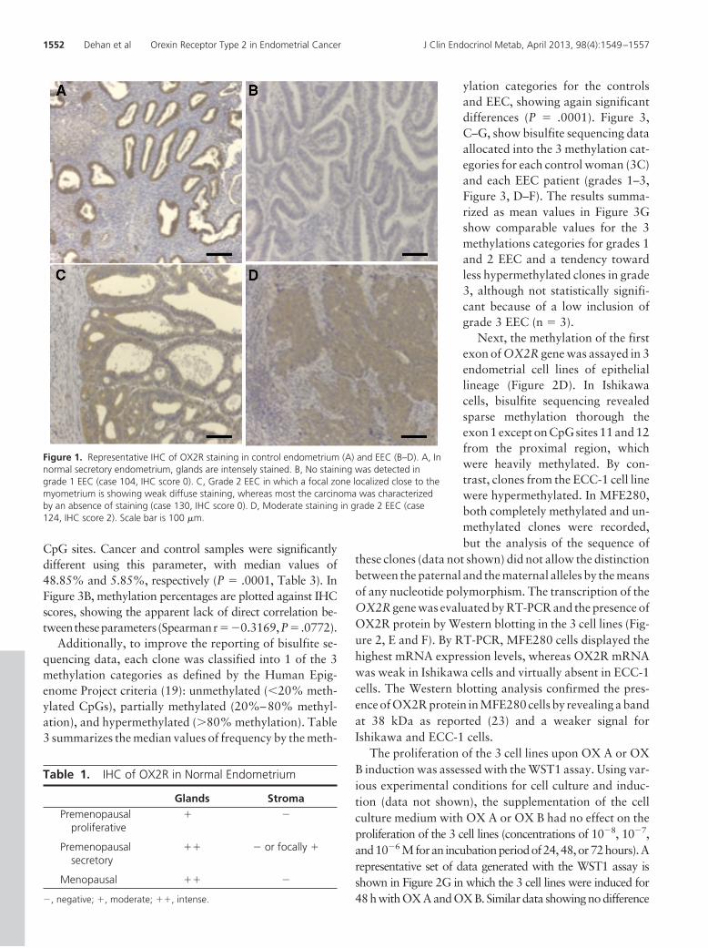
The expression pattern of OX2R protein was first inves-tigated in normal cycling endometrium by immunohisto-chemistry (IHC) using an anti-OX2R monoclonal anti-body. We tested a panel of 24 normal endometrial tissuesfrom untreated women covering the entire menstrual cycleas well as postmenopausal endometrium. An intense tomoderate immunoreactivity with an OX2R antibody wasfound in glandular cells. The most intense staining wasdetected in the premenopausal secretory phase as well asin the menopausal condition (Figure 1A and Table 1). Bycontrast, when the OX2R antibody was applied on the 32EEC cases collected for this study (Table 2), 21 samples(65.6% of the total) obtained an IHC score 0 correspond-ing to an absence of staining either complete either withminor area of labeling, which often localized close to theinterface with the myometrium (Figure 1, B and C). For theremaining 11 cases of EEC (34.4% of the total), a weak ormoderate immunostaining was recorded on the tumoraltissue (IHC scores 1 or 2, Figure 1D).
To explain the frequent loss of OX2R immunostaining,the methylation status of the CpG island located in exon1 of the OX2R gene was evaluated on 32 EEC samplestested on IHC and on 18 controls endometria using bisul-fite sequencing. Figure 2A shows the sequence of OX2Rexon 1 locus with the localization of the 27 CpG sitesinvestigated (14 in the proximal portion and 13 in thedistal portion). Two different PCRs were used to cover thewhole exon 1 locus (proximal and distal portion) with theexception of 90 nucleotides comprising 6 CpG sites. Those90 nucleotides were excluded for technical reasons (in-ability to successfully amplify these regions by PCR).
Representative bisulfite sequencing charts of individualCpG sites located on OX2R exon 1 are displayed in Figure2, B and C, for patient 119 (grade 1) and control 15. Thecomplete bisulfite sequencing data for EEC and controlssamples are available in Supplemental Figures 1 and 2,respectively, published on The Endocrine Society’s Jour-nals Online web site at http://jcem.endojournals.org. Ta-ble 2 and Figure 3A display the percentages of methylated
doi: 10.1210/jc.2012-3263 jcem.endojournals.org 1551
CpG sites. Cancer and control samples were significantlydifferent using this parameter, with median values of48.85% and 5.85%, respectively (P � .0001, Table 3). InFigure 3B, methylation percentages are plotted against IHCscores, showing the apparent lack of direct correlation be-tweentheseparameters (Spearmanr��0.3169,P� .0772).
Additionally, to improve the reporting of bisulfite se-quencing data, each clone was classified into 1 of the 3methylation categories as defined by the Human Epig-enome Project criteria (19): unmethylated (�20% meth-ylated CpGs), partially methylated (20%–80% methyl-ation), and hypermethylated (�80% methylation). Table3 summarizes the median values of frequency by the meth-
ylation categories for the controlsand EEC, showing again significantdifferences (P � .0001). Figure 3,C–G, show bisulfite sequencing dataallocated into the 3 methylation cat-egories for each control woman (3C)and each EEC patient (grades 1–3,Figure 3, D–F). The results summa-rized as mean values in Figure 3Gshow comparable values for the 3methylations categories for grades 1and 2 EEC and a tendency towardless hypermethylated clones in grade3, although not statistically signifi-cant because of a low inclusion ofgrade 3 EEC (n � 3).
Next, the methylation of the firstexon of OX2R gene was assayed in 3endometrial cell lines of epitheliallineage (Figure 2D). In Ishikawacells, bisulfite sequencing revealedsparse methylation thorough theexon 1 except on CpG sites 11 and 12from the proximal region, whichwere heavily methylated. By con-trast, clones from the ECC-1 cell linewere hypermethylated. In MFE280,both completely methylated and un-methylated clones were recorded,but the analysis of the sequence of
these clones (data not shown) did not allow the distinctionbetween the paternal and the maternal alleles by the meansof any nucleotide polymorphism. The transcription of theOX2R gene was evaluated by RT-PCR and the presence ofOX2R protein by Western blotting in the 3 cell lines (Fig-ure 2, E and F). By RT-PCR, MFE280 cells displayed thehighest mRNA expression levels, whereas OX2R mRNAwas weak in Ishikawa cells and virtually absent in ECC-1cells. The Western blotting analysis confirmed the pres-ence of OX2R protein in MFE280 cells by revealing a bandat 38 kDa as reported (23) and a weaker signal forIshikawa and ECC-1 cells.
The proliferation of the 3 cell lines upon OX A or OXB induction was assessed with the WST1 assay. Using var-ious experimental conditions for cell culture and induc-tion (data not shown), the supplementation of the cellculture medium with OX A or OX B had no effect on theproliferation of the 3 cell lines (concentrations of 10�8, 10�7,and10�6 Mforan incubationperiodof24,48,or72hours).Arepresentative set of data generated with the WST1 assay isshown in Figure 2G in which the 3 cell lines were induced for48 h with OX A and OX B. Similar data showing no difference
Figure 1. Representative IHC of OX2R staining in control endometrium (A) and EEC (B–D). A, Innormal secretory endometrium, glands are intensely stained. B, No staining was detected ingrade 1 EEC (case 104, IHC score 0). C, Grade 2 EEC in which a focal zone localized close to themyometrium is showing weak diffuse staining, whereas most the carcinoma was characterizedby an absence of staining (case 130, IHC score 0). D, Moderate staining in grade 2 EEC (case124, IHC score 2). Scale bar is 100 �m.
Table 1. IHC of OX2R in Normal Endometrium
Glands StromaPremenopausal
proliferative� �
Premenopausalsecretory
�� � or focally �
Menopausal �� �
�, negative; �, moderate; ��, intense.
1552 Dehan et al Orexin Receptor Type 2 in Endometrial Cancer J Clin Endocrinol Metab, April 2013, 98(4):1549–1557

betweentreatedanduntreatedhavebeencollectedatothertimepoints (24 hours, 72 hours, etc). To further evaluate the apo-ptosis in the same culture conditions, a caspase-3/7 assay wasused. No evidence of apoptosis was demonstrated after 48hours of induction with OXA or OXB (Figure 2H).
DiscussionDiscovered 15 years ago, the orexinergic system has at-tracted major interest with now more than 2500 publica-tions recorded in PubMed web site with the keywordorexin. First analyzed in the brain, the orexins and orexin
Table 2. Clinical Features, Methylation Status of OX2R, and IHC Scores for Controls (n � 18) and EEC PatientsListed Separately After the Pathological Grade: Grade 1 (n � 21); Grade 2 ( n � 10); Grade 3 (n � 3)
CaseNumber Menopause
Age,y
Methylation,%
IHCScore
Controls 1 No 40 3.4 ND2 No 48 7.9 ND3 No 40 4.8 ND4 No 37 2.6 ND5 No 42 10.9 ND6 No 39 2.7 ND7 No 38 5.9 ND8 No 32 7.2 ND9 Yes 52 7.5 ND
10 Yes 54 5.8 ND11 Yes 55 16.7 ND12 Yes 50 10.7 ND13 Yes 50 5.3 ND14 Yes 54 2.2 ND15 Yes 54 5.5 ND16 Yes 49 5.9 ND17 Yes 75 4.7 ND18 Yes 54 7.3 ND
EEC grade 1 101 Yes 60 50.0 1102 Yes 78 10.9 1103 Yes 54 47.7 1104 Yes 69 67.1 0105 Yes 60 38.7 1106 Yes 81 80.9 0107 Yes 74 35.2 0108 Yes 65 43.2 0109 Yes 58 25.4 1110 Yes 68 13.4 0111 Yes 78 32.2 0112 Yes 80 72.4 0113 Yes 60 44.0 0114 Yes 70 72.1 0115 No 39 39.8 0116 Yes 58 61.6 0117 Yes 75 60.4 0118 Yes 74 42.8 0119 Yes 65 41.1 1120 Yes 69 68.6 0121 Yes 72 66.8 ND
EEC grade 2 122 Yes 69 64.5 2123 Yes 74 50.7 0124 Yes 64 25.2 2125 Yes 65 53.8 0126 No 47 69.5 1127 Yes 55 5.4 0128 Yes 67 65.9 0129 Yes 61 33.1 0130 Yes 81 63.6 0131 Yes 84 64.7 1
EEC grade 3 132 Yes 86 36.0 1133 Yes 54 23.8 0134 Yes 79 50.0 ND
Abbreviation: ND, not determined. IHC scores include the following: 0, general absence of staining; 1, global weak staining; and 2, globalmoderate staining.
doi: 10.1210/jc.2012-3263 jcem.endojournals.org 1553

Figure 2. Methylation on OX2R locus in normal endometrium, EEC and cell lines. A, The sequence of exon 1 is presented with open arrows to indicate theexon’s limits. CpG sites are numbered as appearing in the methylation patterns below. Horizontal arrows indicate the position of primers used for proximal anddistal PCR amplification. B and C, Representative methylation patterns of control and EEC cases. Each numbered column represents a CpG site and every rowrepresents a unique allele. Blue, red, and white squares, respectively, represent unmethylated, methylated, or undetermined CpG sites. The pattern in panel Bshows mostly unmethylated alleles in control endometrium (control 15), whereas EEC patient 119 in panel C shows intense methylation, which is maximal inCpG sites 10–13 from the proximal portion. D, Methylation pattern of OX2R exon 1 in MFE 280, ECC-1, and Ishikawa cells. In MFE 280, hypermethylated as wellas completely unmethylated clones are observed. In ECC-1 cells, the intense methylation pattern is recorded. In Ishikawa cells, methylated CpG sites areconcentrated at positions 11 and 12 from the proximal portion. RT-PCR for OX2R (E) and Western blot (F) using anti-OX2R antibody for the 3 endometrial celllines. Only MFE 280 showed strong signals in E and F. For ECC-1 and Ishikawa cells, absent or very weak signals are recorded. Loading controls are given byGAPDH RT-PCR (E) and Western blot (F). G, Representative experiment showing the values (percentage of control) of WST1 tests performed with cell cultures(ECC-1, MFE280, and Ishikawa) induced for 48 hours with graded concentrations of OX A and OX B. Orexins have no effect on cell proliferation. H,Representative caspase-3/7 assays performed with cell cultures (ECC-1, MFE280, and Ishikawa) induced for 48 hours with graded concentrations of OX A andOX B. The values expressed in the percentage of noninduced controls show no effect of both orexins on apoptosis.
1554 Dehan et al Orexin Receptor Type 2 in Endometrial Cancer J Clin Endocrinol Metab, April 2013, 98(4):1549–1557

receptors have been recently shown to have a broader rolein peripheral organs and in cancer. Many aspects of theirphysiological roles are probably still to be uncovered.
We described here the localization of OX2R protein inthe epithelial compartment of normal endometrium dur-
ing the whole menstrual cycle and inmenopausal conditions. In EEC,65.6% of 32 tumoral samples inves-tigated were devoid of OX2R stain-ing either completely or with mini-mal focal labeling. Additionally,hypermethylation of the first exon ofOX2R was demonstrated in EEC bycomparison with normal endome-trium (median methylation percent-age of 48.85% and 5.85%, res-pectively). Comparisons betweenmethylation categories for the 3grades of EEC in Figure 3G showedsimilar methylation for grades 1 and2 and a tendency toward lower per-centages of the partially methylatedand hypermethylated categories forgrade 3, although this has to be con-firmed on a larger number of clinicalsamples. Similar observations re-porting reduced methylation rates inadvanced tumor stages have alreadybeen done as, for example, in colo-rectal cancer, in which O6-methyl-guanine DNA methyltransferase(MGMT) hypermethylation is moreintense in T1/T2 carcinomas than inthe more advanced T3/T4 stages (24).The MGMT protein has then been re-ported to be reexpressed in colon ad-enocarcinomas (25), whereas it wasabsent in MGMT-methylated adeno-mas (26) from which carcinomas aresupposed to derive.
This is the first time that epige-netic regulation is identified forOX2R. Its genomic locus was re-cently studied and revealed the pres-ence of 3 nontranslated 5� exons andthe use of alternative promoters (27).Here in vitro studies using cell linesallowed the establishment of a cor-relation between the methylation ofthe first exon and the lack of geneexpression. The methylation of firstexons have indeed been recentlyidentified as major gene-silencing
events (28). In ECC-1 cells, an intense methylation of exon1 was correlated with the absence of signal in RT-PCR andin OX2R immunoblotting. In Ishikawa cells, weak OX2Rtranscription and protein expression was associated with
Figure 3. Quantitative methylation of OX2R first exon. A, Percentages of methylation for cancer(n � 34) and controls (n � 18) samples. B, EEC patients (n � 34): plotting of methylationpercentages and IHC scores showing the absence of correlation between them. C–G, Allocationof OX2R bisulfite sequencing clones to the 3 methylations categories in controls (C; n � 18) andEEC grade 1 (D; n � 21), grade 2 (E; n � 10), grade 3 (F; n � 3). G, Mean values for controlsand EEC of all 3 grades are displayed. In panel C, clones are mostly unmethylated. In panels Dand E, most of the samples show a high proportion of hypermethylated clones, which tends tobe lower in panel F for grade 3 EEC.
doi: 10.1210/jc.2012-3263 jcem.endojournals.org 1555

sparse methylation except around CpG sites 11 and 12 in theproximal zone. In the MFE280 cells, for which a moderateOX2Rexpressionisdetected,bothcompletelyunmethylatedand methylated clones were identified making the locushemizygous and permissive for OX2R expression. Unfortu-nately, no nucleotide polymorphism could be found in thesequenced fragments, and it was not possible to establishwhether theobservedheterogeneousmethylationwouldseg-regate together with paternal and maternal alleles. This ob-servation is interesting because it affects MFE280, a poorlydifferentiated cell line. This could be related to the tendencyto lower methylation density observed in grade 3 EEC. Het-erogeneousmethylationappears tobe frequent in thehumangenome and is not always connected to gene imprinting (29).The OX2R gene has not been reported to be imprinted, al-though in a recent computational study (30), it was includedin a list of imprinted gene candidates.
By contrast, in vivo, no clear correlation could beestablished between the methylation percentages andthe IHC scores evaluated on tumoral tissues (Figure 3B).However, the samples with lower methylation levels(�50%) had a mean IHC score higher than sampleswith high (�50%) methylation levels (0.50 vs 0.29).This failure to reach statistical significance for the cor-relation could stem from either the low number of sam-ples of highest tumor grade or the sampling methodapplied. Indeed, the material used for DNA extractionand the methylation study comes from a different partof the tumor than the one used for OX2R IHC. In agree-ment with our hypothesis, data from Faquin et al (31)and Feng et al (32) also showed substantial heteroge-neity in EEC.
In our study, the bisulfite sequencing after bacterialcloning allowed the quantification of methylation at thelevel of individual alleles later allocated into 3 methylationcategories. This approach is particularly necessary whenanalyzing heterogeneous material as tumoral samples(33). The identification of OX2R methylation in EECcould be prolonged by the establishment of an OX2R-based test applicable in clinical diagnostic situations, us-ing a methylation-specific PCR technology on endome-trial tissue obtained by curettage as starting material.
WST1 proliferation assays in the presence of OX A andOX B or the caspase-3/7 tests did not show any differencecompared with controls, demonstrating the absence oforexin-induced apoptosis. This result was obtained for the 3cell lines including MFE280 with demonstrated expressionof OX2R. An alternative explanation could be that the levelof expression of OX2R in MFE280 cells would be too low totrigger orexin-induced apoptosis. In other cancer models ex-pressing OX2R, such an apoptosis has been reported in apancreatic rat cell line (34), whereas for adrenocortical ad-enoma cells, a stimulation of growth and cortisol secretionwas described upon orexin induction (35). Thus, in the con-text of endometrial cancer, the direct implication of OX2Rin the control of cell proliferation is not established and theepigenetic OX2R down-regulation reported here may im-pact cancer cells by other means that have yet to be uncov-ered. De Carvalho et al (36) recently studied the methylationof promoter regions that were necessary for cancer cell sur-vival. Intriguingly, no classical tumor suppressor gene wasevidenced by their screening, which identified many cell sig-naling molecules, notably several G protein-coupled recep-tors, leading the authors to conclude that the role of GPRCsin tumor progression was probably underestimated. In thisstudy the OX2R silencing by DNA methylation might cer-tainly be considered on the same line.
In the normal endometrium, the physiological role ofOX2R and the orexinergic system also remains to be betterunderstood. Very recently the presence of OX A and OXB was established in the porcine endometrium (37), which,if confirmed in human tissues, could suggest an autocrine/paracrine interaction of the orexins with OX2R as fore-seen in the male genital tract (7).
In conclusion, the OX2R was shown to be present in nor-mal endometrium, whereas in EEC, a frequent OX2R loss isassociated with the frequent hypermethylation of its firstexon. The methylation intensities are similar in grades 1 and2 EEC and tend to regress in grade 3 clinical samples, al-though more grade 3 cases would be required to confirm thetendency. Heterogeneous methylation with the presence ofcompletely methylated and unmethylated alleles was alsoobserved on the MFE280 cell line characterized by poordifferentiation.
Table 3. Global and Detailed Methylation Evaluation in Controls and EEC of All Grades
Controls(n � 18)
All Grades Cancer(n � 34)
PValue
Global methylation, % 5.85 (4.05–7.70) 48.85 (34.15–65.30) a
Detailed distribution of clonesUnmethylated (�20%) 0.9348 (0.8254–0.9479) 0.2944 (0.1232–0.5040) a
Partially methylated (from 20% to 80%) 0.0652 (0.0521–0.1623) 0.4063 (0.2971–0.6181) a
Hypermethylated (�80%) 0.0 (0.0–0.0) 0.1585 (0.0729–0.3750) a
Median values (25th and 75th percentile) are given. P values were calculated using the Mann-Whitney test.a P � .0001.
1556 Dehan et al Orexin Receptor Type 2 in Endometrial Cancer J Clin Endocrinol Metab, April 2013, 98(4):1549–1557

Acknowledgments
We thank Professors F. Kridelka and F. Goffin for referring clin-ical samples (Hôpital ND des Bruyères and Hôpital de la Cita-delle, respectively) and Drs K. Delbecque and B. Bisig for exper-tise in pathology. The technical assistance of N. Krusy, I. Dasoul,M.-R. Pignon, and E. Dortu is also acknowledged.
Address all correspondence and requests for reprints to:Pierre Dehan, PhD, Department of Experimental Pathology,University of Liège, Tour de Pathologie (B23 � 4), Boulevardde l’Hôpital 1, B 4000 Liege Belgium. E-mail:[email protected].
This work was supported by an Oncomethylomic grant fromthe Région Wallonne (Convention C5745). C.M. is a researchassociate from the Fonds National de la Recherche Scientifique(FNRS, Belgium).
Disclosure Summary: The authors declare there is no conflictof interest.
References
1. de Lecea L, Kilduff TS, Peyron C, et al. The hypocretins: hypothal-amus-specific peptides with neuroexcitatory activity. Proc NatlAcad Sci USA. 1998;95:322–327.
2. Sakurai T, Amemiya A, Ishii M, et al. Orexins and orexin receptors:a family of hypothalamic neuropeptides and G protein-coupled re-ceptors that regulate feeding behavior. Cell. 1998;92:573–585.
3. Heinonen MV, Purhonen AK, Makela KA, Herzig KH. Functions oforexins in peripheral tissues. Acta Physiol (Oxf). 2008;192:471–485.
4. Kirchgessner AL, Liu M. Orexin synthesis and response in the gut.Neuron. 1999;24:941–951.
5. Mitsuma T, Hirooka Y, Kayma M, et al. Radioimmunoassay forhypocretin-2. Endocr Regul. 2000;34:23–27.
6. Mitsuma T, Hirooka Y, Kayama M, et al. Radioimmunoassay fororexin A. Life Sci. 2000;66:897–904.
7. Karteris E, Chen J, Randeva HS. Expression of human prepro-orexinand signaling characteristics of orexin receptors in the male repro-ductive system. J Clin Endocrinol Metab. 2004;89:1957–1962.
8. Tafuri S, Muto RL, Pavone LM, et al. Novel localization of orexinA in the tubular cytotypes of the rat testis. Regul Pept. 2010;164:53–57.
9. Silveyra P, Lux-Lantos V, Libertun C. Both orexin receptors areexpressed in rat ovaries and fluctuate with the estrous cycle: effectsof orexin receptor antagonists on gonadotropins and ovulation.Am J Physiol Endocrinol Metab. 2007;293:E977–E985.
10. Laburthe M, Voisin T, El Firar A. Orexins/hypocretins and orexinreceptors in apoptosis: a mini-review. Acta Physiol (Oxf). 2010;198:393–402.
11. Rouet-Benzineb P, Rouyer-Fessard C, Jarry A, et al. Orexins actingat native OX(1) receptor in colon cancer and neuroblastoma cells orat recombinant OX(1) receptor suppress cell growth by inducingapoptosis. J Biol Chem. 2004;279:45875–45886.
12. Nakabayashi M, Suzuki T, Takahashi K, et al. Orexin-A expressionin human peripheral tissues. Mol Cell Endocrinol. 2003;205:43–50.
13. Hecht JL, Mutter GL. Molecular and pathologic aspects of endo-metrial carcinogenesis. J Clin Oncol. 2006;24:4783–4791.
14. You JS, Jones PA. Cancer genetics and epigenetics: two sides of thesame coin? Cancer Cell. 2012;22:9–20.
15. Tao MH, Freudenheim JL. DNA methylation in endometrial cancer.Epigenetics. 2010;5:491–498.
16. Esteller M. Epigenetics in cancer. N Engl J Med. 2008;358:1148–1159.
17. Sharma S, Kelly TK, Jones PA. Epigenetics in cancer. Carcinogen-esis. 2010;31:27–36.
18. Laird PW. The power and the promise of DNA methylation mark-ers. Nat Rev Cancer. 2003;3:253–266.
19. Eckhardt F, Lewin J, Cortese R, et al. DNA methylation profiling ofhuman chromosomes 6, 20 and 22. Nat Genet. 2006;38:1378–1385.
20. Mo B, Vendrov AE, Palomino WA, DuPont BR, Apparao KB,Lessey BA. ECC-1 cells: a well-differentiated steroid-responsive en-dometrial cell line with characteristics of luminal epithelium. BiolReprod. 2006;75:387–394.
21. Hackenberg R, Loos S, Nia AH, Kunzmann R, Schulz KD. Expres-sion of placental protein 14 by the new endometrial cancer cell lineMFE-280 in vitro and by endometrial carcinomas in vivo. Antican-cer Res. 1998;18:1153–1158.
22. Rohde C, Zhang Y, Reinhardt R, Jeltsch A. BISMA—fast and accuratebisulfite sequencing data analysis of individual clones from unique andrepetitive sequences. BMC Bioinformatics. 2010;11:230.
23. Randeva HS, Karteris E, Grammatopoulos D, Hillhouse EW. Ex-pression of orexin-A and functional orexin type 2 receptors in thehuman adult adrenals: implications for adrenal function and energyhomeostasis. J Clin Endocrinol Metab. 2001;86:4808–4813.
24. Nagasaka T, Goel A, Notohara K, et al. Methylation pattern of theO6-methylguanine-DNA methyltransferase gene in colon during pro-gressive colorectal tumorigenesis. Int J Cancer. 2008;122:2429–2436.
25. Jass JR, Whitehall VL, Young J, Leggett BA. DNA methylation incolorectal cancer. In: Szyf M, ed. DNA Methylation and CancerTherapy. Georgetown and New York: Kluwer Academic/Plenumand Landes Biosciences/Eurekah.com Publishers; 2005:59–68.
26. Rashid A, Shen L, Morris JS, Issa JP, Hamilton SR. CpG islandmethylation in colorectal adenomas. Am J Pathol. 2001;159:1129–1135.
27. Chen J, Randeva HS. Genomic organization and regulation of thehuman orexin (hypocretin) receptor 2 gene: identification of alter-native promoters. Biochem J. 2010;427:377–390.
28. Brenet F, Moh M, Funk P, et al. DNA methylation of the first exonis tightly linked to transcriptional silencing. PloS One. 2011;6:e14524.
29. Meaburn EL, Schalkwyk LC, Mill J. Allele-specific methylation inthe human genome: implications for genetic studies of complex dis-ease. Epigenetics. 2010;5:578–582.
30. Luedi PP, Dietrich FS, Weidman JR, Bosko JM, Jirtle RL, HarteminkAJ. Computational and experimental identification of novel humanimprinted genes. Genome Res. 2007;17:1723–1730.
31. Faquin WC, Fitzgerald JT, Boynton KA, Mutter GL. Intratumoralgenetic heterogeneity and progression of endometrioid type endo-metrial adenocarcinomas. Gynecol Oncol. 2000;78:152–157.
32. Feng YZ, Shiozawa T, Horiuchi A, et al. Intratumoral heteroge-neous expression of p53 correlates with p53 mutation, Ki-67, andcyclin A expression in endometrioid-type endometrial adenocarci-nomas. Virchows Archiv. 2005;447:816–822.
33. Mikeska T, Candiloro IL, Dobrovic A. The implications of heter-ogeneous DNA methylation for the accurate quantification of meth-ylation. Epigenomics. 2010;2:561–573.
34. Voisin T, Firar AE, Avondo V, Laburthe M. Orexin-induced apo-ptosis: the key role of the seven-transmembrane domain orexin type2 receptor. Endocrinology. 2006;147:4977–4984.
35. Spinazzi R, Rucinski M, Neri G, Malendowicz LK, Nussdorfer GG.Preproorexin and orexin receptors are expressed in cortisol-secret-ing adrenocortical adenomas, and orexins stimulate in vitro cortisolsecretion and growth of tumor cells. J Clin Endocrinol Metab. 2005;90:3544–3549.
36. De Carvalho DD, Sharma S, You JS, et al. DNA methylation screen-ing identifies driver epigenetic events of cancer cell survival. CancerCell. 2012;21:655–667.
37. Nitkiewicz A, Smolinska N, Maleszka A, Kiezun M, Kaminski T.Localization of orexin A and orexin B in the porcine uterus. ReprodBiol. 2012;12:135–155.
doi: 10.1210/jc.2012-3263 jcem.endojournals.org 1557


