Expression of Stromelysin-3 in the Human Placenta and Placental Bed 1997.pdf · 278 Placenta...
9
Placenta (1997), 18, 277-285 Expression of Stromelysin-3 in the Human Placenta and Placental Bed E. Maquoi a, M. Polette b, B. Nawrocki b, P. Bischof c, A. Noi~l a'd, A. Pintiaux e, M. Santavicca f, J.-P. Schaaps e, R. Pijnenborg f, P. Birembaut b and J.-M. Foidart a'g a Laboratory of Biology, University of Liege, Sart Tilman, Belgium b INSERM U-314, CHU Maison Blanche, Reims, France Department of Obstetrics and Gynecology, University of Geneva, Geneva, Switzerland d Department of Obstetrics and Gynecology, University of Liege, CHR Citadelle, Liege, Belgium e INSERM U-184, CNRS IGBMC, Facult~ de M6decine, IIIkirch, France f Department of Obstetrics and Gynecology, U.Z. Gasthuisberg, Leuven, Belgium Paper accepted 1 7 December 1 996 Human placentation is mediated by fetal trophoblastic cells which penetrate into the decidualized uterine endometrium. Trophoblast invasion requires the precisely regulated secretion of specific proteinases able to degrade the endometrial basement membranes and extracellular matrix. To document further the involvement of these proteinases during human placentation, we evaluated in vivo the expression of stromelysin-3, a member of the metalloproteinase family, during the first and third trimesters of pregnancy, by means of immunohistochemistry, in situ hybridization and Northern blot analysis. Human extravillous trophoblasts invading the maternal decidua produced stromelysin-3 during both, the first and third trimesters of pregnancy, but to a lesser extent during the latter. In floating villi, stromelysin-3 expression was restricted to the syncytiotrophoblasts that line intervillous vascular spaces. In conclusion, stromelysin-3 is expressed by differentiated, non-proliferative villous and extravillous trophoblastic cells in early and late placental beds and villi, and its pattern of expression evolves during pregnancy. Our observations suggest that stromelysin-3 could play a role in human placentation. © 1997 W. B. Saunders Company Ltd Placenta (1997), 18, 277-285 INTRODUCTION Implantation of the human blastocyst and the subsequent placental development are dependent on trophoblast invasion of the decidualized endometrium. As implantation proceeds, undifferentiated villous cytotrophoblast stem cells (CTB) originating from the outer cell layer of the blastocyst divide and differentiate into morphologically and functionally distinct cell populations. In the villi, they fuse to form hormonally active villous syncytiotrophoblast (STB) which controls the transfer of solutes between maternal and fetal blood. Where villi contact the uterine wall, multilayered trophoblast cell columns (TCC) physically anchor the embryo to the decidua. In these columns, CTB differentiate progressively into non- proliferative, invasive and migratory intermediate trophoblasts (IT) (Damsky et al., 1992; Kliman and Feinberg, 1992; Damsky, Sutherland and Fisher, 1993). During the first 4 months of gestation [Figure l(a)], these IT sprout from the TCC and migrate through the decidua and the inner third of the myometrium. They infiltrate the walls of the spiral arteries and replace the endothelial lining as far as the myometrial segment of the vessels. This invasive trophoblastic activity g To whom correspondence should be addressed. O143~I.004/97/040277 + 09 S12.00/0 is characterized by the breaching of multiple basement membranes (including those of the endometrial glands, blood vessels and decidual cells) and the degradation of the inter- stitial extracellular matrix (ECM) of the decidua (Boyd and Hamilton, 1967; Brosens, Robertson and Dixon, 1967; Pijnenborg et al., 1980; Tuttel et al., 1985; Aplin, 1991; Foidart et al., 1992). The human placenta thus functions as an invasive tissue, analogous to a locally invasive tumour (Liotta, 1986). How- ever, unlike tumour invasion, trophoblast penetration is regulated precisely, both spatially and temporally, during gestation. Several in vitro and in vivo studies have revealed that trophoblast cells have a regulated capacity to produce different matrix-degrading enzymes (Lala and Graham, 1990; Bischof and Martelli, 1992; Graham and Lala, 1992). Production of serine proteinases, such as urokinase-type plasminogen acti- vator (u-PA) by trophoblastic cells correlates temporally with blastocyst invasion (Sherman, Strickland and Reich, 1976; Strickland, Reich and Sherman, 1976; Sappino et al., 1989; Behrendtsen, Alexander and Werb, 1992). The presence of u-PA, plasminogen activator inhibitor type 1 and u-PA receptor has been associated with infiltrating trophoblasts ~t~1997w. B. Saundcrs CompanyLt(I
Transcript of Expression of Stromelysin-3 in the Human Placenta and Placental Bed 1997.pdf · 278 Placenta...

Placenta (1997), 18, 277-285
Expression of Stromelysin-3 in the Human Placenta and Placental Bed
E. Maquo i a, M. Polet te b, B. Nawrock i b, P. Bischof c, A. Noi~l a'd, A. Pint iaux e, M. Santavicca f, J.-P. Schaaps e, R. Pi jnenborg f, P. B i rembaut b and J . - M . Foidart a'g
a Laboratory of Biology, University of Liege, Sart Tilman, Belgium
b INSERM U-314, CHU Maison Blanche, Reims, France
Department of Obstetrics and Gynecology, University of Geneva, Geneva, Switzerland
d Department of Obstetrics and Gynecology, University of Liege, CHR Citadelle, Liege, Belgium
e INSERM U-184, CNRS IGBMC, Facult~ de M6decine, IIIkirch, France
f Department of Obstetrics and Gynecology, U.Z. Gasthuisberg, Leuven, Belgium
Paper accepted 1 7 December 1 996
Human placentation is mediated by fetal trophoblastic cells which penetrate into the decidualized uterine endometrium. Trophoblast invasion requires the precisely regulated secretion of specific proteinases able to degrade the endometrial basement membranes and extracellular matrix. To document further the involvement of these proteinases during human placentation, we evaluated in vivo the expression of stromelysin-3, a member of the metalloproteinase family, during the first and third trimesters of pregnancy, by means of immunohistochemistry, in situ hybridization and Northern blot analysis. Human extravillous trophoblasts invading the maternal decidua produced stromelysin-3 during both, the first and third trimesters of pregnancy, but to a lesser extent during the latter. In floating villi, stromelysin-3 expression was restricted to the syncytiotrophoblasts that line intervillous vascular spaces. In conclusion, stromelysin-3 is expressed by differentiated, non-proliferative villous and extravillous trophoblastic cells in early and late placental beds and villi, and its pattern of expression evolves during pregnancy. Our observations suggest that stromelysin-3 could play a role in human placentation. © 1997 W. B. Saunders Company Ltd Placenta (1997), 18, 277-285
I N T R O D U C T I O N
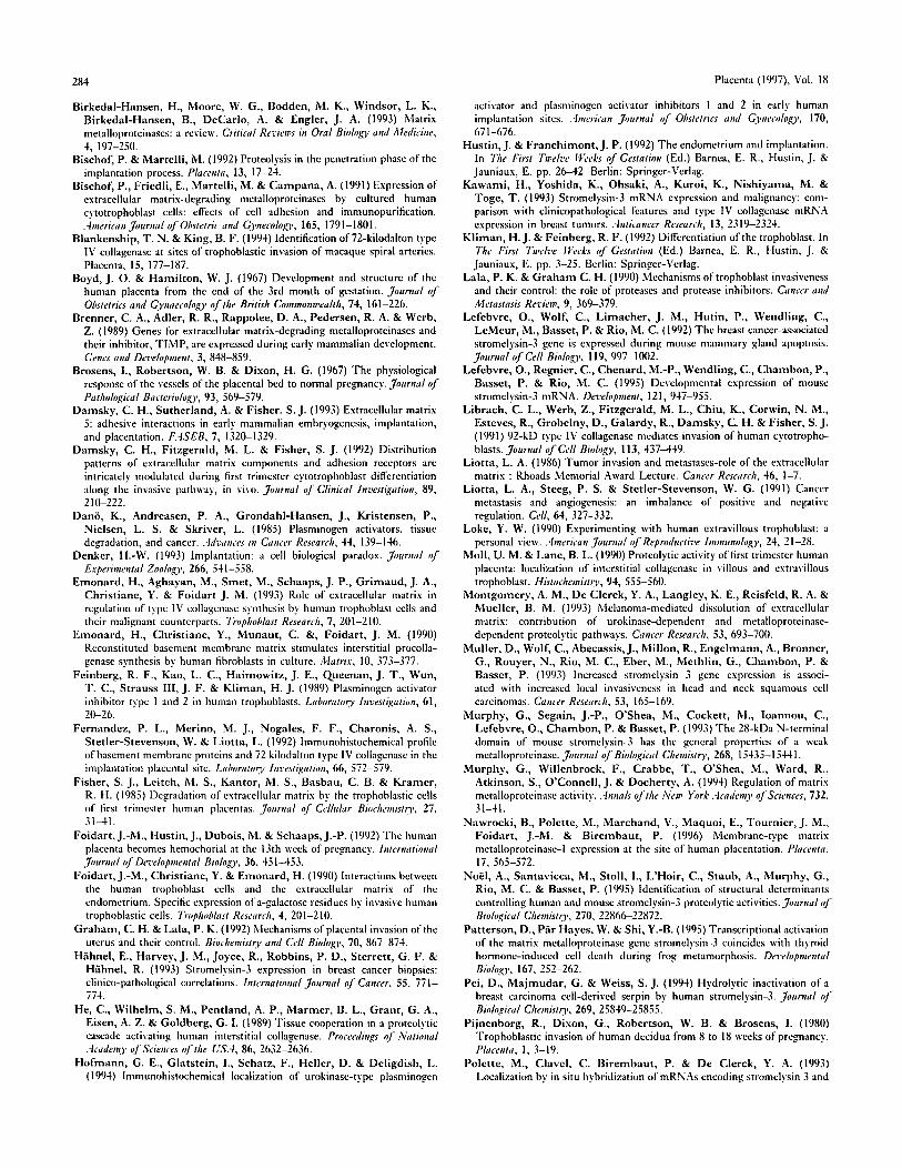
Implantation of the human blastocyst and the subsequent placental development are dependent on trophoblast invasion of the decidualized endometrium. As implantation proceeds, undifferentiated villous cytotrophoblast stem cells (CTB) originating from the outer cell layer of the blastocyst divide and differentiate into morphologically and functionally distinct cell populations. In the villi, they fuse to form hormonally active villous syncytiotrophoblast (STB) which controls the transfer of solutes between maternal and fetal blood. Where villi contact the uterine wall, multilayered trophoblast cell columns (TCC) physically anchor the embryo to the decidua. In these columns, CTB differentiate progressively into non- proliferative, invasive and migratory intermediate trophoblasts (IT) (Damsky et al., 1992; Kliman and Feinberg, 1992; Damsky, Sutherland and Fisher, 1993). During the first 4 months of gestation [Figure l(a)], these IT sprout from the TCC and migrate through the decidua and the inner third of the myometrium. They infiltrate the walls of the spiral arteries and replace the endothelial lining as far as the myometrial segment of the vessels. This invasive trophoblastic activity
g To whom correspondence should be addressed.
O143~I.004/97/040277 + 09 S12.00/0
is characterized by the breaching of multiple basement membranes (including those of the endometrial glands, blood vessels and decidual cells) and the degradation of the inter- stitial extracellular matrix (ECM) of the decidua (Boyd and Hamilton, 1967; Brosens, Robertson and Dixon, 1967; Pijnenborg et al., 1980; Tuttel et al., 1985; Aplin, 1991; Foidart et al., 1992).
The human placenta thus functions as an invasive tissue, analogous to a locally invasive tumour (Liotta, 1986). How- ever, unlike tumour invasion, trophoblast penetration is regulated precisely, both spatially and temporally, during gestation.
Several in vitro and in vivo studies have revealed that trophoblast cells have a regulated capacity to produce different matrix-degrading enzymes (Lala and Graham, 1990; Bischof and Martelli, 1992; Graham and Lala, 1992). Production of serine proteinases, such as urokinase-type plasminogen acti- vator (u-PA) by trophoblastic cells correlates temporally with blastocyst invasion (Sherman, Strickland and Reich, 1976; Strickland, Reich and Sherman, 1976; Sappino et al., 1989; Behrendtsen, Alexander and Werb, 1992). The presence of u-PA, plasminogen activator inhibitor type 1 and u-PA receptor has been associated with infiltrating trophoblasts
~t~ 1997 w. B. Saundcrs Company Lt(I

278 Placenta (1997), Vol. 18
A
Figure 1. Localization of ST3 in a first trimester (11 weeks) placental bed. (A) Diagram showing the spatial organization of the fetal-maternal interface at the end of the first trimester. D, decidua; CTB, cytotrophoblast; IT, intermediate trophoblast; IVC, intervillous chamber; IVT, intravascular trophoblast; MBV, maternal blood vessel; PBGC, placental-bed giant cell; S, stroma; STB, s.vncytiotrophoblast bordered by sinusoids; TCC, trophoblastic cell column. (B) Transverse section througb a villus showing STB strongly labelled by the anti-ST3 antibody. Scale bar=10l) p.m. (C) Longitudinal section through the placental bed. Strong STB positivity (arrows) for ST3 is observed. ST3 is also expressed by some spindle-shaped IT (arrowheads) that are derived from negative TCC, become detached and infiltrate the maternal decidua. Scale bar=lS0 lam. (D) Deeper in the decidua, numerous elongated IT, interspersed among negative decidual cells, were recognized by the anti-ST3 antibody. Scale bar=200 ~tm. (El Some IT fused to form PBGC which also express ST3. Scale bar=15(I p-m. (17) The endothelial lining of the maternal blood vessels has been replaced by ST3-positive trophohlasts. Scale bar=8ll ~lm.

Maquoi et al.: Stromelysin-3 during Human Placentation
(Feinberg et al., 1989; Zini et al., 1992; Hoffman et al., 1994). Members of the matrix metalloproteinases (MMP) family also appear to play a pivotal role during placentation. The acqui- sition of an invasive phenotype by early trophoblasts has been correlated with the ability of these cells to synthesize a large array of MMP: interstitial collagenase (Yagel et al., 1988; Moll and Lane, 1990), gelatinase A (Bischof et al., 1991; Autio- Harmeinen et al., 1992; Fernandez et al., 1992; Blankenship and King, 1994; Polette et al., 1994), gelatinase B (Fisher et al., 1985; Emonard et al., 1990; Bischof et al., 1991; Librach et al., 1991; Behrendtsen, Alexander and Werb, 1992; Emonard et al., 1993; Polette et al., 1994), stromelysin-1 and stromelysin-2 (Brenner et al., 1989), as well as a membrane- type MMP potentially involved in gelatinase A activation (Nawrocki et al., 1996). The invasive trophoblasts and the maternal decidual cells also secrete specific inhibitors of these proteinases: plasminogen-activator inhibitors 1 and 2 (Feinberg et al., 1989; Hofmann et al., 1994), and tissue inhibitors of metalloproteinases 1 and 2 (Lala, and Graham, 1990; Polette et al., 1994). Thus, trophoblast invasiveness appears to be regulated tightly by the balance between the activated enzymes and their inhibitors.
A few years ago, another member of the MMP family, stromelysin-3 (ST3), was identified (Basset, Wolf and Chambon, 1990). ST3 exhibits the same general features as previously described MMP, including a typical zinc-binding site and a conserved cysteine residue characteristic of the MMP prodomain (Basset, Wolf and Chambon, 1993). Although its physiological substrate is still unknown, ST3 is probably a proteinase, because the purified protein exhibits proteolytic activities (Murphy et al., 1993). However, ST3 displays functional properties and an activation mechanism different from that of other MMP (Birkedal-Hansen et al., 1993; Murphy et al., 1993; Pei, Majmudar and Weiss, 1994; NoEl et al., 1995; Santavicca et al., 1996). Wolf and co-workers demonstrated that st3 transcription in embryonic fibroblasts can be induced by 12-o-tetradecanoyl phorbol 13-acetate and growth factors, thereby suggesting a transcriptional regulation similar to that of interstitial collagenase, stromelysin-1 and gelatinase B (Wolf et al., 1992). According to those data, ST3 appears to belong to a new MMP subfamily that exhibits its proper enzymatic characteristics and narrow substrate specificity (Basset et al., 1993; Murphy et al., 1993; Pei, Majmudar and Weiss, 1994; Noel et al., 1995). The st3 gene is known to be expressed in the stromal compartment of most invasive human carcinomas and the highest levels of ST3 transcripts are found in tumours demonstrating high local invasiveness (Urbanski et al., 1992; Wolf et al., 1992; Basset, Wolf and Chambon, 1993; Birkedal-Hansen et al., 1993; H~ihnel et al., 1993; Kawami et al., 1993; Muller et al., 1993; Polette et al., 1993; Segain et al., 1993). However, ST3 transcripts are also present in healthy situations in which extensive ECM remodelling occurs: embryonic devel- opment (Basset et al., 1990), frog metamorphosis (Patterson, Par Hayes and Shi, 1995), mammary gland involution (Lefebvre et al., 1992), normal uterus and, interestingly, at
279
very high levels in human term placenta (Basset et al., 1990).
To localize ST3 in placental tissues and to investigate the potential role of this enzyme during human placentation, immunohistochemical labelling, in situ hybridization and Northern blot analysis were used to evaluate the expression of ST3 in first and third trimester placental tissues from normal pregnancies.
MATERIALS AND METHODS
Tissue preparation
Samples of human placental villi obtained from 16 first trimester therapeutic abortions and 10 term third trimester placentae immediately after delivery were examined. Twelve placental-bed biopsies were obtained in situ with echoguided sampling forceps during the first and third trimesters of normal pregnancies. This procedure allows precise anatomical localization (placental bed) of the sample and prevents the tissue disruption observed during abortion or delivery (Hustin and Franchimont, 1992). Finally, four full-thickness implan- tation sites in hysterectomy specimens taken at 8.5-13 weeks of pregnancy were also studied. One part of each sample was frozen in liquid nitrogen for Northern blot analysis, while the remainder was fixed in formalin and embedded in paraffin for in situ hybridization and immunohistochemical studies.
Immunohistochemistry
Immunolabelling of ST3 was accomplished according to the following procedure. Endogenous peroxidase activity was quenched by a 10-min incubation with 3 per cent hydrogen peroxide in phosphate-buffered saline (PBS). Tissues sections were subjected to three 5-min heating cycles in citric acid buffer (pH 6) in a microwave oven (Philips, power setting: 750 W). Non-specific antibody binding was blocked by incubation in 7 per cent bovine serum albumin-PBS (BSA- PBS) for 30 min. Sections were incubated overnight at room temperature with a mouse monoclonal antibody raised against the haemopexin domain of ST3 (5ST-4A9) (Wolf et al., 1993; Santavicca et al., 1995), grown as an ascites fluid and used diluted 1/4000 in 3 per cent BSA-PBS. A biotinylated second- ary antibody (LSAB 2 Kit, Dako, Denmark) was applied for 30min prior to incubation with streptavidin-horseradish peroxidase (LSAB 2 Kit) for an additional 30 rain. Antibody localization was visualized with a solution of diaminobenzidine and 0.03 per cent hydrogen peroxide. Each step of the procedure was followed by three washes in PBS.
To evaluate the specificity of immunolabelling, the primary antibody was replaced by either an ascites fluid containing 5ST-4A9 which was preincubated with immobilized recom- binant ST3 (Santavicca et al., 1995) or an isotype-matched

280 Placenta (1997), Vol. IS
,~ . ~ ,~ '5; :.i "', . . . . - '¢.::~v'~;-~ .
~a:i,,~{ ¢:~:~'z*,, ~ .g. r e : ,
' ~ , ' - ' . ' - ~ . ? # " " ~ " • L
~ ' ~ i ~ ~ '~/':~ ":: ' , ~ , " '~ ~ ' ! ~ ~ " ~=" .,~,..',C.. ~ ~ ' , .:~:~? :;'=./;:' C ~" ~, . ,,~ ,,, % ~ V ~
Figure 2. l,ocalization of ST3 mRNA in first trimester ~illi and placental beds. (A) mRNA transcripts of ST3 are detected in the S'I'B (arrmvheads) of ~illi. The decidua (1)) does not express ST3. Scale bar=200 ~.tm. (B) "I'(X: arising from Ihc villus do nm c(mtain ST3 mRNA while the STB (arrmvheads) is clearly labelled. Scale bar=l()0 )_tin. (C) ['|" (arrowheads) expressing S'1'3 mRNA have replaced the endothelial lining of .\II3V. Scale bar=50 lain. (I)) A seeti(m incubated ~itll die sense RNA pr()bc. Scale har=2()(lFtm. (1':) IT (arnmhcads) expressing S'F3 mRNA are obser',ed among decidual cells. Scale bar=25 ).tin.
i r re levant an t ibody (a mouse ascitcs fluid raised against
hams te r CD-3) . No immunoreac t iv i ty was observed in the
controls.
Epithelial cells and t , 'ophoblasts were identified by i m m u n o -
his tochemical labell ing with a mtmochmal an t ibody to cvto-
kcm'atins 8 / 1 8 ( C A M 5.2, Bec ton- l ) i ck inson) revealed with a
rabbi t an t i -mouse an t ibody conjugated to horseradish peroxi-
dasc ( 1 / 5 0 in PBS; Dako). All sect ions were counte rs ta ined
with hematox.vlin, moun ted , and examined unde r an OI.vmpus
AH3 microscope.
In situ hybridizat ion
Paraftin was r emoved from 5-).ml thick tissue sect ions whicb
were then rehvdra tcd and t reated with 0.2 M HCI for 20 min at
room tempera tu re , fi)llowed by a 15-min incubat ion at 37°(i
with prote inase K (1 I.tg/ml in Tr i s -e thv lcned iamine te t l ' aace t i c
ac id-NaCl ; S igma Chemical Co., St l ,ouis, M O , U S A ) to
remove basic prote ins . T h e sections were washed in 2 x S S C
(sa l ine-sodium citrate), acetvlated in 0.25 per cent acetic
a n h v d r i d c in 0.1 M t r i c thano lamine for 10 min and hybr id ized

Ma,.lUOi ct al.: Stromdvsin-3 during I lurnan Plac,,:ntation 281
Figure 3. Term placental hod. (:\) Immunolahclling of cytokcratins t'c~ cak'd thdr presence h'~ S'FB, a t'L'~ CTIL TCC (arrmvs) and IT. Scale har=150 vml. (B) The S'l'13 and a suhpopulation of the IT in this serial section reacted poshivcl.v ~ith the anri-NT3 antflmd~. The 'I'(X~ arc negative (arums). Scale har=l 5(1 [am.
overnight with SsS-labellcd antisense RNA t,anscripts. ST3 eDNA insert (160()-bp Z IV probe; Basset et al., 1993) was subcloncd into the Blt, escript plasmid (Stratagene, l,a Jolla, CA, USA) and used to prcpare ~sS-lahcllcd RNA probes. Hybridizations were followed by RNase treatment (20 l.tg/ml, 111, 37°(") to remove unhvbridizcd probes. Four stringent washes (2 x SSC and 1 x SSC, 15 rain at room temperature) were performed and autoradiography was carried out by using 1)19 emulsion (Kodak, Vilvorde, Belgium). Slides were exposed fi)r 21 days prior to development. The controls were performed under the same cnnditions using ~SS-labelled sense RNA probes. All slides were countcrstained with haema- toxylin, mountcd and examined under an Olympus AH3 microscope. Control slides were negative.
Northern blot analysis
Total RNA was extracted fl'om tissues by RNAzol treatment (Biogenesis, Bournemouth, UK). An aliquot of RNA (15 l J-g) was electrophorescd on 1 per ccnt agarnsc gels, containing 10 per cent formaldehyde and t,'ansfcrrcd onto a nylon membrane (Hybond"%N; Amersham, Gent, Belgium). The membrane was hybridized with a cDNA probe (as described above), that had been labelled with .;2p using randonl priming synthesis (Bochringcr .Xlannheim, Mannheim, Germany). The filter was exposed for 1 day for floating villi and 7 days for placental beds (six independent samples were analysed for each gestational stage). The membrane was rehvbridized to an oligonuclcotide probe of human 28 S rRNA (Chmcth, Palo Alto, CA, USA), which served as a control. The amounts of ST3 transcripts were quantified by densitometric analysis ofautoradiographs of the Northern blots. All results were corrected for RNA loading by dcnsitometric data obtained for the 28 S rRNA signals.
RESULTS
Immunolabelling of the first trimes'ter placental-bed biopsies and implantation-site sections with anti-cvtokeratin antibody
revealed mainly t w o different trophoblastic populations: the anchoring TCC (composed of rounded unitm'nl cohesive cells) that sprouted from the base of the villi and penetrated into the decidua; and |usitbrm I'1" that rose fi'om the tips of the TCC and infiltrated the placental bed as deep as the mw)mctrium. Some of these IT were located in the maternal arterial walls anti lumen as intravascular trophoblasts (IVT), while others fused with each other to form multinucleated placental-bed giant cells (PBGC) (Loke, 1990). \\;hen sections were incu- bated with anti-ST3 antibody, the villous C'I'B anti their developing TCC anchors were t, nreactive. In cont,'ast, clearly positive IT and PBGC were interspersed among negative decidual cells throughout the placental-bed thickness [Figure I(C), (D) and (E)]. In some maternal blood vessels, the endothelial lining" had been replaced by ST3-positive trnpho- blasts [Figure I(F)]. Deeper in the myometrium, very few ST3-positivc trophoblastic cells were observed (data not shown).
In situ hybridization data clearly indicated that ST3 mRNA was localized in the same immunoreactive cell populations (i.e. IT, PBGC and IVT) in the first trimester placental beds (Figure 2).
Anahsis of first trimester floating villi by both immuno- histochemistrv anti in situ hybridization revealed that the STB layer, which borders the villous surface, was strongly positive f'or ST3 protein and mRNA transcripts. In contrast, no detectable label was observed in the underlying villous CTB or in the stromal core (including the fetal fibroblasts, Hotbauer's cells and vascular endothelial cells) [(Figures I(B) and 2(A)].
As gestation progresses, placental morphology changes. In floating villi, the vilhms SUl'fhce expands and the CTB become widely separated. When term floating villi were examined, only STB were positive for ST3; CTB and the stromal core were negative [Figure 3(B)]. These findings were confirmed by hybridization studies (data not shown). Ii1 term placental beds, the TCC had fi'equcntly lost their structure anti were limited to a few cell layers. Their precise delimitation required cvtokeratin imnmnolabelling [Figure 3(A)]. As illustrated

282
in Figure 3(B), the T C C were negative for ST3. In the decidua, cytokeratin-positive IT were present [Figure 3(A)] but they had assumed a more rounded shape than during the first trimester, an observation that reflects their diminished migratory capacity. Immunolabelling of the third trimester placental beds did not reveal any significant modification in the expression pattern of ST3. No immunoreactivity was observed in TCC, while positivity was only seen in I T [Figure 3(B)]. ST3 mRNA localization was restricted to I T (data not shown).
These histological studies were complemented by Northern blot analysis in order to obtain a semiquantitative evaluation of ST3 mRNA expression [Figure 4(A)]. Densitometric scanning of the blots, after standardization for the total amount of mRNA deposited on the gel, indicated that in floating villi, the level of ST3 mRNA increased threefold from the first trimester to term [Figure 4(B)]. In contrast, the level of ST3 mRNA measured in placental beds was 4.5-fold lower at term than during the first trimester [Figure 4(B)].
DISCUSSION
< Z 2
© ~ 1 Z
O3
© 0
(A)
/ /
(B)
Fv"
1 3 4 5 6
m - o o O
/ /
Placenta (1997), Vol. 18
PB 8 9 10 11 12
O ~ ~ ~- ST-3
/ / *-28 S
First trimester
6 4
Third trimester
In this study, the in vivo expression of ST3, a member of the M M P family, was analysed in human placenta during the first and third trimesters of gestation. To the best of our knowl- edge, this is the first demonstration of the spatiotemporal distribution of this enzyme during human pregnancy.
The processes of implantation and placentation both depend on the penetration and remodelling of the uterine endo- metrium and vasculature by invasive trophoblasts. The migra- tory nature of human trophoblasts resembles closely that of highly invasive tumours, so that the normal trophoblasts have been called 'pseudomalignant ' (Strickland and Richards, 1992). In the pregnant endometrium, the decidual cells surround themseh, es with a sparse fibrillar network of different matrix proteins, including laminin, fibronectin, collagens I, III, IV, V, entactin, heparan sulfate proteoglycan, etc. (Wewer et al., 1985; Foidart et al., 1990; Damsky et al., 1992). To degrade these matrix components, human first trimester invasive trophoblasts have been shown to secrete a large array of proteolytic enzymes including u-PA (Hofmann et al., 1994), interstitial collagenase (Moll and Lane, 1990), gelatinase A (Fernandez et al., 1992; Polette et al., 1994), gelatinase B (Polette et al., 1994), and membrane-type M M P (Nawrocki et al., 1996).
Immunohistochemical labelling and in situ hybridization showed that, in addition to these previously described pro- teinases, extravillous invasive trophoblasts also express ST3 during the first and third trimesters of pregnancy. The level of ST3 expression was lower during the third trimester as revealed by Northern blot analysis. However, we can not completely exclude that this decreased ST3 mRNA level could result from a reduced number of IT present in term placental bed biopsies.
Interestingly, CTB which proliferate to form T C C are in- itially noninvasive villous cells that progressively differentiate
3
r/?
~ 2 ©
o9
o 0
(C) 7
Y///~t / / / / / . . . . . .
~////A
First trimester
12
10 ..
Third trimester
Figure 4. (A) Northern blot analysis of ST3 mRNA and 28 S rRNA in first and third trimester floating villi (FV) and placental beds (PB). Total RNA was isolated, elcctrophoresed and blot hybridized as described in 'Materials and .Methods'. Lanes 1, 2, 3: first trimester FV (9, 8, and 10 weeks, respectively); lanes 4, 5, 6: third trimester FV (38, 40, and 40 weeks, respectively); lanes 7, 8, 9: first trimester PB (9, 8, and 10 weeks, respectively); lanes lfl, 11, 12: third-trimester PB (38, 41), and 40 weeks, respectively). (B) and (C) Densito- metric analysis of the Northern blot. Levels of ST3 transcripts in (B) floating villi and (C) placental beds of first and third trimesters were quantified by densitometric analysis of autoradiographs of the Northern blots. All rcsuhs were corrected for RNA loading by densitometric data obtained for the 28 S rRNA signals. Results are expressed as optical density (OD) of ST3 signal/OD of 28 S rRNA. Relative OD of individual samples (hatched bars) and mean values fi)r first and third trimesters (black bars) are presented. Gestation ages are identical to those described in (A).
into highly invasive I T (Damsky et al., 1992; Damsky, Sutherland and Fisher, 1993; Denker, 1993). They express ST3 only in their most distal regions, where they become embedded into the decidual compartment as IT. This spatially regulated onset of ST3 expression coincides with the down- regulation of the cc6134 integrin and the concomitant appearance of the cql31 integrin, which mediates the interaction of trophoblasts with different ECM ligands (Damsky et al., 1992; Damsky, Sutherland and Fisher, 1993). This switch in

Maquoi et al.: Stromelysin-3 during 1 luman Placentation
adhesion-molecule expression has been shown to contribute to the acquisition of an invasive phenotype by cultured CTB (Damsky et al., 1992; Damsky, Sutherland and Fisher, 1993; Denker, 1993). Moreover, integrin-ligand interactions have been reported previously to modulate the expression of M M P in several cell lines (Werb et al., 1989; Seftor et al., 1993; Seltzer et al., 1994). Therefore, we hypothesize that during the differentiation of CTB into invasive IT, ST3 expression could be induced by specific integrin-mediated interactions with ECM components. Our observations indicate that the expres- sion of ST3 by these cells may contribute to the extensive basement membrane and stroma remodeling associated with first trimester trophoblast invasion.
In addition to the extravillous trophoblasts, we also localized ST3 in the villous STB which line the intervillous vascular spaces. Previous studies demonstrated the presence of u-PA, interstitial collagenase, gelatinase A and B and their corre- sponding inhibitors (plasminogen-activator inhibitors 1 and 2 and tissue inhibitor of metalloproteinase 1) in the villous STB (Feinberg et al., 1989; Moll and Lane, 1990; Fernandez et al., 1992; Hofmann et al., 1994; Polette et al., 1994). In this particular location, it is possible that ST3, in conjunction with the other proteinases, may play a role by preventing extensive perivillous deposition of fibrin. Such deposits would reduce the solute transfer between the maternal and fetal blood, thus dangerously limiting the exchange of nutrients and gases to the embryo.
It is interesting to note that the ST3 distribution pattern is very similar to that previously reported for u-PA (Hofmann ct al., 1994). These two proteinases are expressed mostly in the same trophoblastic cells (STB, IT and IVT), suggesting that both enzymes may cooperate during placenta formation and functioning, possibly through a proteinase cascade. A similar colocalization of these two proteinases has already been reported in breast carcinomas (Wolf et al., 1993), as well as during mouse embryo implantation (Lefebvre et al., 1995). Recently, Pei, Majmudar and Weiss (1994) demonstrated that purified human ST3 is able to cleave ct2-macroglobulin, an
283
inhibitor of all proteinases, as well as two serine proteinase inhibitors: cq-proteinase inhibitor and the c~_,-antiplasmin. This demonstration reinforces the hypothesis that ST3 and u-PA could be involved in a proteolytic cascade. Indeed, cell-bound u-PA is a key enzyme in the initiation of the plasminogen activation cascade, a major pathway of extra- cellular proteolysis. Also, u-PA can convert the widely occur- ring zymogen plasminogen into enzymatically active plasmin (Dano et al., 1985; Liotta, Steeg and Stetler-Stevenson, 1991; P611~inen, Stephens and Vaheri, 1991), which can directly degrade various ECM glycoproteins (Montgomery et al., 1993) and activate other matrix-degrading enzymes, such as pro- interstitial collagenase (He et al., 1989; Murphy et al., 1994), pro-gelatinase B and pro-stromelysin-1 (Murphy et al., 1994) which mediate this matrix degradation. As ST3 is able to cleave and inactivate the proteinase inhibitors cited above, the concomitant production of ST3 and plasmin (via u-PA activity) by trophoblastic cells could be seen as a prerequisite for initiating a potent proteolytic cascade that enables the degradation of most ECM components. However, the capacity of other M M P to cleave plasmin inhibitors (Zhang et al., 1994; Noi~l et al., 1995) suggests that ST3 might also have another proteolytic activity against a more specific substrate, presently unknown (Noi~l et al., 1995). Our results also demonstrate that, in contrast to epithelial tumor cells which induce the synthesis of ST3 in adjacent fibroblasts (Basset, Wolf and Chambon, 1993), the invasive trophoblasts themselves produce ST3 and other proteinases which are involved in the infiltration of the maternal decidua.
From these different observations, we can conclude that ST3 expression is spatially restricted to differentiated, non- proliferative, villous (STB) and extravillous (IT, PBGC and IVT) trophoblastic populations, both of which are involved in important steps of placentation. This demonstration consti- tutes the first non-fibroblastic localization to be reported for ST3. All previous studies in human tissues indicated that ST3 was exclusively expressed by stromal cells (for review, see Basset, Wolf and Chambon, 1993).
A C K N O W L E D G E M E N T S
We gratefully thank Professors P. Chambon anti P. Basset for the generous gifts of the ST3 eDNA probe and 5ST-4A9, and L. Voiders Ibr technical assistance in Northern blot analysis. EM is the beneficiary of grant from the Belgian National Fund tbr Scientific Research-T,31t3vic Foundation (no.7.4567.95). This work was supported by grants from the 'Communautt3 Franqaisc dc Belgique' (Actions de Recherche Conccrtt3es 93/98-171 and 95/110-1911, a grant (rom the Commission of European Communities (Concerted European Action, BIOMED 1 no. PL931346), a grant from the FNRS-I,OTTO (no.9.4561.94), a grant from the 'CGER-Assuranccs' and 'ash VIVA' 1993/1996, grants from the 'Fonds dc la Recherche Scientifique 316dicale' (no. 3.4573.95), 'Fonds National de la Recherche Scientifique-Fondation Tt316vie' (no.7.4535.93), the 'Association contrc le Cancer', the 'Association Sportive contrc Ic Cancer', all in Belgium and a grant from the Industry (Bochringer Mannheim GmbH, Germany).
REFERENCES
Aplin, J. D. (1991) Implantation, trnphoblast differentiation and haemochorial placcntation: mechanism evidence in vivo and in vitro. Jourmd of Cell Science, 99, 681-692.
Autio-Harmeinen, H., Hurkainen, T., Niskasaari, K., H6yhtya, M. & Tryggvason, K. (19921 Simultaneous expression of 70 kilodalton type IV collagcnasc anti type IX; collagen alpha I (IX") chain genes by cells of early human placenta and gestational endometrium. Laborato O, Investigation, 67, 191-200.
Basset, P., Wolf, C. & Chambon, P. (19931 Expression of the stromel.vsin-3 gone in fibroblastic cells of invasivc carcinomas of the breast and other human tissues: a review. Bix'ast Cancer Research aml Treatment, 24, 185-193.
Basset, P., Bellocq, J. P., Wolf, C., StoII, I., Hutin, P., Limacher, J. 3'1., Podhajeer, O. L., Chenard, M. P., Rio, M. C. & Chambon, P. (19911) A novel metalloproteinase gene specifically expressed in stromal cells of breast carcinomas. A:atm'c, 348, 699-704.
Behrendtsen, O., Alexander, C. M. & Werb, Z. (1992) Metallnproteinases mediate extraccllular matrix degradation by cells from mnuse blastocyst outgrowths. Development, 94, 555-560.
284
Birkedal-Hansen, H., Moore, W. G., Bodden, M. K., Windsor, L. K., BirkedaI-Hansen, B., DeCarlo, A. & Engler, J. A. (1993) Matrix metalloproteinases: a review. Critical Reviems in Oral Biology and Medicine, 4, 197-250.
Bischof, P. & Martelli , M. (19921 Proteolysis in the penetration phase of the implantation process. PlaceNta, 13, 17-24.
Bischof, P., Friedli, E., Martelli , M. & Campana , A. (19911 Expression of extracellular matrix-degrading metalloproteinases by cultured human cytotrophoblast cells: effects of cell adhesion and immunopurification. =lmerica n Journal of Obstetric and Gynecologo,, 165, 1791-180 I.
Blankenship, T. N. & King, B. F. (1994) Identification of 72-kilodalton type IV collagenase at sites of trophoblastic invasion of macaque spiral arteries. Placenta, 15, 177-187.
Boyd, J. O. & Hamil ton, W. J. (19671 Development and structure of the human placenta from the end of the 3rd month of gestation. JourNal of Obstetrics and G.),naecolo~), of the British Commonwealth, 74, 161-226.
Brenner, C. A., Adler, R. R., Rappolee, D. A., Pedersen, R. A. & Werb, Z. (19891 Genes tbr extracellular matrix-degrading metalloproteinases and their inhibitor, TIMP, are expressed during early mammalian development. GeNes and Development, 3, 848-859.
Brosens, I., Robertson, W. B. & Dixon, H. G. (1967) The physiological response of the vessels of the placental bed to normal pregnancy. JourNal of Pathological Bacteriology,, 93, 569-579.
Damsky, C. H., Sutherland, A. & Fisher. S. J. (1993) Extracellular matrix 5: adhesive interactions in early mammalian embryogcnesis, implantation, and placcntation. F.4SEB, 7, 1320-1329.
Damsky, C. H., Fitzgerald, M. L. & Fisher, S. J. (19921 Distribution patterns of extracellular matrix components and adhesion receptors are intricately modulated during first trimester cytotrophoblast differentiation along the invasive pathway, in vivo. Journal o/" Clinical Investtgatioo, 89, 210-222.
Dan6, K., Andreasen, P. A., Grondahl-Hansen, J., Kristensen, P., Nielsen, L. S. & Skriver, L. (1985) Plasminogen activators, tissue degradation, and cancer..4dvances in Cancer Research, 44, 139-146.
Denker, H.-W. (1993) Implantation: a cell biological paradox. JourNal of ExperimeNtal Zoology, 266, 541-558.
Emonard, H., Aghayan, M., Smet, M., Schaaps, J. P., Gr imaud, J. A., Christ iane, Y. & Foidart J. M. (1993) Role of extracellular matrix in regulation of type IV collagenase synthesis by human trophoblast cells and their malignant counterparts. Trophoblast Research, 7, 201-210.
Emonard, H., Christ iane, Y., Munaut , C. &, Foidart, J. M. (1990) Reconstituted basement membrane matrix stimulates interstitial procolla- genase synthesis by human fibrohlasts in culture. Matrix, 10, 373-377.
Feinberg, R. F., Kao, L. C., Haimowitz, J. E., Q_ueenan, J. T., Wun, T. C., Strauss III, J. F. & Kliman, H. J. (19891 Plasminogen activator inhibitor type 1 and 2 in human trophoblasts. Laborato o, INvestigatioN, 61, 2(I--26.
Fernandez, P. L., Merino, M. J., Nogales, F. F., Charonis, A. S., Stetler-Stevenson, W. & Liotta, L. (1992) Immunohistochemical profile of basement membrane proteins and 72 kilodalton type IV collagenase in the implantation placental site. Laboratoq., INvestigatioN, 66, 572--579.
Fisher, S. J., Leitch, M. S., Kantor, M. S., Basbau, C. B. & Kramer , R. H. (1985) Dcgradation of extracellular matrix by the trophoblastic cells of first trimester human placentas. JourNal ~" Celhdar Biochemisto,, 27, 31-41.
Foidart, J.-M., Hustin, J., Dubois, M. & Schaaps, J.-P. (1992) The human placenta becomes hemochorial at thc 13th wcek of prcgnanc.v. INterNatioNal JourNal of Developmental Biology, 36, 451-453.
Foidart , J.-M., Christ iane, Y. & Emonard, H. (1990) Interactions between the human trophoblast cells and the extracellular matrix of the endometrium. Specific expression ofa-galactose residues by invasive human trophoblastic cells. Trophoblast Research, 4, 201-210.
Graham, C. H. & Lala, P. K. (1992) Mechanisms of placental invasion ofthc uterus and their control. Biochemisto, and Cell Biology, 70, 867-874.
Hfihnel, E., Harvey, J. M., Joyce, R., Robbins, P. D., Sterrett , G. F. & Hfihnel, R. (19931 Stromelysin-3 expression in breast cancer biopsies: clinico-pathological correlations. INternational JourNal of CaNcer, 55, 771- 774.
He, C., Wilhelm, S. M., Pentland, A. P., Mariner , B. L., Grant , G. A., Eisen, A. Z. & Goldberg, G. I. (1989) Tissue cooperation in a protcolytic cascade activating human interstitial collagenase. ProceediNgs o[" NatioNal .-lcaden O, of Sciences o/'the US.4, 86, 2632-2636.
Hofmann, G. E., Glatstein, I., Schatz, F., Heller, D. & Deligdish, L. (19941 Immunohistochemical localization of urokinase-type plasminogen
Placenta (1997), Vol. 18
activator and plasminogcn activator inhibitors 1 and 2 in early human implantation sites..4merican Journal of Obstetrics and Gynecology,, 170, 671-676.
Hustin, J. & Franchimont , J. P. (1992) The endomctrium and implantation. In The First Twelve H;eeks of Gestation (Ed.) Barnea, E. R., Hustin, J. & Jauniaux, E. pp. 26-42. Berlin: Springer-Verlag.
Kawami, H., Yoshida, K., Ohsaki, A., Kuroi, K., Nishiyama, M. & Toge, T. (1993) Stromelysin-3 mRANA expression and malignancy: com- parison with clinicopathological features and type IV collagenase mRNA expression in breast tumors..-Inticancer Research, 13, 2319-2324.
Kl iman, H. J. & Feinberg, R. F. (1992) Differentiation of the trophoblast. In The First Tweh'e Weeks of Gestation (Ed.) Barnea, E. R., Hustin, J. & Jauniaux, E. pp. 3-25. Berlin: Springer-Verlag.
Lala, P. K. & G r a h a m C. H. (19911) Mechanisms of truphoblast invasivcncss and their control: the role of proteases and prote-ase inhibitors. CaNcer and Metastasis Review, 9, 369-379.
Lefebvre, O., Wolf, C., Limacher , J. M., Hutin, P., Wendling, C., LeMeur, M., Basset, P. & Rio, M. C. (19921 The breast cancer-associated stromelysin-3 genc is expressed during mouse mammary gland apoptosis. JourNal of Cell Biolog),, 119, 997-1002.
Lefebvre, O., Regnier, C., Chenard, M.-P., Wendling, C., Chambon , P., Basset, P. & Rio, M. C. (1995) Developmental expression of mouse strumelysin-3 mRNA. Development, 121,947-955.
Librach, C. L., Werb, Z., Fitzgerald, M. L., Chiu, K., Corwin, N. M., Esteves, R., Grobelny, D., Galardy, R., Damsky, C. H. & Fisher, S. J. (19911 92-kD type IV collagenase mediates invasion of human cytotropho- blasts. JourNal of Cell Biologv, 113, 437-449.
Liotta, L. A. (1986) Tumor invasion and metastases-role of the extracellular matrix : Rhoads Memorial Award Lecture. Cam'er Research, 46, 1-7.
Liotta, L. A., Steeg, P. S. & Stetler-Stevenson, W. G. (1991) Cancer metastasis and angiogencsis: an imbalance of positive and negative regulation. Cell, 64, 327-332.
Loke, Y. W. (1990) Experimenting with human extravillous trophoblast: a personal view..'lmerican JourNal of Reproductive ImNmnolo~),, 24, 21-28.
Moll, U. M. & Lane, B. L. (1990) Proteolytic activity of first trimester human placenta: localization of interstitial collagenase in villous and cxtravillous trophoblast, llistochemiso3,, 94, 555-56(I.
Montgomery, A. M., De Clerek, Y. A., Langley, K. E., Reisfeld, R. A. & Mueller, B. M. (1993) Melanoma-mediated dissolution of extracellular matrix: contribution of urokinase-dependent and metalloproteinase- dependent proteolytic pathways. Cancer Research, 53, 693-700.
Muller, D., Wolf, C., Abecassis, J., Millon, R., Engelmann, A., Bronner, G., Rouyer, N., Rio, M. C., Eber, M., Methlin, G., Chambon , P. & Basset, P. (1993) Increased stromelysin 3 gene expression is associ- ated with increased local invasiveness in head and neck squamous cell carcinomas. Cam'er Research, 53, 165-169.
Murphy, G., Segain, J.-P., O'Shea, M., Coekett, M., Ioannou, C., Lefebvre, O., Chambon, P. & Basset, P. (1993) The 28-kDa N-terminal domain of mouse stromelysin-3 has the general properties of a weak metallopruteinase. JourNal of Biological Chemisto,, 268, 15435-15441.
Murphy, G., Willenbrock, F., Crabbe, T., O'Shea, M., Ward, R., Atkinson, S., O'Connell , J. & Docherty, A. (1994) Regulation of matrix metallopruteinase activity..'lnnals of the Nero York Academy oJ'Sciences, 732, 31-41.
Nawrocki, B., Polette, M., Marchand, V., Maquoi, E., Tournier , J. M., Foidart , J.-M. & Birembaut , P. (1996) Membrane-type matrix metalloproteinase-I expression at the site of human placentation. PlaceNta, 17, 565-572.
NoEl, A., Santavicca, M., Stoll, I., L'Hoir, C., Staub, A., Murphy, G., Rio, M. C. & Basset, P. (19951 Identification of structural determinants controlling human and mouse stromelysin-3 proteolytic activities. JourNal of Biological Chemisto,, 270, 22866-22872.
Patterson, D., Pfir Hayes, W. & Shi, Y.-B. (1995) Transcriptional activation of the matrix mctalloproteinase gene stromclysin-3 coincides with thyroid hormone-induced cell death during frog metamorphosis. DevelopmeNtal Biology, 167, 252-262.
Pei, D., Majmudar , G. & Weiss, S. J. (19941 Hydrolytic inactivation of a breast carcinoma cell-derived serpin by human stromelysin-3. JoNraal of Biological Chemisto,, 269, 25849-25855.
Pijnenborg, R., Dixon, G., Robertson, W. B. & Brosens, I. (1980) Truphoblastic invasion of human decidua from 8 to 18 weeks of pregnancy. PlaceNta, I, 3-19.
Polette, M., Clavel, C. Birembaut , P. & De Clerck, Y. A. (1993) Localization by in situ hybridization of mRNAs encoding stromelysin 3 and

Maquoi ct al.: Stromelysin-3 during Human Placentation
tissue inhibitors of mctallo-proteinascs TIMP-1 and TIMP-2 in human head and neck carcinomas. Pathology Research and Practice, 189, 1052-1057.
Polette, M., Nawrocki, B., Pintiaux, A., Massenat , C., Maquoi, E., Voiders, L., Schaaps, J. P., Bi rembaut , P. & Foidart , J. M. (1994) Expression of gclatinases A and B and their tissue inhibitors by cells of carl.v and term human placenta and gestational endometrium. Laboratory Investt~,ation, 71, 838-846.
PSII/inen, J., Stephens, R. W. & Vaheri, A. (1991) Plasminogen activation at the surface of normal and malignant cells. Advances ht Cam'er Research, 57, 273-280.
Santavicea, M., NoiH, A., Lutz, Y., Stoil, I., Segain, J.-P., Rouyer, N. & Basset, P. (1995) Characterization of monoclonal antibodics against stromel.vsin-3 and their use to evaluate stromelysin-3 levels in human breast carcinomas. International Journal o/'Cancer, 64, 33(~341.
Santavicca, M., NoEl, A., Angliker, H., Stoll, I., Segain, J.-P. & Anglard, P. (1996) Characterization of structural determinants and molecular mech- anisms involved in pro-stromelysin-3 activation by 4-aminophenylmercuric acetate and furin-type convertascs. Biochemical Journal, 315, 953-958.
Sappino, A. P., Huarte, J., Belin, D. & Vassali, J. D. (1989) Plasminogen activator in tissue remodeling and invasion: mRNA localization in mouse ovaries and implanting embryos. Journal of Cell Biology,, 109, 2471-2479.
Seftor, R. E., Seftor, E. A., Stetler-Stevenson, W. G. & Hendrix, M. J. (1993) The 72 kDa type IV cullagenase is modulated via differential expression of av133 and a5131 integrins during human melanoma cell invasion. Cancer Research, 53, 3411-3415.
Seltzer, J. L., Lee, A. Y., Akers, K. T., Sudbeck, B., Southon, E. A., Wayner, E. A. & Eisen, A. Z. (1994) Activation of 72-kDa type IV collagcnasc/gelatinase by normal fibroblasts in collagen lattices is mediated by integrin receptors but is not related to lattice contraction. Experimental Cell Research, 213, 365-374.
Sherman, M. I., Strickland, S. & Reich, E. (1976) Differentiation in early mouse embryonic and tcratocarcinoma cells in vitro: plasminogen activator production. Cancer Research, 36, 4208~4216.
Strickland, S., Reich, E. & Sherman, M. I. (1976) Plasminogen activator in early embryogcncsis: enzyme production by trophoblast and parietal endo- derm. Cell, 9, 231-24(I.
Strickland, S. & Riehards, W. G. (1992) Invasion of the trnphoblasts. Cell, 71,355-357.
285
Tuttel , S. E., O'Toole, R. V., O'Shaughnessy, R. W. & Zuspan, F. P. (1985) Immunochemical evaluation of human placental implantation: an initial study...lmeriean Journal of Obstetrics and Gynecology,, 153, 239-244.
Urbanski , S.J., Edwards, D. R., Mait land, A., Leco, K.J., Watson, A. & Kossakowska, A. E. (1992) Expression of metalloproteinases and their inhibitors in primary pulmonary carcinomas. British Journal of Cancer, 66, 1188-1194.
Werb, Z., Mainardi , C. L., Vater, C. A. & Harris, E. D. (1977) Endogenous activation of latent collagenases by rheumatoid synovial cells. Evidence tbr a role of plasminogen activator. Nero England J , urnal of Medhine, 296, 1017-1022.
Werb, Z., Tremble , P. M., Behrendtsen, O., Crowley, E. & Damsky, C. H. (1989) Signal transduction through the fibronectin receptor induces collagenase and stromelysin gene expression. Journal of Cell Biolog),, 109, 877-889.
Wewer, U. M., Faber, M., Liotta, L. & Albrechtsen, R. (1985) Immuno- chemical and ultrastructural assessment of the nature of the pcricellular basement membrane of human decidual cells. Laborato,3, Investigation, 53, 624-633.
Wolf, C., Chenard, M.-P., Durand de Grossouvre, P., Bellocq, J.-P., Chambon, P. & Basset, P. (1992) Breast-cancer-associated stromelysin-3 gene is expressed in basal cell c~arcinoma and during cutaneous wound healing. Journal of Investigative Dermatology,, 99, 870-872.
Wolf, C., Rouyer, N., Lutz, Y., Adida, C., Loriot, M., Bellocq, J.-P., Chambon, P. & Basset, P. (1993) Stromclysin-3 belongs to a subgroup of proteinascs expressed in breast carcinoma fibroblastic cells and possibly implicated in tumor progression. Proceedings of the National Academy of Sciences of the US.'I, 90, 1843-1847.
Yagel, S., Parhar , R. S., Jeffrey, J. J. & Lala, P. K. (1988) Normal non-metastatic trophoblast cells share in vitro invasivc properties of malignant cells. Journal of Cell Plo,siologl,, 136, 455~1-64.
Zhang, Z., Winyard, P. G., Chidwiek, K., Murphy, G., Wardell, M., Carrell, R. W. & Blake, D. R. (1994) Proteolysis of human native and oxidised alpha l-proteinase inhibitor by matrilysin and strnmelysin. Biochemh'a et Biophysica .4cta, ! 199, 224-228.
Zini, J.-M., Murray, S. C., Graham, C. H., Lala, P. K., Karik6, K. & Barnathan , E. S. (1992) Characterization of urokinasc receptor expression by human placental trophoblasts. Bhwd, 79, 2917-2929.