EXPRESSION OF AMPHIPATHIC PROTEIN (SAP1) FROM SWEET …
66
EXPRESSION OF AMPHIPATHIC PROTEIN (SAP1) FROM SWEET PEPPER FOR INDUCTION OF RESISTANCE TO Xanthomonas Campestris pv. Musacearum IN BANANA NAMUKWAYA BETTY BSc. LUM (Hons, MAK) Reg No. 2006/5016U Students No. 202015466 A THESIS SUBMITTED TO THE SCHOOL OF GRADUATE STUDIES IN PARTIAL FULFILLMENT FOR THE AWARD OF THE DEGREE OF MASTER OF SCIENCE IN CROP SCIENCE OF MAKERERE UNIVERSITY MARCH, 2011
Transcript of EXPRESSION OF AMPHIPATHIC PROTEIN (SAP1) FROM SWEET …

EXPRESSION OF AMPHIPATHIC PROTEIN (SAP1) FROM SWEET PEPPER FOR
INDUCTION OF RESISTANCE TO Xanthomonas Campestris pv. Musacearum IN
BANANA
NAMUKWAYA BETTY
BSc. LUM (Hons, MAK)
Reg No. 2006/5016U
Students No. 202015466
A THESIS SUBMITTED TO THE SCHOOL OF GRADUATE STUDIES IN PARTIAL
FULFILLMENT FOR THE AWARD OF THE DEGREE OF MASTER OF SCIENCE
IN CROP SCIENCE OF MAKERERE UNIVERSITY
MARCH, 2011

ii
DECLARATION
I, Namukwaya Betty declare that this study is original and has not been published or
submitted for any other degree award to any other university.
Signed…………………………………………………
Date……………………………………………………
Namukwaya Betty
BSc. LUM (Hons, MAK)
This thesis has been submitted for examination with our approval as the University appointed
supervisors.
Signed…………………………………………………
Date……………………………………………………
Dr. Settumba B. Mukasa (PhD)
BSc Agric (MAK); MSc (QLD-Australia); PhD (SLU-Sweden).
Signed ………………………………………………..
Date…………………………………………………...
Dr. Arinaitwe Geoffrey (PhD)
BSc Agric (MAK); MSc (MAK); PhD (KUL –Belgium); DBA (UMI).

iii
DEDICATION
This thesis is dedicated to my dear son Shafic Muwulya and my beloved father Mr. Ezra
Lutakoome, all my friends and whoever contributed to this book, and most of all to the
Almighty God who strengthened me all through.

iv
ACKNOWLEDGEMENT
I wish to thank NARO and IITA for providing funds for this study through the National
Banana Research Program. I am grateful to the National Banana Research Program for
permission granted to me to use the program’s time and facilities for my studies.
I wish to thank Dr. Settumba B Mukasa, Department of Crop Science, Makerere University,
Dr. Geoffrey Arinaitwe (NARO) and Dr. Leena Tripathi (IITA), under whose supervision this
research was conducted. Dr. Wilberforce Tushemereirwe, the Head of National Banana
Research Program for all the necessary assistance he gave me. Special thanks particularly go
to Dr. Geofrey Arinaitwe who pioneered my work and aided me to develop it to a research
topic. I further thank Dr. Andrew Kiggundu, Dr. Charles Changa, Mr. David Talengera and
Dr. Jai Tripathi for all the technical expertise rendered and for the encouragement given at the
time of need.
To all staff of the National Banana Research Program, IITA and CIAT Biotechnology unit,
for all forms of assistance, whenever approached. Special thanks to Mr. Henry Basheja, Mr.
Gilbert Gumisiriza, Mr. Richard Echodu, Mr. Abubaker Muwonge, Ms. Betty Magambo and
Ms Pamela Lamwaka for their invaluable time they rendered. I’m indebted to the banana cell
culture, transformation, micro-propagation, and pathology groups for all their support and
contribution to the success of this work.
Finally, I glorify the Almighty God who has taken me through up to the end of this study.
v
TABLE OF CONTENTS
DECLARATION....................................................................................................... ii
DEDICATION .........................................................................................................iii
ACKNOWLEDGEMENT ........................................................................................ iv
TABLE OF CONTENTS........................................................................................... v
ABSTRACT............................................................................................................. xi
CHAPTER ONE ....................................................................................................... 1
INTRODUCTION..................................................................................................... 1
1.1 Background ......................................................................................................... 1
1.2 Banana Xanthomonas wilt and its economic impact............................................. 2
1.3 Statement of the Problem..................................................................................... 4
1.4 Justification ......................................................................................................... 4
1.5 Objectives............................................................................................................ 5
1.5.1 Specific objectives ............................................................................................ 5
1.5.2 Hypotheses ....................................................................................................... 5
CHAPTER TWO....................................................................................................... 6
LITERATURE REVIEW .......................................................................................... 6
2.1 Banana and its Importance in East Africa............................................................. 6
2.2 Banana Production Constraints ............................................................................ 7
2.2.1 History of Banana Xanthomonas wilt disease.................................................... 8
2.3.1 Etiology of Banana Xanthomonas Wilt disease ................................................ 8
2.3.1.1 Banana Xanthomonas Disease symptoms....................................................... 9
2.4 Resistance to BXW............................................................................................ 10
2.5 Management of Banana Xanthomonas Wilt diseases.......................................... 11

vi
2.6 Genetic engineering for resistance to bacterial diseases in plants........................ 11
2.7 Plant defense mechanisms ................................................................................. 12
2.8 Sap1 mediated resistance mechanism................................................................ 13
2.8.1 Transformation studies with sap1.................................................................... 14
2.9 Genetic transformation of banana....................................................................... 14
CHAPTER THREE................................................................................................. 16
MATERIALS AND METHODS............................................................................. 16
3.1 Study1. Genetic transformation of banana cultivars `Sukali Ndiizi’ and
`Nakinyika’ ............................................................................................................. 16
3.1.1 Plant material.................................................................................................. 16
3.1.2 Agrobacterium strains and binary vectors........................................................ 16
3.1.3 Bacterial growth and plasmid isolation…………………………………………17
3.1.4 Transformation of Agrobacterium……………………………………………...18
3.1.5 Growth and isolation of Plasmid DNA from EHA 105.................................... 20
3.1.6 Restriction digestion ....................................................................................... 20
3.1.7 Preparation of bacterial media and Agrobacterium culture for transformation...21
3.1.7.1 Transformation of banana embryogenic cells ............................................... 22
3.1.7.2 Histochemical Gus assay.............................................................................. 23
3.1.7.3 Selection and regeneration of transgenic banana plants ................................ 23
3.2. Study 2: Molecular characterization of transgenic lines..................................... 24
3.2.1 DNA extraction............................................................................................... 24
3.2.2 PCR Analysis.................................................................................................. 25
3.2.3 Southern blot analysis ..................................................................................... 26
vii
3.3. Study 3: Evaluation of transgenic lines resistance to BXW among regenerated
lines......................................................................................................................... 26
3.3.1 In vitro screening ............................................................................................ 26
3.3.2 Preparation of bacterial suspensions................................................................ 27
3.3.3 Inoculation of in- vitro plantlets ...................................................................... 27
3.3.4 Disease assessment ......................................................................................... 27
3.3.5 Data collection................................................................................................ 28
CHAPTER FOUR ................................................................................................... 29
RESULTS ............................................................................................................... 29
4.1 Transformation and regeneration ....................................................................... 29
4.1.1 Restriction Analysis of Plasmid DNA………………………………………….29
4.1.2 Transformation efficiency………………………………………………………29
4.1.3 Selection and regeneration of putatively transformed banana lines ................. 31
4.2 Mocular characterization of transgenic lines ...................................................... 33
4.2.1 PCR Analysis.................................................................................................. 33
4.2.2 Southern blot analysis ..................................................................................... 34
4.3 Efficacy of sap1 gene ........................................................................................ 36
CHAPTER FIVE…………………………………………………………………….39
DISCUSSION ......................................................................... …………………….39
5.1 Transformation and regeneration…………………………………………………39
5.1.1 Transformation efficiency………….…………………………………………...39
5.1.2 Selection of transformed lines…………………………………………………..40
5.1.3 Shoot regeneration………………………………………………………………41

viii
5.2 Molecular characterization of transgenic lines……………………………………42
5.2.1 PCR ANALYSIS ………………………………………………………………….42
5.2.2 Southern blot analysis…………………………………………………………..42
5.3 Efficacy of sap gene ……………………………………………………………..43
CHAPTER SIX………………………………………………………………………45
CONCLUSIONS AND RECOMMENDATIONS.................................................... 45
REFERENCES........................................................................................................ 47
ix
LIST OF TABLES
Table1 Showing transformation efficiency after Gus assay…………………………30
Table 2 . PCR analysis and integration patterns of randomly selected transgenic lines
of ‘Sukali Ndiizi’ and ‘Nakinyika’containing sap1and npt11 genes ........................ 36
Table 3. Evaluation of transgenic lines for enhanced resistance to Xanthomonas campestris
pv. musacearum using in vitro plants…………………………………………………38

x
LIST OF FIGURES
Figure 1. BXW symptoms; wilting of leaves (A), premature ripening of the bunch (B) and
Cross-section of banana pseudo stem showing yellow oozing. .............................................. 10
Figure 2. Schematic representation T-DNA regions of the two binary vectors used in the
transformation………………………………………………………………………...17
Figure 3. Binary vector map (pBISAP1, 11729bp) containing sap1 gene .............................. 17
Figure 4. Restriction digests of binary vector pBISAP1 on a 1% agarose gel ........................ 29
Figure 5. Schematic representation of histochemical GUS assays ......................................... 30
Figure 6. Transformed (A) and non transformed (B) embryogenic cells ................................ 31
Figure 7. Number of embryogenic cell clones (ECs) ............................................................. 32
Figure 8. Embryo initiation (A) and shoot development (B) of transformed ECS .................. 32
Figure 9. Shoot regeneration in cultivars ‘Nakinyika’ (A) and ‘Sukali Ndizi’ (B) ................. 33
Figure 10. PCR products for sap1 primers ............................................................................ 34
Figure 11. PCR products for nptII primers ............................................................................ 34
Figure 12. Southern blot analysis using genomic DNA of transgenic banana plants .............. 35
Figure 13. In vitro screening of transgenic lines for resistance against BXW ........................ 37

xi
ABSTRACT
Bananas and plantains constitute the most important staple food crop in sub-Saharan Africa.
They are also a source of income for millions of people in this region. The livelihoods of
millions of Ugandan farmers have been threatened by the recent outbreak of the Banana
Xanthomonas wilt (BXW) disease caused by Xanthomonas campestris pv.musacearum. BXW
is currently the most destructive emergent disease in Uganda, causing up to 100% plantation
loss where no control measures are employed. It attacks all banana cultivars, including East
African Highland Bananas (EAHBs) and there is no banana cultivar that is resistant to BXW.
Due to lack of resistant cultivars in the available germplasm, coupled with the high infertility
of the triploid banana cultivars, genetic engineering seems to be the most feasible way of
introducing resistance into banana germplasm. This study reports Agrobacterium-mediated
transformation of embryogenic cell suspensions of two banana cultivars ‘Nakinyika’ (EA-
AAA) and ‘Sukali Ndiizi’ (ABB), using amphipathic protein gene (sap1) that confers
resistance against plant Banana Xanthomonas Wilt diseases. Transformed cells were selected
on kanamycin supplemented medium and regeneration frequencies of 62% and 38% were
observed for cultivars ‘Nakinyika’ and ‘Sukali Ndiizi,’ respectively. The presence of sap1
gene was confirmed by PCR analysis of transformed lines. The integration of sap1 gene into
the plant genome was confirmed using Southern blot analysis. The efficacy of sap1 gene was
tested by evaluating the transgenic lines for resistance against BXW using in vitro plantlets
under laboratory conditions. The preliminary results obtained suggest that sap1 gene could
provide significant resistance to BXW in banana.
1
CHAPTER ONE
INTRODUCTION
1.1 Background
Bananas and plantains constitute the most important staple food crop globally. They are
cultivated in over 100 countries covering about 10 million hectares, with annual production of
88 million tones (Sharrock and Frison, 1999). In Africa, bananas and plantains provide more
than 25% of food energy requirement for more than 100 million people of whom 20 million
are from East Africa alone. Uganda ranks second after India in the world in banana
production with an annual output of 10.5 million tones with 90% consumed locally
(FAOSTAT 2004). In terms of revenue, banana is one of the most important cash crops
contributing up to 22% of national agricultural revenue (Kalyebala et al., 2007). Despite the
importance of bananas, the crop is threatened by various production constraints such biotic
and abiotic factors (Ortiz et al., 2002).
Biotic factors in general significantly reduce yield of banana in comparison to abiotic factors.
The major biotic factors include banana weevils (Cosmopolites sodidus), nematodes
(Rodopholus similis), black sigatoka disease (Mycosphaerella fijiensis), Fusarium wilt
(Fusarium oxysporum f.sp cubense) and banana Xanthomonas wilt disease (Xanthomonas
campenstris pv.musacerum). The abiotic factors include low soil fertility with associated yield
loses typically in the range of 30-60%. Overall, low soil fertility and Banana Xanthomonas
wilt (BXW) appear to pose the most damaging threat to banana productivity. Banana
Xanthomonas wilt has become a new threat and is currently rated the most serious constraint
to banana production in Uganda.

2
The Banana Xanthomonas Wilt (BXW) disease caused by the bacterium Xanthomonas
campestris pv. musacearum endangers the livelihood of millions of farmers in East Africa
(Tushemereirwe et al., 2004). BXW is threatening the banana production in the Great Lakes
region of Eastern Africa including Burundi, Rwanda, Democratic Republic of Congo,
Uganda, Kenya, and Tanzania (Kalyebala et al., 2007). The disease was first reported about
40 years ago in Ethiopia on Ensete, which is closely related to banana (Yirgou et al., 1968).
Outside Ethiopia, BXW was first reported in Uganda in 2001 (Tushemereirwe et al., 2004)
and has now spread to almost all major banana producing districts of the country (Tripathi et
al., 2009). The disease has contributed to decreased household and national food security and
income (Tushemereirwe et al 2004; Tushemereirwe et al., 2003). The disease has also been
reported in Democratic Republic of Congo (Ndungo et al., 2006), Rwanda (Reeder et al.,
2007), Tanzania (Mgenzi et al., 2006), Kenya (Mbaka et al., 2007) and Burundi (Carter et al.,
2009).
1.2 Banana Xanthomonas wilt and its economic impact
Banana Xanthomonas Wilt (BXW) is an emerging disease of banana in East Africa. It is a
vascular disease that results in permanent wilting and eventual death of the plant. All banana
cultivars are susceptible to BXW, although field observations suggest that the disease appears
to be more prevalent on ‘Pisang‘Awak’ (ABB commonly known as Kayinja). In susceptible
banana cultivars yield losses of up to 100% have been reported (Tushemereirwe et al., 2004;
2003). By the end of 2004, the disease had infected an average of 33% mats nationwide
(Karamura et al., 2006). The total banana yield loss due to BXW infection was estimated at
30-52 % between 2001 and 2004 and it was estimated that Uganda would lose up to 4 billion

3
US shillings by end 2010 if the epidemic is not arrested (Karamura et al., 2006). Most
susceptible cultivars have been reported to include ‘Bogoya,’ ‘Pisang Awak’ and ‘Sukali
Ndiizi’ (Tushemereirwe et al., 2006).
The impact of BXW is both extreme and rapid, unlike those of other diseases, which cause
gradual losses over years. The economic impact of BXW is due to death of the mother plant
that would otherwise contribute to the ratoon plant production cycles (Tripathi et al., 2007).
Fields infested with X. campestris pv. musacearum cannot be replanted with bananas for at
least 6 months due to carryover of soil borne inoculum (Turyagyenda et al., 2007). BXW has
similarities to other bacterial wilts of banana caused by Rastonia solanacealum, including
moko, blood, and bugtok diseases (Thwaites et al., 2000). Once these pathogens have
established, disease control is very difficult (Eden- Green et al., 2004).
All banana types are susceptible to the BXW disease, and no resistance gene has been
identified in bananas (Tripathi et al., 2008; Sekiwoko et al., 2006). The disease has spread
throughout the major banana producing districts of Uganda, causing losses of up to 100% in
poorly managed banana plantations (Tushemereirwe et al., 2004). Yield losses are associated
with early ripening and rotting of fruits even in the absence of apparent external symptoms of
the disease, and wilting and death of the banana plants. As a result of its spread within the
eastern, northern and central districts of Uganda, several farmers have abandoned banana
cultivation (Tripathi et al., 2009).

4
1.3 Statement of the Problem
Banana Xanthomonas Wilt (BXW) is the most devastating disease of banana in the entire
Great lakes region of Africa. So far, No naturally occurring resistance to BXW has been
identified in banana, yet banana breeding is very difficult because of polyploidy, long
generation cycles, sterility in most edible cultivars, and small gene pool resulting in lack of
resistance in cultivated Musa sp (Stover and Simmond, 1987). Therefore, genetic engineering
is considered the most feasible approach since genes with potential resistance against most of
the banana diseases and pests using genes from other plant species (Tripathi et al., 2008) can
be obtained from other plant species.
1.4 Justification
The use of genetic engineering approach has been identified as a potential option that could be
utilized to facilitate and/or enhance the process of developing resistant banana cultivars to
BXW without significantly changing their original characteristics. Developing transgenic
plants with effective resistance to bacterial diseases is generally difficult to accomplish.
However, efforts including the expression of pathogenesis related proteins and others with
antimicrobial effects have been reported. Antimicrobial proteins so far reported to enhance
resistance against bacterial diseases include ferredoxin-like protein (sap1) in rice, cana lily,
sweet pepper and Oncidium orchid (Liau et al., 2003, Yip et al., 2006). Sweet pepper
amphipathic protein (sap1), previously isolated and cloned from sweet pepper (Capsicum
annuum), is reported to significantly enhance harpin-mediated hypersensitive response
(Dayakar et al., 2003). Although the sap1 gene enhanced plant protection against gram-
negative bacteria, there is no information concerning the use of this gene in the management
5
of bacterial diseases in banana. However, based on the available information, sap1 has
potential of enhancing banana’s resistance against BXW.
1.5 Objectives
The main objective of this study was to investigate whether the expression of sap1 gene from
sweet pepper can be used in the control of BXW.
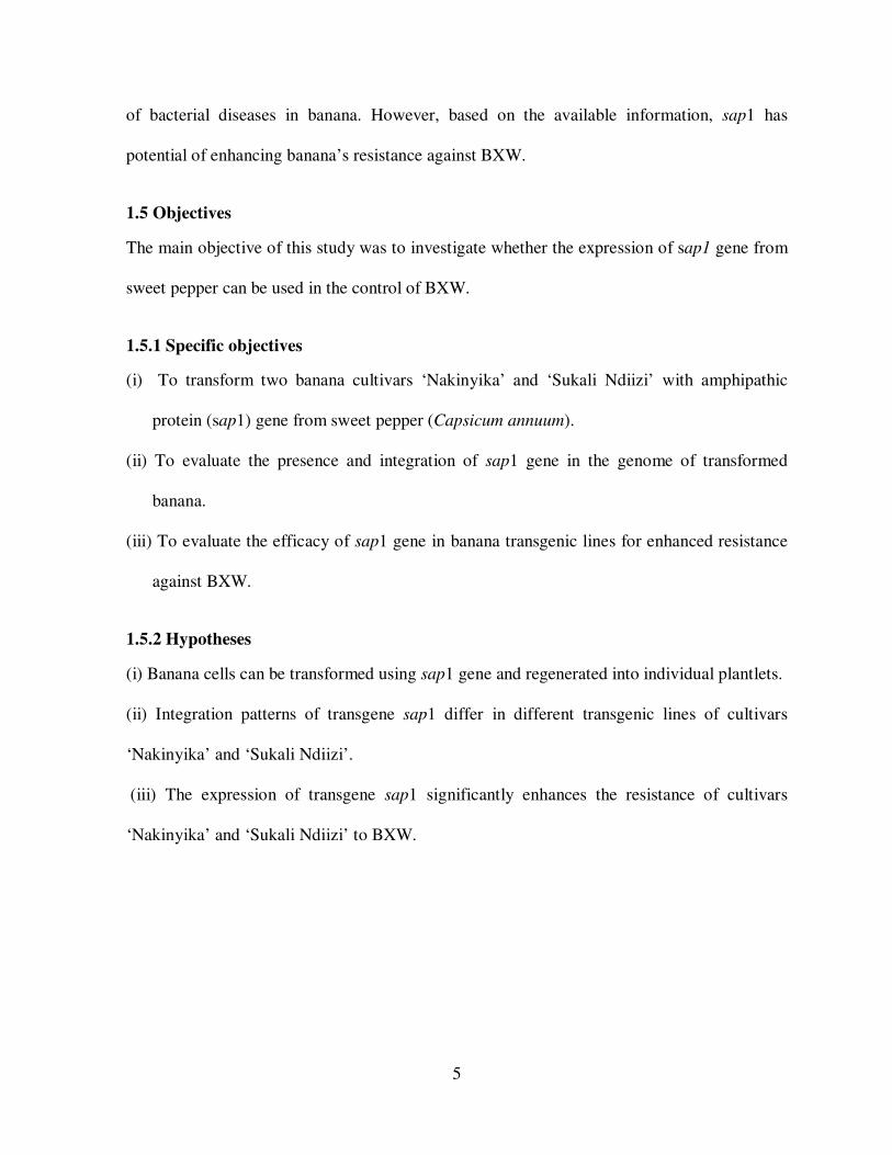
1.5.1 Specific objectives
(i) To transform two banana cultivars ‘Nakinyika’ and ‘Sukali Ndiizi’ with amphipathic
protein (sap1) gene from sweet pepper (Capsicum annuum).
(ii) To evaluate the presence and integration of sap1 gene in the genome of transformed
banana.
(iii) To evaluate the efficacy of sap1 gene in banana transgenic lines for enhanced resistance
against BXW.
1.5.2 Hypotheses
(i) Banana cells can be transformed using sap1 gene and regenerated into individual plantlets.
(ii) Integration patterns of transgene sap1 differ in different transgenic lines of cultivars
‘Nakinyika’ and ‘Sukali Ndiizi’.
(iii) The expression of transgene sap1 significantly enhances the resistance of cultivars
‘Nakinyika’ and ‘Sukali Ndiizi’ to BXW.
6
CHAPTER TWO
LITERATURE REVIEW
2.1 Banana and its Importance in East Africa
Bananas belong to genus Musa, family Musaceae and order Zingiberales. Bananas are
believed to have originated from South East Asia and Indochina (Simmonds, 1962) where the
earliest domestication of bananas is also believed to have happened. From here, they were
introduced to all tropical and subtropical regions of the world thus gaining great importance.
It is suggested that edible bananas originated from two wild seed forming species, Musa
acuminata Colla (2n = 2X= 22) and Musa balbisiana Colla (2n=22) and provide “A” and “B”
genomes of bananas, respectively. Bananas were introduced into East Africa by Arab traders
between India and East Africa, or could have reached East Africa via the west coast of Africa.
Somatic mutations gave rise to the large variability in the East African highland banana
(EAHB) cultivars making East Africa the secondary center of diversity for this group.
Banana has many attributes that makes it an important crop. The progressive conversion of
starch into sugars after harvest makes some banana cultivars to be consumed as fruits (for
example, Cavendish) (Bagamba et al., 2006) while others like plantain are considered to be a
carbohydrate staple. Depending on the juice yield, some fruit type cultivars are used to
produce wine and gin. East African highland bananas are mainly produced as a starch staple
that compete with other crops such as cereals (like maize and millet) and tubers (like sweet
potatoes and cassava) (Bagamba et al., 2006). In addition to providing a reliable source of
food, banana is an important source of income with excess production sold in local markets.
Average per capita annual consumption of bananas in Uganda is the largest in the world,

7
estimated to be about 1kg per person per day. Banana are consumed as fruit, prepared by
cooking, roasting or drying, used for production of banana juice and fermented for production
of alcoholic beverage (beer, wine and gin) (Edmeades et al., 2006). Bananas are also a source
of animal feeds (fresh pseudostems, male buds, banana peelings and by-products of
fermentation), wrapping material for produce in storage, construction materials (thatch and
binding ropes) and handicrafts (mats, baskets, hand bags, necklaces and decorations)
(Karugaba and Kimaru, 1999). It also provides soil surface cover, reduces soil erosion on
steep slopes and a principal source of mulch for maintaining and improving soil fertility.
2.2 Banana Production Constraints
In Africa, bananas and plantains provide more than 25% of food energy requirement
(Robinson, 1996) for more than 100 million people of which 20 million are from East Africa
alone. In terms of revenue, banana is one of the most important cash crops contributing up to
22% of national agricultural revenue (Kalyebala et al., 2007). In Uganda, the East African
highland banana (Musa spp. cv. EA-AAA), popularly known as ‘matooke’, provide staple
food for over 70% (Picq et al., 1998) of the population. Banana production is threatened by
various biotic constraints such as pests (banana weevil and nematodes) and diseases (Ortiz et
al., 2002). Of the diseases, Black Sigatoka, Fusarium wilt and Banana Xanthomonas Wilt
(BXW) cause significant yield losses in farmers fields with yield losses of up to 90% (Aritua
et al., 2007). In 2006, the estimated loss if BXW was not controlled was 295 million USD
worth of banana output valued at farm gate (Kalyebara et al., 2006). This expected loss
translates to around 200 USD of food and income per household. As reported previously
(Tushemereirwe et al., 2006), all banana varieties so far screened are susceptible to BXW.
Once the pathogen (Xanthomonas campestris pv. musacearum) has established, the disease
8
control is very difficult and eradication impossible (Eden-Green, 2004). Management of
BXW in banana is challenging due to lack of resistance in the available banana gene pool and
continuous association of host and inoculum over a long period of time (Tripathi et al., 2009).
Recommended BXW control measures are mainly cultural practices. However, these
measures were reported to reduce 10% of the losses to BXW (Tushemereirwe et al., 2006).
Thus there is a strong need to use non conventional disease control strategies for example
genetic engineering with genes that have potential resistance against BXW.
2.2.1 History of Banana Xanthomonas wilt disease
The Banana Xanthomonas wilt (BXW) disease caused by the bacterium Xanthomonas
campestris pv musacearum (Xcm), has threatened millions of Ugandan farmers due to its
severe effect on banana production (Tushemereirwe et al., 2004). The disease was first
reported in Ethiopia in 1968 on Enset (wild/false banana) and then in banana in 1974 (Yirgou
and Bradbury 1968, 1974). It was later described on bananas in the Keffa, Shoa and Sidamo,
Harerge and Game-Goffa regions of Ethiopia, on cultivar Casse Hybrid (Yirgou and
Bradbury, 1974), with incidence between 70 and 80% (Korobko et al., 1987). In Ethiopia
where BXW disease is considered important on Enset, banana cultivation is less extensive and
therefore its destructive nature on the latter may be difficult to determine. In Uganda it was
first reported in 2001(Tushemereirwe et al., 2003) and has since spread rapidly with
plantation incidence of up to 70% and affecting both EAHB and exotic (dessert/ beer)
bananas.
2.3.1 Etiology of Banana Xanthomonas Wilt disease
Little is known about the etiology of BXW. It is reported that the pathogen enters the host
through wounds on roots, pseudostems and leaves (Yirgou and Bradbury, 1968, 1974;

9
Korobko et al., 1987). It is suspected that the bacterium also enters the plant through the male
buds as reported for the Moko disease (Korobko et al., 1987). According to Yigou and
Bradbury (1974) long distance transmission of the disease is aided through: contaminated
farming tools such as, pangas and pruning knives, which transmit the bacteria through injuries
on roots and aerial parts and movement of infected plant materials (suckers, bunches, leaves).
The major transmitters of the disease are the insects as they move from one plant to another
looking for nectar in flowers (Sekiwoko et al., 2006).
Xanthomonas campestris pv. musacearum (Xcv) attacks the vascular system of both banana
and Ensete ventricosum (Enset) causing wilting and death of the plants. Xcm is motile, Gram-
negative rod shaped, possesses a single polar flagellum and produces typically yellow,
convex, mucoid, slimy colonies on nutrient agar and other media (Yirgou and Bradbury,
1968, 1974; Tripathi et al., 2007). Phylogenetic relationships were evaluated for 20 isolates of
the bacterium collected within a period of about four decades, between 1968 and 2005, from
Ethiopia, Uganda, Democratic Republic of Congo, Tanzania and Rwanda. Sequence analyses
of the internally transcribed spacer (ITS) locus (Aritua et al., 2008) and the gyrase B (gyrB)
gene revealed only limited (<2%) nucleotide divergence among the isolates (Aritua et al.,
2008).
2.3.1.1 Banana Xanthomonas wilt Disease symptoms
Affected banana plants develop symptoms characterized by a progressive yellowing and
wilting of leaves, with fruits ripening prematurely and unevenly with internal brown
discoloration (Fig .1). When stems are cut, a pocket of pale yellow bacterial ooze appears
within 5-15 min (Yirgou and Bradbury, 1974; Tushemereirwe et al., 2004). Yellow or brown

10
streaks occur in the vascular tissues of infected plants. Other symptoms on the floral parts
include wilting of bracts, shriveling and rotting of the male buds, and flower stalks turning
yellow-brown (Yirgou and Bradbury, 1968; Tushemereirwe et al., 2004). Plant death
commonly results from infection.
Figure 1. BXW symptoms; wilting of leaves (A), premature ripening of the bunch (B) and(C)
cross-section of banana pseudostem showing yellow oozing.
2.4 Resistance to BXW
Banana Xanthomonas Wilt (BXW) attacks banana cultivars of all genome types including
East African Highland bananas. The EAHBs are reported to be slightly tolerant than plantain
and dessert cultivars (Karamura et al., 2006). Some of the EAHB cultivars like
‘Mbwazirume’ and ‘Nakitembe’ have been reported to escape the infection due to the
persistence male bracts (Tripathi et al., 2009). Cultivar ‘Bluggoe’ was most infected (74%),
followed by ‘Pisang Awak’ (30%), EA Highland bananas (12%) and lastly ‘Sukali Ndizi’
(3%) (Tushemereirwe et al., 2003). A study done on germplasm screening in Mukono district
A B C

11
on 42 genotypes identified no resistant cultivar among the edible ones except Musa balbisiana
which is a natural wild type (Ssekiwoko et al., 2006).
2.5 Management of Banana Xanthomonas Wilt diseases
Management of diseases in tropical perennial crops such as banana is a challenge due to
continuous association of host and inoculum over a long period of time (Ploetz et al., 2007).
The recommended measures for BXW management involve a mixture of approaches
combining exclusion, eradication, host resistance, and crop protection. Control of BXW and
similar bacterial diseases of banana depends on prevention of disease spread (containment),
reduction of disease impact in affected farms (management), and rehabilitation of previously
affected areas. In Uganda, BXW is mainly controlled by improved cultural practices in well
organized banana production areas (Tushemereirwe et al., 2003). Cultural practices that have
been used so far include the use of clean planting materials, clean tools which are sterilized in
fire or diluted sodium hypochloride, de-budding by breaking the male buds with a forked
stick, cutting and burying of diseased plants, and crop rotation (Tushemereirwe et al., 2004).
These cultural practices have not been used effectively by farmers as they are expensive,
labour intensive and time consuming. As a result, low level of adoption has been observed
which has led to continuous disease development, outbreak and further spread (Ssekiwoko et
al., 2006). Therefore, this calls for use of genetically engineered genotypes whose
development and management are cheap and less intensive.
2.6 Genetic engineering for resistance to bacterial diseases in plants
Genetic transformation approach has been used to control bacterial wilts in many crops
(Huang et al., 2004). The path system-specific plant resistance (R) genes that mediate
resistance to bacterial, fungal, viral and nematode pathogens have been cloned from several
12
plant species (Bent, 1996). Many of these R gene products share structural motifs, which
indicate that disease resistance to diverse pathogens may operate through similar pathways.
For example, the Bs2 resistance gene of pepper specifically recognizes and confers resistance
to strains of X. campestris pv. vesicatoria (Xcv) (Wang et al., 1996) that contain the
corresponding bacterial a virulence gene, avr Bs2 (Tai et al., 1999). Transgenic tomato plants
expressing the pepper Bs2 gene suppress the growth of Xcv. The Xa1 gene in rice confers
resistance to Japanese race 1 of X. oryzae pv. oryzae, the causal pathogen of bacterial blight
(Yoshimura et al., 1998). Transgenic bananas expressing Hrap gene conferred resistance to
BXW (Tripathi et al., 2010). Sap1 is an amphipathic protein isolated from the sweet pepper,
Capsicum annum (Lin et al., 1997). The use of Sap1 has been shown to delay the
hypersensitive response induced by Pseudomonas syringae pv. syringae in non-host plants
through the release of the proteineous elicitor, harpins (Lin et al., 1997). Further analysis also
showed that Sap1 functioned in a dose-dependent manner by competitively inhibiting the
interaction between harpins and its receptor on the plant cells and consequently suppressed
bacterial growth. This transgene has showed enhanced hypersensitive response against
various pathogens in many dicot and monocot crops (Tripathi et al., 2009).
2.7 Plant defense mechanisms
Different steps in plant defense pathways have been evaluated to get a better understanding of
defense responses and potentially produce transgenic plants with improved defense systems.
One step in the defense-signaling cascade that has been evaluated is the NPR1 gene from
Arabidopsis, which regulates salicylic acid signaling (Chern et al., 2001). When NPR1 was
over expressed in Arabidopsis as well as in rice, plants with stronger protein induction and

13
enhanced bacterial and fungal resistance were generated. When bacterial salicylic acid (SA)
generating enzymes were expressed in transgenic tobacco, salicylic acid (SA) accumulation
was substantially increased and proteins were constitutively expressed conferring enhanced
resistance to fungal as well as viral infections (Verberne et al., 2000).
Expression of an amphipathic protein (sap1) which was isolated from sweet pepper, capsicum
annuum has been shown to delay the hypersensitive response induced by Pseudomonas
syringae pv. syringae in non-host plants through the release of the proteineous elicitor harpin
(Lin et al., 1997). Therefore, since previous reports in other studies have shown that Sap1
inhibited the interaction between Xanthomonas and the host plant in other species. It is
expected that sap1 might cause the same interaction between Xanthomonas campestris
pv.musacearum (Xcm) and the host plant so that transgenic banana plants expressing the sap1
gene show resistance to BXW. SAP1 is a protein designated as PFLP (Plant ferredoxin-like
protein) by virtue of its high homology with plant ferredoxin protein containing an N-terminal
signal peptide responsible for chloroplast targeting and a putative 2Fe-2S domain responsible
for ferredox activity.
2.8 Sap1 mediated resistance mechanism
Sap1 is a ferredoxin-like protein that is involved in many redox reactions leading to the
production of Reaction Oxygen Species (ROS) a characteristic of plant defense response. It
intensifies the harpin-mediated HR. Inhibition of the harpin-mediated HR by sap1 was shown
to be dosage-dependent and revealed a competitive pattern. Sap1 may interact with harpin as a
putative receptor so as to prevent the binding between receptor and the active fragment of
harpin. In this way, harpin may retain its ability to activate an HR via the signal transduction

14
system (Lin et al., 1997). The production of reactive oxygen species (ROS) is one of the
earliest events during HR and considered as a characteristic of plant defense responses
(Levine et al., 1994). Moreover, SAP1 protein is a ferredoxin that is involved in many redox
reactions leading to the production of ROS and it is found that sap1 enhances ROS
production, so as a result intensifies the harpin-mediated HR (Dayakar et al., 2003).
2.8.1 Transformation studies with sap1
The sap1 gene enhanced resistance against various pathogens in many plants (Tang et al.,
2001). Transgenic rice expressing sap1 was produced showing enhanced resistance against
one of the most devastating diseases of rice in Africa and Asia, bacterial leaf blight, which is
caused by the Gram-negative bacterium, Xanthomonas oryzae pv. oryzae (Xoo) (Tang et al.,
2001). Another study showed that sap1 confers resistance against soft rot disease which is
caused by Erwinia carotovora in Oncidium (orchid) even when the entire plant was
challenged with the pathogen (Liau et al., 2003; You et al., 2003).
2.9 Genetic transformation of banana
Biotechnological techniques that are required for genetic transformation of banana have been
reported (Sagi et al., 2000). Different embryogenic cell suspensions (ECSs) technologies have
been reported (Cote et al., 1996; Strosse et al., 2006). Genetic transformation using direct
gene transfer methods (Sagi et al., 1995) and Agrobacterium based gene transfer system (May
et al., 1995; Hernández et al., 1999; Ganapathi et al., 2001; Khanna et al., 2004) are used
routinely in different laboratories. Biotechnology, therefore, offers the most feasible and
precise tools to introduce useful genes such as those for pest and disease resistance into
locally available varieties without changing their preferred characteristic.

15
Genetic transformation of banana, which started with the use of micro projectile
bombardment, is now routinely used (Becker et al., 2000; Sagi et al., 1995). Later,
Agrobacterium mediated transformation was reported (May et al., 1995; Sagi et al., 1995;
Hernández et al., 1999; Ganapathi et al., 2001; Khanna et al., 2004). Agrobacterium mediated
transformation system is more efficient than particle bombardment system in bananas. This is
because high transformation frequencies are obtained (Khanna et al., 2004) with higher
frequencies of transgenic lines containing single transgene copy numbers (Tzafira and
Citovsky, 2005). Many banana cultivars of variable genome types have been transformed so
far (Sagi et al., 1995; May et al., 1995; Becker et al., 2000; Ganapathi et al., 2001; Tripathi et
al., 2008; Arinaitwe et al., 2004). Recently, ECSs of highland banana were developed at the
National Agricultural Research Laboratories (NARL) Kawanda. The transformation protocol
of ECSs obtained was also developed, opening several avenues of genetic improvement of
EAHB cultivars. Therefore, this study considers transformation of EAHB cultivars with sap1
gene, so as to increase their resistance to Xanthomonas Wilt.
16
CHAPTER THREE
MATERIALS AND METHODS
3.1 Study 1. Genetic transformation of banana cultivars `Sukali Ndiizi’ and `Nakinyika’
3.1.1 Plant material
The study was carried out at the National Agricultural Research Laboratories (NARL),
Kawanda, Uganda.
Embryogenic cell suspensions (ECS) of the banana cultivars ‘Sukali Ndizi’ (AAB) and
‘Nakinyika’ (AAA-EA) were used in the study. ECS were sub-cultured and maintained as
previously reported Cote et al. (1996).
3.1.2 Agrobacterium strains and binary vectors
Agrobacterium tumefaciens strain EHA105 (Hood et al., 1986), harboring binary vector
pBISAPI, was used in all transformation experiments with pCAMBIA as a positive control.
The construct pBSAP1 was provided by Academia sinica Taiwan through IITA. The T-DNA
of binary vector pBISAPI contains a Sweet pepper amphipathic gene (sap1) gene driven by
Cauliflower Mosaic Virus promoter (CaMV35S) and neomycin phosphotransferase II (nptII)
as a plant cell selectable marker gene (Figure.2). The T-DNA of binary vector
pCAMBIA1305.1, used for control transformation, contains an improved gusA gene version
(GusPlus, www.Cambia.org) isolated from Staphylococcus sp. and hygromycin
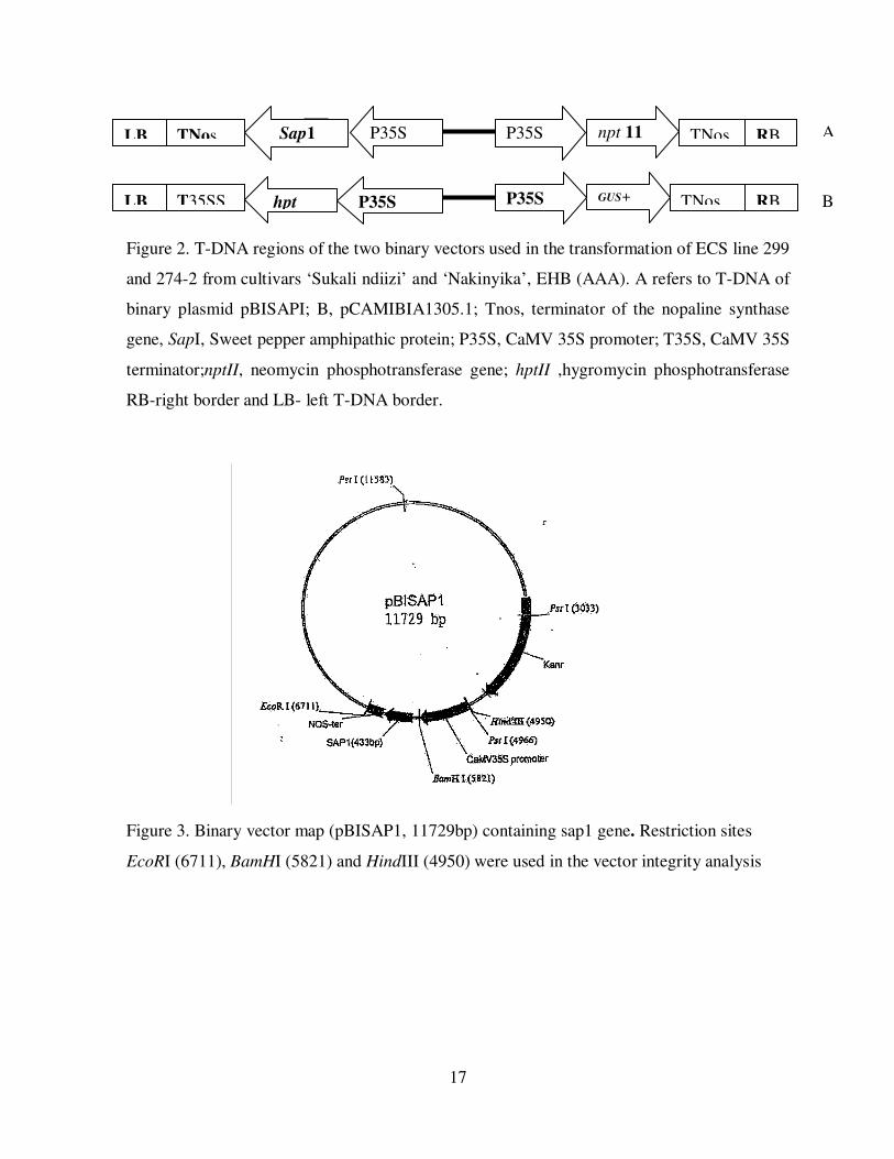
phosphotranferase (hpt) gene for plant cell selection (Figure 2). The size of the Plasmid used
was 11729bp (Figure 3).
17
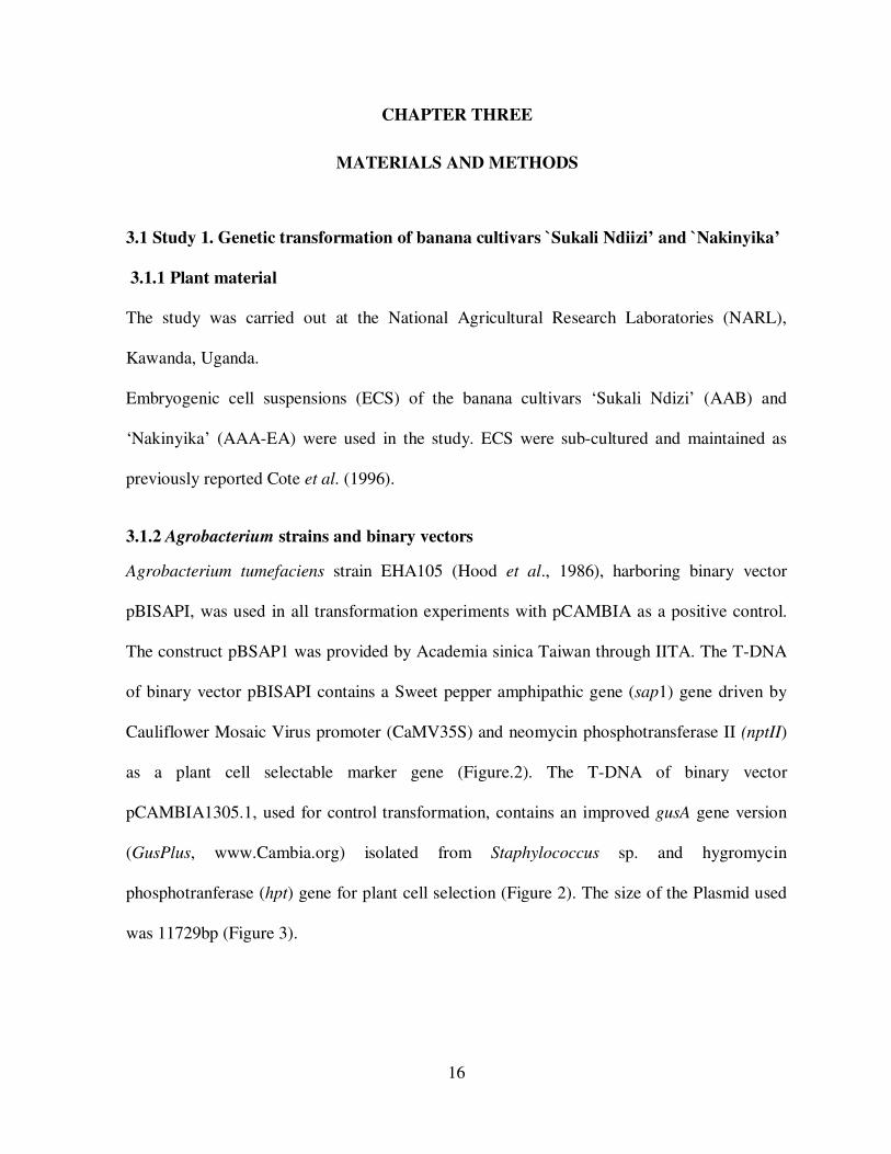
Figure 2. T-DNA regions of the two binary vectors used in the transformation of ECS line 299
and 274-2 from cultivars ‘Sukali ndiizi’ and ‘Nakinyika’, EHB (AAA). A refers to T-DNA of
binary plasmid pBISAPI; B, pCAMIBIA1305.1; Tnos, terminator of the nopaline synthase
gene, SapI, Sweet pepper amphipathic protein; P35S, CaMV 35S promoter; T35S, CaMV 35S
terminator;nptII, neomycin phosphotransferase gene; hptII ,hygromycin phosphotransferase
RB-right border and LB- left T-DNA border.
Figure 3. Binary vector map (pBISAP1, 11729bp) containing sap1 gene. Restriction sites
EcoRI (6711), BamHI (5821) and HindIII (4950) were used in the vector integrity analysis
Sap1 P35S TNos
hpt GUS+ TNos RB P35S P35S T35SS LB
RB npt 11
TNos P35S LB A
B
18
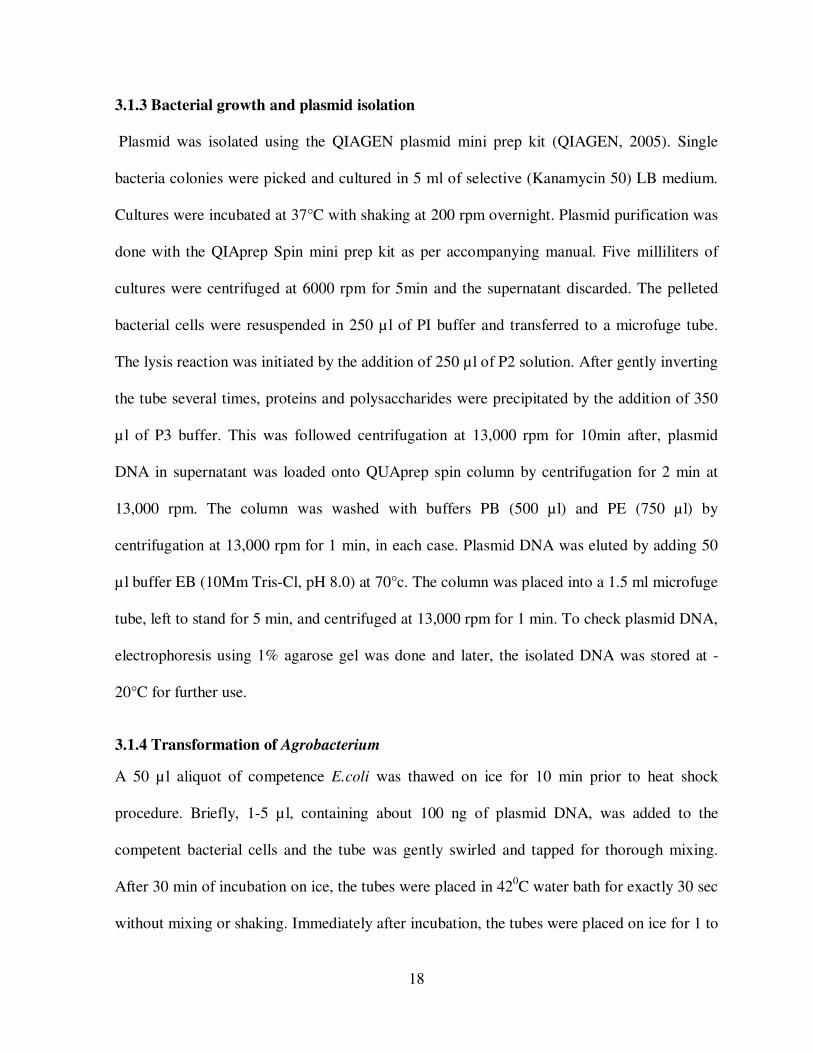
3.1.3 Bacterial growth and plasmid isolation
Plasmid was isolated using the QIAGEN plasmid mini prep kit (QIAGEN, 2005). Single
bacteria colonies were picked and cultured in 5 ml of selective (Kanamycin 50) LB medium.
Cultures were incubated at 37°C with shaking at 200 rpm overnight. Plasmid purification was
done with the QIAprep Spin mini prep kit as per accompanying manual. Five milliliters of
cultures were centrifuged at 6000 rpm for 5min and the supernatant discarded. The pelleted
bacterial cells were resuspended in 250 µl of PI buffer and transferred to a microfuge tube.
The lysis reaction was initiated by the addition of 250 µl of P2 solution. After gently inverting
the tube several times, proteins and polysaccharides were precipitated by the addition of 350
µl of P3 buffer. This was followed centrifugation at 13,000 rpm for 10min after, plasmid
DNA in supernatant was loaded onto QUAprep spin column by centrifugation for 2 min at
13,000 rpm. The column was washed with buffers PB (500 µl) and PE (750 µl) by
centrifugation at 13,000 rpm for 1 min, in each case. Plasmid DNA was eluted by adding 50
µl buffer EB (10Mm Tris-Cl, pH 8.0) at 70°c. The column was placed into a 1.5 ml microfuge
tube, left to stand for 5 min, and centrifuged at 13,000 rpm for 1 min. To check plasmid DNA,
electrophoresis using 1% agarose gel was done and later, the isolated DNA was stored at -
20°C for further use.
3.1.4 Transformation of Agrobacterium
A 50 µl aliquot of competence E.coli was thawed on ice for 10 min prior to heat shock
procedure. Briefly, 1-5 µl, containing about 100 ng of plasmid DNA, was added to the
competent bacterial cells and the tube was gently swirled and tapped for thorough mixing.
After 30 min of incubation on ice, the tubes were placed in 420C water bath for exactly 30 sec
without mixing or shaking. Immediately after incubation, the tubes were placed on ice for 1 to
19
2 min. Then, 250µl of Luria-Bertani (LB) medium (10 g/l tryptone, 5 g/l yeast extract, 10 g/l
NaCl, pH 7.0) were added to the transformation mix and the bacterial cells were incubated for
2 h at 370C with shaking at 210 rpm to allow recovery from the heat shock and start
expression of the selectable marker gene. After 2 h of incubation, 100 µl of the culture was
plated on selective (Rifampicin 50, streptomycin 200 and Kanamycin 50) LB medium pre-
warmed to 370C and incubated overnight at 37
0C. Single colonies were then picked to initiate
cultures for plasmid purification and making glycerol stocks for long-term storage.
For enhanced growth, Agrobacterium tumefaciens strain EHA105 was inoculated on the
nutrient-rich semi-solid LB medium rather than Yeast-Mannitol (YM) (10 g/l mannitol, 0.4
g/l yeast extract, 0.1 g/l K2HPO4, 0.4 g/l KH2PO4, 0.1 g/l NaCl, 0.2 g/l MgSO.7H2O, pH
6.8)YM. Selective LB plates, previously prepared and stored at 4°C in the fridge, were
warmed at 28°C for 2 hrs prior to inoculation. For inoculation, 4 µl of non-transformed and
disarmed Agrobacterium cells (from the glycerol stocks at -80°C) was used as an inoculum.
After inoculation, plates were incubated at 28°C for 3 days. A single colony was picked and
inoculated in 50 ml YM medium (0.4 gL-1 yeast extract, 10 gL-1 mannitol, 0.5 gL-1
K2HPO4, 0.2 gL-1 MgSO4, 0.1gL-1 NaCl, pH 7.0) medium for 3 days at 28°C till OD600
=0.5-0.7. A 50 mL cultures of OD600 =0.6 was spun at 6000 rpm for 5min and the supernatant
was discarded. The pellet was re-suspended in10ml of ice-cold distilled water; centrifuged for
5 min at 6000 rpm/4°C and supernatant discarded. The pellet was re-suspended in 10ml
0.15M CaCl2, cells spun at 500 rpm for 5min and supernatant discarded as above. This was
followed by re-suspending the pellet in 1ml ice-cold 20 mM CaCl2 and was sub-divided into
100µl aliquots. For transformation, an aliquot (100 µl) of cells contained in 1.5ml eppendorf

20
tube was kept on ice for 5min, prior to addition of 10 µl of plasmid DNA (pBISAPI). The
mixture was gently tapped, incubated on ice for 30min, frozen in liquid nitrogen for 1 min and
later flashed in water at 37°C followed by addition of 1ml of warm (28°C) LB medium. The
culture was incubated at 28°C for 4 hrs, with gentle shaking, in order to allow the expression
of selectable marker genes prior to transfer onto selective medium. Finally, aliquots of 200µ l
each were plated on selective semi-solid LB medium (Rifampicin 50, streptomycin 200 and
Kanamycin 50) and incubated at 28°C for 3 days. Single colonies of transformed
Agrobacterium tumefaciens strain EHA105 were picked, cultured in YM in preparation for
transformation of banana cell or preparation of glycerol stocks.
3.1.5 Growth and isolation of Plasmid DNA from EHA 105
A trace of EHA105 containing a plasmid pBISAP1 from the glycerol stock was removed with
a sterile inoculating loop and streaked on semi-solid LB agar supplemented with Rifampicin
(50 ug/ml), Streptomycin (200 µg/ml) and Kanamycin (100 ug/ml). The plates were incubated
for three days at 28oC. A single colony from the putative antibiotic resistant colonies,
appearing on the agar plates, was isolated with the help of a sterile loop and inoculated into
10ml of LB broth containing Kanamycin (50 µg/ml), Rifampicin (50 µg/ml), and
Streptomycin (200 µg/ml) and grown for three days. Plasmid was purified using the QIAGEN
plasmid mini prep kit. Confirmation of plasmid DNA presence was done by gel
electrophoresis using 1% agarose gel.
3.1.6 Restriction digestion
Double restriction digestion of the plasmid DNA using restriction enzymes (BamH1/Hind III
and Hind III /EcoRI) was then done to check the integrity of pBISAPI binary vector
(11729Kb). The restriction reaction (20 µl) `contained 1x NEBB2 (New England BioLabs
21
Buffer2) and 10 Units of each of the restriction enzymes used in double digestion, and 0.8 µg
of plasmid DNA. The reaction mixture was incubated at 37ºC over night. After incubation
the reaction mixture was checked on 1% agarose gel for presence of the expected fragment.
3.1.7 Preparation of bacterial media and Agrobacterium culture for transformation
The method used for plant transformation was the Centrifugation Assisted Agrobacterium-
mediated Transformation system reported by Khanna et al. (2004). Yeast-Mannitol (YM) (10
g/l mannitol, 0.4 g/l yeast extract, 0.1 g/l K2HPO4, 0.4 g/l KH2PO4, 0.1 g/l NaCl, 0.2 g/l
MgSO.7H2O, pH 6.8) and Luria-Bertani (LB) (10 g/l tryptone, 5 g/l yeast extract, 10 g/l NaCl,
pH 7.0), liquid and solid media were used for culturing and selecting A. tumefaciens strains,
EHA105 (pBISAP1) and EHA 105 (pCAMBIA 1305.1). Solid and liquid media was
prepared using de-ionised water autoclaved at 121oC for 15mins. EHA 105 from pBISAPI
glycerol stocks at -80 ºC was streaked on selective YM solid plates with 50 µg/ml kanamycin,
50 µg/ml rifampicin and 200 µg/ml Streptomycin while EHA 105 from (pCAMBIA 1305.1)
glycerol stock at -80 ºC was streaked on selective YM solid plates with 100 µg/ml kanamycin
and grown at 28ºC for 3 days. Single colonies of EHA (pCAMBIA 1305.1) and EHA105
(pBISAP1) bacteria were inoculated in 25 mls of selective liquid YM Rifampicin 50,
streptomycin 200 and Kanamycin 50) and grown on a shaker at 200 rpm and temperature of
28º C for 3days. To increase bacterial cell growth, fresh culture was started by adding 5mls of
Agrobacterium inoculum from the seed culture into 20ml liquid LB medium. The culture was
incubated over night at 28º C and 200 rpm. Prior to transformation, bacterial cells were
pelleted by centrifugation at 5000 rpm for 10 min at room temperature. The bacterial pellet
was re-suspended in the bacteria re-suspension medium (T-MA1) comprised of standard MS
salts and vitamins (Murashige and Skoog, 1962), supplemented with (1 mg/l biotin, 100 mg/l
22
malt extract, 100 mg/l glutamine, 230 mg/l proline, 20 mg/l ascorbic acid, 5 g/l polyvinyl
pyrrolidone 10 (PVP 10), 400 mg/l L-cysteine, 1 mg/l indole acetic acid (IAA), 1 mg/l
naphthalene acetic acid (NAA), 4 mg/l 2, 4- dichlorophenoxyacetic acid (2,4-D), 85.5 g/l
sucrose, pH 5.3) and 300µM Acetosyringone (AS). The bacteria were then activated by gentle
shaking at 25ºC and 70 rpm for 2 hrs until an optical density (OD) of 0.6 (OD600nm) was
reached.
3.1.7.1 Transformation of banana embryogenic cells
Embryogenic cell suspension line ECS-ND 299 of ‘Sukali Ndizi’ and ECS -NAK 274-2 of
‘Nakinyika’ were transformed using T-MA1 containing Acetosyringon as reported by
(Arinaitwe et al., 2008). Banana ECS (0.5 settled cell volume) were re-suspended in 10ml of
pre-warmed liquid MA2 medium consisted of standard MS salts and vitamins (Murashige and
Skoog, 1962), 4.1 µM biotin, 4.5 µM 2, 4-D, 680 µM glutamine, 100 mg/l malt extract, 20
mg/l ascorbic acid, 45 g/l sucrose and the pH of 5.3. Later incubated for 5 min at 45°C (heat
shock) as described by Khanna et al. (2004). The medium was later decanted leaving settled
cells at the bottom of the falcon tubes. The cells were re-suspended in 10ml of Agrobacterium
suspension, adjusted to 0.6 OD600. ECS were allowed to settle and later maintained at 25°C
with shaking at 25 rpm. The infected ECS, drained on 50 µm nylon mesh, were transferred
onto semi-solid T-MA1 medium1 -containing 300 µM AS in 15cm Petri dishes. The ECS
were co- cultivated for 5 days, prior to their transfer onto selective MA3 medium (containing
kanamycin100µg/ml and Timentin 200 µg/ml) in the dark at 22oC. for embryo formation
media (MA3) containing (3.2 g/l SH salts, standard MS vitamins (Murashige and Skoog,
1962), 4.1µM biotin, 100 mg/l malt extract, 680 µM glutamine, 230 mg/l proline, 100 mg/l
myo-inositol, 60 mg/l citric acid, 40 mg/l ascorbic acid, 10 g/l PVP 10, 400 mg/l L-cysteine,

23
1.1 µM NAA, 0.2 µM zeatin, 0.5 µM kinetin, 0.7 µM 2-ip, 45 g/l sucrose, 10 g/l lactose and
2.3 g/l phytagel).
3.1.7.2 Histochemical Gus assay of banana cells
To monitor the transformation conditions and later on transformation efficiency, ECS
transformed with PCAMBIA 1305.1, 0.2 ml SCV, containing gusA gene were assayed for β-
glucurodinase activity (GUS assay). Three samples per cultivar were incubated in a subsrate
solution containing 100 Mm sodium phosphate (pH 7.0), 50 mM ascorbate, and 0.1% Triton
X-100, 0.4 mM potassium ferricyanide, 0.5 mM potassium ferrocyanide and 1mM 5-bromo-
4-chloro-3-indolyl-β-Dglucuronic acid (X-Gluc) (Jefferson, 1987). A sterile sheet of filter
paper was placed at the bottom of each clean and transparent 15-cm diameter Petri dish. A
volume of 1ml of X-Gluc staining solution was added at the centre of each sterile filter paper.
Transformed cells plated on 50 µM nylon mesh, were transferred onto wet filter papers. An
additional 300-700 µl of X-Gluc staining solution was added in each sample, and then Petri
dishes covered, sealed with plastic film, and incubated at 37°C overnight. Blue foci (blue
stained cells or cell clusters) in each sample were observed and counted under a stereo
microscope and their photograph taken using a digital camera.
3.1.7.3 Selection and regeneration of transgenic banana plants
After 5 days of co-cultivation in the dark at 22oC, infected ECS were washed with liquid MA2
medium, supplemented with Timentin at 200 µg/ml, and drained. Infected ECS were then
transferred onto selective semi-solid MA3 media supplemented with Timentin (200 µg/ml) to
kill off Agrobacterium and Kanamycin (100 µg/ml) to kill non transformed banana cells. ECS
were sub- cultured onto selective MA3 medium on 50µM nylon mesh every two weeks until
embryogenic cell clones (EC) were observed. After 3 months of selection in the dark at 26oC,

24
grayish-white masses of cell clusters appeared. These cell clusters were individually
transferred onto selective semi-solid embryo initiating RD1 containing (standard MS salts
and vitamins (Murashige and Skoog, 1962), 20 mg/l ascorbic acid, 100 mg/l myo-inositol, 30
g/l sucrose, pH 5.3 and 2.3 g/l phytagel) supplemented with 300 µg/ml Timentin and 100
µg/ml kanamycin for further clone forming. After one month the clones started forming
shoot-like structure, these were picked and transferred onto selective semisolid MA4,
constituting standard MS salts supplemented with Morel vitamins (Morel and Wetmore,
1951), 0.22 µM 6-benzylaminopurine (6-BAP), 1.14 µM IAA, 30 g sucrose, pH 5.8 and 2.3 g
phytagel, Timentin (200 µg/ml) and Kanamycin (100 µg/ml), for shoot development and
growth. Developing shoots were transferred onto proliferation medium supplemented with 5
mg/l of Benzylamunopurine (BAP). The shoots that developed were multiplied so as to get
enough materials for Molecular analyses and in vitro screening.
3.2. Study 2. Molecular characterization of transgenic lines
3.2.1 DNA extraction
Plant genomic DNA was isolated as describe by Stewart and via, (1993). About 1g of young
leaf tissue from eight putative transformants was homogenized in liquid nitrogen and mixed in
5ml of preheated (65°C) DNA extraction buffer (0.1M Tris-Cl, 20mM Sodium
ethylenediamietetra acetic acid (EDTA), 1.4MNaCl and 20%[w/v] hexadecyltrimethyl
ammonium bromide [CTAB] and 0.2% [v/v] ß-mercaptoethanol [pH 0.8] in sterile 15ml
falcon centrifuge tubes and incubated at 65°C in a water bath for 90min with occasional
gentle swirling. The samples were mixed with chloroform-isoamylalcohol (24:1). The
contents were centrifuged at 6000 rpm for 10min at room temperature, and the aqueous phase
was transferred to fresh sterile centrifuge tubes, 10µl of RNase was added to each tube and
25
incubated at 37ºC for 30 min. DNA precipitation was done by the addition of equal volume of
isopropanol to the supernatant and tubes were inverted for about 20 times to mix. The
contents were left at -20 overnight. The mixture was centrifuged at 6000 rpm for 10min to
pellet the DNA. The pellet was washed with 70% [v/v] ethanol together with 200µl of sodium
acetate and air-dried. The DNA pellet was dissolved in 200µl of Tris-EDTA (TE) buffer (pH
8.0), followed by centrifugation at 13,000 rpm for 15min. Genomic DNA was checked by
running it on 1% agarose gel. The extracted DNA was ready to use for PCR and Southern blot
analyses. DNA was quantified, by using a spectrophotometer, to determine the required
amount of DNA for PCR and Southern blot analyses.
3.2.2 PCR Analysis
To detect the presence of sap1 gene in the plant genome of the randomly selected putatively
transgenic lines, PCR analysis was performed. Plasmid DNA of pB1SAP1 was used as
positive control. PCR analysis was carried out for the randomly selected lines of ‘Nakinyika’
and‘Sukali Ndiizi’ with sap1gene primers (5’ CCCTCAATAATGGCTAGTGTCT-3’), as
forward and (5’ TCAGACTGTGGATAAGCAGCAACAC-3’) as reverse, were used in the
PCR amplification process. The PCR reactions, each 20 µl, contained 2 µl of plant DNA
template and 18 µl of master mix. The master mix consisted of 1x Buffer PCR buffer, 200 µM
dNTPs, 1.5 mM MgCl2, 0.5 µm of each primer, 0.5 U/reaction of Taq DNA polymerase
(Promega, USA). Reactions were started with initial (94oC for 2min) and subjected to 35
cycles as 1 min at 94o C denaturing, 1 min at 55
o C annealing, and 1 min of extension at 72
oC.
The final extension phase was prolonged to 7 min at 72o
C. For controls, the plasmid vector
was used as a positive control and non-transformed plant DNA as negative control. The PCR
26
products were separated by electrophoresis in a 1.0% agarose gel and DNA fragments were
visualized under UV transluminator after ethidium bromide staining.
3.2.3 Southern blot analysis
To determine the integration profiles of these transgenic lines, Southern blot analyses were
performed on selected lines. Restriction digestion of individual DNA samples was carried out
under the suitable conditions depending on restriction enzyme. The choice of enzyme was
based on the vector map and an enzyme that cuts once within the T-DNA was selected (Hind
III). To perform Southern analysis of sap1 gene, 10 µg of total banana genomic DNA were
digested with 6µl of the restriction enzyme overnight at 37° C. Southern hybridization
analysis was done following the standard protocols which include, electrophoresis on 0.8%
agarose gel for 5 hrs at 50 V to separate the digested DNA fragments, transfer of separated
fragments to a positively charged nylon membrane by upward capillary blotting (Southern,
1975). DIG-labeled probe, specific for the transgene was prepared using a PCR DIG probe
synthesis kit (Boehringer Mannheim, Germany). Membranes were probed using a DIG
luminescence detection kit (Boehringer Mannheim) as per manufacturer’s instructions. The
prehybridization, hybridization and high stringency washing conditions of (2x SSC, 0.1%
SDS) were developed using the DIG luminescent detection kit (Boehringer Mannheim,
Germany). Detection of the hybridized fragments on the nylon membrane was done with CPD
according to the manufacturer’s instructions.
3.3. Study 3: Evaluation of transgenic lines for resistance to BXW
3.3.1 In vitro screening
Thirty transgenic lines containing sap1gene were randomly selected. These lines were
artificially inoculated with Xanthomonas campestris pv. musacearum (Xcm) as previously

27
reported (Tripathi et al., 2008). Prior to screening, preparation of bacterial suspension was
performed as described below.
3.3.2 Preparation of bacterial suspensions
A single colony of Xanthomonas campestris pv. musacearum bacterial isolate was inoculated
into 25 ml of YTS medium and cultured at 28°C with shaking at 150 rpm for 48 hrs. The
bacterial culture was centrifuged at 5000 rpm for 5 min and the pellet was re-suspended in
sterile double distilled water. The optical density (OD600) of the bacterial suspension was
checked and bacterial concentration was adjusted to 108 cfu / ml with sterile water. Fresh
inoculum was used for all the experiments in order to increase virulence of the pathogen.
3.3.3 Inoculation of in- vitro plantlets
Thirty (30) transgenic lines of banana cultivars ‘Nakinyika’ and ‘Sukali Ndiizi’ (fifteen lines
each cultivar) were randomly selected and tested with artificial inoculation. The inoculum
was injected into the pseudostem of in vitro plantlets as described by (Tripathi et al., 2008).
Two shoots per line were inoculated with the fresh culture of Xanthomonas campestris pv.
musacearum bacteria. For comparison purposes, two non-transformed shoots were inoculated
with water and the other two with the Xanthomonas campestris pv. musacearum bacteria. The
plants were inoculated by drawing 100 µl of bacterial suspension into a micro syringe and
later injected into the pseudostem of the plantlets, using a hypodermic needle. The inoculated
plantlets were put into the baby jars fitted with lid and incubated at room temperature for
eight weeks.
3.3.4 Disease assessment due to BXW
Plantlets incubated under invitro conditions were monitored and assessed everyday for eight
weeks for disease symptoms. The disease symptoms included chlorosis, necrosis and finally
28
complete wilting and death of plants. Wilt incidence was measured as number of wilted plants
over total number of plants inoculated. Observations were made regularly and the data was
recorded on each plantlet. The relative resistance of transgenic lines to BXW was evaluated
eight weeks after inoculation. Plantlets were categorized based on disease severity as:
resistant if a plant did not show any disease symptoms; moderately resistant, if a plant showed
delayed symptoms; and susceptible, if a plant got symptoms within 12-20 days after
inoculation similar to control non-transgenic plants.
3.3.5 Data collection
Data on regenerated shoots was collected by counting shoots per medium type and the data
was presented in form of pictures and graphs. Data on disease incubation and complete
wilting was collected basing on chlorosis and necrosis of leaves and complete wilting of
plants, and entered into a table. Average incubation and average wilting was computed
basing on number of days for appearing of the symptom and death of the plant. The means
presented were for two replicates. Analysis of variance (ANOVA) was conducted using SAS,
and interaction between means was by least significant difference (LSD) at P= 0.05 (SAS,
2003).
29
CHAPTER FOUR
RESULTS
4.1 Transformation and regeneration
4.1.1 Restriction Analysis of Plasmid DNA
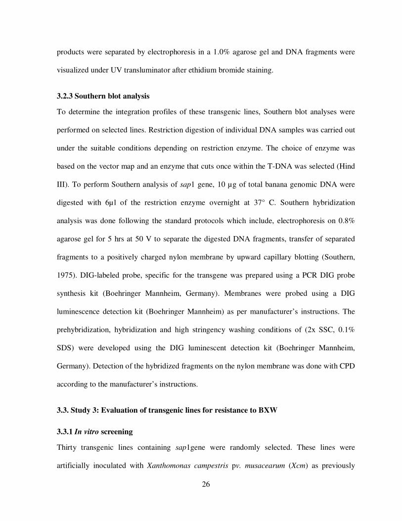
Double restriction digest to confirm presence of the binary vector pBISAP1, using restriction
enzyme combinations, gave variable fragment sizes (Fig 4). These were; 10,858bp and 871bp
for combinations HindIII/BamHI; 9968bp and 1761bp, HindIII/EcoRI; and 890bp and
10839bp fragments for combination BamHI/EcoRI.
M 1 2 3 4
Figure 4. Restriction digests of binary vector pBISAP1 on a 1% agarose gel, M- DNA-
Marker; 1,undigested DNA; and lanes 2-4 represent results of restriction analyses of
pBISAP1 digested with HindIII/BamHI, HindIII/EcoRI, and BamHI/EcoRI respectively.
4.1.2 Transformation efficiency
Six days after infection, randomly selected samples of, embryogenic cell lines ECS-274 NAK
and ECS-299 ND, lines transformed with PCAMBIA 1305.1 were assayed histochemically
10,000bp
4,000bp
2,000bp
1000bp
500bp

30
for gusA expression. For each sample, approximately 80% of the ECSs turned blue after
incubation. Corresponding ECS in the non transformed samples remained creamish (Fig 5).
Results indicated successful transformation and high transfer efficiency.
Figure 5. Schematic presentation of histochemical GUS assays, A) Blue foci represent the
transformed ECSs of cultivar ‘Nakinyika’ following Agrobacterium-mediated transformation.
B) Stained non transformed banana cell.
Table 1. Transformation efficiency of ‘Nakinyika’ and ‘Sukali ndiizi’ after Gus assay
Cell line code Total amount of
cells used
No. of blue focil Transformation efficiency
ND 299 24,000 18960 79%
NAK 274-2 24000 19200 80%
B A

31
4.1.3 Selection and regeneration of Putatively transformed banana lines
After 3 weeks of culturing on MA3, infected cells turned brown (Fig 6A). Three months later,
whitish-grey masses of cell clusters (Cell clones) appeared and continued growing. The cell
clusters continued developing and formed embryo-like structures and the remaining cells
turned black and died. The non- transformed control cells that had been transferred to non
selective medium (MA3 without selection) developed embryos and later into plantlets. (Fig
6B).
After three months on MA3, individual putatively transformed cell clones that survived were
picked and transferred onto selective medium RD1 supplemented with appropriate antibiotics.
For cultivar ‘Nakinyika’ 600 cell clones were singly transferred onto fresh selective medium
whereas a total of 400 cell clones from cultivar ‘Sukali Ndiizi’ were transferred. Of the initial
total number of cell clones, of both cultivars, only 460 and 320 cell clones of ‘Nakinyika’ and
‘Sukali Ndiizi’ respectively, survived and were transferred onto selective embryo initiation
Figure 6. Transformed (A) and non transformed (B) embryogenic cells of banana on
selective and non selective medium respectively. The pictures were taken three months
of culturing.
B A

32
medium (RD1 fig.7). The rest of the cell clones turned brown blackened and finally died (Fig
8A). On selective embryo initiation medium RD1, 260 shoot-like structures of cultivar
‘Nakinyika’ and 160 of cultivar ‘Sukali Ndiizi’ survived. These surviving individual shoot-
like structures were transferred onto shoot growth and development medium (MA4) from
which 135 and 83 shoots of ‘Nakinyika’ and ‘Sukali Ndiizi,’ respectively, developed. (Figure
8 B). Although more shoot-like structures were observed on RD1, some never developed into
shoots (Figure 8 B). Some started germinating after two weeks and others took longer.
Figure 7. Number of embryogenic cell clones (ECs), in cultivar ‘Nakinyika’ and
‘Sukali Ndiizi’ at different stages of selection and shoot regeneration.
0
100
200
300
400
500
600
700
MA3 R D1 MA4
medium type
Nakinyika
S ukalindiiz i
Num
ber
of
Clo
nes
A B
Figure 8. Embryo initiation (A) and shoot development (B) of transformed ECS of
cultivar ‘Nakinyika’ and ‘Sukali ndizi’ after 2-3 months of selection on kanamycin
(50µg/ml) supplemented MA3 and MA4 media, respectively.

33
Regeneration of putatively transgenic lines was done using (Murashige and Skoog, 1962),
medium. The numbers of shoots that were regenerated from shoot-like structures, for both
cultivars, are shown in (Fig 7). For the cell lines used (ECS-274 NAK and ECS-299 ND),
transformed cell clones of cultivar ‘Nakinyika’ had the highest regenerability (62%)
compared to 38% in cultivar ‘Sukali Ndiizi’ (Figs 9).
4 .2 Mocular Characterization of transgenic lines
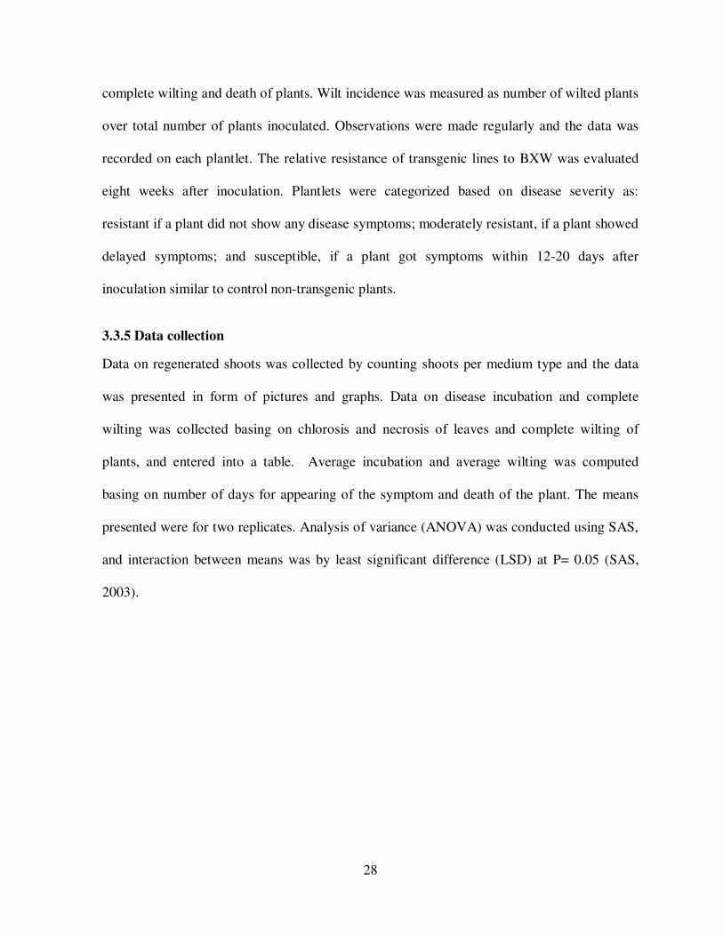
4.2.1 PCR Analysis
Molecular characterization of putatively transgenic lines was done using PCR and Southern
blot analyses. PCR analysis, using gene specific primers, gave a 420 bp and 600-bp fragments
for coding regions of sap1 and nptII genes respectively. Twenty nine (29) putatively
transformed plants that were randomly selected and tested, 26lines were PCR positive (Figure
10 and 11). These lines included ND 39, ND 49, and ND 44 for ‘Sukali Ndiizi’ and for
‘Nakinyika’ lines were NAK 9, NAK 14, and NAK 12. However, three lines did not show any
B A
Figure 9. Shoot regeneration in cultivars ‘Nakinyika’ (A) and ‘Sukali Ndiizi’ (B) following
the 2-3 months of selection process.
B A
34
amplification. This could be due to the presence of contaminants within the DNA samples or
these could have been escapes.
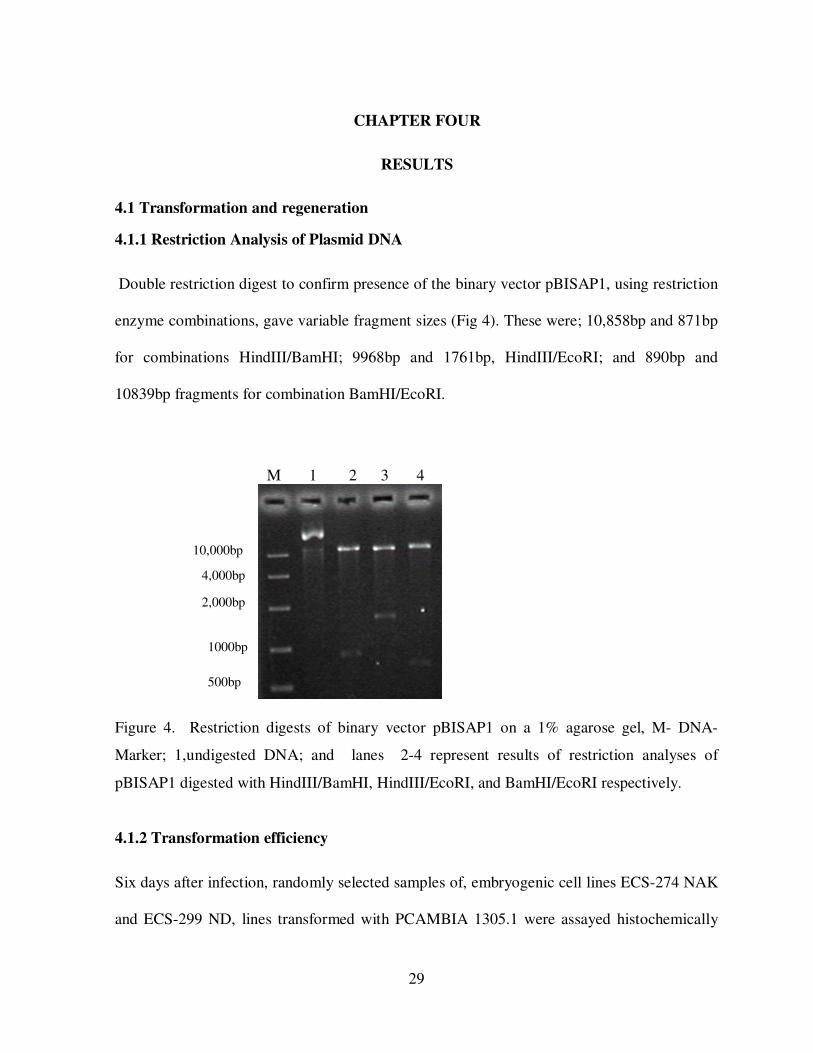
4.2.2 Southern blot analysis
Southern blot analysis was done to confirm the integration patterns of sap1 in cultivars
‘Nakinyika’ and ‘Sukali Ndiizi’. Six randomly selected transgenic lines of both cultivars were
analyzed using sap1 specific probes. Different integration patterns were observed in different
600bp
Figure 10. PCR products for sap1 primers; M- DNA marker, Lines 9, 14, 39, 49, 12 and 44 are
transgenic samples, N-control (non-transformed), W-water p-positive control (plasmid). The
arrows indicate the expected PCR products of sap1.
420bp
M 9 14 39 49 12 44 N W P
M 9 14 39 49 12 44 N W P
Figure 11. PCR products for nptII primers; M- DNA marker, Lines 9, 14, 39, 49, 12 and 44 are
transgenic samples, N-control (non-transformed), W-water P-positive control (plasmid). The
arrows indicate the expected PCR products of ntpII.
npt11
sap1

35
lines, indicating that these lines were stably transformed. Transgenic lines NK9, NK14, ND39
and ND49, had at least 1- 4 integration loci (Figure 12). Non-transformed line (NT) did not
show any signal, indication that the observed signals were not from the endogenous banana
sap1 gene or genes (Figure 12).
Figure 12. Southern blot analysis using genomic DNA of transgenic banana plants digested
with Hind III. Plant genomic DNA pBISAP1 DNA was digested with HindIII. MIII:
molecular weight marker; P: pBISAP1 (positive control); Lines NK14, ND39, ND49, NK12,
ND44 and NK9 are transgenic plants; NT Non-transgenic plants
21kb
2kb
4.9kb
MIII P NK14 ND39 ND49 NK12 ND44 NK9 NT

36
Table 2. PCR analysis and integration patterns of randomly selected transgenic lines of
‘Sukali Ndiizi’ and ‘Nakinyika’containing sap1and npt11 genes
Cultivar and line no. PCR
analysis
Southern blot
analysis
No. of
integration loci
Sap1 Npt11 Sap1
Untransformed control
NAK 9
NAK 14
NAK 12
ND 39
ND 49
ND44
pB1SAP1
-
+
+
-
+
+
-
+
-
+
+
-
+
+
-
+
-
+
+
-
+
+
-
+
-
1
1
0
3
4
0
1
- and + denote the absence and presence of PCR (or Southern blot) signal respectively
4.3 Efficacy of sap1 gene
After in vitro screening, plantlets were categorized into three classes. These categories were
(1) plantlets with no symptoms (resistant), (2) plantlets with delayed (tolerant/moderately)
symptoms (after 37-43 days) and (3) plantlets that quickly developed (susceptible) BXW
symptoms (after 12-20 days) (Fig 13). The variable responses to BXW were observed in
different transgenic lines. On average, non transgenic plantlets started showing leaf wilting
symptoms 16 days after inoculation. Transgenic lines of ‘Sukali Ndiizi’ showed symptoms
within 12-14 days after inoculation. In cultivar ‘Nakinyika’ symptoms became visible within
16-17 days after inoculation. Three out of fifteen transgenic plants of cultivar ‘Nakinyika’ did
not show BXW symptoms (resistant) whereas for ‘Sukali Ndiizi’, Two out of fifteen lines
were symptom-less. For both cultivars, a total of 7 lines appeared to be tolerant with delayed

37
symptoms. Other lines which were inoculated quickly got the symptoms and finally wilted.
Results of in vitro BXW screening are presented in Table 3.
Figure 13. In vitro screening of transgenic lines for resistance against BXW after artificial
inoculation with Xanthomonas campestris pv. musacearum (Xcm). A represents inoculated
untransformed plantlet; B to D show inoculated transgenic plantlets. These pictures were
taken 8 weeks after inoculation.
C D
B A
38
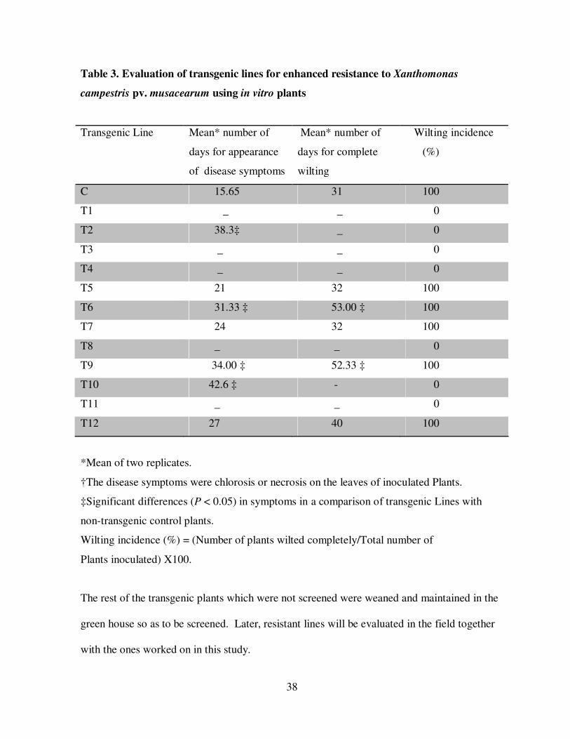
Table 3. Evaluation of transgenic lines for enhanced resistance to Xanthomonas
campestris pv. musacearum using in vitro plants
Transgenic Line
Mean* number of
days for appearance
of disease symptoms
Mean* number of
days for complete
wilting
Wilting incidence
(%)
C 15.65 31 100
T1 _ _ 0
T2 38.3‡ _ 0
T3 _ _ 0
T4 _ _ 0
T5 21 32 100
T6 31.33 ‡ 53.00 ‡ 100
T7 24 32 100
T8 _ _ 0
T9 34.00 ‡ 52.33 ‡ 100
T10 42.6 ‡ - 0
T11 _ _ 0
T12 27 40 100
*Mean of two replicates.
†The disease symptoms were chlorosis or necrosis on the leaves of inoculated Plants.
‡Significant differences (P < 0.05) in symptoms in a comparison of transgenic Lines with
non-transgenic control plants.
Wilting incidence (%) = (Number of plants wilted completely/Total number of
Plants inoculated) X100.
The rest of the transgenic plants which were not screened were weaned and maintained in the
green house so as to be screened. Later, resistant lines will be evaluated in the field together
with the ones worked on in this study.

39
CHAPTER FIVE
DISCUSSION
5.1 Transformation and regeneration
5.1.1 Transformation efficiency
Transient transformation frequency, using gusA or gfp reporter gene, is indicative of gene
transfer efficiency (Arinaitwe et al., 2004). For example, high histochemical GUS expression
is indicative of high gene transfer efficiency. In this study, over 80% of the cells transformed
with gusA gene turned blue after the histochemical GUS assay; indicating over 80% gene
transfer efficiency (Figure 5). High gene transfer efficiency in banana cells transformed using
Agrobacterium-mediated transformation system was previously reported by (Mohanty et al.,
1999), and is associated with high ECS line competency (Yip et al., 2006), actively dividing
cell-status, and cells on auxin rich medium (Sagi et al., 2000). The results obtained confirm
that the two ECS lines used were highly competent to Agrobacterium infection with high gene
transfer efficiency. Since transformation with the sap1 gene and gusA reporter gene were
done under the same conditions, gene transfer efficiency with sap1 gene was equally high.
Higher gene transfer efficiency in banana cells has been reported by several authors
(Ganapathi et al., 2001; Khanna et al., 2004). High transformation frequency was also
confirmed in numerous embryogenic cell clones (stable transformation stage) that survived
after the selection process (Fig. 6) and is influenced by the plant genotype, explants type,
inoculation and co-culture conditions, transformation competency (Cheng et al., 2004) and
inoculation or co-cultivation length (Amoah et al., 2000).
40
5.1.2 Selection of transformed lines
After Agrobacterium co-cultivation, the banana cells were transferred onto MA3 medium
supplemented with 100mg/l Kanamycin. A month after infection, masses of cells turned
brown and finally died. Browning is reported to be due to necrotic effects of the selective
agent (Sagi, 2000). Although many ECSs are transformed by Agrobacterium, not all cells
develop into embryos. At each phase of selection the number of surviving cell clones
decreased. For example from selective MA3 to RD1 the number of surviving cell clones
decreased from 600 to 400 in cultivar ‘Nakinyika’ as compared to 400-320 in ‘Sukali Ndiizi’.
This observation implies that a proportion of these cell clones were not transformed. While
monitoring gfp gene expression in banana cells from transformation till shoot regeneration,
Arinaitwe et al. (2004), observed similar trends where the cell clones expressing gfp gene
decreased with time. The explanation given was that some clones were escapes and others
were not stably transformed (transiently transformed). Thus cell clones where the gene was
successfully integrated into the genome survived and subsequently developed into shoots.
Selection at RD1 too provides enhanced selection pressure since single cell clones are
transferred onto freshly prepared selective medium.
Development of cell clones depends mainly on the embryogenicity of a particular ECS line.
ECS lines that readily for embryos generate numerous embryos during selection and thus
enhances the number of surviving cell clones. Variable embryogenicity and survival rates
among transformed banana cells were previously observed in different banana cultivars
(Arinaitwe et al., 2004). Variation of embryogenicity, within the ECS lines may be due to
variable GA/ABA rations among individual cells. These ratios determine whether tissue

41
develops as embryonic or postembryonic (Braybrook et al., 2006; Zheng et al., 2009).
Although not all cell clones subsequently develop into shoots, high embryogenicity and
survival rates increase the number of shoots regenerated after the transformation cycle.
According to the segregation analysis, genetic control of regeneration by two-dominant genes
has been proposed for both the re-generation percentage or efficiency (Reisch and Bing-ham
1980; Wan et al., 1988) and the number of embryos (Hernandez-Fernandez and Christie
1989). However, the ability to form embryos (embryogenicity) seems to be independently
controlled (Zheng et al., 2009).
5.1.3 Shoot regeneration
Shoot initiation and development from embryogenic cell clones remain a challenging step in
plant genetic engineering. Although hundreds of cell clones are recovered after the selection
process, a small proportion of up to 70% develop into shoots (Sagi et al., 1995). In the current
study, shoot regeneration frequencies of 62% and 38% were observed in cultivars ‘Nakinyika’
and ‘Sukali Ndiizi’ respectively. Shoot regenerability in plant cell cultures is reported to be
influenced by embryogenicity and the duration in culture (Arinaitwe et al., 2008). Thus
freshly initiated ECS lines are highly regenerable than ECS lines that have been in culture for
over a year. The number of shoots regenerated per cultivar is known to be influenced by the
number of clones picked from embryo germination medium onto shoot germination medium,
success of the transformation event and effective control of Agrobacterium by using an
appropriate antibiotic at lower concentrations, during selection (Yip et al., 2006; Liau et al.,
2003). The results of this study were similar to previous studies where shoot basal discs were
used and exposed to same selection. In that study, a total of 28 out of 260 (10.8%) were
regenerated (Yip et al., 2006).

42
5.2 Molecular characterization of transgenic lines
5.2.1 PCR analysis
Using gene specific primers (sap1-specific primers) and npt11, PCR analyses were
performed to confirm its presence in the banana genome. Out of 29 the lines randomly
selected and tested, 26 lines showed the presence of sap1 and npt11gene; a frequency of 89%.
Three lines didn’t amplify for both npt11 and sap1. Probably the DNA contained Taq
polymerase inhibitors or this could be due to the presence of contaminants within the DNA
samples or these could have been escapes. No amplification was identified in the non
transformed plants for both transgenes, hence the primers were gene specific.
5.2.2 Southern blot analysis
The results show that sap1 gene was integrated into the banana genome with lines 9, 14, 39
and 49 having at least 1- 4 integration sites, thereby confirming their transgenic nature. The
pattern of bands differed in these transgenic lines, indicating that these lines represent
different transgenic events. The number of integrations varied between one and four. Similar
results were reported in several other monocots including rice, wheat and barley by Dai et al.
(2001). Line ND 49 gave the highest number of integrations followed by ND 39 whereas
Lines NK 14 and ND 9 both had one integration locus of sap1 gene. Integration patterns of 1
to 4 have been observed in different banana genome types transformed using Agrobacterium-
mediated transformation procedure (Arinaitwe et al., 2008). In this study, the integration
pattern observed was simpler than what is observed (over 7 integrations) in transformants
generated through particle-mediated transformation system (Wakita et al., 1998). Although
previous studies reported the ferredoxin-like gene as ubiquitous in the plant kingdom (Yip et
43
al., 2006), it was not detected in non transformed banana genomic DNA. Southern blot
analysis of the putative positives confirmed stable integration of the sap1gene into the banana
genome. The transgenic lines showed 1 to 4 integration patterns which are characteristic of
Agrobacterium- mediated transformation. The integration patterns were also generally
random, implying different transformation events.
5.3 Efficacy of sap1 gene
The efficacy of sap1 gene was tested using in vitro assay of the transgenic lines of the banana
cultivars ‘Nakinyika’ and ‘Sukali Ndiizi’ for resistance against BXW. Two weeks after
inoculation, leaves of transgenic plants exhibited HR-like symptoms. These results were
similar to (Yip et al., 2006) who observed the same. In contrast, randomly selected lines of
the two cultivars that were inoculated with 100µl of the bacterial suspension Xanthomonas
campestris pv. musacearum produced symptoms of chlorosis and necrosis within 12-14 days
for ‘Sukali Ndiizi’ and 16-17 days for ‘Nakinyika’ after inoculation. Susceptible lines wilted
and died 30-40 days and 32-47 days after inoculation for cultivars ‘Sukali Ndiizi’ and
‘Nakinyika,’ respectively. Control plants, inoculated with sterile water remained healthy. This
was similar to what was observed by (Tripathi et al., 2008; Tripthi et al., 2010). Previous
studies report that sap1 transgene activity against Erwinina carotovora (EC) and
Pseudomonas syringae pv tabaci in tobacco plants have shown hypersensitive response (HR)
and suppressed bacterial growth during compatible pathogen attack (Huang et al., 2004). In
this study, inoculated transgenic lines demonstrated resistance to Xcm at variable levels (Yip
et al., 2006; Tang et al; 2001). The strongest resistance was observed in two lines of ‘Sukali
Ndiizi’, and three of ‘Nakinyika’. Basing on the previous reports (Tang et al., 2001; Liau et
44
al., 2003; Tripathi et al., 2008), and the results of this study, sap1 gene provides variable
protection to plants against wilt-causing pathogens.
Similar resistance trends were previously reported in transgenic lines of rice containing sap1
gene (Tang et al., 2001). In that study, only lines 2, 6, 8, and 9 showed strong resistance
whereas other lines showed variable resistance levels. Lines of Cana lily and rice containing
sap1 gene exhibited HR-like necrosis as observed by Yip et al., (2006), and Tang et al.,
(2001) respectively. Results obtained in this study and previous reports in rice (Tang et al.,
2001), and Cana lily (Yip et al., 2006) indicate that sap1 gene provides variable resistance,
thus evaluation of substantial number of transgenic lines is desirable.
Though some transformed lines did not show resistance, this does not mean that they didn’t
carry the gene. The possible reasons for this could be attributed to the way the gene was
positioned in the plant. In that, if the gene is positioned in the non coding region it will not be
transcribed so in this way, the gene will not be active. Unlike on the other hand if it’s in a
coding region, then it will be transcribed and therefore active and so resistance will be
expressed. The other reason could be based on the integration site in that the lesser the
number of integration sites, the more stable it becomes and vice versa.

45
CHAPTER SIX
CONCLUSIONS AND RECOMMENDATIONS
Banana Xanthomonas wilt, caused by the gram-negative bacterium, is a devastating disease of
banana causing yield losses of up to 100% in Uganda (Tushemereirwe et al., 2003). For many
years, researchers have used conventional breeding to improve banana yields, especially by
looking for traits which confer resistance to banana pests (Nematodes and weevils) and
diseases like, Black Sigatoka, and Fusarium wilt. No edible banana cultivar is resistant to
BXW (Tripathi et al., 2008) hence resistance is needed from external sources. Wide crossing
with distant banana cultivars, for example Calcutta 4, generates hybrids with inferior cooking
characteristics.
The results from this study showed successful transformation of ECSs from cultivars ‘Sukali
Ndizi’ and ‘Nakinyika’ with sap1 gene from sweet pepper. Invitro BXW-screening results
show enhanced protection from BXW. Transient gusA expression confirmed ECS lines (ECS-
299 ND) and (ECS-274 NAK) from cultivars ‘Sukali Ndizi’ and ‘Nakinyika’ respectively,
were competent to Agrobacterium infection. Shoot development from transformed
Embryogenic Cell Clones (ECs) confirmed the regenerability of both ECS lines used.
In this study, 83 and 135 transgenic lines of banana cultivars ‘Nakinyika’ and ‘Sukali Ndiizi’
were generated.
Molecular characterization of selected transgenic lines confirmed both the presence and stable
integrations of transgene sap1 in the banana genome. PCR analyses showed the presence of
sap1 gene in the genome of the regenerated banana lines. Integration sites of two to four were
46
observed, after Southern blot analyses with gene specific probes. Sap1 gene from sweet
pepper can protect banana plants from BXW.
Invitro evaluation of randomly selected regenerated banana lines, containing sap1 gene from
sweet pepper showed enhanced resistance against BXW. Thus sap1 gene has a potential to
protect banana plants against Banana Xanthomonas wilt. However, it is important to evaluate
these lines under natural Xcm infection in the field since the development of transgenic lines
resistance against BXW is highly desirable.
Furthermore, a few lines have been characterized, so it is important to do molecular
characterization of all the lines prior to their evaluation in the field. Combination of Sap1
gene with other genes that have different modes of action would provide strong and durable
resistance against Banana Xanthomonas Wilt (BXW).

47
REFERENCES
Amoah, B.K., Wu, H., Sparks, C and Jones, H.D. 2001. Factors influencing Agrobacterium-
mediated transient expression of uidA in wheat inflorescence tissue. Journal of
Experimental Botany, 52 (358): 1135-1142.
Arinaitwe, G., Remy, S., Strosse, H., Swennen, R and Sagi, L. 2004. Agrobacterium and
particle bombardment-mediated transformation of a wide range of banana cultivars. In:
Cellular Molecular Biology, and Induced mutations. Science Publishers Inc., Enfield,
HN, USA, pp351-357. http://.www.Scipub.net/agriculture/anana-Improvement-Induced-
mutations.html.
Arinaitwe, G. 2008. Improved Agrobacterium- mediated Transformation Methods for Banana
and Plantain. PhD Thesis Katholic University of Luven Belgium, pp158.
Aritua, V., Parkison, N., Thwaites, R., Tushemereirwe, W and Smith, J. 2007. Molecular
epidemiology of Xanthomonas campestris pv. musacearum, the causal agent of
Xanthomonas wilt of banana and enset. In: program and Abstracts of ISHS/ProMusa
symposium: Recent Advances in Banana Crop Production for Sustainable Production and
Improved Livelihoods. Green way Woods Resort, White River, South Africa, September
10-14, 2007, pp 32.
Aritua, V., Parkison, N., Thwaites, R., Heehey, J.V., Jones, D. J., Tushemereirwe, W.,
Crozier, J., Reeder, R., Stead, D. E and Smith, J. 2008. Characterization of Xanthomonas
sp. Causing wilt of enset and banana and its proposed reclassification as a strain of X.
vasicola. Plant pathology, 57: 170-177.
Bagamba, F., Kikulwe, E., Tushemereirwe, W. K., Ngambeki, D., Muhangi, J., Kagezi, G.,
Ragama, P. E and Eden- Green, S. 2006. Farmers awareness of Banana Bacterial wilt
and its Control in Uganda. African Crop Science Journal, 14: 157- 165.
Becker, D.K., Dugdale, B., Smith, M.K., Harding, R.M and Dale, J.L. 2000. Genetic
transformation of Cavendish banana (Musa spp. AAAgroup) cv 'Grand Nain' via micro
projectile bombardment. Plant Cell Reports, 19:229-234.
Bent, A. F. 1996. Plant disease resistance genes: Function meets structure. Plant Cell,8: 1757-
1771
Braybrook, S.A., Stone, S.L., Park, S., Bui, A.Q., Le, B.H., Fisher, R.L., Goldberh, R.B and
Harada, J.J. 2006. Genes directly regulated by leaf cotyledon provide insight into the

48
control of embryo maturation and somatic embryogenesis. Natural Academia Science
USA, 103 : 3468-3473.
Carter, B.A., Reeder, R., Mgenzi, S.R., Kinyua, Z.M., Mbaka, J.N., Doyle, K., Mwangi, M.,
Valentine, G., Aritua, V., Ivey, M.L., Miller, S.A and Smith, J. 2009. Confirmation of
Xanthomonas vasicola causative organism of Banana Xanthomonas wilt in Tanzania,
Burundi and Kenya. Plant Pathology, 57: 170-177.
Chern, M.S., Fitzgerald, H.A., Yadav, R.C., Canvas, P.E., Dong, X and Ronald, P.C. 2001.
Evidence for a disease-resistance pathway in rice Similar to the NPR1-mediated signaling
pathway in Arabidopsis. The Plant Journal, 27: 101-113.
Cheng, M., Lowe, A., Spencer, T.M., Ye, X and Armstrong, C.L. 2004. Factors influencing
Agrobacterium-mediated transformation of monocotyledonous species. In Vitro Cellular
and Developmental Biology Plant, 40: 31-45.
Cote, F.X., Domergue, M. R., Monmarson, S., Schwendiman, J., Tiesson, C and Escalant, J.
V. 1996. Embryogenic cell Suspensions from male flowers of Musa AAA cv. Grand
naine. Plant Physiology, 97: 285-290.
Dai, S., Zheng, P., Marmey, P., Zhang, S., Tian, W., Chen, S., Beachy, R. N. and Fauquet, C.
2001. Comparative analysis of transgenic rice plants obtained by Agrobacterium-
mediated transformation and particle bombardment. Molecular Breeding. 7: 25-33.
Dayakar, B.V., Lin, H.J., Chen, C.H., Ger, M.J., Lee, B.H., Pai, C.H., Chow, D., Huang, H.E.,
Hwang, S.Y., Chung, M.C and Feng, T.Y. 2003. Ferredoxin from sweet pepper
(Capsicum annuum L,) intensifying Harpins-mediated hypersensitive response shows an
enhanced production of active oxygen species (AOS). Plant Molecular Biology, 51:913–
924.
Eden-Green, S. 2004. Focus on bacterial wilt. How can the advance of banana Xanthomonas
wilt be halted? Infomusa, 13:38- 41.
Edmeades, S., Smale, M., Kikulwe, E. M., Nkuba, J and Byabachwezi, M. S. R. 2006.
Characteristics of banana – growing households and banana cultivars in Uganda and
Tanzania. In: (Eds). An Economic Assessment of Banana Genetic Improvement and
Innovation in the Lake Victoria Region of Uganda and Tanzania. [M. Smale and W.K
Tushemereirwe, eds]. IFPRI Research Report 155. IFPRI, Washington, DC, pp 49-74.
FAOSTAT Agriculture Data (2004). http:// apps.fao.org

49
Ganapathi, T. R., Higgs, N.S., Balint-Kurti, P.J., Arntzen, C.J., May, G.D and Van Eck,
J.M.2001 Agrobacterium-mediated transformation of embryogenic cell suspensions of
the banana cultivar ‘Rasthali’ (AAB). Plant Cell Reports, 20: 157-162.
Hernández, P. J. B., Swennen, R., Galan Sauco, V And Sagi, L. 1999. Agrobacterium-
mediated transformation of banana embryogenic cell Suspension cultures. International
Symposium on the molecular and cellular Biology of Banana. Ithaca, U.S.A., 22-25
March 1999 Infomusa 8: (1) PROMUSA XIII Abstract. http:bananas Bioversity
International.
Hernandez-Fernandez, M.M., and Christie, B.R. 1989. Inheritance of somatic embryogenesis
in alfalfa (Medicago sativa L.). Genome, 32: 318-321.
Hoods, E.E., Helmer, G.L., Fraley, R.T., and Chilton, M.D. 1986. The hyper virulence of
Agrobacterium tumefaciens 1281 is encoded in region of pTiBo542 outside of T-DNA.
Journal Bacteriology, 168: 1291-1301.
Huang, S.N., Chen, C.H., Lin, H. J., Ger, M. J., Chen, Z. I and Feng, T. Y. 2004. A
hypersensitive response was induced by virulent bacteria in transgenic tobacco plants
over expressing a plant ferredoxin-like protein (PFLP). Physiological Molecular Plant
Pathology, 64:103-110.
Jefferson, R.A. 1987. Assaying chimeric genes in plants: the GUS gene fusion system. Plant
Molecular Biology, 5:387-405.
Kalyebara, M.R., Ragama, P.E., Kagezi, G.H., Kubiriba, J., Bagamba, F., Nankinga, K. C and
Tushemereirwe, W. 2006. Economic importance of the banana Xanthomonas wilt in
Uganda. Africana Crop Science Journal, 14: 93-103.
Kalyebara, R., Wood, S and Abodi, P. 2007. Assessing the potential impact of selected
technologies on the banana industry in Uganda. Chapter 10 in an economic assessment of
banana genetic improvement and Innovations in the Lake Victoria Region of Uganda and
Tanzania, pp 141-156.
Karamura ,E., Kayobyo, G., Blomme, G., Benin, S., Eden Green, S. and Markham, R. 2006.
Impacts of BXW epidemic on the Livelihoods of rural communities in Uganda. Abstracts
of the 4th International Bacterial Wilt Symposium, 17- to 20
th July 2006. Central Science
Laboratory York, UK, pp 57

50
Karugaba, A and Kimaru, G., 1999. Banana production in Uganda. An essential food and
cash crop. Regional Land management unit (RELMA) Technical Hand book Series 18.
Khanna, H., Becker, D., Kleidon, J. and Dale, J. 2004. Centrifugation Assisted
Agrobacterium tumafaciens
mediated transformation (CAAT) of embryogenic cell suspensions of banana (Musa spp.
Cavendish AAA and Lady finger (AAB). Molecular Breeding, 14: 239 - 252.
Korobko, A., Wondimagegneg, E., and Dilo, C. 1987. Survey and identification of Bacterial
diseases on banana in Ethiopia.In: Scientific Phytopathological Laboratory of the state
Agro-Industrial Committee of the USSR. Progress Report for the period 1986/87.
Ambo, Ethiopia, pp 387-390.
Levine, A., Tenhaken, R., Dixon, R and Lamb, C. 1994. H2O2 from the oxidative burst
orchestrates the plant hypersensitive disease resistance response. Plant cell, 79:583–593.
Liau, C.H., Lu, J.C., Prasad, V., Hsiao, H.H., You, S.J., Lee, J.T., Yang, N.S., Huang, H.E.,
Feng, T.Y., Chen, W.H and Chan, M.T. 2003. The sweet pepper ferredoxin-like protein
(PFLP) conferred resistance against soft rot disease in oncidium orchid. Transgenic,
12:329–36.
Lin, H. J., Cheng, H.Y Fig.12.., Chen, C.H., Huang, H.C and Feng, T.Y.1997. Plant
amphipathic proteins. Delay hypersensitive response caused by hairpins and
Pseudomonas syringae pv. syringae. Physiological Molecular Plant Pathology, 51: 367-
376.
Martin, G., Brommonschenkel, S., Chunwongse, J., Frary, A., Ganal, M., Spivey, R., Wu, T.,
Earle, E., Tanksley, S. 1993. Map-based cloning of a protein kinase gene conferring
disease resistance in tomato. Science, 262:1432-1436.
May, G. D., Rownak, A., Mason, H., Wiecko, A., Novak, F.J. and Artzen, C. J., 1995.
Generation of transgenic banana (Musa acuminata). Plants via Agrobacterium-mediated
transformation. Biotechnology, 13: 486- 492.
Mbaka, J., Ndungu, V and Mwangi, M. 2007. Outbreak of Xanthomonas wilt Program and
Abstracts of ISHS/ProMusa symposium: Recent advances in Banana Crop Protection for
Sustainable production and Improved Livelihoods. Greenway woods Resort, White
River, and South Africa, and September 10-14, 2007, pp58-59.

51
Mgenzi, S. R. B., Eden-Green, S. J and Peacock, J. 2006. Overview of banana Xanthomonas
wilt in Tanzania. In: G. Saddler, J. Elphinstone, and J. Smith, (eds) Abstract Book of the
4th International Bacterial Wilt Symposium, 17- 20 July 2006. Central Science
Laboratory, York, UK, pp 107.
Mohanty, A., Sarma, N.P., and Tyagi, A.K. 1999. Agrobacterium-mediated high frequency
transformation of an elite indica rice variety pusa Basmati 1 and transmission of the
transgenes to R2 progeny. Plant Science, 147: 127-137.
Morel, G and Wetmore, R.H. 1951. Tissue culture of monocotyledons. American Journa of
Botany, 38: 138-140.
Murashige, T and Skoog, F.A. 1962. A revised medium or rapid growth and bioassay with
tobacco tissue cultures. Physiology Plant 15:473-497.
Ndungo, V., Eden-Green, S., Blomme, G., Crozier, J and Smith, J. 2006. Presence of banana
Xanthomonas wilt (Xanthomonas campestris pv.musacearum) in the Democratic
Republic of Congo (DRC). Plant Pathology, 55:294.
Ortiz, R., Frison, E and Sharrock, S. 2002. The CGIAR – Future Harvest program for Musa in
Africa. Chronica Horticulture, 42: 18-24.
Picq, C., Fouré, E.and Frison, E.A., 1998. Bananas and Food Security. In: The Proceedings of
the International Symposium, Douala, Cameroon, 10-14 November, 1998.pp 21-35.
Ploetz, R. C. 2007. Diseases of tropical perennial crops: Challenging problems in diverse
environments. Plant Disease, 91:644-663.
Reeder, R., Opolot, O., Muhinyuza, J., Aritua, V., Crozier, J and Smith, J. 2007. Presence of
Banana Bacterial Wilt (Xanthomonas campestris pv. musacearum) Rwanda.
http://www.bspp.org.uk/ndr/jn2007/2007-01. Asp
Reisch, B and Bingham, E.T. 1990. The genetic control of bud formation from callus cultures
of diploid alfalfa. Plant Science Letter, 20: 71-77.
QIAGEN. 2005. Plasmid purification Handbook, 3rd edition. QIAGEN Benelux B.V. The
Netherlands, pp.52.
Robinson, J.C. 1996. Bananas and Plantains. Crop Production Science in Horticulture 5.
Wallingford, Oxfordshire: CAB International

52
Sagi, L. 2000. Genetic Engineering of Banana for Disease Resistance-\Future Possibilities.
Jones D. R. (ed), Diseases of Banana, Abaca and Ensete. CABI, Wallingford, U.K, pp
465-515.
Sagi, L., Panis, B., Remy, S., Schoofs, H., De Smith, K., Swennen, R and Cammue, B. P. A.
1995. Genetic transformation of banana and plantain (Musa spp.) by particle
bombardment. Biotechnology, 13: 481-485.
SAS (2003). SAS System for windows, version 9.1. Cary, NC: SAA Institute.
Sekiwoko, F., Tushemereirwe, W.K., Batte, M., Ragama, P.E and Kumaketch, A. 2006.
Reaction of Local banana germplasm to inoculation with Xanthomonas Campestris
pv.musacearum. African Crop Science Journal, 14. 151-155.
Sharrock, S. I and Frison, E. A. 1999. In: Networking banana and Plantain. INIBAP Annual
Report. 1999.
Simmonds, N.W.1962. The evolution of the bananas. Longmans, pp 170.
Southern, E.M. 1975. Detection of specific sequences among DNA fragments Separated by gel
electrophoresis. Journal of Molecular Biolology, 98: 503 – 517.
Stewart, N. C. Jr. and Via, L. E. 1993. A rapid CTAB DNA Isolation technique. Useful for
RAPD fingerprinting and other PCR applications. Biotechniques, 14: 748 - 749.
Stover, H and Simmonds, N. W. 1987. Bananas; Tropical Agriculture series; Longmans, U.K,
Third edition.
Strosse, H., Schoofs, H., Panis, B., André, E., Reyniers, K and Swennen, R. 2006.Development
of embryogenic cell suspensions from shoot meristematic tissue in bananas and plantains
(Musa spp.). Plant Science, 170 (1):104-112.
Tai, T.H., Dahlbeck, D., Clark, E.T., Gajiwala, P., Pasion, R., Whalen, M.C., Stall, R.E and
Staskawicz, B.J. 1999. Expression of the Bs2 pepper gene confers resistance to bacterial
spot disease in tomato. Nature Science, 23:14153-14158.
Tang, K.X., Sun, X.F., Hui, Q.N., Wu, A.Z., Lin, C.H., Lin, H.J., Twyman, R.M., Christou, P
and Feng, T.Y. 2001. Transgenic rice plants expressing the ferredoxin-like protein (AP1)
from sweet pepper show enhanced resistance to Xanthomonas oryzae pv.oryzae. Plant
Science, 160:1035–1042.
53
Thwaites, R., Eden-Green, S and Black, R. 2000. Diseases caused by Bacteria. In: D. R. Jones,
ed Diseases of Banana, Abaca and Enset. CABI Publishing, Walling-ford, UK, pp 213-
239.
Tripathi, L., Mwangi, M., Abele, S., Aritua, V., Tushemereirwe, W .K and Bandyopadhyay, R.
2009. A threat to banana Production in East and Central Africa. Plant Disease, 93(5):
440-451.
Tripathi, L. 2005. Transgenic technologies for developing bacterial disease resistance in plants.
In: eds. Regency. Genetic Resources and Biotechnology, vol. 3.D. Thangadurai, T.,
Pullaiah, and L. Tripathi., Publications, New Delhi, pp 200-220.
Tripathi, L., Tripathi, N., Tushemereirwe, W.K and Bandyopadhyay, R. 2007. Development of
a semi-selective Medium for isolation of Xanthomonas Campestris pv.musacearum from
banana plants. European journal Plant Pathology, 117:177-186.
Tripathi, L., Odipio, J., Tripathi, J. N and Tusiime, G. 2008. A rapid technique for screening
banana cultivars for resistance to Xanthomonas wilt. European Journal of Plant
Pathology, 121:9-19
Tripathi, L., Mwaka, H., Tripathi, J.N. and Tushemereirwe, W.K. 2010. Expression of Sweet
pepper Hrap gene in banana enhances resistance to Xanthomonas campestris pv.
Musacearum. Molecular Plant Pathology, 10, 1-11
Turyagyenda, L.F., Blomme, G., Sekiwoko, F and Eden-Green, S. 2007. Determination of the
appropriate fallow period to control Xanthomonas wilt following infection of banana. In:
program and Abstracts of ISHS /Promusa Symposium: Recent Advances in Banana
Crop protection for Sustainable Production and Improved livelihoods. Green way woods
Rosort Whute River, South Africa, September 10-14, 2007, pp 58-59.
Tushemereirwe, W.K., Kangire, A., Smith, J., Ssekiwoko, F., Musiitwa, C and Karyeija, R.
2003. An outbreak of Banana Bacterial wilt in Uganda. Infomusa, 12:2-8
Tushemereirwe, W. K., Kangire, A and Ssekiwoko, F. 2004. First report of Xanthomonas
campestris pv.musacearum on banana in Uganda. Plant Pathology, 53: 802.
Tushemereirwe, W. K., Okasai, O., Kubiriba, J., Yanking, C., Muhangi, J., Odoi, N and Opio,
F. 2006. Status of Banana Bacterial Wilt in Uganda. Abstracts of the 4th
International
Bacterial Wilt Symposium, 17- to 20th July 2006, York, UK.

54
Tzfira, T and Citovsky, V. 2005. Agrobacterium-mediated genetic transformation of plants:
biology and biotechnology. Current Opinion in Biotechnology, 17:1-8.
Verberne, M.C., Verpoorte, R., Bol, J.F., Mercado-Blanco, J and Linthorst, H.J. 2000. Over
production of salicylic acid in plants by bacterial transgenes enhances pathogen
resistance. Nature Biotechnology, 18: 77-78.
Wakita, Y., Otani, M., Iba, K., and Shimada, T. 1998. Co-integration, co-expression and Co-
segregation of an unlinked selectable marker gene and NtFAD3 gene in transgenic rice
plants produced by particle bombardment. Genes and Genetic Systems 73: 219:226
Wan, Y., Sorensen, E.L and Liang., G.H. 1988. Genetic control of in vitro regeneration in
alfalfa (Medicago sativa L.). Euphytica, 39:3-9.
Wang, G. L., Song, W. Y., Ruan, D. L., Sideris, S and Ronald, P.C. 1996. The cloned gene,
X21, confers resistance to Multiple Xanthomonas Oryae pv.Oryzae Isolates in transgenic
plants. Molecular Plant Microbiology Interaction, 9: 850- 855.
Yip, M.K., Huang, H.E., Ger, M.J., Chiu., S.H., Tsai, Y.C., Lin, C.I and Feng, T. Y. 2006.
Production of soft rot resistant calla lily by expressing a ferredoxin-like protein gene
(pflp) in transgenic plants. Plant Cell Reports, (26): 449 – 457.
Yirgou, D and Bradbury, J.F. 1968. Bacterial wilt of ensete (EnseteVentricosum). Incited by
Xanthomhonas musacearum. Phytopathology, 59:111-112.
Yorgou, D and Bradbury, J.F. 1974. A note on wilt of banana caused by the ensete wilt
organism Xanthomonas musacearum. East Africa Agricultural and Forestry Journal, 40:
11-14.
Yoshimura, S., Yamanouchi, U., Katayose, Y., Toki, S., Wang, Z. X., Kono1, Kurata. N.,
Yano, M., Lwata, N. and Sasaki, T. 1998. Expression of Xa1, a bacterial blight-
resistance gene in rice. Induced by bacterial inoculation. Proceedings of National
Academy of Science of USA 95: 1663-1668.
You, S.J., Liau, C.H., Huang, H.E., Feng, T.Y., Prasad, V., Hsiao, H., Lu, J.C and Chan, M.T.
2003. Sweet pepper ferredoxin-like protein (pflp) gene as a novel selection marker for
orchid transformation. Planta, 217:60-65.
Zheng, Y., Ren, N., Wang, H., Stromberg, A.J and Perry, S.E. 2009. Global Identification of
Targets of the Arabidopsis MADS Domain Protein AGAMOUS-like. The Plant Cell,
21:2563-2577.

55