The Amphipathic Helix of Influenza A Virus M2 Protein Is Required ...
10
The Amphipathic Helix of Influenza A Virus M2 Protein Is Required for Filamentous Bud Formation and Scission of Filamentous and Spherical Particles Kari L. Roberts, a,b George P. Leser, a,b Chunlong Ma, a Robert A. Lamb a,b Department of Molecular Biosciences a and Howard Hughes Medical Institute, b Northwestern University, Evanston, Illinois, USA Influenza virus assembles and buds at the infected-cell plasma membrane. This involves extrusion of the plasma mem- brane followed by scission of the bud, resulting in severing the nascent virion from its former host. The influenza virus M2 ion channel protein contains in its cytoplasmic tail a membrane-proximal amphipathic helix that facilitates the scission process and is also required for filamentous particle formation. Mutation of five conserved hydrophobic residues to ala- nines within the amphipathic helix (M2 five-point mutant, or 5PM) reduced scission and also filament formation, whereas single mutations had no apparent phenotype. Here, we show that any two of these five residues mutated together to ala- nines result in virus debilitated for growth and filament formation in a manner similar to 5PM. Growth kinetics of the M2 mutants are approximately 2 logs lower than the wild-type level, and plaque diameter was significantly reduced. When the 5PM and a representative double mutant (I51A-Y52A) were introduced into A/WSN/33 M2, a strain that produces spheri- cal particles, similar debilitation in viral growth occurred. Electron microscopy showed that with the 5PM and the I51A- Y52A A/Udorn/72 and WSN viruses, scission failed, and emerging virus particles exhibited a “beads-on-a-string” morphol- ogy. The major spike glycoprotein hemagglutinin is localized within lipid rafts in virus-infected cells, whereas M2 is associated at the periphery of rafts. Mutant M2s were more widely dispersed, and their abundance at the raft periphery was reduced, suggesting that the M2 amphipathic helix is required for proper localization in the host membrane and that this has implications for budding and scission. I nfluenza A virus, a member of the Orthomyxoviridae, is an en- veloped virus with a negative-strand segmented RNA genome. In virions, the eight RNA segments are decorated with the nucleo- capsid protein (NP) and an RNA-dependent RNA polymerase (Pol) complex. The ribonucleoproteins (RNPs) are enclosed within a lipid bilayer supported by an internal coat of matrix pro- tein (M1). The viral integral membrane proteins hemagglutinin (HA), neuraminidase (NA), and the M2 ion channel protein are anchored in the viral envelope. During infection the viral RNA is replicated in the nucleus, and it subsequently associates with the viral nucleoprotein (NP) and the RNA polymerase subunits (PA, PB1, and PB2). Assembled RNPs exit the nucleus through nuclear pores and traffic along microtubules to the plasma membrane (1). M1 interacts with the viral RNP at neutral pH (2–4) and together with HA, NA, and M2 assembles into viral particles at the plasma membrane. In polarized cells, budding takes place on the apical surface preferentially at highly organized patches of lipids termed lipid rafts (5, 6). Viral envelopes derived from lipid rafts are highly enriched in cholesterol and contain large amounts of glycerophos- pholipids and sphingolipids (7). In the virus-infected cell, HA associates with lipid rafts (8, 9), which coalesce to form “barges” of rafts (from 325 to 500 nm at 4 h postinfection [p.i.] to 425 to 600 nm at 6 h p.i.), with HA colocalizing with the ganglioside lipid raft marker GM1 (9). M2 protein associates mainly with the periphery of lipid rafts and is largely excluded from virions. In contrast, M1 cannot target the plasma membrane in the absence of other viral proteins (10, 11). M1 expressed alone in cells cannot form virus-like particles (12) and likely targets the plasma membrane by associating with the cytoplasmic tails of the viral transmembrane proteins such as HA, NA (13, 14), and M2 (11, 15). Many strains of influenza virus bud as spherical particles (ap- proximately 100 nm in diameter) while others produce long fila- ments (up to 20 m in length). For example, A/WSN/33 (H1N1) (herein designated WSN) buds largely as spherical virions, whereas A/Udorn/72 (H3N2) (herein designated Udorn) pro- duces a mixed population of spherical and filamentous virions in MDCK cells. It is unclear if spherical or filamentous viruses have advantages over one another as both appear to be similarly infec- tious (16) and contain a single copy of the viral genome (17–20). It is noteworthy that spherical and filamentous viruses have separate primary entry pathways (21, 22). The dominant morphology of viruses isolated from the upper respiratory tract of human pa- tients is filamentous (19, 20), suggesting that filamentous influ- enza virus is more relevant clinically and may have a selective advantage in circulating strains. It is well established that serial passage of a filamentous strain in embryonated chicken eggs re- sults in selection of spherical particles (23). Thus, dominance of spherical virus production in cell culture may be caused by a loss in environmental selectors. Previous studies have shown that fil- amentous virus production is dependent upon M1 (16, 24–26) as well as Rab11 and Rab11-interacting proteins (27). In addition it has been shown that conserved residues within an amphipathic helix in the membrane-proximal region of the M2 cytoplasmic tail Received 21 May 2013 Accepted 30 June 2013 Published ahead of print 10 July 2013 Address correspondence to Robert A. Lamb, [email protected]. Copyright © 2013, American Society for Microbiology. All Rights Reserved. doi:10.1128/JVI.01363-13 September 2013 Volume 87 Number 18 Journal of Virology p. 9973–9982 jvi.asm.org 9973 on April 6, 2018 by guest http://jvi.asm.org/ Downloaded from
Transcript of The Amphipathic Helix of Influenza A Virus M2 Protein Is Required ...

The Amphipathic Helix of Influenza A Virus M2 Protein Is Requiredfor Filamentous Bud Formation and Scission of Filamentous andSpherical Particles
Kari L. Roberts,a,b George P. Leser,a,b Chunlong Ma,a Robert A. Lamba,b
Department of Molecular Biosciencesa and Howard Hughes Medical Institute,b Northwestern University, Evanston, Illinois, USA
Influenza virus assembles and buds at the infected-cell plasma membrane. This involves extrusion of the plasma mem-brane followed by scission of the bud, resulting in severing the nascent virion from its former host. The influenza virus M2ion channel protein contains in its cytoplasmic tail a membrane-proximal amphipathic helix that facilitates the scissionprocess and is also required for filamentous particle formation. Mutation of five conserved hydrophobic residues to ala-nines within the amphipathic helix (M2 five-point mutant, or 5PM) reduced scission and also filament formation, whereassingle mutations had no apparent phenotype. Here, we show that any two of these five residues mutated together to ala-nines result in virus debilitated for growth and filament formation in a manner similar to 5PM. Growth kinetics of the M2mutants are approximately 2 logs lower than the wild-type level, and plaque diameter was significantly reduced. When the5PM and a representative double mutant (I51A-Y52A) were introduced into A/WSN/33 M2, a strain that produces spheri-cal particles, similar debilitation in viral growth occurred. Electron microscopy showed that with the 5PM and the I51A-Y52A A/Udorn/72 and WSN viruses, scission failed, and emerging virus particles exhibited a “beads-on-a-string” morphol-ogy. The major spike glycoprotein hemagglutinin is localized within lipid rafts in virus-infected cells, whereas M2 isassociated at the periphery of rafts. Mutant M2s were more widely dispersed, and their abundance at the raft periphery wasreduced, suggesting that the M2 amphipathic helix is required for proper localization in the host membrane and that thishas implications for budding and scission.
Influenza A virus, a member of the Orthomyxoviridae, is an en-veloped virus with a negative-strand segmented RNA genome.
In virions, the eight RNA segments are decorated with the nucleo-capsid protein (NP) and an RNA-dependent RNA polymerase(Pol) complex. The ribonucleoproteins (RNPs) are enclosedwithin a lipid bilayer supported by an internal coat of matrix pro-tein (M1). The viral integral membrane proteins hemagglutinin(HA), neuraminidase (NA), and the M2 ion channel protein areanchored in the viral envelope. During infection the viral RNA isreplicated in the nucleus, and it subsequently associates with theviral nucleoprotein (NP) and the RNA polymerase subunits (PA,PB1, and PB2). Assembled RNPs exit the nucleus through nuclearpores and traffic along microtubules to the plasma membrane (1).M1 interacts with the viral RNP at neutral pH (2–4) and togetherwith HA, NA, and M2 assembles into viral particles at the plasmamembrane. In polarized cells, budding takes place on the apicalsurface preferentially at highly organized patches of lipids termedlipid rafts (5, 6). Viral envelopes derived from lipid rafts are highlyenriched in cholesterol and contain large amounts of glycerophos-pholipids and sphingolipids (7).
In the virus-infected cell, HA associates with lipid rafts (8, 9),which coalesce to form “barges” of rafts (from �325 to 500 nm at4 h postinfection [p.i.] to �425 to 600 nm at 6 h p.i.), with HAcolocalizing with the ganglioside lipid raft marker GM1 (9). M2protein associates mainly with the periphery of lipid rafts and islargely excluded from virions. In contrast, M1 cannot target theplasma membrane in the absence of other viral proteins (10, 11).M1 expressed alone in cells cannot form virus-like particles (12)and likely targets the plasma membrane by associating with thecytoplasmic tails of the viral transmembrane proteins such as HA,NA (13, 14), and M2 (11, 15).
Many strains of influenza virus bud as spherical particles (ap-proximately 100 nm in diameter) while others produce long fila-ments (up to 20 �m in length). For example, A/WSN/33 (H1N1)(herein designated WSN) buds largely as spherical virions,whereas A/Udorn/72 (H3N2) (herein designated Udorn) pro-duces a mixed population of spherical and filamentous virions inMDCK cells. It is unclear if spherical or filamentous viruses haveadvantages over one another as both appear to be similarly infec-tious (16) and contain a single copy of the viral genome (17–20). Itis noteworthy that spherical and filamentous viruses have separateprimary entry pathways (21, 22). The dominant morphology ofviruses isolated from the upper respiratory tract of human pa-tients is filamentous (19, 20), suggesting that filamentous influ-enza virus is more relevant clinically and may have a selectiveadvantage in circulating strains. It is well established that serialpassage of a filamentous strain in embryonated chicken eggs re-sults in selection of spherical particles (23). Thus, dominance ofspherical virus production in cell culture may be caused by a lossin environmental selectors. Previous studies have shown that fil-amentous virus production is dependent upon M1 (16, 24–26) aswell as Rab11 and Rab11-interacting proteins (27). In addition ithas been shown that conserved residues within an amphipathichelix in the membrane-proximal region of the M2 cytoplasmic tail
Received 21 May 2013 Accepted 30 June 2013
Published ahead of print 10 July 2013
Address correspondence to Robert A. Lamb, [email protected].
Copyright © 2013, American Society for Microbiology. All Rights Reserved.
doi:10.1128/JVI.01363-13
September 2013 Volume 87 Number 18 Journal of Virology p. 9973–9982 jvi.asm.org 9973
on April 6, 2018 by guest
http://jvi.asm.org/
Dow
nloaded from

are required for viral filament formation (28) as well as facilitatingfission of budding membrane in vitro (29).
The M2 protein is a homotetramer of 97 amino acids permonomer consisting of a small N-terminal ectodomain (24 resi-dues), a transmembrane domain (residues 25 to 43), and a longcytoplasmic tail (residues 44 to 97) (30). M2 is a multifunctionalprotein and has a pH-activated ion channel activity that selectivelygates protons into the viral interior during uncoating when thevirion has been endocytosed and is in the acidic environment ofthe endosomal lumen. Acidification of the virion causes dissocia-tion of M1 protein from the viral RNP, a prerequisite for transportof the RNPs into the nucleus (31). M2 is also involved in virionbudding and scission during viral assembly. M2 residues 45 to 62form an amphipathic helix (32–34) and make up a critical regionfor filamentous budding and efficient scission. In many influenzavirus strains, M2 contains up to four possible cholesterol recogni-tion/interaction amino acid consensus (CRAC) motifs that over-lap the helix domain; however, Udorn virus is a rare example of astrain that does not contain a CRAC motif that fits the L/V-X1–5-Y-X1–5-R/K consensus sequence. A cysteine residue at position 50is covalently modified with palmitate and could play an role inanchoring M2 to the membrane though this modification hasbeen shown to have only minor effects on virulence in mice (35)and membrane targeting in mammalian cells (36, 37).
Previous reports from our laboratory have demonstrated thatfive highly conserved residues within the hydrophobic face of theM2 amphipathic helix (shown in red text in Fig. 1A and B) arerequired for viral filament formation in cells infected with theUdorn strain. A mutant virus containing alanine substitutions forall five of these conserved residues (M2 five-point mutant, or5PM) forms almost entirely spherical virions, whereas single ala-nine mutations within the set of five bulky residues are insufficientto alter virion morphology (28). These studies additionally dem-onstrated that these five hydrophobic residues within the M2amphipathic helix are required for proper scission of the emergingviral envelope. Virus with single alanine mutations within this setof five residues exhibited a normal M2 phenotype (28). To deter-mine the minimum region of the amphipathic helix required forscission, the five conserved hydrophobic residues were mutatedtwo at a time, and their effect on virus growth and morphologywas examined. The data show that any two of these hydrophobicresidues mutated together to alanines in the Udorn virus strainresult in a loss of filamentous virus formation, reduced viral fit-ness, and scission.
MATERIALS AND METHODSCells and reagents. Madin-Darby canine kidney (MDCK) and 293T cellswere maintained in Dulbecco’s modified Eagle’s medium (DMEM) sup-plemented with 10% fetal bovine serum (FBS). MDCK cells stably ex-pressing wild-type (wt) Udorn M2 protein (M2-MDCK) were maintainedin DMEM with 10% FBS, 200 �g/ml Geneticin (G418; InvivoGen, SanDiego, CA), and 2 �M amantadine (Sigma-Aldrich, St. Louis, MO). Allcells were maintained in a humidified incubator containing 5% CO2 at37°C.
Mutant virus construction. Recombinant Udorn and WSN viruseswere constructed by reverse genetics, as described previously (15, 38).Briefly, eight plasmids with the eight influenza virus RNA segments underthe control of a Pol I promoter and four plasmids encoding PB1, PB2, PA,and NP under the control of a Pol II promoter were used to transfectsubconfluent 293T cells in six-well dishes. At 15 h posttransfection, themedium was replaced with 2 ml of OptiMEM containing 3 �g/ml N-acetyl
trypsin (NAT; Sigma-Aldrich). Six hours after the medium was changed,the cells had detached from the dish, and clumps were disrupted by up anddown pipetting. The cells and medium were transferred to subconfluentMDCK cells grown in six-well dishes. After 48 to 60 h, the supernatant wascollected and clarified by centrifugation, and virus titers were determinedby plaque assay (described below) on M2-MDCK cells. The Udorn virusM2 five-point mutant (5PM) was constructed previously (28). To createthe additional M2 mutants, the M segment pHH21 plasmid was mutatedusing QuikChange mutagenesis (Stratagene, La Jolla, CA). To ensure thatthe mutant viruses contained the expected mutation, viral RNA was ex-tracted using a QIAamp viral RNA kit (Qiagen, Valencia, CA) and tran-scribed to DNA using Super Reverse Transcriptase (Molecular GeneticResources, Tampa, FL) and amplified with AmpliTaq DNA polymerase(Applied Biosystems, Foster City, CA). The nucleotide sequence of thecomplete RNA segment 7 M gene was determined by using a 3100-Avantgenetic analyzer (Applied Biosystems).
Immunofluorescence microscopy. MDCK cells were grown on glasscoverslips and infected with wt Udorn virus or viruses containing the M2mutations at a multiplicity of infection (MOI) of 3. Infection was synchro-nized by allowing viral attachment at 4°C for 1 h, followed by a wash inphosphate-buffered saline (PBS) and overlay of warmed (37°C)DMEM–1% penicillin and streptomycin supplemented with 1 �g/mlNAT. Infected cells were fixed with 10% formalin (Electron Microscopy
FIG 1 M2 amphipathic helix mutants. (A) Map of M2 structure depicting theectodomain (ecto), transmembrane domain (TM), and cytoplasmic tail. Theamphipathic helix located between the transmembrane domain and cytoplas-mic tail is underlined. Amino acid sequence is of the A/Udorn/72 strain. (B)Sequence of M2 amphipathic helix, with residues mutated to alanines in the5PM virus shown in red. The residues in red were mutated two at a time toalanines. Amino acid sequence is that of the A/Udorn/72 strain. (C) Helicalwheel plots illustrating the basic and polar faces of the M2 amphipathic helixseparated by a red line. The image was created using HeliQuest software(http://heliquest.ipmc.cnrs.fr/).
Roberts et al.
9974 jvi.asm.org Journal of Virology
on April 6, 2018 by guest
http://jvi.asm.org/
Dow
nloaded from

Sciences, Hatsfield, PA) at 8.5 h postinfection (p.i.). Cell surfaces wereblocked with 10% normal donkey serum (Sigma-Aldrich) in PBS, fol-lowed by immunostaining (without permeabilization) with goat anti-H3HA antibody (derived from influenza A/Aichi/2/68 virus; National Insti-tute of Allergy and Infectious Disease Repository, Bethesda, MD) at aconcentration of 1:400 in PBS–1% bovine serum albumin (BSA). Boundprimary antibody was labeled with Alexa Fluor 488-conjugated anti-goatIgG (Invitrogen, Eugene, OR) at a concentration of 1:200 in PBS–1% BSA.Coverslips were mounted onto glass slides using Prolong Gold (Invitro-gen) and left to set overnight in the dark. Immunostained cells were im-aged with an LSM 5 Zeiss (Thornwood, NY) confocal microscope using a63� objective. Collected images were modified using Adobe PhotoshopCS3.
Thin-section transmission electron microscopy. To visualize bud-ding complexes on cell surfaces, MDCK cells were mock infected or in-fected with the wt, the 5PM, or the I51A-Y52A mutant Udorn or WSNvirus at an MOI of 3 as described above. At 12 h p.i. (Udorn) or 6 h p.i.(WSN), cells were transferred to 4°C and subsequently stained with anti-bodies, embedded in resin, and processed for transmission electron mi-croscopy as previously described (9). The antibodies used were specific forM2 (monoclonal antibody 14C2) (39), for Udorn HA (goat antiserumspecific for A/Equine/Miami/1/63 HA [3]), or for WSN HA (chicken an-tiserum against A/Puerto Rico/8/34 HA [1]). Antibody binding was visu-alized by using host-specific secondary antibodies raised in donkeys andconjugated with either 6-nm (M2) or 12-nm (HA) gold particles (JacksonImmunoResearch Laboratories, Inc., West Grove, PA). Thin sectionswere contrasted by staining with uranyl acetate and lead citrate. Sampleswere examined in a JEOL 1230 electron microscope operating at 80 kVand imaged with a Gatan 831 charge-coupled-device (CCD) camera.
Coassociation of HA and M2 in planar sheets of plasma membrane.MDCK cells were infected with Udorn at an MOI of 3. At 12 h p.i., the cellswere placed at 4°C and blocked with 0.2% ovalbumin and 0.1% normaldonkey serum in 100 mM NaCl, 5 mM KCl, 1 mM CaCl2, 1 mM MgSO4,and 1 mM NaH2PO4, pH 7.4. Cells were stained with antibodies specificfor Udorn HA and M2, followed by host-specific secondary immunoglob-ulin conjugated to 12-nm (HA) or 6-nm (M2) gold particles. In all cases,antibodies were diluted in blocking buffer with 0.1% BSA-c (ElectronMicroscopy Sciences, Hatfield, PA). Preparation of membrane sheets wasdone as described originally by Sanan and Anderson (40) and was carriedout essentially as described previously (15). Images of samples were takenat a magnification of 40,000 and encompassed approximately 11.1 �m2 ofplasma membrane. At least 10 images were analyzed for each experimen-tal condition, with each image representing a different cell plasma mem-brane.
The location of gold particles was marked manually using Adobe Pho-toshop CS3 (Adobe Systems Inc., San Jose, CA). The resulting images weresegmented and an x/y coordinate list for all the gold markers in an imagewas generated using ImageJ (http://rsb.info.nih.gov/ij/). The Ripley anal-ysis (41), which examines the expected number of neighbors within aspecified distance given the number of points in the sampled area andcompares these to a sample random distribution, was done using theSpatStat library (42) for the statistical analysis of spatial data in R (http://www.R-project.org). Simulations of 100 populations of random pointswere used to generate envelopes describing complete spatial randomness.
Plaque assays of influenza Udorn and WSN viruses. To determineviral titers and plaque diameter, confluent MDCK or M2-MDCK cellsgrown in 60-mm dishes were infected with virus suspended in 500 �l ofDMEM–1% BSA and incubated with agitation at 4°C for 1 h. Virus inoc-ulum was removed, and cells were washed in PBS and overlaid with a 1:1mixture of 2.4% Avicel (FMC, Newark, DE) and 2� DMEM–10 mMHEPES–2% penicillin and streptomycin plus 1 �g/ml L-1-tosylamide-2-phenylmethyl chloromethyl ketone (TPCK)-treated trypsin (Worthing-ton, Lakewood, NJ). At 48 h p.i., the overlay was removed, and cells werestained with blue-black stain (1 g naphthalene black, 110 ml glacial aceticacid, 13.6 g of sodium acetate, anhydrous, and H2O to 1 liter) for 30 min.
After incubation, the stain was removed, and cells were gently washedwith water and left to dry. Plaque diameters were measured using ImageJ.
Two-electrode voltage clamp analysis of Xenopus oocytes express-ing wt and mutant M2 proteins. To construct the 10 unique alaninesubstitution mutants in the M2 amphipathic helix mutants in a suitablevector for expression of mRNA in oocytes of Xenopus laevis, the wt M2open reading frame was cloned into the pSUPER vector; M2 was subjectedto mutagenesis by using QuikChange mutagenesis, and the sequence wasverified. Detailed methods of oocyte preparation and maintenance and of
FIG 2 M2 helix mutants do not produce filamentous virus. Images are ofMDCK cells infected with wt or mutant Udorn virus at an MOI of 3. Cells werefixed at 8.5 h p.i. and stained with antibody to HA (green).
Influenza Virus M2 Protein Amphipathic Helix
September 2013 Volume 87 Number 18 jvi.asm.org 9975
on April 6, 2018 by guest
http://jvi.asm.org/
Dow
nloaded from

mRNA synthesis and microinjection were described previously (43). Inthis study, 50 ng of mRNA was injected into each oocyte. At 48 h afterinjection, whole-cell surface currents were measured by using a two-elec-trode voltage clamp apparatus. Currents were acquired and analyzed us-ing pClamp, version 10.0, software (Axon Instruments).
RESULTSDouble mutation of the amphipathic helix of M2 ablates bud-ding of filamentous virions and restricts virus growth. To deter-mine the minimum region of the amphipathic helix necessary for
filamentous budding and for efficient scission, reverse genetics(15, 38) was used to make mutations in the amphipathic helix.Two hydrophobic residues were changed simultaneously suchthat all possible combinations of pairs of the five hydrophobicresidues mutated in the 5PM M2 protein were mutated to alanine(Fig. 1B). To ensure that there were no unanticipated mutations,the entire RNA segment 7 encoding the M1 and M2 proteins foreach mutant virus was sequence verified (data not shown). A he-lical wheel plot (Fig. 1C) illustrates the location of these alanine
FIG 3 M2 (Udorn) helix mutants produce smaller plaques than the wt virus and have reduced growth kinetics. (A and B) The plaque assay of Udorn virus wasperformed using MDCK cells infected with wt or mutant influenza virus. Cells were overlaid with Avicel-DMEM following a 1-h incubation at 4°C to allow viraldetachment. After 48 h of incubation at 37°C, cells were stained to reveal plaques. Plaque diameters were measured using ImageJ, and a t test was performed tomeasure differences between diameters. Asterisks indicate a statistical difference in diameter compared to the wt (P � 0.05). (C) MDCK cells were infected withwt or mutant virus at an MOI of 0.001. The supernatant was collected at the shown times p.i., and the titer was determined by plaque assay. All data points weredone in triplicate. hpi, hours postinfection.
Roberts et al.
9976 jvi.asm.org Journal of Virology
on April 6, 2018 by guest
http://jvi.asm.org/
Dow
nloaded from

substitutions for the 5PM compared to wild-type (wt) Udorn M2protein. Basic residues are shown in blue, acidic are in red, hydro-phobic are in yellow, and alanine and glycine are in gray. A red lineseparates the polar and hydrophobic faces of the helix.
Immunostaining and confocal fluorescence microscopy wereused to visualize the phenotype of budding virus from wt Udornand M2 mutant virus-infected MDCK cells using antibody spe-cific for hemagglutinin (HA). It was observed that MDCK cellsinfected with the wt virus contain long filamentous virions ex-tending from the cell surface as well as a lesser amount of sphericalbudding (Fig. 2). The M2 double mutants exhibited a very differ-ent morphology from wt virus in that very few viral filaments wereobserved budding from the cell surface, and the vast majority ofthese virus particles budded as small spheroid-like buds. M2 dou-ble mutant viruses that emerged from the surface of MDCK cellswere indistinguishable in morphology from budding particlesfrom cells infected with 5PM virus (Fig. 2).
To address whether the mutant M2 viruses were attenuated for
growth, a plaque assay was performed in MDCK cells, and theplaque sizes of the mutant viruses were compared to the plaquesize of wt virus. As shown in Fig. 3A and B, wt Udorn virus formedlarge (4-mm) plaques, whereas the 5PM and all the M2 doublemutants had a reduced diameter (Fig. 3A and B). A t test verifiedthat the mutant M2 virus plaque sizes were statistically differentfrom wt plaques (P of �0.05) and that there was no statisticaldifference between mutant M2 plaque sizes.
In addition to differences in average plaque diameters, it wasnoted that all viruses produced a mixed population of large andsmall plaques within a single dish of infected cells. One interpre-tation of this difference in plaque sizes is that genetically distinctpopulations of virus were present within the virus stock. To testthis notion, large and small plaques were picked from the wt and5PM and from a representative M2 double mutant (I51A-Y52A)virus and replaqued. In every case, virus from individual pickedplaques formed both large and small plaques in the assay (data notshown), indicating that the trait bred true.
FIG 4 pH-activated proton current and amantadine sensitivity of wt M2 and mutant ion channels. wt Udorn M2 and mutant M2 ion channels were expressedin oocytes of Xenopus laevis, and surface currents were measured using a two-electrode voltage clamp apparatus. Ion channels were activated by using a bathingsolution of pH 5.5. Representative recordings of these ion channels are shown. Amantadine sensitivity was evaluated by bathing oocytes in pH 5.5 solutioncontaining 100 �M amantadine when the surface current reached maximum.
Influenza Virus M2 Protein Amphipathic Helix
September 2013 Volume 87 Number 18 jvi.asm.org 9977
on April 6, 2018 by guest
http://jvi.asm.org/
Dow
nloaded from

The growth kinetics of wt and mutant M2 viruses were deter-mined by performing a multistep growth curve. The data indicatethat all M2 mutants were debilitated in growth at every tested timepoint compared to wt virus (Fig. 3C). Despite the more extensivemutations in the 5PM M2, its growth curve fell within the middleof the M2 double mutants. This 1- to 2-log decrease in titer for the5PM is less severe than the 5-log decrease from a previous reportwith this recombinant Udorn M2 mutant (28); the result was alsomore comparable to the reported growth kinetics in atranscomplementation assay though we did not observe recoveryto wt titers at 48 h p.i. as was reported using this different assay(44). The growth curves of the mutant viruses correlate with thesizes of the plaques formed by the mutant viruses.
The M2 amphipathic helix mutations do not affect M2 ionchannel activity. To determine if the M2 amphipathic helix dou-ble mutants affected the M2 proton-selective ion channel activity,which in turn could affect virus growth, the ion channel activitiesof the mutant M2 proteins were measured using oocytes of Xeno-pus laevis expressing M2 and a two-electrode voltage clamp pro-cedure. The M2-specific current was measured after bathingoocytes in pH 5.5 buffer to activate the channel. Subsequent ad-dition of the M2 ion channel blocker amantadine (100 �M for 2min) demonstrated sensitivity since proton movement ceased af-ter drug treatment (Fig. 4). All double M2 mutants demonstratedion channel activity similar to that of the wt, including sensitivityto amantadine (Fig. 4). The ion channel activity of the 5PM M2had been shown previously to be indistinguishable from that ofthe wt M2 (28). The data suggest that the growth defects cannot bea result of a defect in ion channel activity.
M2 double mutants in the influenza WSN virus genetic back-ground. In tissue culture, the Udorn strain buds predominantly aslong filaments, whereas the WSN strain buds nearly exclusively asspherical particles. The Udorn M2 mutant growth defect is con-comitant with a loss in filamentous morphology, which raises thepossibility that the M2 mutations may be relevant only to filamen-tous strains. To test this hypothesis, the 5PM M2 and the M2I51A-Y52A, as a representative of the double M2 mutants, wereintroduced into the influenza WSN virus genetic background byreverse genetics. The plaque sizes and growth kinetics of the wtWSN and WSN M2 mutant viruses are shown in Fig. 5A and B.
wt WSN virus forms plaques with an average diameter of 2 mmafter 48 h p.i. The WSN 5PM and I51A-Y52A M2 mutant virusesmade plaques approximately 1 and 1.5 mm in diameter, respec-tively, at 48 h p.i. Both the WSN 5PM and I51A-Y52A M2 mutantviruses had plaque diameters that were statistically different fromthe diameter of wt WSN virus plaques, as indicated by a t test (P �0.05). The smaller plaque size observed with the WSN 5PM isapproximately the same percent decrease (50%) in diameter ob-served with the Udorn 5PM compared to their respective wt virusplaque sizes (Fig. 3).
In parallel to the reduced plaque diameter, WSN M2 mutantswere attenuated for viral growth kinetics in a multistep growthcurve. MDCK cells were infected at an MOI of 0.001 with wt WSN,WSN 5PM, or WSN M2 I51A-Y52A, and virus titers were deter-mined at the times indicated in Fig. 5C. The WSN 5PM grewapproximately 1 to 2 logs more slowly than the wt WSN virus at alltime points. The WSN M2 I51A-Y52A mutant virus was less at-tenuated, reaching wt titers by 48 h p.i., but was debilitated ingrowth to a similar extent as the WSN 5PM virus at earlier timepoints (1 to 2 logs lower than the wt from 18 to 39 h p.i.). Thus,
whereas the M2 I51A-Y52A mutation in the WSN background issomewhat less attenuated than the 5PM virus, both viruses haveconsiderable growth defects compared to wt WSN virus.
Both Udorn M2 virus mutants and WSN M2 virus mutantsexhibit incomplete scission. To examine the morphology of bud-ding virions, MDCK cells were infected with wt Udorn or wt WSNvirus or with the WSN mutant 5PM or M2 I51A-Y52A (as a rep-resentative double mutant) virus, and at 12 h p.i. (Udorn) or 6 hp.i. (WSN) the cells were prepared for thin sectioning and immu-nostaining using gold-conjugated antibody to HA and M2(12-nm and 6-nm gold particles, respectively). Representative im-ages are shown in Fig. 6A to D (Udorn) and 7 A to D (WSN).
Thin sections of the cells infected with wt Udorn virus exhib-ited long filamentous particles budding from cells, with the viral
FIG 5 M2 (WSN) amphipathic helix mutants produce smaller plaques thanthe wt virus and have reduced growth kinetics. (A and B) The plaque assay wasperformed using MDCK cells infected with wt WSN or M2 mutant influenzavirus. Cells were overlaid with Avicel-DMEM following a 1-h incubation at4°C to allow viral detachment. After 48 h of incubation at 37°C, cells werestained to reveal plaques. Plaque diameters were measured using ImageJ, and at test was performed to measure differences between diameters. Asterisks in-dicate a statistical difference in diameter compared to the wt (P � 0.05). (C)MDCK cells were infected with wt or mutant WSN virus at an MOI of 0.001.The supernatant was collected at the listed times, and the titer was determinedby plaque assay. All data points were done in triplicate.
Roberts et al.
9978 jvi.asm.org Journal of Virology
on April 6, 2018 by guest
http://jvi.asm.org/
Dow
nloaded from
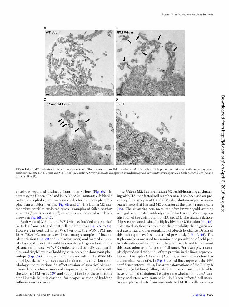
envelopes separated distinctly from other virions (Fig. 6A). Incontrast, the Udorn 5PM and I51A-Y52A M2 mutants exhibited abulbous morphology and were much shorter and more pleomor-phic than wt Udorn virions (Fig. 6B and C). The Udorn M2 mu-tant virus particles exhibited several examples of failed scissionattempts (“beads on a string”) (examples are indicated with blackarrows in Fig. 6B and C).
Both wt and M2 mutant WSN viruses budded as sphericalparticles from infected host cell membranes (Fig. 7A to C).However, in contrast to wt WSN virions, the WSN 5PM andI51A-Y52A M2 mutants exhibited many examples of incom-plete scission (Fig. 7B and C, black arrows) and formed clump-like layers of virus that could be seen along large sections of theplasma membrane. wt WSN tended to bud as individual parti-cles, and single layers of budding virus were the dominant phe-notype (Fig. 7A). Thus, while mutations within the WSN M2amphipathic helix do not result in alterations to virion mor-phology, the mutations do affect scission of spherical virions.These data reinforce previously reported scission defects withthe Udorn 5PM virus (29) and support the hypothesis that theamphipathic helix is essential for proper scission of buddinginfluenza virus virions.
wt Udorn M2, but not mutant M2, exhibits strong cocluster-ing with HA in infected cell membranes. It has been shown pre-viously from analysis of HA and M2 distribution in planar mem-brane sheets that HA and M2 cocluster at the plasma membrane(15). The clustering was measured after immunogold stainingwith gold-conjugated antibody specific for HA and M2 and quan-tification of the distribution of HA and M2. The spatial relation-ship was measured using the Ripley bivariate K function (41, 45),a statistical method to determine the probability that a given ob-ject exists near another population of objects by chance. Details ofthis technique have been described previously (15, 40, 46). TheRipley analysis was used to examine one population of gold par-ticle density in relation to a single gold particle and to representthis association as a function of distance. For example, a com-pletely random distribution of two proteins in the linear represen-tation of the Ripley K function [L(r) � r, where r is the radius] hasa theoretical value of 0. In Fig. 8 dashed lines represent the 99%confidence interval; thus, linear transformations of the Ripley Kfunction (solid lines) falling within this region are considered tohave random distribution. To determine whether or not HA sim-ilarly coclusters with mutant M2 in Udorn-infected cell mem-branes, planar sheets from virus-infected MDCK cells were im-
FIG 6 Udorn M2 mutants exhibit incomplete scission. Thin sections from Udorn-infected MDCK cells at 12 h p.i. immunostained with gold-conjugatedantibody indicate HA (12 nm) and M2 (6 nm) localization. Arrows indicate an apparent joined membrane between two virus particles. Scale bars, 0.2 �m (A) and0.1 �m (B to D).
Influenza Virus M2 Protein Amphipathic Helix
September 2013 Volume 87 Number 18 jvi.asm.org 9979
on April 6, 2018 by guest
http://jvi.asm.org/
Dow
nloaded from

munostained with gold particles (12- and 6-nm for HA and M2,respectively), and spatial distribution was analyzed using Ripley’sbivariate K function. Data representing a comparison of HA andM2 clustering from at least 10 membrane sheets of different in-fected cells are presented in Fig. 8.
Planar membrane sheets from wt Udorn-infected cells showedthat HA and M2 associate nonrandomly (Fig. 8, solid black line).This nonrandom clustering increases sharply with increased dis-tance and supports previous observations (15). In contrast, anal-ysis of planar membrane sheets from Udorn 5PM and M2 I51A-Y52A mutant virus-infected cells indicates markedly reducedcoclustering of HA and mutant M2 though there is a very weakcoclustering beginning at 75 to 100 nm. This reduced associationof HA and mutant M2 is consistent with the notion that mutantM2 may not be localized in the correct domain of the host lipidbilayer and/or in the necessary density for proper budding andscission.
DISCUSSION
Segment 7 (M segment) of the influenza virus RNA genome en-codes two transcripts through alternative splicing. UnsplicedmRNA encodes the matrix protein M1, and spliced mRNA en-
FIG 8 Statistical analysis of HA and M2 clustering. Apical membranes fromMDCK cells infected with wt Udorn or M2 mutant Udorn virus were immuno-stained for HA and M2, and relative distributions were analyzed using the Ripleybivariate analysis. Dashed lines indicate the 99% confidence interval, and solidlines represent the Ripley K function. HA and M2 are considered to be coclusteredat distances where the solid line is above the 99% confidence interval. 51-52, I51A-Y52A mutation.
FIG 7 WSN M2 mutants exhibit incomplete scission. Thin sections from WSN-infected MDCK cells at 6 h p.i. immunostained with gold-conjugated antibodyindicate HA (12 nm) and M2 (6 nm) localization. Arrows indicate an apparent joined membrane between two virus particles. Scale bars, 0.1 �m (A and C) and0.2 �m (B and D).
Roberts et al.
9980 jvi.asm.org Journal of Virology
on April 6, 2018 by guest
http://jvi.asm.org/
Dow
nloaded from

codes M2. M1 and M2 expressed in cells coprecipitate, and aminoacids 71 to 73 and 74 to 76 within M2 are required for both effi-cient interaction with M2 and viral filament formation (15). Sev-eral genetic studies of M1 have demonstrated a role for the matrixprotein in filamentous budding (16, 24–26). Exposure of filamen-tous virus to the monoclonal antibody 14C2, which binds theectodomain of M2 (39), perturbs filament formation, and escapemutants contain amino acid changes in M1 or the C-terminal tailof M2 (16, 47). Further, treatment with 5 �g/ml of 14C2 IgGrestricts growth (as measured by plaque size) of the filamentousUdorn strain but does not inhibit growth of the spherical WSNinfluenza virus strain (39). Additional evidence to support theinvolvement of M2 with filamentous budding was demonstratedby experiments with Udorn virus expressing M2 truncated atamino acid 70, which results in loss of filament formation (48).Surprisingly, when this same M2 truncation mutation is intro-duced into the WSN background, budding virus exhibits a fila-mentous rather than spherical morphology (48, 49). The latterphenotype, however, is believed to be the result of delayed scissionrather than true filamentous budding (48).
Amphipathic helices have long been recognized as generatorsand sensors of membrane curvature (50–52). Residues on the hy-drophobic face of the helix insert into the membrane and anchorthe helix in place while polar residues are involved in electrostaticinteractions with the membrane and potentially curvature induc-tion. While full-length M2 is a transmembrane protein and istherefore always associated with membranes, the 17-amino-acidsequence of the wt M2 amphipathic sequence is sufficient to asso-ciate with giant unilamellar vesicles (GUVs) and drive budding invitro (29).
Phase-separated GUVs containing sphingomyelin, polyunsat-urated phosphocholine, and cholesterol segregate into lipid-or-dered (LO) and lipid-disordered (LD) phases that are analogous tothe lipid raft (LO) and nonraft (LD) regions of apical membrane incells. A fluorescently labeled peptide corresponding to the M2amphipathic helix peptide associates with the LD face of phase-separated GUVs and clusters at the phase boundary where thepeptide drives budding of the LO phase. Additionally, the M2 cy-toplasmic tail of A/Duck/Ukrane/1/63 (H3N8) fused at its N ter-minus to yellow fluorescent protein was able to associate withmembrane when expressed in mammalian cells though this wasdependent upon an intact CRAC motif and, to a lesser extent,acylation (36). Therefore, it is tempting to speculate that the am-phipathic helix of full-length M2 in virus-infected cells drives itslocalization to the boundary of lipid rafts, where it influences bud-ding and scission of nascent virions.
Other recent in silico data support the ability of M2 to causemembrane curvature as well as demonstrate its sensitivity topenta-alanine mutations within the amphipathic helix (53). It wasfound that the M2 amphipathic helix is sufficient to cause negativeGaussian curvature (NGC), which is a prerequisite alteration ofmembrane shape for budding and scission mechanisms, similar tothat proposed for influenza virus virions (28). Based on these data,M2 is capable of generating NGCs that create a neck diameter 10times smaller than the diameter of an emerging influenza virusparticle. Additionally, M2 protein-containing membrane-inter-acting domains and a cytoplasmic domain cooperatively promotean enhanced ability to generate NGC.
The peripheral localization of M2 near lipid rafts has been doc-umented previously (9), and disruption of the amphipathic na-
ture of the M2 helix may interfere with the innate ability of thehelix to recognize and associate with boundaries of LO domains,resulting in mislocalization and reduced efficacy of budding andscission. This is supported by the electron microscopy images ofmutant virus particles that undergo incomplete scission (Fig. 6and 7) and also by the membrane distribution (Ripley analysis)indicating that the M2 mutant had only a weak association withHA, a lipid raft resident protein (Fig. 8).
The data presented here demonstrate that the amphipathic he-lix within the M2 protein cytoplasmic tail is necessary for filamen-tous virus production and normal viral growth. Mutation of as fewas two conserved hydrophobic residues (to alanines) resulted in aloss of filamentous budding for Udorn virions. Both WSN andUdorn strains containing the 5PM or a double mutation in the M2amphipathic helix are attenuated for growth and scission. Becausethese defects were seen in both filamentous and spherical strains,the M2 amphipathic helix is thought to play an important role inbudding for all influenza A virus strains.
ACKNOWLEDGMENTS
We thank members of the Lamb laboratory for valuable discussions andinsight. Transmission electron microscopy was performed in Northwest-ern University’s Biological Imaging Facility.
This research was supported by grant R01 AI-20201 (R.A.L.) from theNational Institutes of Allergy and Infectious Diseases. K.L.R. is an associ-ate, G.P.L. is a specialist, and R.A.L. is an investigator of the HowardHughes Medical Institute. C.M. is a postdoctoral associate at Northwest-ern University.
REFERENCES1. Amorim MJ, Bruce EA, Read EKC, Foeglein Á, Mahen R, Stuart AD,
Digard P. 2011. A Rab11- and microtubule-dependent mechanism forcytoplasmic transport of influenza A virus viral RNA. J. Virol. 85:4143–4156.
2. Ye Z, Liu T, Offringa DP, McInnis J, Levandowski RA. 1999. Associa-tion of influenza virus matrix protein with ribonucleoproteins. J. Virol.73:7467–7473.
3. Fontana J, Cardone G, Heymann JB, Winkler DC, Steven AC. 2012.Structural changes in influenza virus at low pH characterized by cryo-electron tomography. J. Virol. 86:2919 –2929.
4. Elster C, Larsen K, Gagnon J, Ruigrok RW, Baudin F. 1997. Influenzavirus M1 protein binds to RNA through its nuclear localization signal. J.Gen. Virol. 78:1589 –1596.
5. Nayak DP, Balogun RA, Yamada H, Zhou ZH. 2009. Influenza virusmorphogenesis and budding. Virus Res. 143:147–161.
6. Rossman JS, Lamb RA. 2011. Influenza virus assembly and budding.Virology 411:229 –236.
7. Gerl MJ, Sampaio JL, Urban S, Kalvodova L, Verbavatz J-M, Binning-ton B, Lindemann D, Lingwood CA, Shevchenko A, Schroeder C,Simons K. 2012. Quantitative analysis of the lipidomes of the influenzavirus envelope and MDCK cell apical membrane. J. Cell Biol. 196:213–221.
8. Hess ST, Kumar M, Verma A, Farrington J, Kenworthy A, ZimmerbergJ. 2005. Quantitative electron microscopy and fluorescence spectroscopyof the membrane distribution of influenza hemagglutinin. J. Cell Biol.169:965–976.
9. Leser GP, Lamb RA. 2005. Influenza virus assembly and budding inraft-derived microdomains: a quantitative analysis of the surface distribu-tion of HA, NA and M2 proteins. Virology 342:215–227.
10. Thaa B, Herrmann A, Veit M. 2009. The polybasic region is not essentialfor membrane binding of the matrix protein M1 of influenza virus. Virol-ogy 383:150 –155.
11. Wang D, Harmon A, Jin J, Francis DH, Christopher-Hennings J,Nelson E, Montelaro RC, Li F. 2010. The lack of an inherent membranetargeting signal is responsible for the failure of the matrix (M1) protein ofinfluenza A virus to bud into virus-like particles. J. Virol. 84:4673– 4681.
12. Chen BJ, Leser GP, Morita E, Lamb RA. 2007. Influenza virus hemag-
Influenza Virus M2 Protein Amphipathic Helix
September 2013 Volume 87 Number 18 jvi.asm.org 9981
on April 6, 2018 by guest
http://jvi.asm.org/
Dow
nloaded from

glutinin and neuraminidase, but not the matrix protein, are required forassembly and budding of plasmid-derived virus-like particles. J. Virol.81:7111–7123.
13. Barman S, Ali A, Hui EKW, Adhikary L, Nayak DP. 2001. Transportof viral proteins to the apical membranes and interaction of matrixprotein with glycoproteins in the assembly of influenza viruses. VirusRes. 77:61– 69.
14. Jin H, Leser GP, Zhang J, Lamb RA. 1997. Influenza virus hemagglutininand neuraminidase cytoplasmic tails control particle shape. EMBO J. 16:1236 –1247.
15. Chen BJ, Leser GP, Jackson D, Lamb RA. 2008. The influenza virus M2protein cytoplasmic tail interacts with the M1 protein and influences virusassembly at the site of virus budding. J. Virol. 82:10059 –10070.
16. Roberts PC, Lamb RA, Compans RW. 1998. The M1 and M2 proteins ofinfluenza A virus are important determinants in filamentous particle for-mation. Virology 240:127–137.
17. Calder LJ, Wasilewski S, Berriman JA, Rosenthal PB. 2010. Structuralorganization of a filamentous influenza A virus. Proc. Natl. Acad. Sci.U. S. A. 107:10685–10690.
18. Noda T, Sagara H, Yen A, Takada A, Kida H, Cheng RH, Kawaoka Y.2006. Architecture of ribonucleoprotein complexes in influenza A virusparticles. Nature 439:490 – 492.
19. Chu CM, Dawson IM, Elford WJ. 1949. Filamentous forms associatedwith newly isolated influenza virus. Lancet i:602.
20. Kilbourne ED, Murphy JS. 1960. Genetic studies of influenza viruses. I.Viral morphology and growth capacity as exchangeable genetic traits.Rapid in ovo adaptation of early passage Asian strain isolates by combina-tion with PR8. J. Exp. Med. 111:387– 406.
21. de Vries E, Tscherne DM, Wienholts MJ, Cobos-Jimenez V, Scholte F,Garcia-Sastre A, Rottier PJ, de Haan CA. 2011. Dissection of the influenzaA virus endocytic routes reveals macropinocytosis as an alternative entrypathway. PLoS Pathog. 7:e1001329. doi:10.1371/journal.ppat.1001329.
22. Rossman JS, Leser GP, Lamb RA. 2012. Filamentous influenza virusenters cells via macropinocytosis. J. Virol. 86:10950 –10960.
23. Choppin PW, Murphy JS, Tamm I. 1960. Studies of two kinds of virusparticles which comprise influenza A2 virus strains. III. Morphologicalcharacteristics: independence of morphological and functional traits. J.Exp. Med. 112:945–952.
24. Bourmakina SV, Garcia-Sastre A. 2003. Reverse genetics studies on thefilamentous morphology of influenza A virus. J. Gen. Virol. 84:517–527.
25. Burleigh LM, Calder LJ, Skehel JJ, Steinhauer DA. 2005. Influenza Aviruses with mutations in the M1 helix six domain display a wide variety ofmorphological phenotypes. J. Virol. 79:1262–1270.
26. Elleman CJ, Barclay WS. 2004. The M1 matrix protein controls thefilamentous phenotype of influenza A virus. Virology 321:144 –153.
27. Bruce EA, Digard P, Stuart AD. 2010. The Rab11 pathway is required forinfluenza A virus budding and filament formation. J. Virol. 84:5848 –5859.
28. Rossman JS, Jing X, Leser GP, Balannik V, Pinto LH, Lamb RA. 2010.Influenza virus M2 ion channel protein is necessary for filamentous virionformation. J. Virol. 84:5078 –5088.
29. Rossman JS, Jing X, Leser GP, Lamb RA. 2010. Influenza virus M2protein mediates ESCRT-independent membrane scission. Cell 142:902–913.
30. Lamb RA, Zebedee SL, Richardson CD. 1985. Influenza virus M2 proteinis an integral membrane protein expressed on the infected-cell surface.Cell 40:627– 633.
31. Pinto LH, Lamb RA. 2007. Controlling influenza virus replication byinhibiting its proton channel. Mol. Biosyst. 3:18 –23.
32. Nguyen PA, Soto CS, Polishchuk A, Caputo GA, Tatko CD, Ma C,Ohigashi Y, Pinto LH, DeGrado WF, Howard KP. 2008. pH-inducedconformational change of the influenza M2 protein C-terminal domain.Biochemistry 47:9934 –9936.
33. Schnell JR, Chou JJ. 2008. Structure and mechanism of the M2 protonchannel of influenza A virus. Nature 451:591–595.
34. Tian C, Gao PF, Pinto LH, Lamb RA, Cross TA. 2003. Initial structuraland dynamic characterization of the M2 protein transmembrane and am-phipathic helices in lipid bilayers. Protein Sci. 12:2597–2605.
35. Grantham ML, Wu WH, Lalime EN, Lorenzo ME, Klein SL, Pekosz A.2009. Palmitoylation of the influenza A virus M2 protein is not requiredfor virus replication in vitro but contributes to virus virulence. J. Virol.83:8655– 8661.
36. Thaa B, Levental I, Herrmann A, Veit M. 2011. Intrinsic membraneassociation of the cytoplasmic tail of influenza virus M2 protein and lateralmembrane sorting regulated by cholesterol binding and palmitoylation.Biochem. J. 437:389 –397.
37. Thaa B, Tielesch C, Möller L, Schmitt AO, Wolff T, Bannert N,Herrmann A, Veit M. 2012. Growth of influenza A virus is not impededby simultaneous removal of the cholesterol-binding and acylation sites inthe M2 protein. J. Gen. Virol. 93:282–292.
38. Hoffmann E, Neumann G, Kawaoka Y, Hobom G, Webster RG. 2000.A DNA transfection system for generation of influenza A virus from eightplasmids. Proc. Natl. Acad. Sci. U. S. A. 97:6108 – 6113.
39. Zebedee SL, Lamb RA. 1988. Influenza A virus M2 protein: monoclonalantibody restriction of virus growth and detection of M2 in virions. J.Virol. 62:2762–2772.
40. Sanan DA, Anderson RG. 1991. Simultaneous visualization of LDL re-ceptor distribution and clathrin lattices on membranes torn from the up-per surface of cultured cells. J. Histochem. Cytochem. 39:1017–1024.
41. Ripley BD. 1979. Tests of “randomness” for spatial point patterns. J. R.Stat. Soc. Series B Stat. Methodol. 41:368 –374.
42. Baddeley A, Turner R. 2005. Spatstat: an R package for analyzing spatialpoint patterns. J. Stat. Softw. 12:1– 42.
43. Ma C, Soto CS, Ohigashi Y, Taylor A, Bournas V, Glawe B, Udo MK,Degrado WF, Lamb RA, Pinto LH. 2008. Identification of the pore-liningresidues of the BM2 ion channel protein of influenza B virus. J. Biol.Chem. 283:15921–15931.
44. Stewart SM, Pekosz A. 2011. Mutations in the membrane-proximal re-gion of the influenza A virus M2 protein cytoplasmic tail have modesteffects on virus replication. J. Virol. 85:12179 –12187.
45. Ripley BD. 1977. Modelling spacial patterns. J. R. Stat. Soc. Series B Stat.Methodol. 39:172–212.
46. Wilson B, Pfeiffer J, Raymond-Stintz M, Lidke D, Andrews N, Zhang J,Yin W, Steinberg S, Oliver J. 2007. Exploring membrane domains usingnative membrane sheets and transmission electron microscopy. MethodsMol Biol. 398:245–261.
47. Hughey PG, Roberts PC, Holsinger LJ, Zebedee SL, Lamb RA, Com-pans RW. 1995. Effects of antibody to the influenza A virus M2 protein onM2 surface expression and virus assembly. Virology 212:411– 421.
48. McCown MF, Pekosz A. 2006. Distinct domains of the influenza A virusM2 protein cytoplasmic tail mediate binding to the M1 protein and facil-itate infectious virus production. J. Virol. 80:8178 – 8189.
49. Iwatsuki-Horimoto K, Horimoto T, Noda T, Kiso M, Maeda J, Wa-tanabe S, Muramoto Y, Fujii K, Kawaoka Y. 2006. The cytoplasmic tailof the influenza A virus M2 protein plays a role in viral assembly. J. Virol.80:5233–5240.
50. Drin G, Antonny B. 2010. Amphipathic helices and membrane curvature.FEBS Lett. 584:1840 –1847.
51. Shen H, Pirruccello M, De Camilli P. 2012. SnapShot: membrane cur-vature sensors and generators. Cell 150:1300 –1300.e2. doi:10.1016/j.cell.2012.08.017.
52. Jensen MB, Bhatia VK, Jao CC, Rasmussen JE, Pedersen SL, Jensen KJ,Langen R, Stamou D. 2011. Membrane curvature sensing by amphipathichelices: a single liposome study using �-synuclein and annexin B12. J. Biol.Chem. 286:42603– 42614.
53. Schmidt NW, Mishra A, Wang J, Degrado WF, Wong GCL. Influenzavirus A M2 protein generates negative Gaussian membrane curvature nec-essary for budding and scission. J. Am. Chem. Soc., in press.
Roberts et al.
9982 jvi.asm.org Journal of Virology
on April 6, 2018 by guest
http://jvi.asm.org/
Dow
nloaded from