Excavations at Carmarthen Greyfriars 1983-1997 1(discussion).pdf · Excavations at Carmarthen...
50
Excavations at Carmarthen Greyfriars 1983-1997 Analysis of Skeletal Remains VOLUME I: Summary of Findings & Photographs Dr. J. L. Wilkinson, formerly of the Anatomy Dept., University College, Cardiff TEXT EDITED BY TERRY JAMES for CAMBRIA ARCHAEOLOGY 2001
Transcript of Excavations at Carmarthen Greyfriars 1983-1997 1(discussion).pdf · Excavations at Carmarthen...

Excavations at Carmarthen Greyfriars 1983-1997
Analysis of Skeletal Remains
VOLUME I:Summary of Findings & Photographs
Dr. J. L. Wilkinson,formerly of the Anatomy Dept., University College, Cardiff
TEXT EDITED BY TERRY JAMES f o r CAMBRIA ARCHAEOLOGY 2001

Volume I. Page 2 of 50
CONTENTS
VOLUME I—SUMMARY OF FINDINGS
Hints on using this document—3
Editorial Note—4
Summary of Findings from the Cloisters and Chapter House (1984-1997)—5
Summary of Findings from the Church (1988 & 1990)—13
Summary Table (1988/1990 Seasons)—22
Summary of 1997 Findings from the Choir—30
Photographs—36
VOLUME II—DETAILED DESCRIPTIONS

Volume I. Page 3 of 50
Other reports in this series, published herewith in electronic format, are: small finds; pottery and floor tiles as well as the1997 excavation report. The main structural report is published in Medieval Archaeology, Vol. xli 1997 pp. 100-194.The electronic publications canbe read using Adobe Acrobat. This can be freely download and used from http://www.adobe.com/products/acrobat/readstep2.html.
Hints on using this document:
Take a moment to learn how Acrobat works. Time spent now will help you harness the power and ease of moving around this electronic document.Text Marked like this is a ‘hot link’ (see the cursor changes to a POINTING HAND when you hover over red words). Click on any red words andletters: these will take you to the appropriate cross reference or illustration (then use the right mouse button to return to original page)
USE THESE THREEBUTTON TORESCALE THE PAGEOR ILLUSTRATIONTO YOUR LIKING
THISBUTTONTO FIND
TEXT
THISBUTTON TOENLARGE
(LEFTMOUSE
BUTTON TOGO BACK)
THISBUTTONTO ‘PAN’WHEN
IMAGE ISTOO BIG
FORSCREEN
THESE THREEBUTTONS
WILL DISPLAYCONTENTS
ANDSECTIONS INLEFT MARGIN
START, END OFDOCUMENT,
PREVIOUS ANDNEXT PAGE
Note you can rescale a page and move to another page number by clicking on the relevant part of the lower left of the screen.

Volume I. Page 4 of 50
Editorial Note
The following bone reports were compiled by Dr Wilkinson at particular periods of the Greyfriars excavations. Given the rollingnature of redevelopment of the site it was never clear when the excavations would terminate. His reports were designed tostand on their own. The task of text editor has been to try to present a cohesive report, but neither time nor finance allows acomplete restructuring. The individual synopses have been extracted and presented first (Volume I). Detailed descriptions arein Volume II. The method of analysing the bones was undertaken according to a skeleton's completeness. Complete skeletons,(or assemblages of individuals clearly identified on site), were examined first. Mixed assemblages of bone from commoncontexts followed after these. Because of this the description do not follow a Context Number sequence, although within theworking process described here numbers like, complete skeletons, tend to be sequential. After study the bones were interred inthe graveyard of St. Mary’s RC Church, Carmarthen, and Dr Wilkinson's typescript was deposited with the rest of the archiveand finds in Carmarthen County Museum. Please note that Context Numbers for the 1997 report start afresh at No.1.
Conventions used for Dentition (Volume II)
The following notation has been adopted throughout this publication
Usual convention Meaning Adopted Convention Exampleχ loss a.m. double strikethrough 8– or / loss p.m. single strikethrough 8U unerupted inferior/subscript figure 8
o erupting superior/superscript fig. 8
underlined
ul=upper left ( for MS. ) ll=lower left (for MS. )ur=upper right ( for MS. ) lr=lower right (for MS. )

Volume I. Page 5 of 50
1984 to 1987 Excavations
SUMMARY OF FINDINGS FROM THE CLOISTERS AND CHAPTER HOUSE
There were 29 graves, of which 24 contained only one body. In 5 there were remains of another individual; four of theseconsisted of one or two bones only and were probably an accidental inclusion from an adjacent burial, either duringinhumation or exhumation. Thus 34 individuals were represented in graves.
Apart from the grave burials there were 6 localized aggregations with a regional body representation adequate enough toindicate the burial of one individual in 4 sites and two individual admixed in each of 2 sites, i.e. eight burials. Altogether 42people have been identified so far.
There were 17 other contexts in which small amounts of bone were found, most commonly from hand (4), foot (6) or skull(6).Two fragment s were unidentified. These remains are regarded as either having been derived from a grave such as thosealready mentioned, or alternatively they may be the scattered remains from other disturbed graves.
In one location (context 871) there were the partial remains of an adult and an adolescent and in addition there was the verysingular discovery of five left great tow metatarsal bones from adults. It is difficult to explain this. Obviously it is not theaccidental residuum of five burials. It is most unlikely o have resulted from amputation or any other surgical intervention;the find is too selective and no operative procedure requires the individual removal of this bone. Occasionally, for example insome Anglo-Saxon cemeteries, the feet are buried separately presumable as a penal act or for reasons of superstition (seeWilkinson 1980).
It may be noted that here also, in context 722 there were the partial remains of a pair of feet, probably the result of gravedisturbance. Occasionally, particularly in Roman or Roman- British remains, there are aggregations of identical animal bones,

Volume I. Page 6 of 50
particularly the astragali of sheep; these were used as counters, but there is no record of human bones being used as items ofutility in Western societies. It may be some most unusual form of token burial or the result of some curious whim or custom.
Individual ages
It was possible to determine the ages of 34 individuals; these included 28 from graves and 6 not in graves. The ageing of adultswas largely dependent upon the degree of dental attrition. Evidence of advancing age was supported by the presence ofdegenerative disease of joint surfaces, loss of teeth and changes in the he mandible. the degree of fusion of cranial sutures isnot generally a reliable guide to the ageing process.
In immature skeletons there was a lack of fusion of long bone epiphyses, the bones also being of small size; the permanentdentition was incomplete and occasionally there was a persistence of deciduous teeth into adolescence. At intermediate agesbetween adolescence and adult life the fusion, or otherwise of the spheno-occipital synchondrosis, the development and fusion ofthe late accessory epiphyses of iliac crest, ischial tuberosity and vertebral bodies, the degree of fusion of sacrum or body of thesternum were of assistance in determining age.
The age range of 34 individuals was a follows:
-15
-20
-25
-30
-35
-40
+45
4 7 4 1 2 8 8

Volume I. Page 7 of 50
It is surprising that 11 individuals (32%) were adolescents or young adults under the age of 20 years; this age range is furtheranalyzed as follows:
-10
-11
-12
-13
-14
-15
-16
-17
-18
-19
-20
1 1 3 4 1 1
There were no skeletal remains of young children apart from an isolated finding of one deciduous tooth (context 300). Only 7adults (20%) were between the ages of 20 and 35 years. Sixteen (47%) were over 35 years old, and this group was equallydivided between those just under 40 years and those who were more elderly.
Sex
Skeletal sexing was determined in 23 adults. Twenty-two were male. Only one (context 573) was regarded as female and inthis elderly individual the evidence was not totally conclusive because the skeleton was incomplete, in particular there were nopelvic bones, the ends of the long bones were missing and the height could be determined.
In most cases it was not possible to be certain of the sex of the adolescents. This was partly because sexual characteristics wereincompletely developed, partly because significant areas, particularly the pelvis, were missing. Most appeared to be male; inonly two, both aged about 15 years were there features suggesting a female. The first (context 757) was of small stature (154-159cm) as compared with degree of dental development, but some 15 year old boys are of this height. the second (context 801)had bones of a generally slender build, but the remains were partial. In neither was there pelvic evidence.
Volume I. Page 8 of 50
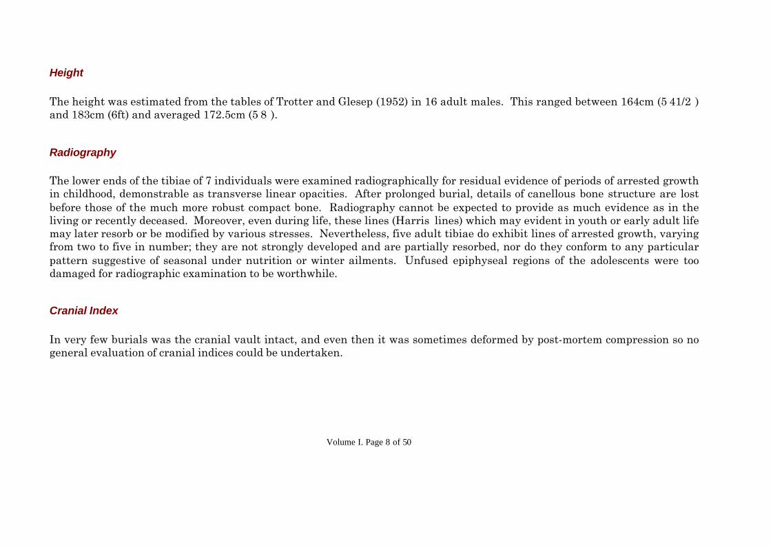
Height
The height was estimated from the tables of Trotter and Glesep (1952) in 16 adult males. This ranged between 164cm (5’41/2”)and 183cm (6ft) and averaged 172.5cm (5’8”).
Radiography
The lower ends of the tibiae of 7 individuals were examined radiographically for residual evidence of periods of arrested growthin childhood, demonstrable as transverse linear opacities. After prolonged burial, details of canellous bone structure are lostbefore those of the much more robust compact bone. Radiography cannot be expected to provide as much evidence as in theliving or recently deceased. Moreover, even during life, these lines (Harris’ lines) which may evident in youth or early adult lifemay later resorb or be modified by various stresses. Nevertheless, five adult tibiae do exhibit lines of arrested growth, varyingfrom two to five in number; they are not strongly developed and are partially resorbed, nor do they conform to any particularpattern suggestive of seasonal under nutrition or winter ailments. Unfused epiphyseal regions of the adolescents were toodamaged for radiographic examination to be worthwhile.
Cranial Index
In very few burials was the cranial vault intact, and even then it was sometimes deformed by post-mortem compression so nogeneral evaluation of cranial indices could be undertaken.

Volume I. Page 9 of 50
Pathology
Infective and Degenerative Conditions
The dental condition was, in general, very poor even by the standards of the time. Periodontal recession was presentthroughout and was quite advanced in one youth age 16 years. In nine cases there were dental abscesses and commonly thesewere multiple; they are a reflection of the severity of the periodontal disease. Caries was present in ten individuals; usuallythis affected the cervical region of the teeth and followed periodontal recession. In one person there was hypercementosis ofupper molar roots due to excessive mobility. Attrition of molar teeth, accompanied by the development of secondary dentine, isevidence of a coarse diet. Apart from the obvious neglect of dental hygiene, the degree of dental disease and the apparentlyearly age of onset may point to a poor quality diet, perhaps lacking vitamin C but possibly including a certain amount of sugar.There were a few exceptions to this pattern, one 25 year old man (context 987) had good teeth, no caries and only slightattrition.
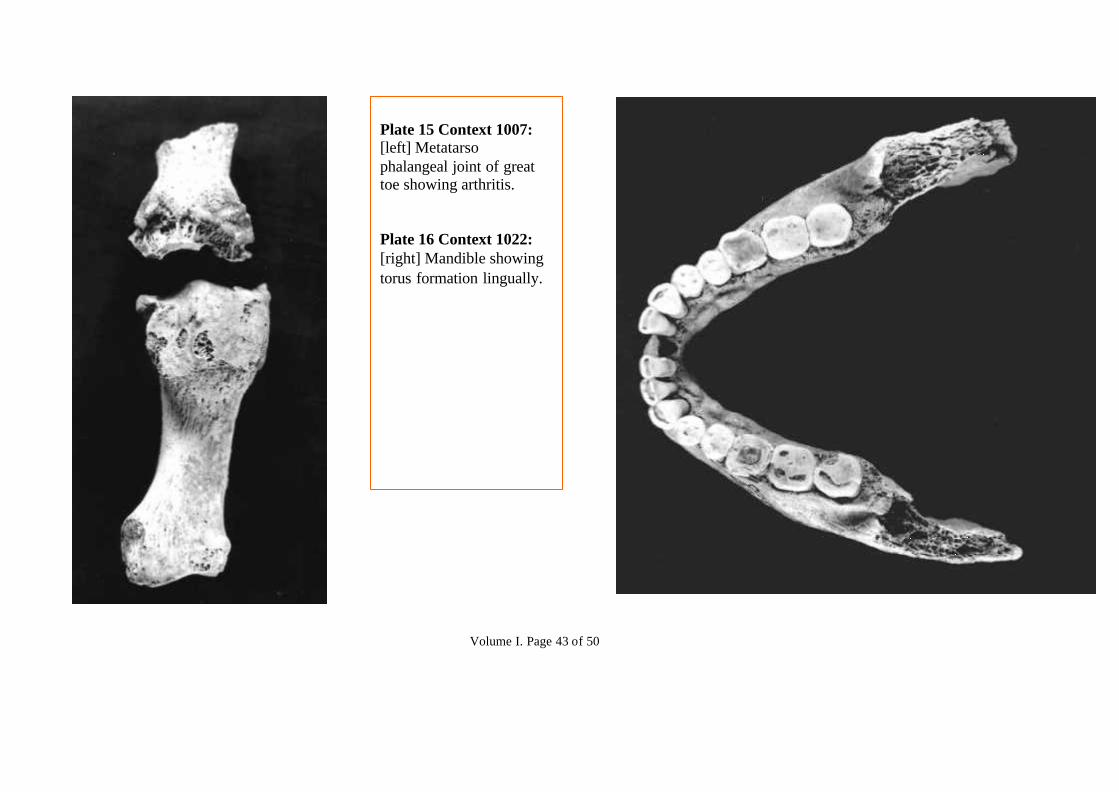
Apart from dental conditions the most prevalent disorder was arthritis. This was noted in 11 individuals; 8 were aged 40 ormore, 2 were 30-35 years old. In eight this principally affected the spine , resulting in osteophyte overgrowth (‘lipping’) of thebodies of the vertebrae, often with fusion of adjacent bones. All regions of the spine were affected, the most mobile areas,cervical and thoraco-lumbar maximally so. In 5 of these there was also evidence of more diffuse collagenous degeneration,affecting the bony attachments of tendons and ligaments. Calcification of these has produced exaggerated roughening andridging at these sites, its degree also influenced by differential muscle bulk and its usage. This condition is also known asdiffuse idiopathic skeletal hyperostosis (‘DISH’); its was apparently most associated with considerable pain, stiffness andrestriction of spinal movement. Sometimes several adjacent vertebral bodies were fused together, a condition known as‘bamboo spine’ (context 966, photoarchive). Osteoarthritis affected the acromio-clavicular joints in two cases. The metatarso-phalangeal joints of the great toes were involved in two men, with gross bony overgrowth in one (context 1007, photograph).The individual thought to be an elderly lady had moderate arthritis of the metacarpo-phalangeal joint of her right thumb; thiswas evidently rheumatoid arthritis and did not affect the larger joints. One elderly male had arthritis of the spine, acromio-clavicular and temporo-manidbular joints. It is noteworthy that, apart from minor lipping of joint margins there was very little

Volume I. Page 10 of 50
arthritic change in the major limb joints - hop, knee and shoulder in particular. This, together with characteristic features of‘DISH’ distinguish the arthritic patterns in this population from that seen in this country now. Cervical spondylitis is acommon condition in the elderly nowadays, then it occurred at an earlier age and as part of a diffuse and gross degenerativecondition.
One man had disease of the right elbow joint. This had caused overgrowth of bone and great distortion of joint contours(context 576, photograph). Movements would have been extremely painful and markedly restricted, the elbow beingpermanently flexed and incapable of extension. This was a suppurative arthritis, infective in origin, but not tuberculous. Itmay have been caused by a penetrating in jury, for example by a thorn. Alternatively it may have been blood-spread from afocus elsewhere in the body, most probably from his gross dental abscesses.
Tumours
There was residual evidence of a tumour in only one case. The surface of a tibial shaft (an isolated find) was indented over anarea 5.5 x 2.7cm, the edge of the depression being hypertrophied and raised. This was evidently due to pressure from a slowgrowing soft tissue tumour, possibly a fibroma or an enlargement (aneurysm) of an adjacent artery.
Fractures
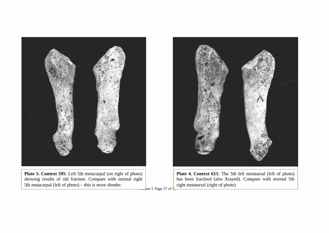
The shaft of one 5th metatarsal had been fractured; this was probably caused by direct violence, such as a rock fall and its isnot typical of a ‘march fracture’, a stress-fatigue lesion. One left 5th metacarpal shaft had been broken as the result of a blow,possibly in boxing. The ‘surgical neck’ of one humerus had been fractured, probably as the result of a fall on the elbow; it hadimpacted and had healed in al almost perfect position. No deformity or disability had resulted from any of these fractures.

Volume I. Page 11 of 50
One elderly man had sustained a dislocation of his left ankle joint, tearing the tibio-fibular ligaments and probably fracturingthe fibula at a higher level (this part is now missing). Following this there was an extravasation of blood between the tibia andfibula which subsequently became ossified, within which a new inferior tibio-fibular joint (or ‘pseudarthrosis’) formed. Thisnow presents a most unusual appearance (context 957, photographed). A bony outgrowth (an exostosis) from the tibiaarticulates by an irregular toothed joint with a similar outgrowth from the fibula above the level of the ankle joint. Despite thisattempt by Nature to restore the situation, there would have been gross permanent instability of the ankle joint and pain onwalking.
Congenital abnormalities and variations
there are a number of anatomical variations but only one was sufficiently abnormal to have given rise to symptoms. In a 25year old man the 5th lumbar vertebrae was in two parts, the body, pedicle and upper articular facets being separated from thelaminae and lower articular facets by a joint on each side which, in life would have been cartilaginous (context 992,photographed). This development abnormality, spondylolisthesis, results from each side of the neural arch having twoossification centers instead of one, and these do not fuse. The bones are usually held in place by the cartilage and by fibroustissue. Sometimes asymptomatic, it is commonly associated with low back pain. As age advances the two parts may separatebecause of the very considerable forces transmitted through this region,; untreated this would produce disability.
Other variations were noted in single individuals apart from the relatively common woriman bones, most prevalent in thelambdoid sutures. There was a persistent metopic suture in the frontal bone of the skull of a 25 year old man. In anotheryoung man the arch of the atlas vertebra had remained ununited in the midline posteriorly and in a more elderly person thegroove for the vertebral artery on the upper surface of the atlas was bridged over by a spur of bone extending to the posteriorarch from the lateral mass. One 35 year old man had bilateral mandibular torus formation lingually between the canine andfirst molar teeth, probably a congenital and hereditary condition. The left femur of an elderly man had a supracondylar spur ofbone postero-medially, extending upwards from the lower insertion of adductor magnus to form a boundary for the opening

Volume I. Page 12 of 50
through which the femoral artery passed; the right femur is damaged and it is not possible to determine whether this featurewas present bilaterally.
General Conclusions
These burials evidently represent a selected group. It is possible that one individual was an elderly woman but the evidence forsexing of this skeleton is not conclusive; apart from that, all other burials of adults were of men. There are no children underthe age of 10 years. There is an unusually large group of adolescents, the sexing of whom is largely indeterminate for thereasons given. There are few individuals in the 20-35 year age group, yet a relatively large number aged approximately 35-45years.
Apart from one person, there is no evidence that these people were of a high social standing. Their dental hygiene wasparticularly bad even for that era, their diet was generally coarse, as was usual. Their muscular development, where this couldbe studied, was quite strong and suggestive of labouring work, walking, stooping, lifting; the prevalence of spinal arthritissupports this pattern of activity. The frequency with which spinal arthritis was associated with collagenous degeneration oftendons and ligaments probably reflects cold, damp working and living conditions. It is noted that amongst the isolated finds,there is a pair of feet, not of itself unusual in a disturbed site; however the present of five left adult great toe metatarsals inone context present an enigma.
References
Trotter, M & Glese, G.C. (1952) Estimation of stature from long bones of American Whites and Negroes. Amer. J.Phys.Anthrop., Washington, 10: 463-514Wilkinson, J.L. (1980) Problems of analysis and interpretation of skeletal remains, in ‘Anglo-Saxon Cemeteries, 1979’ B.A.R.Series 82,228.
Volume I. Page 13 of 50
1988 and 1990 Excavations
SUMMARY OF FINDINGS FROM THE CHOIR OF THE CHURCH AND NORTHERN EXTENSIONOF THE NAVE
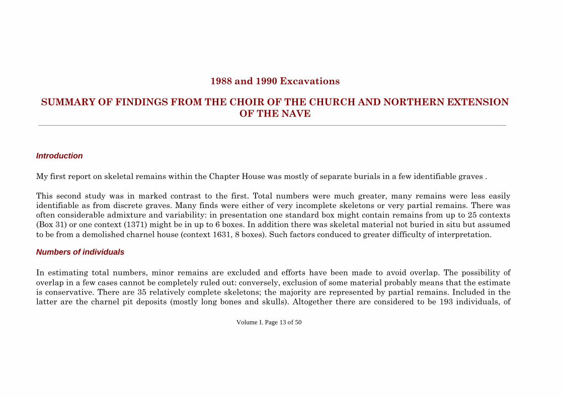
Introduction
My first report on skeletal remains within the Chapter House was mostly of separate burials in a few identifiable graves .
This second study was in marked contrast to the first. Total numbers were much greater, many remains were less easilyidentifiable as from discrete graves. Many finds were either of very incomplete skeletons or very partial remains. There wasoften considerable admixture and variability: in presentation one standard box might contain remains from up to 25 contexts(Box 31) or one context (1371) might be in up to 6 boxes. In addition there was skeletal material not buried in situ but assumedto be from a demolished charnel house (context 1631, 8 boxes). Such factors conduced to greater difficulty of interpretation.
Numbers of individuals
In estimating total numbers, minor remains are excluded and efforts have been made to avoid overlap. The possibility ofoverlap in a few cases cannot be completely ruled out: conversely, exclusion of some material probably means that the estimateis conservative. There are 35 relatively complete skeletons; the majority are represented by partial remains. Included in thelatter are the charnel pit deposits (mostly long bones and skulls). Altogether there are considered to be 193 individuals, of
Volume I. Page 14 of 50
which 142 are adults, 51 are juvenile (under 20yrs.). Of the total, 163 are in situ burials, 30 are from the charnel housesecondary burial. Detailed individual conclusions are set out at the end of context reports: these are briefly aggregated here.
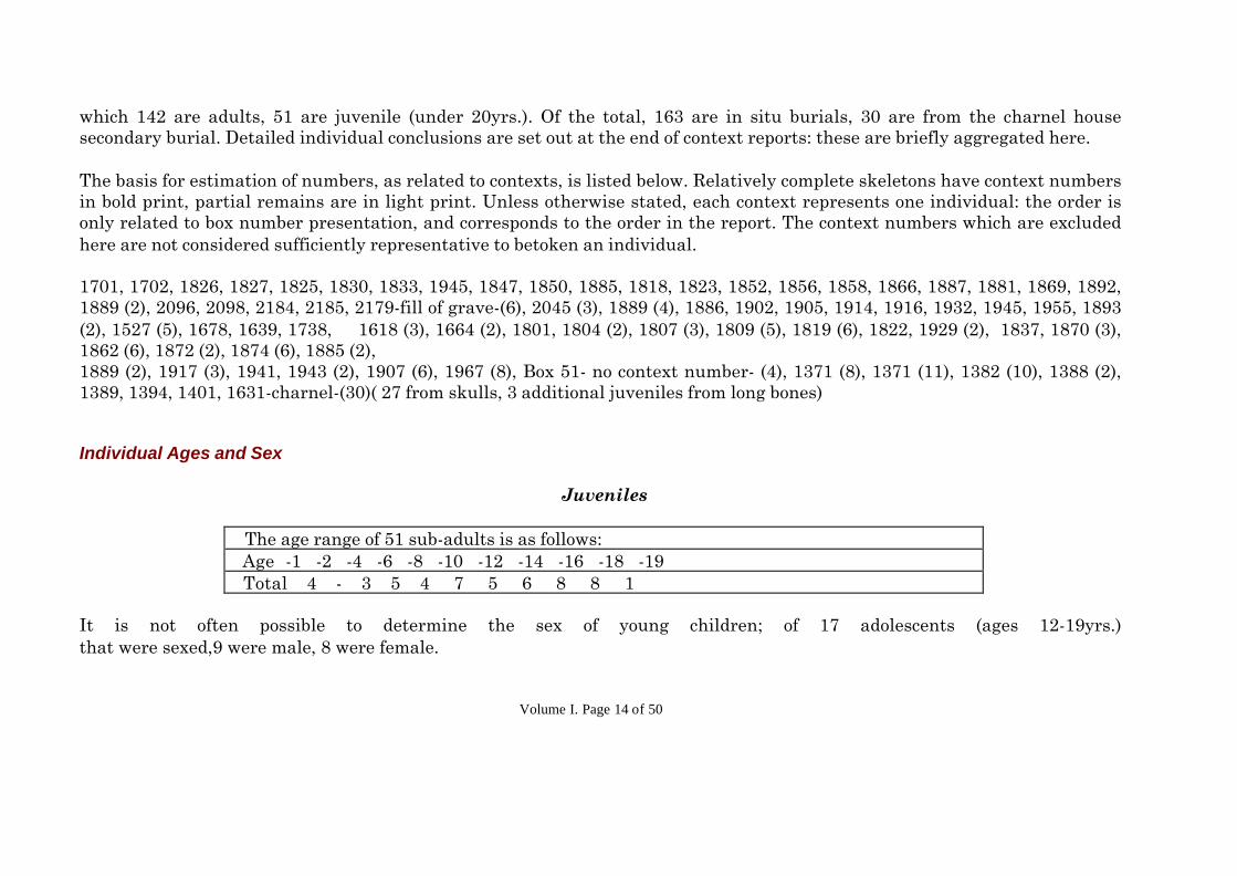
The basis for estimation of numbers, as related to contexts, is listed below. Relatively complete skeletons have context numbersin bold print, partial remains are in light print. Unless otherwise stated, each context represents one individual: the order isonly related to box number presentation, and corresponds to the order in the report. The context numbers which are excludedhere are not considered sufficiently representative to betoken an individual.
1701, 1702, 1826, 1827, 1825, 1830, 1833, 1945, 1847, 1850, 1885, 1818, 1823, 1852, 1856, 1858, 1866, 1887, 1881, 1869, 1892,1889 (2), 2096, 2098, 2184, 2185, 2179-fill of grave-(6), 2045 (3), 1889 (4), 1886, 1902, 1905, 1914, 1916, 1932, 1945, 1955, 1893(2), 1527 (5), 1678, 1639, 1738, 1618 (3), 1664 (2), 1801, 1804 (2), 1807 (3), 1809 (5), 1819 (6), 1822, 1929 (2), 1837, 1870 (3),1862 (6), 1872 (2), 1874 (6), 1885 (2),1889 (2), 1917 (3), 1941, 1943 (2), 1907 (6), 1967 (8), Box 51- no context number- (4), 1371 (8), 1371 (11), 1382 (10), 1388 (2),1389, 1394, 1401, 1631-charnel-(30)( 27 from skulls, 3 additional juveniles from long bones)
Individual Ages and Sex
Juveniles
The age range of 51 sub-adults is as follows: Age -1 -2 -4 -6 -8 -10 -12 -14 -16 -18 -19 Total 4 - 3 5 4 7 5 6 8 8 1
It is not often possible to determine the sex of young children; of 17 adolescents (ages 12-19yrs.)that were sexed,9 were male, 8 were female.
Volume I. Page 15 of 50
Adults
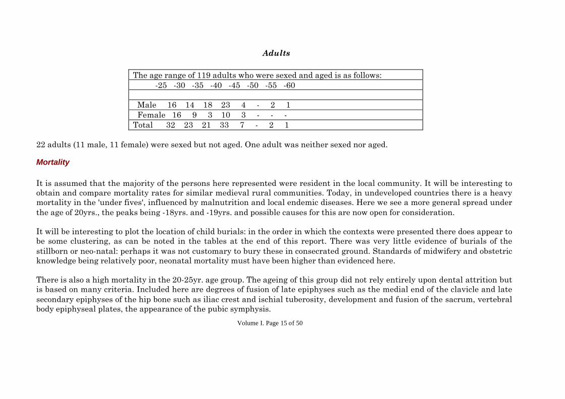
The age range of 119 adults who were sexed and aged is as follows: -25 -30 -35 -40 -45 -50 -55 -60
Male 16 14 18 23 4 - 2 1 Female 16 9 3 10 3 - - -Total 32 23 21 33 7 - 2 1
22 adults (11 male, 11 female) were sexed but not aged. One adult was neither sexed nor aged.
Mortality
It is assumed that the majority of the persons here represented were resident in the local community. It will be interesting toobtain and compare mortality rates for similar medieval rural communities. Today, in undeveloped countries there is a heavymortality in the 'under fives', influenced by malnutrition and local endemic diseases. Here we see a more general spread underthe age of 20yrs., the peaks being -18yrs. and -19yrs. and possible causes for this are now open for consideration.
It will be interesting to plot the location of child burials: in the order in which the contexts were presented there does appear tobe some clustering, as can be noted in the tables at the end of this report. There was very little evidence of burials of thestillborn or neo-natal: perhaps it was not customary to bury these in consecrated ground. Standards of midwifery and obstetricknowledge being relatively poor, neonatal mortality must have been higher than evidenced here.
There is also a high mortality in the 20-25yr. age group. The ageing of this group did not rely entirely upon dental attrition butis based on many criteria. Included here are degrees of fusion of late epiphyses such as the medial end of the clavicle and latesecondary epiphyses of the hip bone such as iliac crest and ischial tuberosity, development and fusion of the sacrum, vertebralbody epiphyseal plates, the appearance of the pubic symphysis.

Volume I. Page 16 of 50
Dental attrition assists in ageing up to 40yrs., but beyond this age the general criteria are less reliable.
There are marked contrasts with the Chapter House series: for example, a higher mortality in the 25-35yr. groups
Body Height
The body height was determined from the tables of Trotter and Gleser (1952) in 58 adults. This ranged between 157 and182cm. for males and between 156 and 166cm. for females.
The body height of 32 adult males are as follows (<1cm. ignored):
-167 -170 -173 -175 -178 -180 -183 cm. (5'6") (6'7") (5'8") (5'9") (5'10") (5'11") (6'0") 3 8 6 5 2 5 3
The body heights of 22 adult females are as follows: (<1cm. ignored)
-157 -160 -162 -165 -167(5'2") (5'3") (5'4") (5'5") (5'6")4 5 5 6 2

Volume I. Page 17 of 50
Physique and Socio-economic factors
Of those evaluated for apparent general physique, as judged by robust or slender bone structure, and the strength of musculartendon insertions, 19 out of 27 were regarded as of robust physique. Their muscle tendon markings were well developed,suggesting of strong sustained physical activity.
Tentative views may be gained as regards socio-economic factors when general physique is considered alongside levels of dentalhygiene. Of 22 thus studied, 14 were regarded as 'working class', used to manual labour, 8 appeared to be of an 'upper class',associated less with manual labour and more observant of hygiene.
Cranial Morphology
In most individuals the skull vault was incomplete, damaged, fragmented or if relatively complete, distorted by post-mortempressure. In only 25 cases was it possible to study cranial indices. These were:
Dolichocephalic 12 : Mesocephalic 8 : Brachycephalic 7
Pathology
Infective and Degenerative Conditions.
The standard of dental hygiene was generally very poor. Periodontal recession was very common and particularly advanced in18 individuals. As a result of poor hygiene and gum recession, dental abscess formation occurred in 16 cases; sometimesmultiple abscesses were present and this was responsible for the loss of teeth. Caries was relatively uncommon by modern
Volume I. Page 18 of 50
standards, only noted in 6 persons: and this is to be expected where the sugar content of the diet is low. Periodontal recessionexposes the neck of teeth and this was the usual site for caries when present. Breakage of teeth, particularly of premolars,resulted from cracking hard objects, probably nuts: some caries could be expected to follow this and would not necessarily beseparately recorded. Attrition of molar teeth was similar in degree to that seen in the first series, indicating a coarse diet: in afew cases the diet appeared to have been more refined. A few individuals had marked wear of the cutting edges of incisor teeth:whilst this can result from biting tough food, unusual incisor wear may be associated with occupation e.g. leather workers,holding a strap with the teeth.
There was one case of suppurative arthritis of a metatarso-phalangeal joint. This may have followed a penetrating injury, suchas by a thorn. A similar case was seen in the Chapter House series and suggests that the individuals may have been unshod.
Osteo-arthritic changes in joints was relatively common. The distribution in order of frequency was as follows:Spine (11), costo-vertebral (transverse) joints (7), Knee (3), metatarsophalangeal (3), hip (2), acromioclavicular (2),sternoclavicular (1), shoulder (1), carpometacarpal (1), sacroiliac (1), tarsal (1)Diffuse idiopathic skeletal hyperostosis (DISH) was noted in 6 cases: ossification of tendon and ligament attachments,accompanied by spinal lipping sometimes involving fusion of adjacent vertebrae but without the disc degeneration of arthritis.It is interesting to note that this is a much lower incidence than was found in the adults from the first series, burials in theChapter House, many of which, it is believed, were monks. There is considerable interest at the present time in an apparentlyhigh incidence of DISH in Priories, and possible reasons for this are under investigation. One modern theory relates to a highfish diet. The nature of DISH was discussed in the first report. In one individual (1829) the coraco-clavicular ligament musthave become ossified and then a very unusual type of joint formed here. There were additional isolated examples of ossificationin tendon attachments, such as a calcaneal spur or a lipping of the olecranon of the ulna at the attachment of the tricepstendon, not uncommon in modern times.
In only 3 cases was there obvious evidence of Schmorl's nodes, central protrusion of intervertebral discs into the adjacentsurfaces of vertebral bodies. Recent investigations reported in the Journal of Anatomy, have suggested that there was a higherincidence of this condition in medieval than in modern times (Fitzpatrick et. al., 1984, 1986). It should be noted, in this regard,

Volume I. Page 19 of 50
that in this series vertebral bodies were not well preserved and often were absent: from the 30 bodies from the Charnel Housethere were only 13.
One individual had a 'squatting facet' on the anterior border of the lower articular surface of the tibia, at the ankle joint.This isquite a common finding amongst some tribal African and Eastern cultures in which squatting is a common posture. It is anuncommon finding in this country.
There were 4 cases of Paget's disease, in which the skull vault was very markedly thickened. It did not appear to have involvedthe post-cranial skeleton.
Trauma.
There was little evidence of injury. One 40-year old lady (context 1945) had sustained a fracture of the right ulna which hadhealed without deformity. A 45-year old lady had fractured an ulna which had united with some overlap, angular androtational deformity. We do not know whether the radius on this side had also been fractured, if so, it is not easy to fix thebones in such a way as to avoid residual deformity. The functional end result in this case was probably fairly good (context1822).
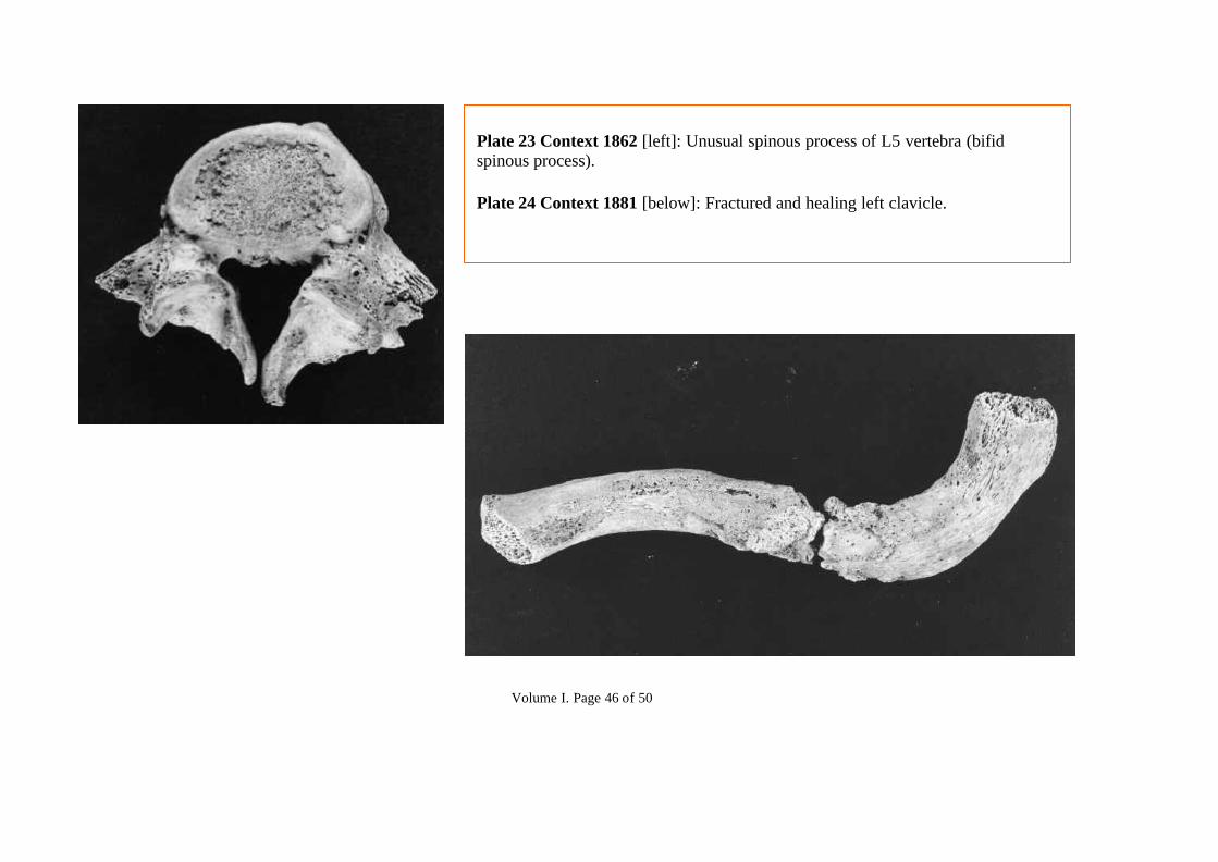
There was one other fracture, of the midshaft of the clavicle, a bone which heals very readily in virtually all cases, but in thisperson had not done so (Plate 24). Healing had begun and there is no evidence that it would not have continued. The patient, ayouth of 16 years, died approximately 7-10 days after the accident. It is probable that there other injuries, possibly to internalorgans which caused death (context 1881).
Of particular interest is a case of trephining of the parietal bone (context 1893). The patient evidently survived the operationbecause the cut edges healed and the spongy diploe are not visible here. This also excludes the possibility of a post-mortem

Volume I. Page 20 of 50
artefact, as does the lack of internal splintering. It is particularly interesting that there are two holes and not just one. Themethod used was that of cutting out a roundel or possibly the use of a single burr drill. Well known in ancient civilizationsranging from Egypt to South American tribes and in Tibet, only about 12 cases are recorded from archaeological sites in thiscountry and these were mostly prehistoric, only 3 Norman and later. The operation is known to have taken place in France inthe 13th. century, and in this country in 16th. and 17th. centuries. As a finding from the medieval period it must be must bevery rare. There appears to be only one recorded find from Wales and that was pre-Norman (Brothwell). It is intended toinvestigate this matter further.
Congenital abnormalities and Variations
Most of these involved either the skull or vertebral column.
Skulls. There were 12 cases of persisting metopic sutures. The average incidence is 2% for most populations. A high incidencemay perhaps indicate a degree of in-breeding. Small accessory sutural (Wormian) bones are not uncommon: in 5 cases Wormianbones were particularly evident: in one individual there were 10 such bones in a row in the occipito-parietal sutures (context1889). Supraorbital foramina in place of the supraorbital notch were quite common: 5 on the left side, 4 on the right, 6 bilateral.There was one case of foramina in the orbital roofs (orbital plates of the frontal bone).Jaws and Teeth there was congenital absence of the 3rd. molars in 7 cases, absence of other teeth in 2, ectopic (extra) teeth in3. There was one instance of fusion of the crowns of premolar teeth (Plate 34). In 3 mandibles a torus was noted.Vertebrae. Imperfect fusion: 1 unfused arch of the atlas vertebra; 1 unfused and bifid spine of a 5th. lumbar vertebra; 1spondylolisthesis of 5th. lumbar vertebra (a case also noted, photographed and described in the Chapter House series); 1unilateral spondylysis of the 5th. lumbar vertebra (an unfused line between upper and lower articular facets on one side of thearch but no displacement). The incidence of spondylolysis in the medieval period was about 5%, less common unilaterally thanbilaterally (Waldron, 1991).Unusual fusion between vertebrae: 2 sacralization of the 5th. lumbar vertebra (fused to the sacrum); 1 congenital fusionbetween two thoracic vertebrae; 2 cases of a bony arch spanning the vertebral artery in the groove on the atlas..

Volume I. Page 21 of 50
Others. Scapula: 2 suprascapular foramina, an arch spanning the suprascapular groove. Sternum: 1 fused manubrio-sternaljoint. Femur: 1 hypotrochanteric fossa, with a 3rd. trochanter. Hip bone: 1 case of unusual ossification of the Y-shapedacetabular cartilage with multiple accessory centres of ossification (os acetabuli).
Note: Unusual congenital anomalies or pathological conditions have been photographed.
References
Brothwell D.R. (1972), 'Digging up bones', 126, Trustees of the British Museum, London.Fitzpatrick K., Bruce M. and Saluja G. (1984), 'Evidence of Schmorl's nodes in medieval Aberdonians' and 'Osteologicalevidence of Schmorl's nodes in two historic British populations', J. Anat. 138, 579 & 600, also J. Anat (1986), 145, 87-96.Waldron H.A. (1991) 'Variations in the prevalence of spondylolysis in early British populations', J. Roy. Soc. Med. 84, 547-549.
Volume I. Page 22 of 50
The 1988-1990 excavations
Summary Table
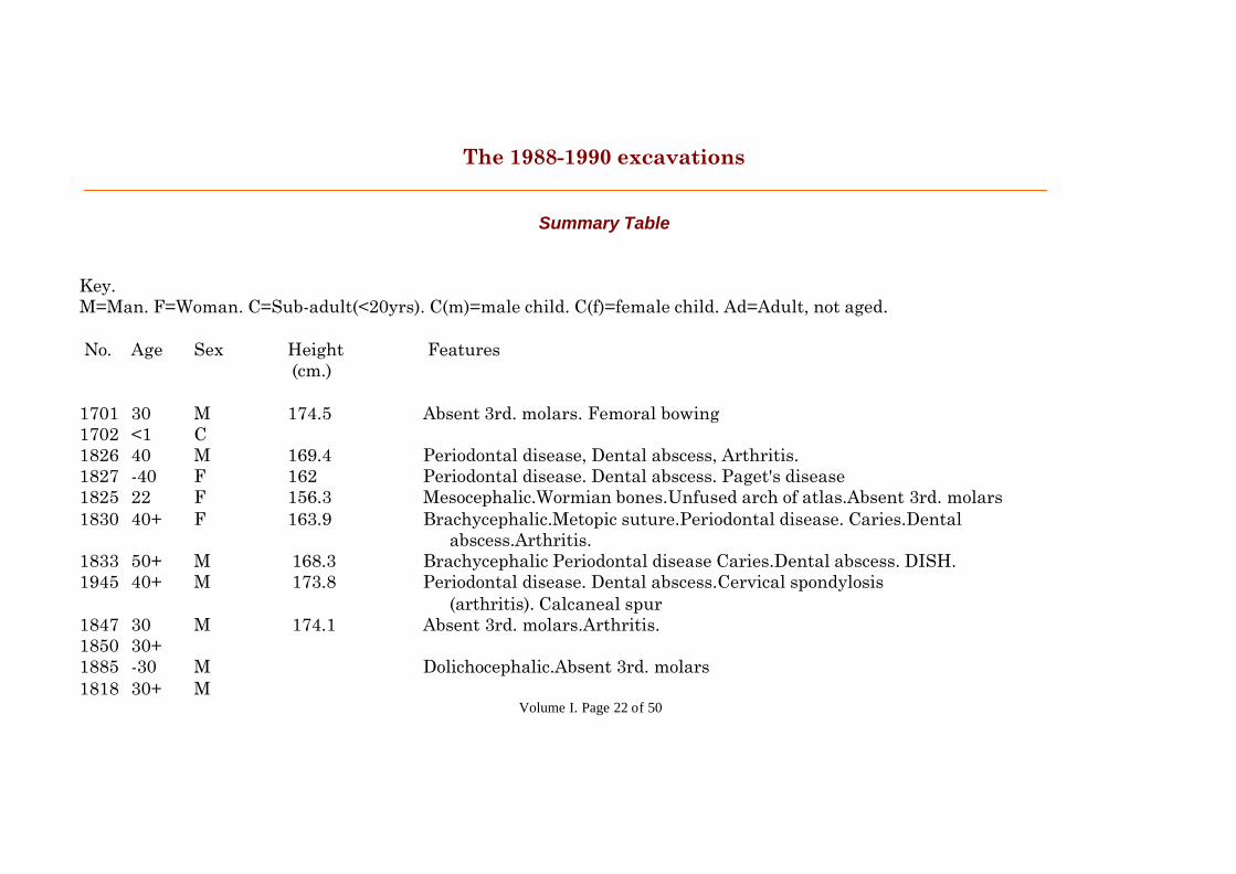
Key.M=Man. F=Woman. C=Sub-adult(<20yrs). C(m)=male child. C(f)=female child. Ad=Adult, not aged.
No. Age Sex Height Features (cm.)
1701 30 M 174.5 Absent 3rd. molars. Femoral bowing1702 <1 C1826 40 M 169.4 Periodontal disease, Dental abscess, Arthritis.1827 -40 F 162 Periodontal disease. Dental abscess. Paget's disease1825 22 F 156.3 Mesocephalic.Wormian bones.Unfused arch of atlas.Absent 3rd. molars1830 40+ F 163.9 Brachycephalic.Metopic suture.Periodontal disease. Caries.Dental abscess.Arthritis.1833 50+ M 168.3 Brachycephalic Periodontal disease Caries.Dental abscess. DISH.1945 40+ M 173.8 Periodontal disease. Dental abscess.Cervical spondylosis (arthritis). Calcaneal spur1847 30 M 174.1 Absent 3rd. molars.Arthritis.1850 30+1885 -30 M Dolichocephalic.Absent 3rd. molars1818 30+ M

Volume I. Page 23 of 50
1823 30 F 157.5 Dolichocephalic.Femoral hypotrochanteric fossa,3rd. trochanter Suppurative arthritis metatarso-phalangeal joint1852 30 M 175.91856 -16 C(f) Dolichocephalic.Wormian bones1858 25 M 181 Dolichocephalic. Absent 3rd. molars1866 60 M 173.8 Mandibular tori.Periodontal disease.Dental abscess.Fused carpo- metacarpal joint1887 40 M 1641881 16 C(m) 175 Unilateral spondylolysis.Periodontal disease1869 40 F 157.2 Brachycephalic.Sacralization L5 vertebra.DISH.Periodontal disease.Dental abscess1892 30 M 176 Periodontal disease.Dental abscess1889 35 F Mesocephalic.Right supraorbital foramen.Periodontal disease .. 25 M Brachycephalic.Multiple Wormian bones.Bilateral supraorbital foramina.Metopic suture.2096 23 M 178.5 Mesocephalic.Wormian bones.Right supraorbital foramen. Spondylolisthesis.Fused manubrio-sternal joint.Schmorl's nodes2098 35 M 173 Absent 3rd. molar&premolar.Incisor attrition.Periodontal disease2184 17 C(m) 176 Mesocephalic.Metopic suture.Wormian bones.Recent fracture clavicle.2185 25 M 169 Brachycephalic.Metopic suture.2179 25 M General for 2179.One of each: dolichocephalic, metopic .. 30 M suture.Ossification supraspinous ligament(DISH).Sacralization L5 .. 45 M vertebra.Tibial squatting facet.Dental abscess. .. 25 F .. 30 F .. 7 C2045 Ad. M .. Ad. F

Volume I. Page 24 of 50
.. 17 C(m)1889 20 F Periodontal disease .. 50+ M 166.6 Paget's disease .. 17 C(m) 174.5 Metopic suture.Ectopic canine tooth .. 25 F 158.41886 35 M 173.4 Brachycephalic.Ectopic unerupted incisor teeth.Periodontal disease
1902 30 M 182 Mandibular torus.DISH.Vertebral fusion.Periodontal disease.Dental abscess1905 28 F 160 Dolichocephalic.Metopic suture.Dental abscess.Periodontal disease.1914 40 F 163 Caries.Periodontal disease1916 30 F 165 Dolichocephalic.Impacted molar.Early arthritis.1932 30 M 180.41945 40 F 161 Arthritis.Old fractured ulna.1955 -30 M 170.8 Absent 3rd. molar1893 20 M Trephine holes in skull. Caries. .. 25 M Right supraorbital foramen1527 30 M .. 20 F .. 6 C .. 10 C .. 15 C1678 Ad. M1639 30 F Dental abscess1738 18 C(f)1618 40+ M .. 30+ M .. 25+ F

Volume I. Page 25 of 50
1664 30 M .. 25 F1801 40 M DISH1804 5½ C .. 10 C1807 3 C .. 11 C .. Ad. F1809 4 ½ C .. 5 ½ C .. 10 C .. 13 C.. Ad. F Metopic suture1819 40 F Dental abscess .. 30 M .. 10 C .. 13 C(f) .. 15 C(m) 165.8 .. 8 C1822 45 F 158.5 Fractured ulna,healed with deformity1829 40 M Paget's disease.DISH.Suprascapular foramen. .. -40 M1837 15 C(m)1870 40+ F 166.2 Arthritis .. 6 C .. 8 ½ C1862 20 M 182.7 Unfused bifid spine 5th. lumbar vertebra .. 35 M 180.4

Volume I. Page 26 of 50
.. Ad. F 162.3 .. <1 C .. 4 C .. Ad M 174 DISH.Periodontal disease.1872 <1 C .. 30 F 1621874 17 C(f) .. 40 M .. 10 C .. 12 C Unusual ossification of acetabulum .. 15 C .. 17 C1885 Ad. M 179 .. 35 F1889 20 F .. 25 M1917 13 C(m) .. 25 F .. 20 M1941 20 F 165.91943 Ad. F .. 40 F1907 <1 C General (1907) one of each-Mesocephalic. Bilateral supraorbital .. 9 C foramina.Absent 3rd. molar.Arthritis. Caries. Dental abscess .. 25 M 168 .. 40 M .. 20 F 156 .. 25 F 161
Volume I. Page 27 of 50
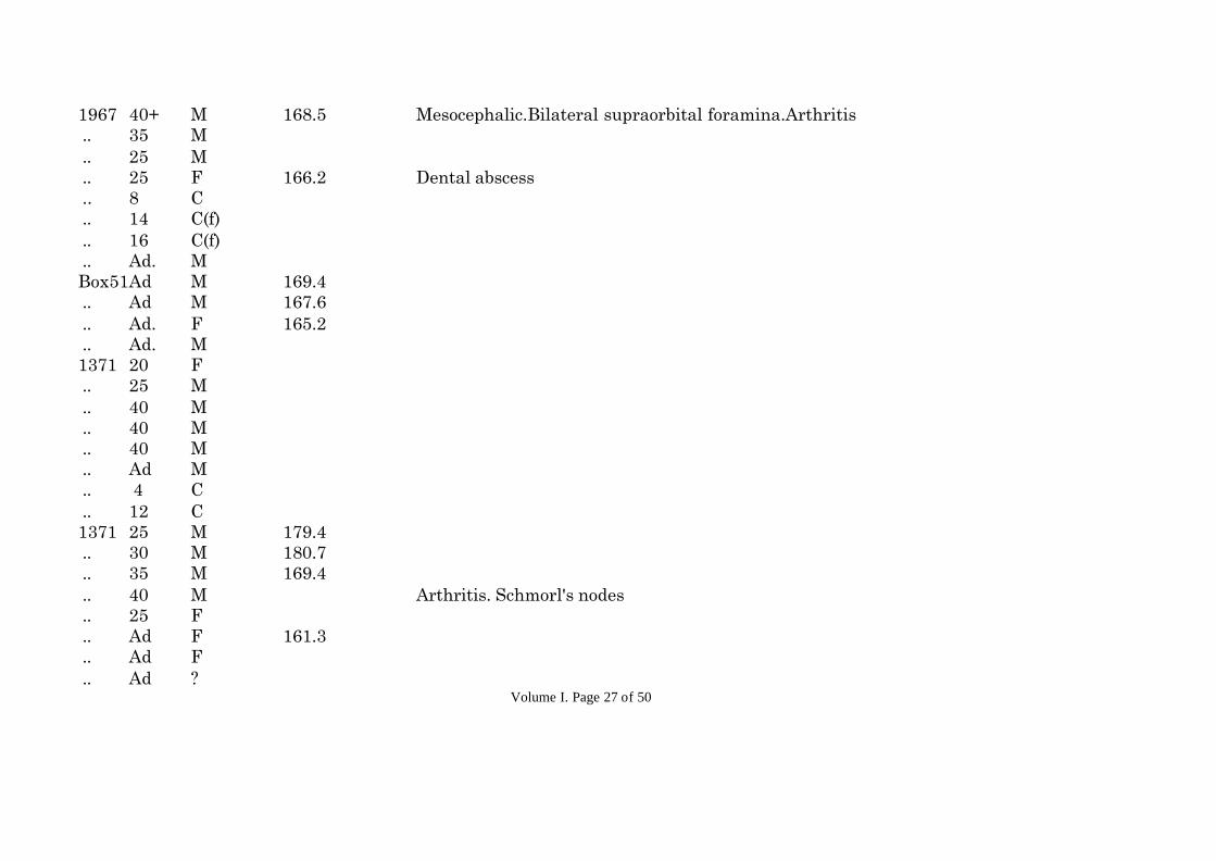
1967 40+ M 168.5 Mesocephalic.Bilateral supraorbital foramina.Arthritis .. 35 M .. 25 M .. 25 F 166.2 Dental abscess .. 8 C .. 14 C(f) .. 16 C(f) .. Ad. MBox51Ad M 169.4 .. Ad M 167.6 .. Ad. F 165.2 .. Ad. M1371 20 F .. 25 M .. 40 M .. 40 M .. 40 M .. Ad M .. 4 C .. 12 C1371 25 M 179.4 .. 30 M 180.7 .. 35 M 169.4 .. 40 M Arthritis. Schmorl's nodes .. 25 F .. Ad F 161.3 .. Ad F .. Ad ?

Volume I. Page 28 of 50
.. 8 C .. 14 C .. 18 C(m)1382 25 M .. 35 M .. 35 M .. Ad M .. Ad M Left supraorbital foramen .. Ad F .. 18 C(f) Dolichocephalic.Bilateral supraorbital foramina .. Ad. F .. Ad F .. 12 C1388 35 M Bilateral supraorbital foramina .. 14 C(f)1389 40 M Periodontal disease. Arthritis1394 35 M Periodontal disease. Dental abscess. Arthritis.1401 40 M Periodontal disease. Caries1631 45 M 171.7 Dolichocephalic.Left supraorbital foramen }These heights correspond .. 40 M 168.7 Dolichocephalic }to the sex but not .. 20 M 174.5 Dolichocephalic.Metopic suture }necessarily to these .. 35 M 171.5 }individuals. .. 30 F }Charnel House long bones .. 40 M }and skulls were unrelated .. 40 M Right supraorbital foramen .. 35 M .. 35 M Metopic suture.Left supraorbital foramen .. 35 F 162.5
Volume I. Page 29 of 50
.. 40 M .. 40 F 159.8 .. 25 F .. 40+ M Paget's disease .. 35 M Left supraorbital foramen .. 30 M .. 35 M .. 45 M .. 45 F .. 30 F Mesocephalic .. 35 M Dolichocephalic .. 40 F Mesocephalic. Metopic suture .. 40 F .. 25 F Metopic suture.Bilateral supraorbital foramina. .. 40 M Brachycephalic .. 40 M Left supraorbital foramen .. 40 M .. 12 C } .. 16 C }Charnel House Skulls.. 19 C }

Volume I. Page 30 of 50
Summary of Finding from the 1997 Excavations (Choir and graveyard)
Introduction
This is my third report, previous accounts relate to the 1984-6 and 1988-90 excavations. The first twenty nine burials werelifted: of these 24 have been examined. Their total individual representation was variable, from a few bones to an almostcomplete skeleton. In six graves there was more than one body. Subsequent skeletons are often represented by 5-30% of a body,up to 3 bodies might be present in one numbered burial: this has a considerable bearing on the amount of detailed informationthat can be acquired, for example in the regional distribution of arthritis, or calculation of cranial indices.
Numbers and sex of individuals
The total number is 111. There were 10 (unsexed) children under the age of 15yrs, the youngest aged 1½yrs. Of 6 adolescents(15-19yrs) 4 were male, 2 female. Of 95 adults 72 were male, 17 female and 6 were of indeterminate sex.
Age structure
In 29 individuals their bone samples were very incomplete: the age was regarded as adult, but more precise detail wasindeterminate. Ages were determined for 78 bodies. For juveniles this related to tooth and bone development. For childrenaccurate tables are available for determining age from the distance between epiphyseal plates, where a long bone is intact. Thedegree of development of deciduous and permanent teeth and their eruption are reliable criteria. As age advances the majorepiphyseal plates fuse at known dates, then between 20 and 25yrs. secondary epiphyseal plates on pelvic crests andtuberosities, vertebral bodies and clavicle unite. The pubic symphyseal surface changes between 18 and 30yrs, and later. Age

Volume I. Page 31 of 50
after 20yrs. is based considerably on dental attrition but a proviso here is that this will depend on the degree of coarseness inthe diet. As age progresses there may be progressive dental loss. The degree of skull suture fusion is of some assistance but avariable factor. As age advances the degree of arthritis becomes more evident. It is generally difficult to give detailed estimatesof age over 45yrs.
TablesTotalAge 5 10 15 20 25 30 35 40 45 45+Adult?No. 3 7 - 6 9 14 13 12 9 529
Children & adolescentsAge 2 3 4-5 6 7 8 9 10 11-14 15 16 17 1819No. 1 2 -- 2 1 1 1 2 -- 1 1 2 11
Body Height5'5" 5'6" 5'7" 5'8" 5'9" 5'10" 5'11" 6'0"6'1" .6'2"Males2 2 4 5 11 5 4 5 11Females4 2 1 1

Volume I. Page 32 of 50
The average male height is not inconsiderable. The peak (27.5%) is at 5'8½" to 5'9"; 67.5% are over 5'8" (5.8½" to 6'2). Takentogether with limited information on cranial indices, this is probably beyond a Celtic pattern. One assumes that the Priorywould attract incomers from a wide geographical area. Was there any Norman influence ?. With a few exceptions the body buildwas robust.
Congenital features
Skull.The cranial index was determined in 8 bodies. The results were; brachycephalic - 1, mesocephalic - 3, dolichocephalic - 4. It wasnoted in the last previous report that dolichocephaly was the most common form.
Only one persistent metopic suture was seen: this contrasts with a much higher incidence in the last report. But it must benoted here that in this series there were relatively very few intact frontal bones survived.
Supraorbital foramen (or foramina) were noted in 4 frontal bones.
Unerupted or absent 3rd. molars : 5 individuals.Accessory teeth: 3 bodiesMandibular torus: 2 bodies
Spine.One one individual the atlas vertebral arterial groove was roofed over to become a canal.
Schmorl's nodes were noted in one vertebral body, but may have been more frequent: relatively few intact vertebral bodies wererepresented throughout. The incidence (as previous noted), is usually higher in medieval skeletons.

Volume I. Page 33 of 50
Pathology
Skull.Hyperostosis frontalis interna, 1 case, is an overgrowth on the inner aspect of the frontal bones, of unknown cause.
Cribra orbitalis, 1 case, porosity of the orbital roof, signifies malnutrition, particularly a shortage of iron.
Birth injury: 1 case of curious flattening of one side of the frontal bone, present since a very early age, perhaps during birth, orearly infancy, when the skull is very thin.
Teeth.Periodontal disease and alveolar recession was very evident in 17 bodies: some degree of it was usually present over the age of20yrs. Its incidence would have been higher had the population been older.
Dental abscess, 6 cases, commonly follows persistent neglect of dental hygiene and often accompanies tooth degeneration andloss.Caries, 4 cases, uncommon in relatively sugar-free diets, was usually in the neck of the tooth, accompanying periodontalrecession.
Dental attrition was often not severe, suggesting that with many the diet was very coarse.
SyphilisA tibia (burials 70/74) and a femur (27) have swellings due to syphilitic periostitis (see below).

Volume I. Page 34 of 50
Trauma.Fracture of the tibia, at the junction of upper three quarters and lower quarter, associated also with a fracture of the upper endof the fibula. This tibial fracture is notoriously slow to heal, partly due to blood supply and lack of local muscle attachments. Ithad healed very soundly with ½" shortening and minimal rotation: it must have been well splinted and rested. (for photograph)Crush fracture of the lower thoracic spine. This was unusual and quite severe. The upper two vertebrae are fused at body andarticular processes, though the disc has partly survived; the lower two have bodies that have been pushed into one another,with vertical crush and rotational deformity. There is also a severe crush fracture of a lumbar vertebral body in an elderlyfemale.
DISH and Arthritis
Diffuse idiopathic skeletal hyperostosis (DISH) was present in 6 cases, with ossification of tendon and ligament insertions andusually vertebral lipping. It was not gross in the sense that it had not caused spinal fusion with ossification of anterolateralligaments, but it was occurring in fairly young individuals and this might have followed had they lived longer. It wasapparently not uncommon in monastic communities and is thought to be related to a high fish diet. Nowadays it is seen mostcommonly in Japan.
Osteoarthritis was noted in 12 bodies. The incidence was as follows:Vertebral bodies (7), costo-vertebral (6), atlanto-axial (2), sacroiliac (1), iliolumbar (1), shoulder (1), radio-ulnar (1), tibio-fibular(1), knee (2), head of 1st. metatarsal (1).
This examination has extended and amplified previous work. There are relatively few females, particularly in the laternumbered burials. On the possibility that burials within the Choir may represent Priors, Novices and perhaps a local highsocial class, further correlation may be of interest.

Volume I. Page 35 of 50
Historical note on syphilis.
There are two opposing views about the antiquity of syphilis. One is that the sailors of Columbus brought it to Europe where itwas spread throughout Italy in 1493 by soldiers of Charles VIII, then rapidly through Europe. The other view is that it existedat much earlier times, was referred to in Egyptian and Assyrian inscriptions and there is evidence of it in mummies. Theoutbreak was virulent in the Middle Ages . It was not then known to be of venereal origin.
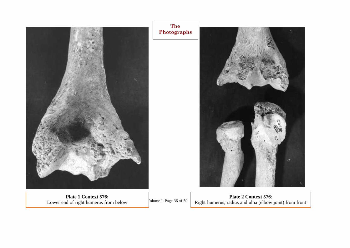
Volume I. Page 36 of 50Plate 2 Context 576:
Right humerus, radius and ulna (elbow joint) from frontPlate 1 Context 576:
Lower end of right humerus from below
ThePhotographs
Volume I. Page 37 of 50
Plate 3. Context 595: Left 5th metacarpal (on right of photo)showing results of old fracture. Compare with normal right5th matacarpal (left of photo) – this is more slender.
Plate 4. Context 615: The 5th left metatarsal (left of photo)has been fractired (also Xrayed). Compare with normal 5thright metatarsal (right of photo)

Volume I. Page 38 of 50
Context 754:
Plate 5 [Left] Left femur showing exaggerated tendoninsulation with the linea aspua (due to DISH)
Plate 6 [Above] Left hip bone, note the roughening andlipping of the muscular attachment to the iliac crest (DISH).

Volume I. Page 39 of 50
Plate 7 Context 957: Pseudarthrosis ofleft tibia and fibia viewed from behind.
Plate 8. Context 957: Pseudarthrosis ofleft tibia and fibia – these have beenseparated to demonstrate the irregulartoothed surfaces of the false new ‘joint’.

Volume I. Page 40 of 50
Plate 9 Context 971: ‘BambooSpine’. Fusion of thoracic vertibralbodies.
Plate 10 Context 971: Fusion of cervicalvertebral. Fusion of lumbar vertebral.

Volume I. Page 41 of 50
Plate 11 Context 972: [left] Earlyfusion of cervical vertebræ -(‘spondylitis’).
Plate 12 Context 980: [right] Iso-lated tibial shaft showing indentationand overgrowth of its margins due toexternal pressure from a soft tissuegrowth.

Volume I. Page 42 of 50
Plate 13 Context 987: [above] Atlas vertebra, failure of fusion of theposterior arch.Plate 14 Context 992: [right] 5th lumbar vertebra showing acongenital anomaly, spondylolisthesis.
Volume I. Page 43 of 50
Plate 15 Context 1007:[left] Metatarsophalangeal joint of greattoe showing arthritis.
Plate 16 Context 1022:[right] Mandible showingtorus formation lingually.

Volume I. Page 44 of 50
Plate 17 Context 1394: [left] Atlasand axis vertibræ. Gross arthriticchange between odontoid process ofaxis and anterior arch of atlas.
Plate 18 Context 1701 [right].
Plate 19 Context 1822: [below]Malunited fracture of ulna.

Volume I. Page 45 of 50
Plate 20 Context 1829 [left] Scapula (I) Acromion – unusual joint forclavicle – DISH; (ii) suprascapular foramen – congenital anomaly.
Plate 21 Context 1829 [above] Under surface of clavicle – a facet forarticulation with acromion of scapula a ligament is attached here but DISHhas caused unusual changes and development of a different type of joint.
Plate 22 Context 1833 [below] Arthritic cercivical vertebræ.
Volume I. Page 46 of 50
Plate 23 Context 1862 [left]: Unusual spinous process of L5 vertebra (bifidspinous process).
Plate 24 Context 1881 [below]: Fractured and healing left clavicle.

Volume I. Page 47 of 50
Plates 25 & 26
Context 1889:
Multiple Wormianbones, persistantmetopic suture.

Volume I. Page 48 of 50
Plates 27 & 28 Context 1893: Trephine holes in right parietal bone of skull.
Volume I. Page 49 of 50
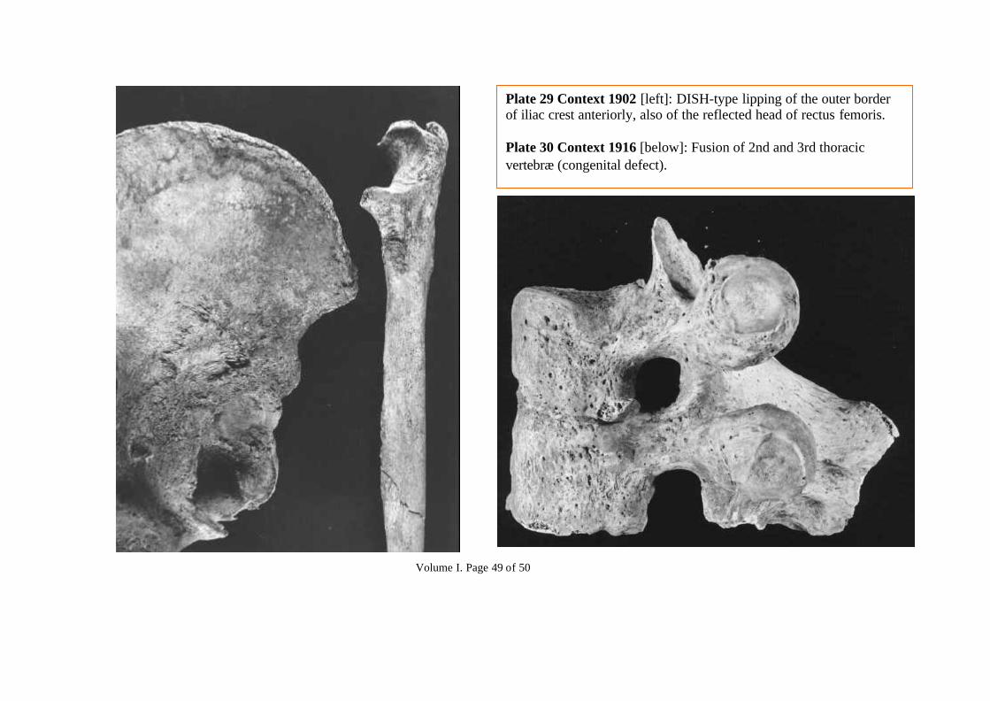
Plate 29 Context 1902 [left]: DISH-type lipping of the outer borderof iliac crest anteriorly, also of the reflected head of rectus femoris.
Plate 30 Context 1916 [below]: Fusion of 2nd and 3rd thoracicvertebræ (congenital defect).
Volume I. Page 50 of 50
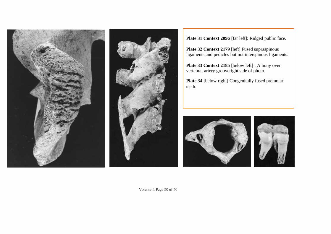
Plate 31 Context 2096 [far left]: Ridged public face.
Plate 32 Context 2179 [left] Fused supraspinousligaments and pedicles but not interspinous ligaments.
Plate 33 Context 2185 [below left] : A bony oververtebral artery grooveright side of photo.
Plate 34 [below right] Congenitally fused premolarteeth.