Enhanced Thermal Destruction of Listeria monocytogenes … · Staphylococci are- a major cause of...
6
Vol. 56, No. 9 APPLIED AND ENVIRONMENTAL MICROBIOLOGY, Sept. 1990, p. 2711-2716 0099-2240/90/092711-06$02.00/0 Copyright C) 1990, American Society for Microbiology Enhanced Thermal Destruction of Listeria monocytogenes and Staphylococcus aureus by the Lactoperoxidase System DAVID N. KAMAU,lt* STEPHANIE DOORES,1 AND KENNETH M. PRUIWTT Department of Food Science, The Pennsylvania State University, University Park, Pennsylvania 16802,1 and Department of Biochemistry, University of Alabama at Birmingham, Birmingham, Alabama 352942 Received 12 April 1990/Accepted 19 June 1990 The lactoperoxidase system (LPS) enhanced thermal destruction of Listeria monocytogenes and Staphylococ- cus aureus. After LPS activation, biphasic survival curves were observed for L. monocytogenes at 57.8°C and for S. aureus at 55.2°C. The data were consistent with a model that assumed two bacterial populations differing in heat sensitivity. The more heat-sensitive fractions (93% of the L. monocytogenes, 92% of the S. aureus) were killed almost instantly. For these biphasic survival curves, D values were based on the much smaller, less-heat-sensitive fractions. For L. monocytogenes, the D522°C values were 30.2 min (untreated milk) and 10.7 min (LPS activated); corresponding D552°C values were 8.2 and 1.6 min; corresponding D57.8.c values were 2.3 and 0.5 min. For S. aureus, the D522°C values were 33.3 min (untreated milk) and 2.2 min (LPS activated), and the corresponding D55s2°C values were 7.6 and 1.1 min, respectively. The most rapid killing of L. monocytogenes occurred when samples were heated soon after activation of the LPS. Activation of the LPS followed by heating can increase the margin of safety with respect to milkborne pathogens. Four major food-borne outbreaks of human listeriosis have been reported: one in Canada (35), two in the United States (14, 24), and one in Switzerland (19). Milk and milk products have been associated with three of these outbreaks (14, 19, 24). In addition to these outbreaks, several dairy products contaminated with Listeria monocytogenes have been recalled (15-18), costing the food industry millions of dollars. The incidence of L. monocytogenes in raw milk has been documented (8, 25). The ability of this pathogen to multiply at refrigeration temperatures (39) and its relatively higher heat resistance compared with other nonsporeforming bac- teria (2, 6, 11, 13) amplify the risk of milkborne listeriosis. Ironically, the main groups at risk include pregnant women and immunocompromised individuals for whom milk con- sumption is generally recommended (21, 24). Staphylococci are- a major cause of mastitis and may account for up to 30% of bovine mastitis (22). Thus, these bacteria can be shed into milk from udders of mastitic cows as well as from the milking environment. However, staph- ylococci are killed during pasteurization (20), and their presence in milk would be indicative of improper pasteur- ization. The lactoperoxidase system (LPS) consists of the enzyme lactoperoxidase (LP), thiocyanate (SCN-), and hydrogen peroxide (H202) and has a broad antimicrobial activity (31). The LP-catalyzed oxidation of SCN- by H202 generates hypothiocyanite anion (OSCN-) which exists in equilibrium with hypothiocyanous acid, PKa 5.3 (1, 37). The hypothio- cyanous acid plus OSCN- can oxidize essential sulfhydryl groups (-SH) in metabolic enzymes, thereby inhibiting bacterial growth (37, 38). Structural damage or changes in bacterial membranes with subsequent leakage or impaired uptake of nutrients, or both have also been reported (29, 31, 33). * Corresponding author. t Present address: Department of Biochemistry and School of Dentistry, SDB Box 26, University of Alabama at Birmingham, UAB Station, Birmingham, AL 35294. In milk, the LPS can be bacteriostatic or bactericidal against a diversity of milkborne spoilage and pathogenic bacteria (5, 7, 12, 31, 33). However, there are no reports on the effect of the LPS on thermal inactivation of bacteria. Consequently, the present study was undertaken to deter- mine the effect of the LPS on thermal resistance of bacteria suspended in milk. We report the dramatic increase in thermal destruction of L. monocytogenes and Staphylococ- cus aureus when exposed to the LPS prior to heating. (Part of this research was previously presented [D. N. Kamau, S. Doores, and K. M. Pruitt, Abstr. Annu. Meet. Am. Soc. Microbiol., 1989, P50, p. 327].) MATERIALS AND METHODS Reagents. Bovine milk lactoperoxidase (EC 1.11.1.7), horseradish peroxidase (EC 1.11.1.7), bovine liver catalase (EC 1.11.1.6), 2-2'-azino-di-(3-ethyl-benzthiozoline-6-sulfo- nic acid) (ABTS), ferric nitrate, leucocrystal violet, 2-mer- captoethanol, and 5-5'-dithiobis-(-2-nitrobenzoic acid) were purchased from Sigma Chemical Co. (St. Louis, Mo.). The purity index of LP was 0.76 (A412/A280) as reported by Sigma. Hydrogen peroxide (30%) and potassium thiocyanate were purchased from Fisher Scientific Co. (Pittsburgh, Pa.). Assay for LPS components. SCN- H202 and OSCN- concentrations were determined calorimetrically by using a Spectronic 21 spectrophotometer (Bausch & Lomb, Inc., Rochester, N.Y.). A Gilford Response UV-VIS spectropho- tometer (Gilford Instrument Laboratories, Inc., Oberlin, Ohio) was used for the LP activity assay. SCN- was assayed by the ferric nitrate method of Betts and Dainton (4). The assay for H202 was the leucocrystal violet method of Mottola et al. (30). To measure the concentrations of OSCN-, the method of Aune and Thomas (1) as modified by Mansson-Rahemtulla et al. (26) was used. For assay of OSCN- in milk, this method was modified as follows: to 6.0 ml of the 5-5'-dithiobis-(-2- nitrobenzoic acid) reagent was added 0.2 ml of the milk sample to be assayed, and the suspension was vortexed. To remove milk proteins, 0.1 ml of commercial cheese rennet (diluted 1:10 in distilled water) was added, followed by 0.1 2711 on January 18, 2020 by guest http://aem.asm.org/ Downloaded from
Transcript of Enhanced Thermal Destruction of Listeria monocytogenes … · Staphylococci are- a major cause of...

Vol. 56, No. 9APPLIED AND ENVIRONMENTAL MICROBIOLOGY, Sept. 1990, p. 2711-27160099-2240/90/092711-06$02.00/0Copyright C) 1990, American Society for Microbiology
Enhanced Thermal Destruction of Listeria monocytogenes andStaphylococcus aureus by the Lactoperoxidase System
DAVID N. KAMAU,lt* STEPHANIE DOORES,1 AND KENNETH M. PRUIWTTDepartment ofFood Science, The Pennsylvania State University, University Park, Pennsylvania 16802,1 and
Department ofBiochemistry, University ofAlabama at Birmingham, Birmingham, Alabama 352942
Received 12 April 1990/Accepted 19 June 1990
The lactoperoxidase system (LPS) enhanced thermal destruction of Listeria monocytogenes and Staphylococ-cus aureus. After LPS activation, biphasic survival curves were observed for L. monocytogenes at 57.8°C andfor S. aureus at 55.2°C. The data were consistent with a model that assumed two bacterial populations differingin heat sensitivity. The more heat-sensitive fractions (93% of the L. monocytogenes, 92% of the S. aureus) were
killed almost instantly. For these biphasic survival curves, D values were based on the much smaller,less-heat-sensitive fractions. For L. monocytogenes, the D522°C values were 30.2 min (untreated milk) and 10.7min (LPS activated); corresponding D552°C values were 8.2 and 1.6 min; corresponding D57.8.c values were 2.3and 0.5 min. For S. aureus, the D522°C values were 33.3 min (untreated milk) and 2.2 min (LPS activated), andthe corresponding D55s2°C values were 7.6 and 1.1 min, respectively. The most rapid killing of L. monocytogenesoccurred when samples were heated soon after activation of the LPS. Activation of the LPS followed by heatingcan increase the margin of safety with respect to milkborne pathogens.
Four major food-borne outbreaks of human listeriosishave been reported: one in Canada (35), two in the UnitedStates (14, 24), and one in Switzerland (19). Milk and milkproducts have been associated with three of these outbreaks(14, 19, 24). In addition to these outbreaks, several dairyproducts contaminated with Listeria monocytogenes havebeen recalled (15-18), costing the food industry millions ofdollars.The incidence of L. monocytogenes in raw milk has been
documented (8, 25). The ability of this pathogen to multiplyat refrigeration temperatures (39) and its relatively higherheat resistance compared with other nonsporeforming bac-teria (2, 6, 11, 13) amplify the risk of milkborne listeriosis.Ironically, the main groups at risk include pregnant womenand immunocompromised individuals for whom milk con-sumption is generally recommended (21, 24).
Staphylococci are- a major cause of mastitis and mayaccount for up to 30% of bovine mastitis (22). Thus, thesebacteria can be shed into milk from udders of mastitic cowsas well as from the milking environment. However, staph-ylococci are killed during pasteurization (20), and theirpresence in milk would be indicative of improper pasteur-ization.The lactoperoxidase system (LPS) consists of the enzyme
lactoperoxidase (LP), thiocyanate (SCN-), and hydrogenperoxide (H202) and has a broad antimicrobial activity (31).The LP-catalyzed oxidation of SCN- by H202 generateshypothiocyanite anion (OSCN-) which exists in equilibriumwith hypothiocyanous acid, PKa 5.3 (1, 37). The hypothio-cyanous acid plus OSCN- can oxidize essential sulfhydrylgroups (-SH) in metabolic enzymes, thereby inhibitingbacterial growth (37, 38). Structural damage or changes inbacterial membranes with subsequent leakage or impaireduptake of nutrients, or both have also been reported (29, 31,33).
* Corresponding author.t Present address: Department of Biochemistry and School of
Dentistry, SDB Box 26, University of Alabama at Birmingham,UAB Station, Birmingham, AL 35294.
In milk, the LPS can be bacteriostatic or bactericidalagainst a diversity of milkborne spoilage and pathogenicbacteria (5, 7, 12, 31, 33). However, there are no reports onthe effect of the LPS on thermal inactivation of bacteria.Consequently, the present study was undertaken to deter-mine the effect of the LPS on thermal resistance of bacteriasuspended in milk. We report the dramatic increase inthermal destruction of L. monocytogenes and Staphylococ-cus aureus when exposed to the LPS prior to heating.
(Part of this research was previously presented [D. N.Kamau, S. Doores, and K. M. Pruitt, Abstr. Annu. Meet.Am. Soc. Microbiol., 1989, P50, p. 327].)
MATERIALS AND METHODSReagents. Bovine milk lactoperoxidase (EC 1.11.1.7),
horseradish peroxidase (EC 1.11.1.7), bovine liver catalase(EC 1.11.1.6), 2-2'-azino-di-(3-ethyl-benzthiozoline-6-sulfo-nic acid) (ABTS), ferric nitrate, leucocrystal violet, 2-mer-captoethanol, and 5-5'-dithiobis-(-2-nitrobenzoic acid) werepurchased from Sigma Chemical Co. (St. Louis, Mo.). Thepurity index of LP was 0.76 (A412/A280) as reported bySigma. Hydrogen peroxide (30%) and potassium thiocyanatewere purchased from Fisher Scientific Co. (Pittsburgh, Pa.).
Assay for LPS components. SCN- H202 and OSCN-concentrations were determined calorimetrically by using aSpectronic 21 spectrophotometer (Bausch & Lomb, Inc.,Rochester, N.Y.). A Gilford Response UV-VIS spectropho-tometer (Gilford Instrument Laboratories, Inc., Oberlin,Ohio) was used for the LP activity assay.SCN- was assayed by the ferric nitrate method of Betts
and Dainton (4). The assay for H202 was the leucocrystalviolet method of Mottola et al. (30).To measure the concentrations of OSCN-, the method of
Aune and Thomas (1) as modified by Mansson-Rahemtulla etal. (26) was used. For assay of OSCN- in milk, this methodwas modified as follows: to 6.0 ml of the 5-5'-dithiobis-(-2-nitrobenzoic acid) reagent was added 0.2 ml of the milksample to be assayed, and the suspension was vortexed. Toremove milk proteins, 0.1 ml of commercial cheese rennet(diluted 1:10 in distilled water) was added, followed by 0.1
2711
on January 18, 2020 by guesthttp://aem
.asm.org/
Dow
nloaded from

APPL. ENVIRON. MICROBIOL.
ml of (NH4)2SO4 (20 mg/ml), and samples were held at 22°Cfor 5 min. The samples were then passed through 0.45-pum-pore-size filters (Metricel membrane filter no. 63068, GelmanSciences Inc., Ann Arbor, Mich.), and the A412 of 3.0 ml ofthe filtrate was measured against a reference milk sample inwhich no OSCN- was generated.The ABTS method of Schindler et al. (34) was used to
assay for LP activity in tryptic soy-yeast extract broth(TSB-YE) and in milk. To eliminate turbidity which caninterfere with this assay, 100-,ul milk samples were added to2.0 ml of ABTS reagent in 0.1 M sodium acetate buffer (pH4.5) and passed through 0.45-Rm-pore-size filters to removeacid-coagulated proteins. The mean LP activity in raw milksamples was 1.93 ABTS units per ml or 29.7 ,ug of LP per mlof milk. In the preheated milk samples, the mean residual LPactivity was 0.60 ABTS units per ml (9.2 jig of LP per ml or31%).Growth media and culture preparation. L. monocytogenes
Scott A (serotype 4b), an isolate from human listeriosisoutbreak, was obtained from M. Doyle, University of Wis-consin, Madison, Wis. S. aureus ATCC 12600 was obtainedfrom the American Type Culture Collection, Rockville, Md.
Tryptic soy broth and agar (Difco Laboratories, Detroit,Mich.) were prepared according to the instructions of themanufacturer with the addition of 0.6% yeast extract (Difco)to yield the TSB-YE or tryptic soy-yeast extract agar (TSA-YE). The pH of TSB-YE was adjusted to that of normal rawmilk (pH 6.6) by the addition of sufficient amounts of 2 Msodium acetate. The volume of sodium acetate added wassmall (a few milliliters per liter of TSB-YE). The resultingincrease in ionic strength would have no appreciable effecton the results since all cultures were grown in the samemedium.
Stock cultures of L. monocytogenes on TSA-YE slantswere incubated for 24 h at 35°C in the presence of 9.5% CO2.Working cultures of L. monocytogenes were prepared bytransferring a loopful from the stock cultures into TSB-YEfollowed by three consecutive 0.1-ml transfers and incuba-tion for 18 to 20 h at 35°C in the presence of 9.5% CO2.Working cultures of S. aureus were prepared as for L.
monocytogenes but with aerobic incubation at 37°C. Theworking cultures were maintained by daily transfers intofresh TSB-YE. Culture slants were refrigerated at 4°C.Raw milk samples. Raw milk samples, obtained from The
Pennsylvania State University dairy farm were asepticallydrawn from healthy cows after the udders were thoroughlycleaned with an iodine solution followed by a 75% ethanolswabbing and a sterile water rinse. After discarding the firstseveral discharges of milk from each teat, milk samples werecollected by hand milking into 500-ml sterile bottles. Theseraw milk samples contained less than 100 CFU/ml as esti-mated by standard plate count procedures (28). These sam-ples were preheated at 57°C for 20 min to eliminate thenatural milk flora as confirmed by spread plating on TSA-YEwith incubation for 48 h at 32°C.
Effect of the LPS on heat resistance of L. monocytogenes andS. aureus. An 18- to 20-h inoculum of L. monocytogenes orS. aureus was decimally diluted in phosphate buffer contain-ing magnesium chloride (3), and 0.1 ml of each of thesediluted inocula was pipetted into 10.0 ml of preheated milk toyield between 105 and 106 CFU/ml. These samples wereseparated into three groups: untreated milk, H202-treatedmilk, and LPS-treated milk.Enough SCN- and H202 to give initial concentrations of
2.4 mM SCN- and 0.6 mM H202 were added to milk for L.monocytogenes studies. For S. aureus studies, initial con-
centrations of 1.2 mM SCN- and 0.3 mM H202 were used.An inherent LP content of about 9.2 jig/ml was measured inpreheated milk. To determine thermal resistance, 2.0-mlportions of each sample were pipetted into sterile glassampoules (Wheaton no. 6202-12D; VWR Scientific, Bridge-port, N.J.) and flame sealed. A temperature-monitoringprobe (Omega Engineering, Inc., Stamford, Conn.) wasplaced in an ampoule (containing uninoculated heating men-struum) and epoxy sealed. Samples were brought to an initialtemperature of 35°C and then heated by complete immersioninto a constant-temperature water bath (Blue M ElectricCo., Blue Island, Ill.) to attain (come-up time of 2 to 3 min)holding temperatures of 52.2, 55.2, and 57.8°C. In prelimi-nary studies, rapid destruction of S. aureus was observed at57.8°C. Consequently, it was only practical to heat samplesat 52.2 and 55.2°C, especially in the presence of the LPS.At holding temperatures of 52.2, 55.2, and 57.8°C, un-
treated and H202-treated samples were removed at 10-, 5-,and 0.5-min intervals, respectively, for L. monocytogenes,while the LPS-treated samples were removed at 5-, 1-, and0.17-min intervals for the above heating temperatures, re-spectively. For S. aureus heated at 52.2°C, untreated, H202-treated, and LPS-treated samples were removed at 10-, 5-,and 1-min intervals, respectively, while at 55.2°C, the re-spective time intervals were 5, 2.5, and 0.42 min. Theampoules were immediately cooled in an ice-water bath,wiped with 95% ethanol, and broken open, and 0.1-mlvolumes were plated or transferred into 9.0 ml of dilutionbuffer for appropriate serial dilutions. Surviving bacteriawere enumerated by spread plating onto TSA-YE and wereincubated for 48 h at 35°C in the presence of 9.5% CO2 for L.monocytogenes and for 48 h at 37°C (aerobically) for S.aureus. For each of these bacteria, these experiments werereplicated four times, with a total of at least 14 observationsper treatment.
Influence of holding time after exposure to the LPS onthermal resistance of L. monocytogenes. This experiment wasdesigned to determine the length of time over which the LPScontinued to affect the heat resistance of L. monocytogenes.Culture preparation, activation of LPS, and heat resistanceprocedures were followed as previously described. How-ever, only LPS-treated milk samples were used since thegoal was to determine whether recovery of L. monocyto-genes from the LPS coincided with a return to normal heatresistance. After inoculation and activation of the LPS, milksamples were incubated at 35°C and decimal reduction times(D values) were determined after 0, 2, 6, 8, 14, and 16 h. Thisexperiment was done in duplicate, with a total of at least 24observations for each holding time.
RESULTSApplication of the logistic model to survival data analyses. D
values are usually reported as the absolute values of thereciprocal of the slope of the plot of log1o survivors versustime (survival curve). However, our data did not fit thesimple logarithmic model of death. Consequently, data wereanalyzed with the logistic model which can be used toanalyze nonlogarithmic survival data (23). The general formof the logistic equation used to analyze our data was:
S = CFU/CFUo= 2/[1 + e(pt)] (1)where S is the surviving fraction at time t, CFU is thenumber of surviving bacteria, CFUO is the initial CFU, e isthe base of the natural logarithm, and
, = 4[(dCFU/dt)max]/CFUO (2)
2712 KAMAU ET AL.
on January 18, 2020 by guesthttp://aem
.asm.org/
Dow
nloaded from

LPS-ENHANCED HEAT DESTRUCTION 2713
The differential (dCFU/dt)max is the slope of the survivalcurve at the point of inflection or the maximum killing rate,and /4 is the maximum specific killing rate. The log trans-form of equation 1 is:
log S = log 2 - log [1 + e(pt)] (3)where log S is the log (CFU/CFUo) at any given time. The Dvalue is given by:
D value = 2/3 (4)For survival curves with no initial lag in killing and only
one distinct killing phase, data were fitted to equation 3.When survival curves displayed an initial lag in death
followed by a distinct one-phase killing, data were fitted tothe following form of equation 3:
log S = log[1 + e(-Pt112)] - log {1 + e[3(t -t12)]} (5)
where t1l2 is the time at which the number of CFU is CFUJ2and is a measure of lag in killing, and the other terms are asdefined for equations 1 through 3.Data for survival curves with no initial lag in killing but
having two distinct killing phases (biphasic) were fitted to thefollowing two-term exponential form of equation 3:
log S = log ({2F1/[1 + e(P1t)]} + {2(1 - F1)l[1 + e(P2t)]}), (6)
where F1 and (1 - F1) represent two fractions of bacteria(differing with respect to heat resistance) and P, and 12 arethe specific killing rates for the two fractions, respectively.This model assumes that the two fractions (populations) arekilled exponentially but at different, independent rates.When the value of CFU is 1, then t is equal to t1, and t1 is
the time required for CFUo to drop to 1. The value of t1 canbe calculated from the respective logistic equation by usingthe values of t1/2 and 1 obtained by nonlinear regression. Thevalue of t1 is a good estimate of the time needed for almostcomplete killing of all of the inoculum.
Regression analysis of data from four separate experi-ments (a total of 40 observations) by the simple log modelgave a mean square error of 1.09 x 10⁻². Analysis of thesame data by use of the log transform of the logistic modelgave a mean square error of 4.76 x 10⁻³. Thus, data analysisby use of the logistic model reduced the mean square errorby more than 40% compared with the results obtained by useof the simple log model. Therefore, we used the appropriatelogistic equations (i.e., equations 3, 5, and 6) for dataanalyses.Data were fitted to the various forms of the logistic
equation by nonlinear regression (32). Analysis of variancewith mean separation by the least significant differenceprocedure (36) was used to determine whether there weresignificant differences among parameter estimates (P0.05).
Influence of the LPS on thermal resistance of L. monocyto-genes. Typical results on the effect of the LPS on thermaldestruction of L. monocytogenes are shown in Table 1 andFig. 1. In milk, the LPS significantly reduced the D values by64.6% at 52.2°C, 80.5% at 55.2°C, and 78.3% at 57.8°C (Table1). Alone, H202 treatment had no significant effect on the Dvalues of L. monocytogenes in milk (Table 1).An initial lag in killing was observed at 52.2 and 55.2°C for
all survival data (Fig. 1A and B), and data were fitted toequation 5. At 57.8°C, survival curves for L. monocytogenesin H202- and LPS-treated milk revealed that there were twokilling phases (biphasic), an initial rapid phase lasting a fewseconds followed by a slower killing phase. To account for
TABLE 1. Effect of the LPS on survival curve parametersa forL. monocytogenes heated in milk at 52.2, 55.2, and 57.8°C
Sample Temp ,B/4 (±SD) t1/2 (±SD) t1e D valuetreatmentb (OC) 10-2/min)' (min)d (min) (minY
Milk 52.2 1.7 ± 0.2 42.0 + 2.1 198 30.2 ± 3.7Milk + HP 52.2 1.7 ± 0.3 25.8 ± 5.3 195 29.4 ± 3.3Milk + LPS 52.2 4.7 ± 0.2 0 70.3 10.7 ± 0.4Milk 55.2 6.1 ± 0.8 16.6 ± 1.2 52.8 8.2 ± 1.1Milk + HP 55.2 4.5 ± 0.6 12.8 ± 1.7 71.4 11.1 ± 1.4Milk + LPS 55.2 30.4 ± 3.5 3.2 ± 0.3 10.6 1.6 ± 0.9Milk 57.8 21.3 ± 1.0 0 15.1 2.3 ± 0.1Milk + HP 57.8 19.2 ± 2.1X 0 15.7 2.6 ± 0.5Milk + LPS 57.8 104.2 ± 14.79 0 2.6 0.5 + 0.1
" Parameter estimates were obtained by fitting survival data to the appro-priate logistic equation by nonlinear regression (32).
b Abbreviations: Milk, untreated milk; Milk + HP, milk + 0.6 mM H202;Milk + LPS, milk + 2.4 mM SCN- + 0.6 mM H202 + 9.2 jig of inherent LPper ml.
' Maximum specific killing rate (1/4) = (dCFU/dt)max/CFUo.d Time at which CFU = CFUJ2, where CFUo = initial CFU/ml.e Predicted time for surviving CFU/ml = 1.fD value = 2/13 at (dCFU/dt)max.g These are 12/4 values, where D value = 2/X2 based on the less-heat-
sensitive fraction 1 - F1 = 0.4 for Milk + HP and 0.07 for Milk + LPS.
this observation, the two-term exponential equation 6 wasused to analyze the data. It was assumed that there were twobacterial fractions, a heat-sensitive fraction (F1) which wasrapidly killed and a less-heat-sensitive fraction (1 - F1)which was killed at a slower rate.For the LPS- and H202-treated samples at 57.8°C, the two
fractions were as follows: F1 = 0.93 and 1 -F1 = 0.07 inLPS-treated milk and F1 = 0.6 and 1 - F1 = 0.4 inH202-treated milk. The P13/4 and P2/4 values represent themaximum specific death rates for the F1 and 1 - F1 frac-tions, respectively. The 1/4 values were so large that theterm {2F1/[1 + e(p1t)]} in equation 6 was zero for all valuesof t > 0, suggesting instant killing of fraction F1. Thisfraction was greater in the LPS-treated (93% of population)than in H202-treated (60%) milk. The D values in LPS- andH202-treated milk heated at 57.80C were estimated from 132,based on the less-heat-sensitive fraction (Table 1).The LPS also significantly increased the maximum specific
killing rates of L. monocytogenes in milk by 176% at 52.2°C,398% at 55.2°C, and 388% at 57.8°C (Table 1). In all cases,the t1/2 and t1 values were the lowest in the presence of theLPS.
Effect of holding time after exposure to the LPS on heatresistance of L. monocytogenes in milk. The data for 0 and 2 hof holding time were consistent with equation 5; 6-, 14-, and16-h holding data fit equation 3; and 8-h holding data fitequation 6. These variations can be attributed to differentkinetics of heat injury and recovery. Data were fitted to theappropriate equations by nonlinear regression (32). Whenthe LPS was activated and samples were held at 35°C priorto heating, the resulting D values increased gradually from1.4 min at 0 h to 6.7 min (a 378.6% increase) after 16 h (Table2). After an 8-h holding time, the D values approached thoseobserved for H202-treated and untreated controls (Table 1).With prolonged holding, a decrease in the maximum specifickilling rate was also observed (Table 2). The highest killingrates and the lowest D values were observed when sampleswere heated immediately after activation of the LPS. Thepredicted time needed to reduce the initial number of CFUper milliliter by half (t112) after 6 h of holding was 0 min,
VOL. 56, 1990
on January 18, 2020 by guesthttp://aem
.asm.org/
Dow
nloaded from
APPL. ENVIRON. MICROBIOL.
I -~~~~~T"- -1- Tr ~ ~~~~~TOuLAXi
2T 0 MILK + HP
-2 A ILK+LPS
f-3- ,-
4- I I4 I j0 10 20 30 40 50 6
Tim (min)
05.
0.01
l , 0.51
h -0.5I00
3 -1.5
B OMILI0 MLK + HP
TH vMIL4-+ LPS
T
T.1.
1+
41
0
0 5 10 15 20 25 30
lnme (min)
0.5.
0.0jIolI -1.0.
i 1.5
° -2.0-
-25-5
. -
0.0 0.5 1.0 1.5 2.0 2.5 30 3.5
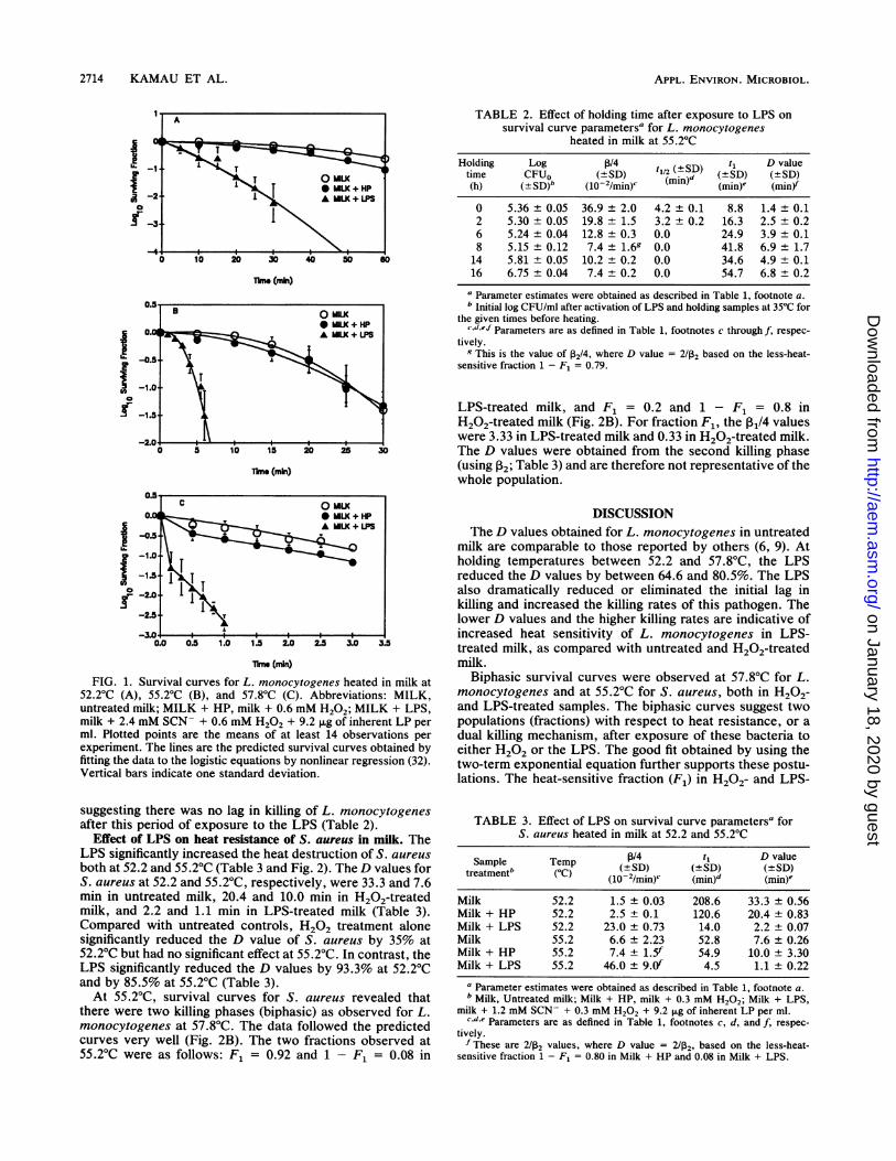
lnme (mlin)FIG. 1. Survival curves for L. monocytogenes heated in milk at
52.2°C (A), 55.2°C (B), and 57.8'C (C). Abbreviations: MILK,untreated milk; MILK + HP, milk + 0.6 mM H202; MILK + LPS,milk + 2.4 mM SCN- + 0.6 mM H202 + 9.2 jig of inherent LP perml. Plotted points are the means of at least 14 observations perexperiment. The lines are the predicted survival curves obtained byfitting the data to the logistic equations by nonlinear regression (32).Vertical bars indicate one standard deviation.
suggesting there was no lag in killing of L. monocytogenesafter this period of exposure to the LPS (Table 2).
Effect of LPS on heat resistance of S. aureus in milk. TheLPS significantly increased the heat destruction of S. aureusboth at 52.2 and 55.2'C (Table 3 and Fig. 2). The D values forS. aureus at 52.2 and 55.2'C, respectively, were 33.3 and 7.6min in untreated milk, 20.4 and 10.0 min in H202-treatedmilk, and 2.2 and 1.1 min in LPS-treated milk (Table 3).Compared with untreated controls, H202 treatment alonesignificantly reduced the D value of S. aureus by 35% at52.2'C but had no significant effect at 55.2'C. In contrast, theLPS significantly reduced the D values by 93.3% at 52.2'Cand by 85.5% at 55.2'C (Table 3).At 55.2'C, survival curves for S. aureus revealed that
there were two killing phases (biphasic) as observed for L.monocytogenes at 57.8'C. The data followed the predictedcurves very well (Fig. 2B). The two fractions observed at55.2'C were as follows: F1 = 0.92 and 1 - F1 = 0.08 in
TABLE 2. Effect of holding time after exposure to LPS onsurvival curve parametersa for L. monocytogenes
heated in milk at 55.2°C
Holding Log t1/4SD t, D valuetime CFUo (±SD) t2 (d)(SD) (SD)(h) (±SD)b (10-2/min)c (min) (min) (min)
0 5.36 ± 0.05 36.9 ± 2.0 4.2 + 0.1 8.8 1.4 ± 0.12 5.30 ± 0.05 19.8 ± 1.5 3.2 ± 0.2 16.3 2.5 ± 0.26 5.24 ± 0.04 12.8 ± 0.3 0.0 24.9 3.9 ± 0.18 5.15 ± 0.12 7.4 ±1.6 0.0 41.8 6.9 ± 1.714 5.81 ± 0.05 10.2 ± 0.2 0.0 34.6 4.9 ± 0.116 6.75 ± 0.04 7.4 ± 0.2 0.0 54.7 6.8 ± 0.2
a Parameter estimates were obtained as described in Table 1, footnote a.b Initial log CFU/ml after activation of LPS and holding samples at 35°C for
the given times before heating.c.d.e.f Parameters are as defined in Table 1, footnotes c through f, respec-
tively.9 This is the value of P2/4, where D value = 2/P2 based on the less-heat-
sensitive fraction 1 - F, = 0.79.
LPS-treated milk, and F1 = 0.2 and 1 - F1 = 0.8 inH202-treated milk (Fig. 2B). For fraction F1, the 131/4 valueswere 3.33 in LPS-treated milk and 0.33 in H202-treated milk.The D values were obtained from the second killing phase(using P2; Table 3) and are therefore not representative of thewhole population.
DISCUSSIONThe D values obtained for L. monocytogenes in untreated
milk are comparable to those reported by others (6, 9). Atholding temperatures between 52.2 and 57.8°C, the LPSreduced the D values by between 64.6 and 80.5%. The LPSalso dramatically reduced or eliminated the initial lag inkilling and increased the killing rates of this pathogen. Thelower D values and the higher killing rates are indicative ofincreased heat sensitivity of L. monocytogenes in LPS-treated milk, as compared with untreated and H202-treatedmilk.
Biphasic survival curves were observed at 57.8°C for L.monocytogenes and at 55.2°C for S. aureus, both in H202-and LPS-treated samples. The biphasic curves suggest twopopulations (fractions) with respect to heat resistance, or adual killing mechanism, after exposure of these bacteria toeither H202 or the LPS. The good fit obtained by using thetwo-term exponential equation further supports these postu-lations. The heat-sensitive fraction (F1) in H202- and LPS-
TABLE 3. Effect of LPS on survival curve parametersa forS. aureus heated in milk at 52.2 and 55.2°C
Sample Temp P/4 t5 D valuetemetb (-C(SD) (±+SD) (-SD)treatmplen (OC) (10-2/min)c (min)d (min)'
Milk 52.2 1.5 ± 0.03 208.6 33.3 + 0.56Milk + HP 52.2 2.5 ± 0.1 120.6 20.4 ± 0.83Milk + LPS 52.2 23.0 ± 0.73 14.0 2.2 ± 0.07Milk 55.2 6.6 + 2.23 52.8 7.6 ± 0.26Milk + HP 55.2 7.4 + 1.5f 54.9 10.0 ± 3.30Milk + LPS 55.2 46.0 ± 9.0f 4.5 1.1 ± 0.22
' Parameter estimates were obtained as described in Table 1, footnote a.b Milk, Untreated milk; Milk + HP, milk + 0.3 mM H202; Milk + LPS,
milk + 1.2 mM SCN- + 0.3 mM H202 + 9.2 ,ug of inherent LP per ml.c.d.e Parameters are as defined in Table 1, footnotes c, d, and f, respec-tively.f These are 2/P2 values, where D value = 2/P2, based on the less-heat-
sensitive fraction 1 - F1 = 0.80 in Milk + HP and 0.08 in Milk + LPS.
A
C OM LK
A MIL + LPS
ITITII1A
2714 KAMAU ET AL.
-.-
on January 18, 2020 by guesthttp://aem
.asm.org/
Dow
nloaded from
LPS-ENHANCED HEAT DESTRUCTION 2715
.0. * MILK + HP
0 0E! -1.5~
.5 -2.0 .
cn -2.5- l l
0 5 10b 15 20 25 30 35 40 45 S0 Se
rime (min)
n_s .
c 0.04
a -0.5-
oo-5 -2.0-
0 2 8 10 12 14 16 15
Time (min)
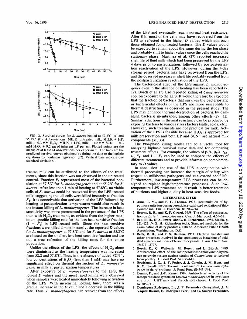
FIG. 2. Survival curves for S. aureus heated at 52.2°C (A) and55.2°C. (B). Abbreviations: MILK, untreated milk; MILK + HP,milk + 0.3 mM H202; MILK + LPS, milk + 1.2 mM SCN- + 0.3mM H202 + 9.2 jig of inherent LP per ml. Plotted points are themeans of at least 14 observations per experiment. The lines are thepredicted survival curves obtained by fitting the data to the logisticequations by nonlinear regression (32). Vertical bars indicate onestandard deviation.
treated milk can be attributed to the effects of the treat-ments, since this fraction was not observed in the untreatedcontrol. Fraction F1 represented most of the bacterial pop-ulation at 57.8°C for L. monocytogenes and at 55.2°C for S.aureus. After less than 1 min of heating at 57.8°C, no viablecells of S. aureus could be recovered from the LPS-treatedmilk, suggesting that all cells were killed instantly as fractionF1. It is conceivable that activation of the LPS followed byheating to pasteurization temperatures would also result inthe instant killing of L. monocytogenes. The increase in heatsensitivity was more pronounced in the presence of the LPSthan with H202 treatment, as evident from the higher max-imum specific killing rate for the less-heat-sensitive fraction(1 - F1) in LPS-treated milk. Since the heat-sensitivefractions were killed almost instantly, the reported D valuesfor L. monocytogenes at 57.8°C and for S. aureus at 55.2°Care based on the smaller, less-heat-sensitive fraction and arenot a true reflection of the killing rates for the entirepopulation.
Unlike the effects of the LPS, the effects of H202 alonewere diminished as the heating temperature was increasedfrom 52.2 and 57.8°C. Thus, in the absence of added SCN-,low concentrations of H202 (less than 1 mM) may have no
significant effect on thermal destruction of L. monocyto-genes in milk at pasteurization temperatures.
After exposure of L. monocytogenes to the LPS, thelowest D values and the most rapid killing were observedwhen samples were heated immediately following activationof the LPS. With increasing holding time, there was a
gradual increase in the D value and a decrease in the killingrate. Thus, L. monocytogenes can recover from the effects
of the LPS and eventually regain normal heat resistance.After 8 h, most of the cells may have recovered from theLPS as reflected in the higher D values which approachthose obtained for untreated bacteria. The D values wouldbe expected to remain about the same during the log phaseand probably shift to higher values once the cells reached thestationary phase. Martinez et al. (27) reported increasedshelf life of fluid milk which had been preserved by the LPS4 days prior to pasteurization, followed by postpasteuriza-tion reactivation of the LPS. However, during the 4-daystorage period, bacteria may have recovered from the LPS,and the observed increase in shelf life probably resulted fromthe postpasteurization reactivation of the LPS.The bactericidal effect of the LPS against L. monocyto-
genes even in the absence of heating has been reported (7,12). Borch et al. (5) also reported killing of Campylobacterspp. on exposure to the LPS. It would therefore be expectedthat the fraction of bacteria that survives the bacteriostaticor bactericidal effects of the LPS are more susceptible tothermal destruction as observed in the present study. TheLPS may enhance thermal destruction of bacteria by dam-aging bacterial membranes, among other effects (29, 31).Similar reductions in thermal resistance can be produced byexposing bacteria to various stress factors (salts, acids, etc.).However, such treatments are not practical for milk. Acti-vation of the LPS is feasible because H202 is approved formilk preservation and both LP and SCN- are natural milkcomponents (31, 33).The two-phase killing model can be a useful tool for
analyzing biphasic survival curve data and for comparingtreatment effects. For example, the values of IB1/4, P2/4, t1/2,t1, F1, and 1 - F1 can be used to compare the effects ofdifferent treatments and to provide information complemen-tary to D values.
In conclusion, the use of the LPS in conjunction withthermal processing can increase the margin of safety withrespect to milkborne pathogens and can extend shelf life.Furthermore, low-temperature LPS processes can be de-signed to replace high-temperature processes. Such low-temperature LPS processes could result in better retentionof nutrients and higher quality in heat-sensitive foods.
LITERATURE CITED1. Aune, T. M., and E. L. Thomas. 1977. Accumulation of hy-
pothiocyanite ion during peroxidase-catalyzed oxidation of thio-cyanate ion. Eur. J. Biochem. 80:209-214.
2. Bearns, R. E., and K. F. Girard. 1958. The effect of pasteuriza-tion on Listeria monocytogenes. Can. J. Microbiol. 4:55-61.
3. Bell, R. H., D. A. Power, and G. H. Richardson. 1985. Media, p.121-122. In G. H. Richardson (ed.), Standard methods for theexamination of dairy products, 15th ed. American Public HealthAssociation, Washington, D.C.
4. Betts, R. H., and F. S. Dainton. 1953. Electron transfer andother processes involved in the spontaneous bleaching of acid-ified aqueous solutions of ferric thiocyanate. J. Am. Chem. Soc.75:5721-5727.
5. Borch, E., C. Wallentin, M. Rosen, and L. Bjorck. 1989.Antibacterial effect of the lactoperoxidase-thiocyanate-hydro-gen peroxide system against strains of Campylobacter isolatedfrom poultry. J. Food Prot. 52:638-641.
6. Bradshaw, J. G., J. T. Peeler, J. J. Corwin, J. M. Hunt, andR. M. Twedt. 1987. Thermal resistance of Listeria monocyto-genes in dairy products. J. Food Prot. 50:543-544.
7. Dennis, F., and J.-P. Ramet. 1989. Antibacterial activity of thelactoperoxidase system on Listeria monocytogenes in trypticasesoy broth, UHT milk and French soft cheese. J. Food Prot.52:706-711.
8. Dominguez Rodriguez, L., J. F. Fernandez Garayzabal, J. A.Vaquez Boland, E. Rodriguez Ferri, and G. Suarez Fernandez.
A O MILK
B O MILK* MILK + HPA MILK + LPS
A\
VOL. 56, 1990
on January 18, 2020 by guesthttp://aem
.asm.org/
Dow
nloaded from

APPL. ENVIRON. MICROBIOL.
1985. Isolation de micro-organismes du genre Listeria a partir delait cru destine a la consommation humaine. Can. J. Microbiol.31:938-941.
9. Donnelly, C. W., and E. H. Briggs. 1986. Psychotrophic growthand thermal inactivation of Listeria monocytogenes as a func-tion of milk composition. J. Food Prot. 49:994-998.
10. Donnelly, C. W., E. H. Briggs, and L. S. Donnelly. 1987.Comparison of heat resistance of Listeria monocytogenes inmilk determined by two methods. J. Food Prot. 50:14-17.
11. Doyle, M. P., K. A. Glass, J. T. Beery, G. A. Garcia, D. J.Pollard, and R. D. Schultz. 1987. Survival of Listeria monocy-togenes in milk during high-temperature, short-time pasteuriza-tion. Appl. Environ. Microbiol. 53:1433-1438.
12. Earnshaw, R. G., and J. G. Banks. 1989. A note on the inhibitionof Listeria monocytogenes NCTC 11994 in milk by an activatedlactoperoxidase system. Lett. Appl. Microbiol. 8:203-205.
13. Fernandez Garayzabal, J. F., L. Dominguez Rodriguez, J. A.Vazquez Boland, E. F. Rodriguez Ferri, V. Briones Dieste, J. L.Blanco Cancelo, and G. Suarez Fernandez. 1987. Survival ofListeria monocytogenes in raw milk treated in a pilot plant sizepasteurizer. J. Appl. Bacteriol. 63:533-537.
14. Fleming, D. W., S. L. Cochi, K. L. MacDonald, J. Brondum, P.S. Hayes, B. D. Plikaytis, M. B. Holmes, A. Audurier, C. V.Broome, and A. L. Reingold. 1985. Pasteurized milk as a vehicleof infection in an outbreak of listeriosis. N. Engl. J. Med.312:404-407.
15. Food and Drug Administration. 1986. Class I recall: milk prod-ucts. FDA Enforcement Report, June 25, 1986.
16. Food Chem. News. 1985. FDA finds Listeria in three brands ofGeneral Foods soft cheeses. Food Chem. News 27(25):33.
17. Food Chem. News. 1986. FDA finds Listeria in brie cheese froma French-certified plant. Food Chem. News, Feb. 17, 1986.
18. Food Chem. News. 1986. Brie cheese recalls extended byGeneral Foods, U.S. importer. Food Chem. News, March 3,1986.
19. Food Chem. News. 1987. N.Y. Times reports Listeria outbreakin Switzerland from soft cheese. Food Chem. News, Dec. 7,1987.
20. Gilmour, A., and M. T. Rowe. 1981. Microorganisms associatedwith milk, p. 35-73. In R. K. Robinson (ed.), Food microbiol-ogy, vol. 1. Applied Science Publishers, New York.
21. Gray, M. L., and A. H. Killinger. 1966. Listeria monocytogenesand listeric infections. Bacteriol. Rev. 30:309-382.
22. Hayes, S. (ed.). 1981. Dairy microbiology. National Dairy Coun-cil, London.
23. Johnson, F. H., H. Eyring, and B. J. Stover (ed.). 1974. Thetheory of rate processes in biology and medicine, p. 2-5,564-574. John Wiley & Sons, Inc., New York.
24. Linnan, M. J., L. Mascola, X. D. Lou, V. Goulet, S. May, C.Salminen, D. Hird, M. L. Yonekura, P. Hayes, R. Weaver, A.Audurier, B. D. Plikaytis, S. L. Fannin, A. Kleks, and C. V.Broome. 1988. Epidemic listeriosis associated with Mexican-style cheese. N. Engl. J. Med. 319:823-828.
25. Lovett, J., D. W. Francis, and J. M. Hunt. 1987. Listeria
monocytogenes in raw milk: detection, incidence, and pathoge-nicity. J. Food Prot. 50:188-192.
26. Mansson-Rahemtulla, B., D. C. Baldone, K. M. Pruitt, and F.Rahemtulla. 1986. Specific assays for peroxidases in humansaliva. Arch. Oral Biol. 13:661-668.
27. Martinez, C. E., P. G. Mendoza, F. J. Alacron, and H. S. Garcia.1988. Reactivation of the lactoperoxidase system during rawmilk storage and its effect on the characteristics of pasteurizedmilk. J. Food Prot. 51:558-561.
28. Messer, J. W., H. M. Behney, and L. 0. Leudecke. 1985.Microbiological count methods, p. 133-150. In G. H. Richard-son (ed.), Standard methods for the examination of dairyproducts, 15th ed. American Public Health Association, Wash-ington, D.C.
29. Mickelson, M. N. 1977. Glucose transport in Streptococcusagalactiae and its inhibition by lactoperoxidase-thiocyanate-hydrogen peroxide. J. Bacteriol. 132:541-548.
30. Mottola, H. A., B. E. Simpson, and G. Gorin. 1970. Absorptio-metric determination of hydrogen peroxide in submicrogramamounts with leucocrystal violet and peroxidase as catalyst.Anal. Chem. 42:410-411.
31. Pruitt, K. M., and B. Reiter. 1985. Biochemistry of peroxidasesystem: antimicrobial effects, p. 143-178. In K. M. Pruitt and J.Tenovuo (ed.), The lactoperoxidase system. Marcel Dekker,Inc., New York.
32. Ralston, R. 1985. Derivative-free nonlinear regression, p. 305-314. In W. J. Dixon (ed.), BMDP statistical software manual.University of California Press, Berkeley, Calif.
33. Reiter, B. 1985. The biological significance of the non-immuno-globulin protective proteins in milk: lysozyme, lactoferrin,lactoperoxidase, p. 281-336. In P. F. Fox (ed.), Developmentsin dairy chemistry, vol. 3. Elsevier Applied Science Publishers,New York.
34. Schindler, J. S., R. E. Childs, and W. G. Bardsley. 1976.Peroxidase from human cervical mucus. The isolation andcharacterization. Eur. J. Biochem. 65:325-331.
35. Schlech, W. F., P. M. Lavigne, R. A. Bortolussi, A. C. Allen,E. V. Haldane, A. J. Wort, A. W. Hightower, S. E. Johnson,S. H. King, E. S. Nicholls, and C. V. Broome. 1983. Epidemiclisteriosis-evidence for transmission by food. N. Engl. J. Med.308:203-206.
36. Steel, R. G. D., and J. H. Torrie (ed.). 1980. Principles andprocedures of statistics: a biometric approach, 2nd ed. p.173-194. McGraw-Hill Book Co., New York.
37. Thomas, E. L. 1985. Products of lactoperoxidase-catalyzedoxidation of thiocyanate and halides, p. 31-53. In K. M. Pruittand J. Tenovuo (ed.), The lactoperoxidase system. MarcelDekker, Inc., New York.
38. Thomas, E. L., and T. M. Aune. 1978. Lactoperoxidase, perox-ide, thiocyanate antimicrobial system: correlation of sulfhydryloxidation with antimicrobial action. Infect. Immun. 20:456-463.
39. Wilkins, P. O., R. Bourgeois, and R. G. E. Murray. 1972.Psychrotrophic properties of Listeria monocytogenes. Can. J.Microbiol. 18:543-551.
2716 KAMAU ET AL.
on January 18, 2020 by guesthttp://aem
.asm.org/
Dow
nloaded from