Isolation & Identification of Staphylococci. Staphylococci Characteristics Staphylococci are often...
34
Isolation & Identification of Staphylococci م ي ح ر ل ا ن م ح ر ل ا ه ل ل ا م س ب
-
Upload
jody-marshall -
Category
Documents
-
view
224 -
download
0
Transcript of Isolation & Identification of Staphylococci. Staphylococci Characteristics Staphylococci are often...
Isolation & Identification of Staphylococci
بسم الله الرحمن الرحيم
Staphylococci Characteristics
Staphylococci are often found in the human nasal cavity (and on other mucous membranes) as well as on the skin.
Gram-positive cocci 0.8-1.0µm in diameter and occur singly, in pairs, in short chains, and most commonly, in irregular grape-like clusters.
Catalase positive.
Oxidase negative
Reduce nitrates to nitrites.
Generally tolerate relatively high concentrations of sodium chloride (7.5-10%) and tellurite. This ability is often employed in preparing media selective for staphylococci.

Gram positive cocci
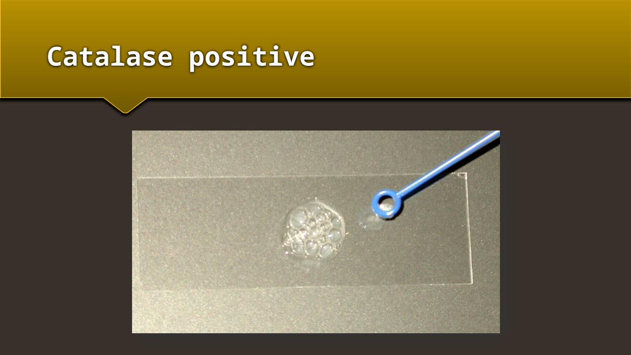
Catalase positive
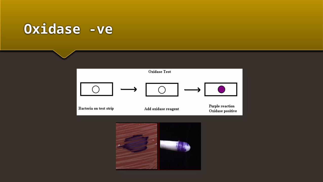
Oxidase -ve
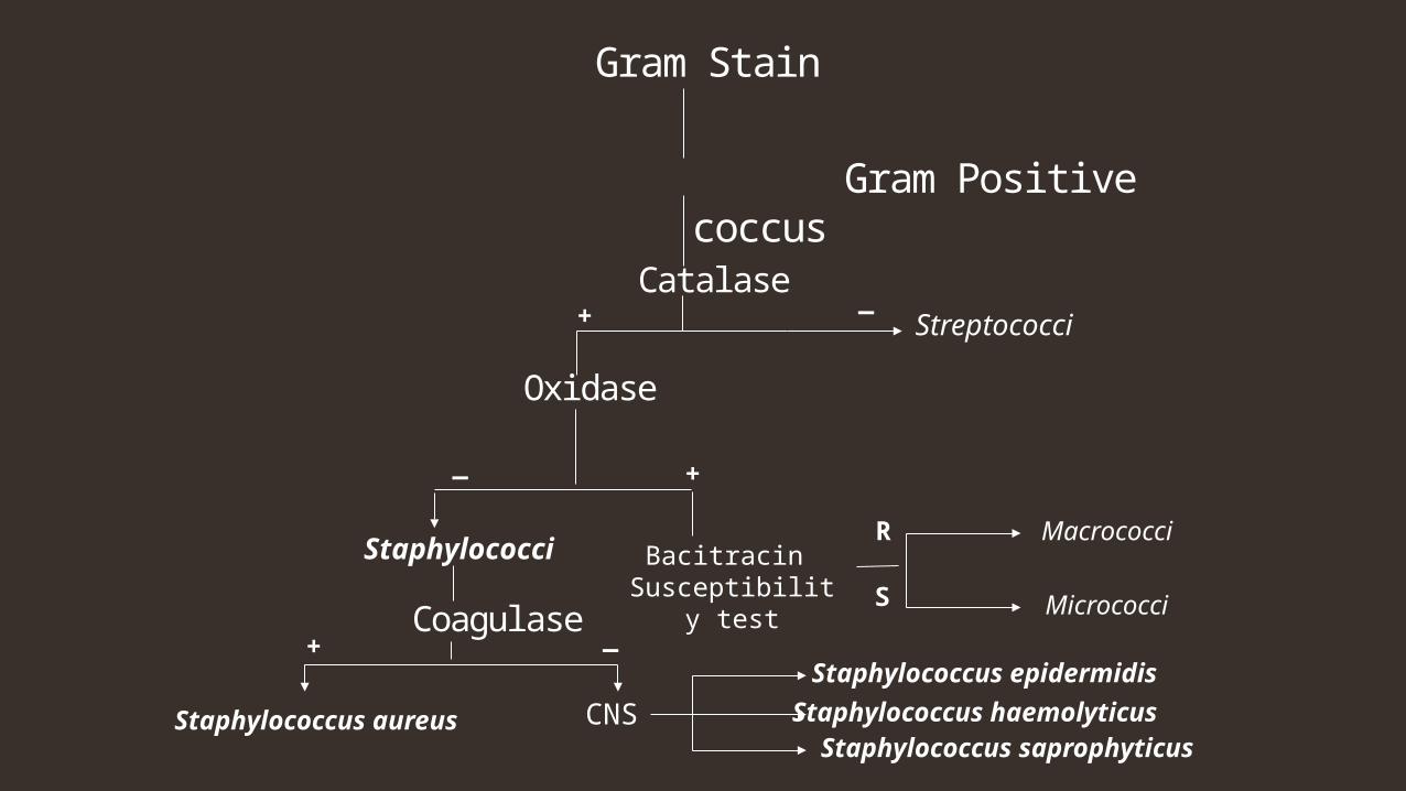
Staphylococci Species
There are five species of staphylococci commonly associated with clinical infections:
1) Staphylococcus aureus.
2) Staphylococcus epidermidis.
3) Staphylococcus haemolyticus.
4) Staphylococcus saprophyticus.
Gram Positive coccus
Catalase
Oxidase
Staphylococci
Streptococci+ _
Gram Stain
_ +
Bacitracin Susceptibility test
Coagulase
Staphylococcus aureus
_+
CNS Staphylococcus epidermidis
Staphylococcus saprophyticus
Macrococci
MicrococciS
R
Staphylococcus haemolyticus
Staphylococcus aureus
Staphylococcus aureus is the most pathogenic species and is implicated in a variety of infections. Approximately 30% of adults and most children are healthy nasal carriers of S. aureus. In the majority of S. aureus infections the source of the organism is either the healthy nasal carrier or contact with an abscess from an infected individual. The portal of entry is usually the skin. S. aureus causes pus-filled inflammatory lesions known as abscesses.
Depending on the location and extent of tissue involvement, the abscess may be called a pustule (an infected hair follicle), a furuncle or boil (if it spreads from the hair follicle to adjacent subcutaneous tissue), or a carbuncle(multiple infection sites involving deeper connective tissue).
It may also spread through soft tissues and cause cellulitis. S. aureus frequently causes infections of accidental wounds and postoperative wounds, although it can also infect healthy intact skin.
Less commonly, S. aureus may escape from the local lesion and spread through the blood to other body areas, causing a variety of systemic Infections that may involve every system and organ. Such systemic infections include septicemia, septic arthritis, endocarditis, meningitis, and osteomyelitis, as well as abscesses in the lungs, spleen, liver, and kidneys.
S. aureus pneumonia may also be a secondary respiratory complication of viral infections such as measles and influenza.
Finally, S. aureus is frequently introduced into food by way of abscesses or the nasal cavity of food handlers. If it is allowed to grow and produces an enterotoxin, it can cause staphylococcal food poisoning.
Virulence Factors for Staphylococcus aureus
Virulence factors for S. aureus include:-
Exotoxins such as:- Leukocidin (kills leukocytes).
Alpha and delta toxins (damage tissue membranes).
Microcapsules (resist phagocytic engulfment and destruction).
Coagulase and protein A (both help resist phagocytic engulfment).
Some strains also produce TSST-1 (toxic shock syndrome toxin-1) and cause toxic shock syndrome, usually associated with tampon use or wounds.
Some strains also produce exfoliatin, an exotoxin which causes scalded skin syndrome, an infection usually seen in infants and young children.
Since most S. aureus strains produce the enzyme coagulase, they are often referred to as coagulase-positive staphylococci.
Coagulase Negative Staphylococci
Clinically common species of staphylococci other than S. aureus are often referred to as coagulase-negative staphylococci.
These staphylococci are normal flora of the skin and, as such, frequently act as opportunistic pathogens, especially in the compromised host.
S. saprophyticus is a relatively common cause of urinary tract infections, especially in healthy young women, but is seldom isolated from other sources.
S. epidermidis is associated with endocarditis.
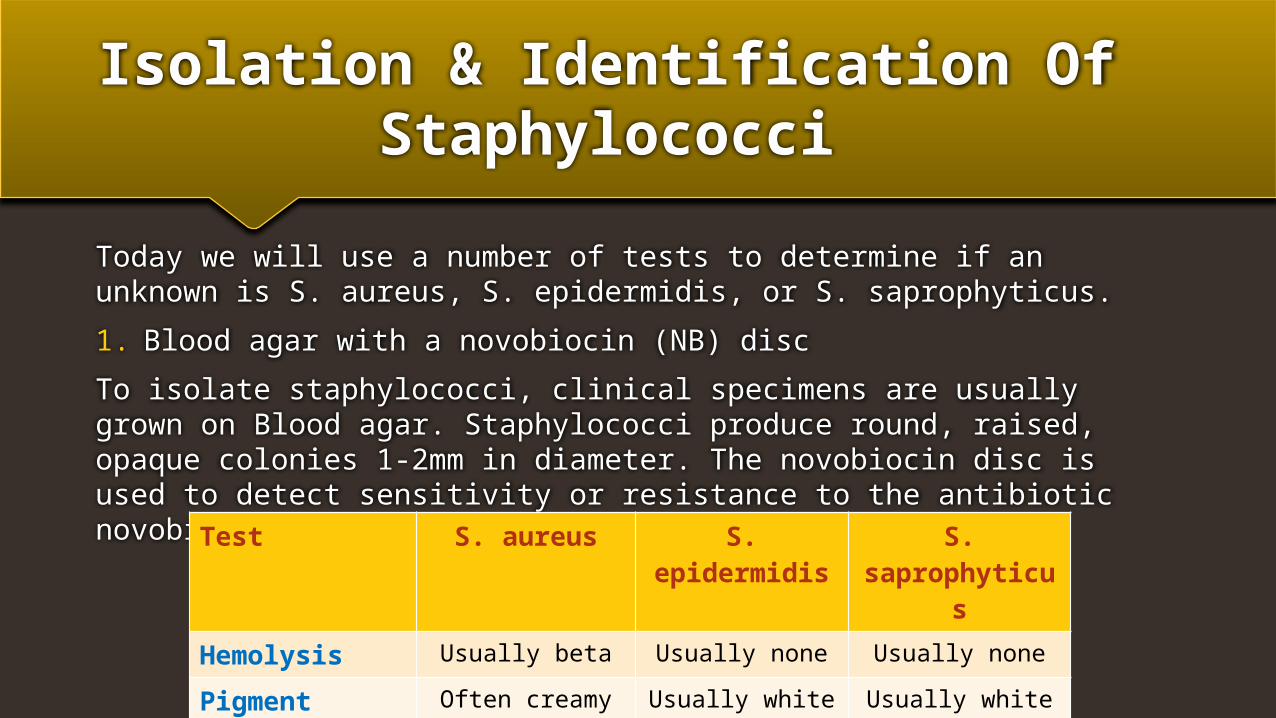
Isolation & Identification Of Staphylococci
Today we will use a number of tests to determine if an unknown is S. aureus, S. epidermidis, or S. saprophyticus.
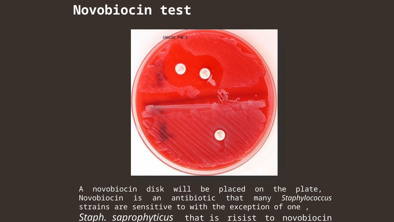
1. Blood agar with a novobiocin (NB) disc
To isolate staphylococci, clinical specimens are usually grown on Blood agar. Staphylococci produce round, raised, opaque colonies 1-2mm in diameter. The novobiocin disc is used to detect sensitivity or resistance to the antibiotic novobiocin.
Test S. aureus S. epidermidis S. saprophyticus
Hemolysis Usually beta Usually none Usually none
Pigment Often creamy gold Usually white Usually white
Novobiocin Test Sensitive Sensitive Resistant
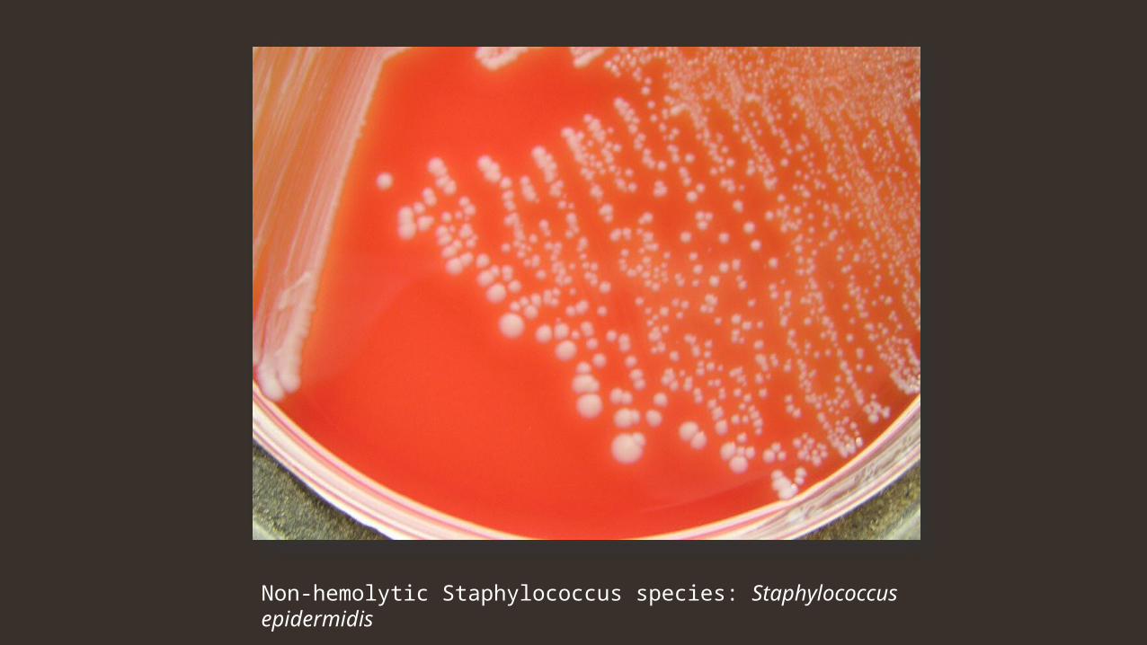
Non-hemolytic Staphylococcus species: Staphylococcus epidermidis
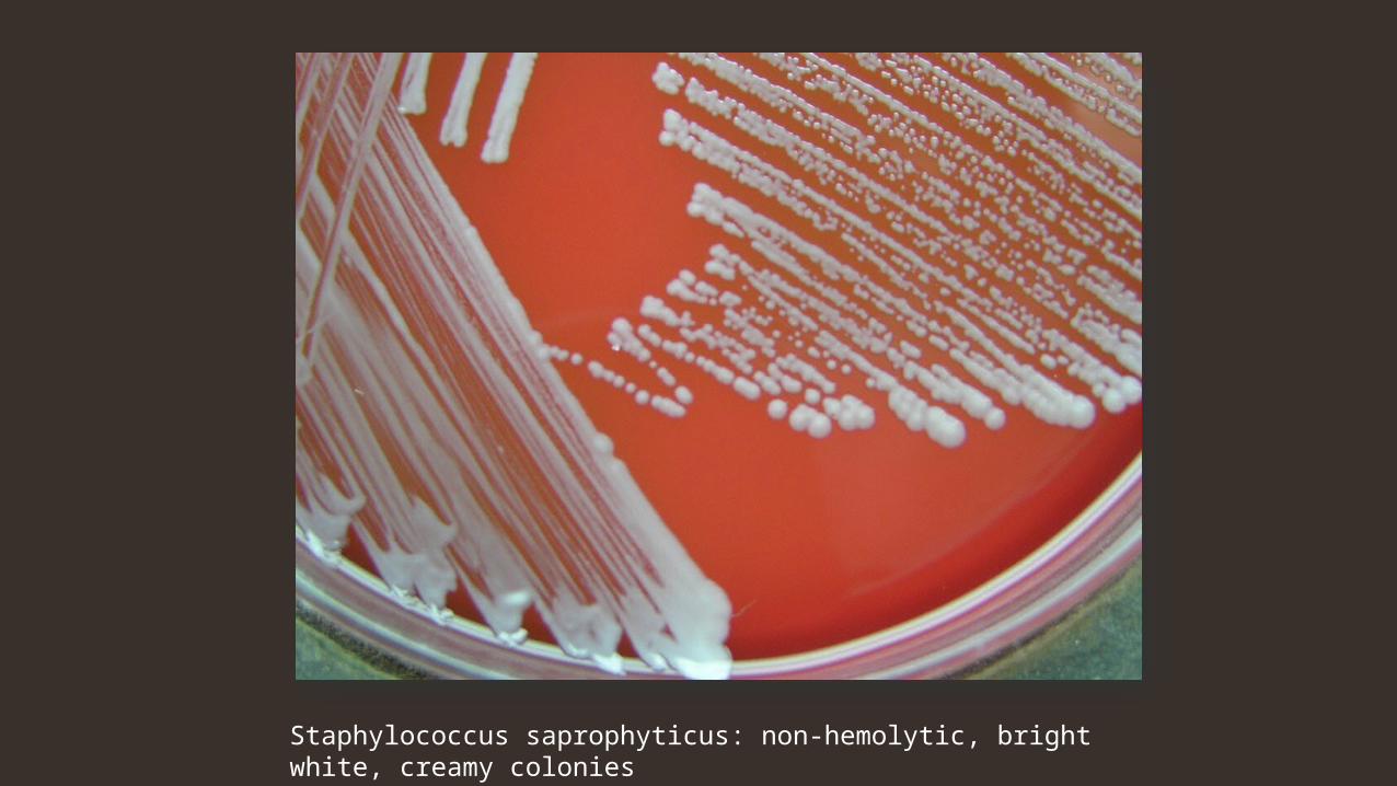
Staphylococcus saprophyticus: non-hemolytic, bright white, creamy colonies
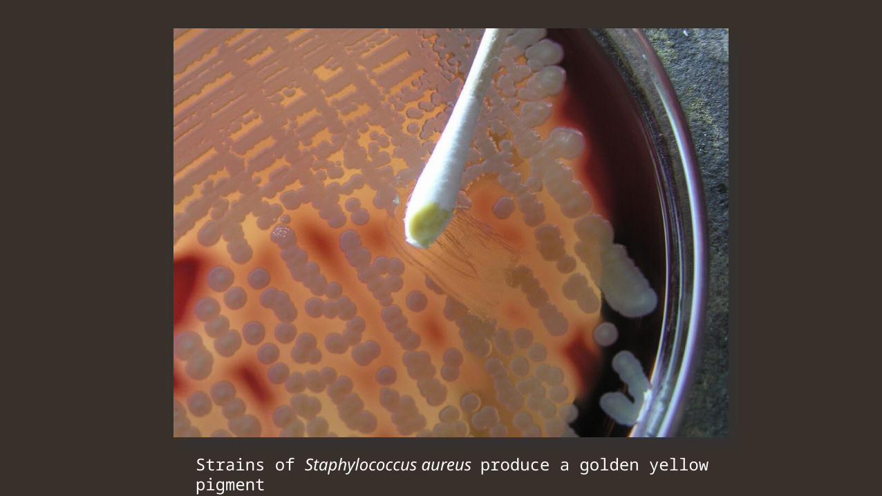
Strains of Staphylococcus aureus produce a golden yellow pigment
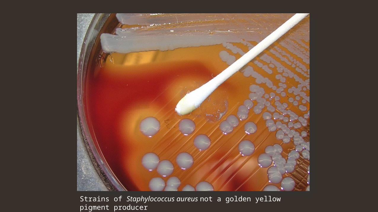
Strains of Staphylococcus aureus not a golden yellow pigment producer
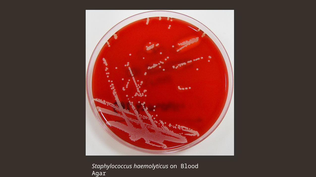
Staphylococcus haemolyticus on Blood Agar
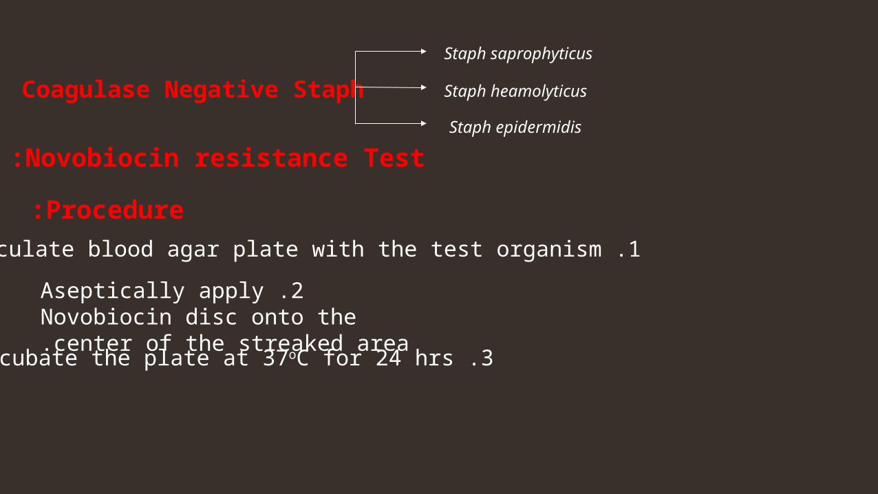
Procedure:1 .Inoculate blood agar plate with the test organism.
2 .Aseptically apply Novobiocin disc onto the center of the streaked area.
3 .Incubate the plate at 37oC for 24 hrs.
Novobiocin resistance Test:
Coagulase Negative Staph
Staph saprophyticus
Staph epidermidis
Staph heamolyticus
Novobiocin test
A novobiocin disk will be placed on the plate, Novobiocin is an antibiotic that many Staphylococcus strains are sensitive to with the exception of one ,
Staph. saprophyticus that is risist to novobiocin antibiotic.
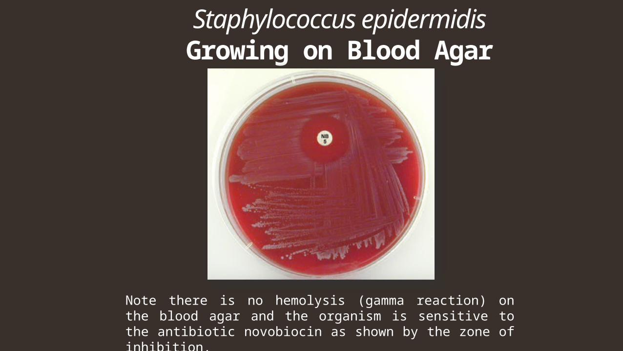
Staphylococcus epidermidis Growing on Blood Agar
Note there is no hemolysis (gamma reaction) on the blood agar and the organism is sensitive to the antibiotic novobiocin as shown by the zone of inhibition.
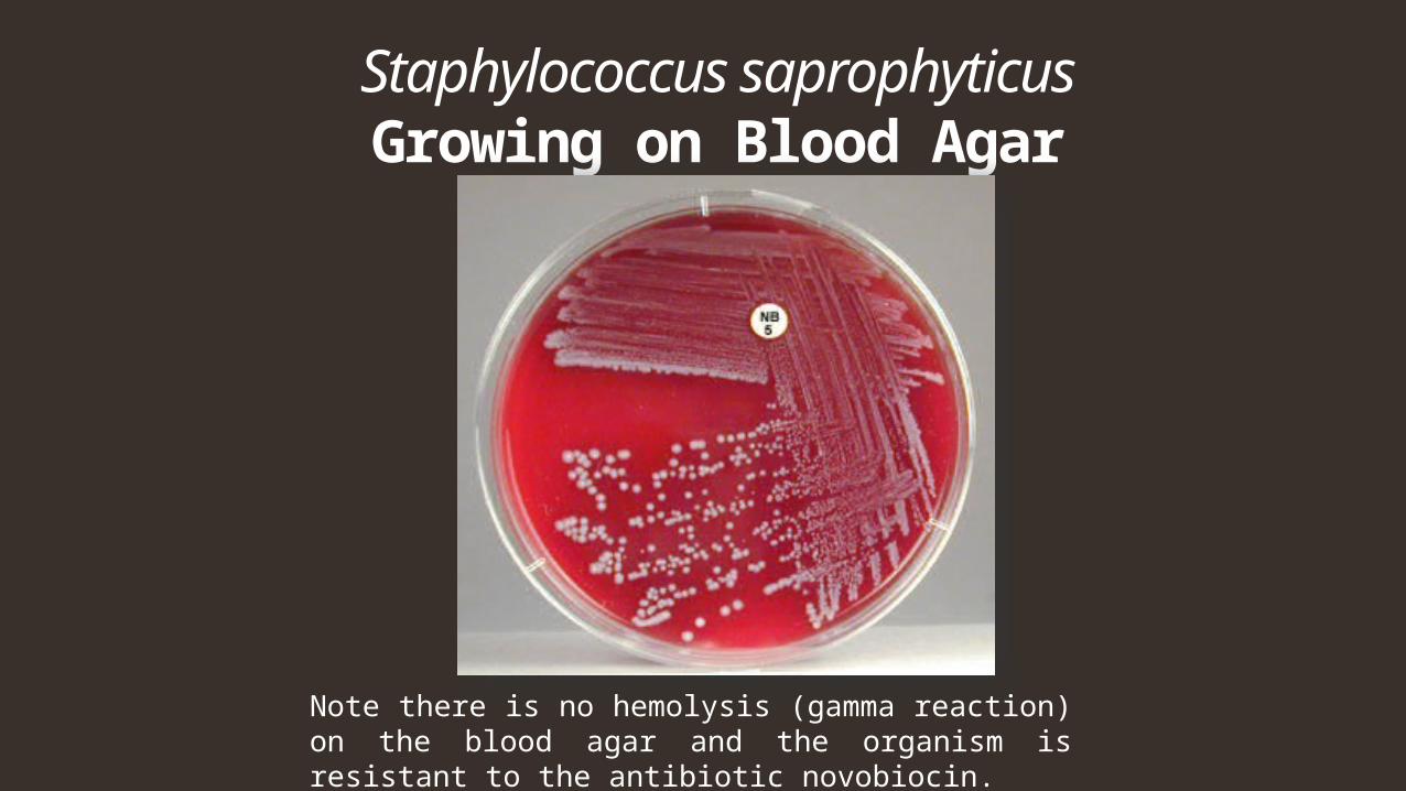
Staphylococcus saprophyticus Growing on Blood Agar
Note there is no hemolysis (gamma reaction) on the blood agar and the organism is resistant to the antibiotic novobiocin.
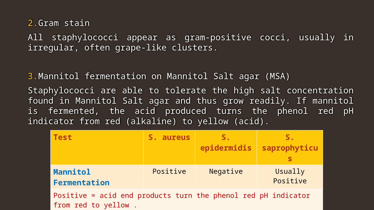
2. Gram stain
All staphylococci appear as gram-positive cocci, usually in irregular, often grape-like clusters.
3. Mannitol fermentation on Mannitol Salt agar (MSA)
Staphylococci are able to tolerate the high salt concentration found in Mannitol Salt agar and thus grow readily. If mannitol is fermented, the acid produced turns the phenol red pH indicator from red (alkaline) to yellow (acid).
Test S. aureus S. epidermidis S. saprophyticus
Mannitol Fermentation Positive Negative Usually Positive
Positive = acid end products turn the phenol red pH indicator from red to yellow .Negative = phenol red remains red .
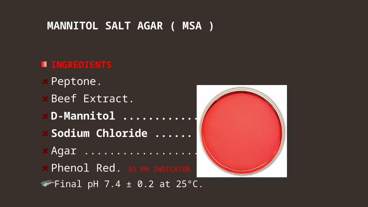
MANNITOL SALT AGAR ( MSA )
INGREDIENTS
Peptone.
Beef Extract.
D-Mannitol .............. 1.0%.
Sodium Chloride ...... 7.5%.
Agar ......................... 1.5%.
Phenol Red. AS PH INDICATOR
Final pH 7.4 ± 0.2 at 25°C.
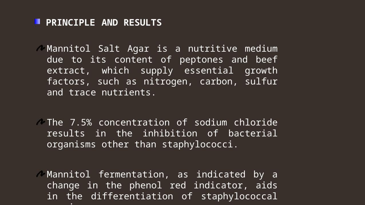
PRINCIPLE AND RESULTS
Mannitol Salt Agar is a nutritive medium due to its content of peptones and beef extract, which supply essential growth factors, such as nitrogen, carbon, sulfur and trace nutrients.
The 7.5% concentration of sodium chloride results in the inhibition of bacterial organisms other than staphylococci.
Mannitol fermentation, as indicated by a change in the phenol red indicator, aids in the differentiation of staphylococcal species.

Mannitol Salt agar

4. Production of deoxyribonuclease (DNase) on DNase agar
DNase agar contains 0.2% DNA. To detect DNase production, the plate is inoculated and incubated. After growth, the plate is flooded with 1N hydrochloric acid (HCl).
Test S. aureus S. epidermidis S. saprophyticus
DNase production Positive Negative Negative
Positive = clear zone around growth after adding 1N HCl (no DNA remaining in the agar). Negative = cloudy around growth after adding 1N HCl (DNA remains in the agar forming a precipitate).
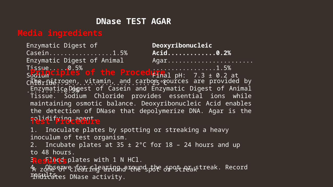
Enzymatic Digest of Casein.................1.5% Enzymatic Digest of Animal Tissue.....0.5% Sodium Chloride...................................0.5%
DNase TEST AGARMedia ingredients
Principles of the Procedure The nitrogen, vitamin, and carbon sources are provided by Enzymatic Digest of Casein and Enzymatic Digest of Animal Tissue. Sodium Chloride provides essential ions while maintaining osmotic balance. Deoxyribonucleic Acid enables the detection of DNase that depolymerize DNA. Agar is the solidifying agent.
Test Procedure 1. Inoculate plates by spotting or streaking a heavy inoculum of test organism.2. Incubate plates at 35 ± 2°C for 18 – 24 hours and up to 48 hours. 3. Flood plates with 1 N HCl. 4. Observe for clearing around the spot or streak. Record results.
Deoxyribonucleic Acid.............0.2% Agar........................................1.5%Final pH: 7.3 ± 0.2 at 25°C
Results A zone of clearing around the spot or streak indicates DNase activity.
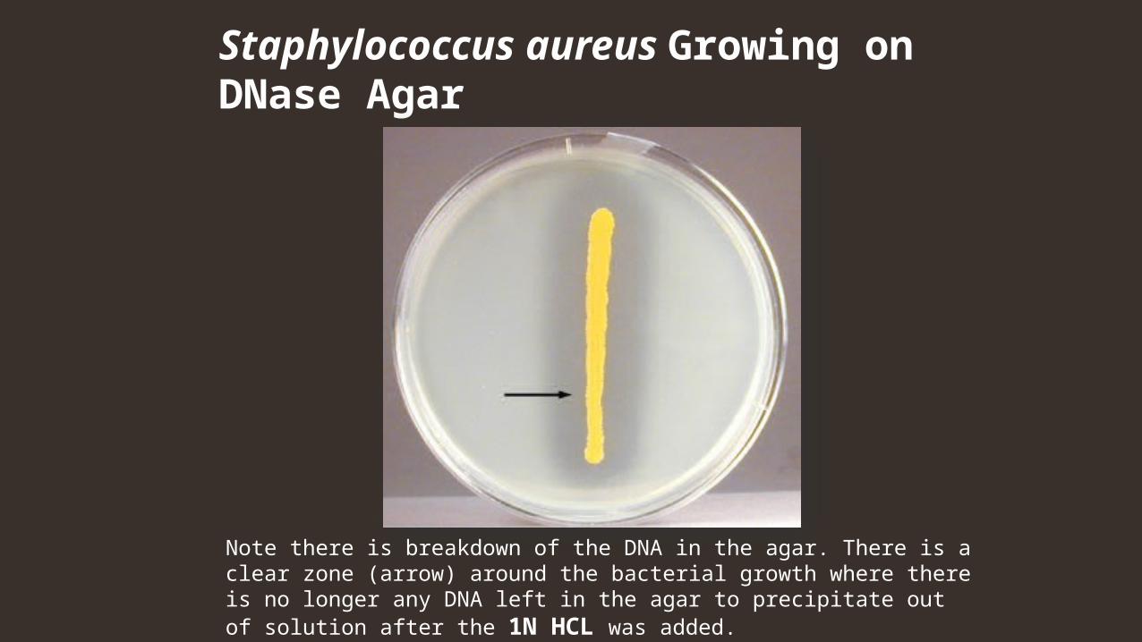
Staphylococcus aureus Growing on DNase Agar
Note there is breakdown of the DNA in the agar. There is a clear zone (arrow) around the bacterial growth where there is no longer any DNA left in the agar to precipitate out of solution after the 1N HCL was added.
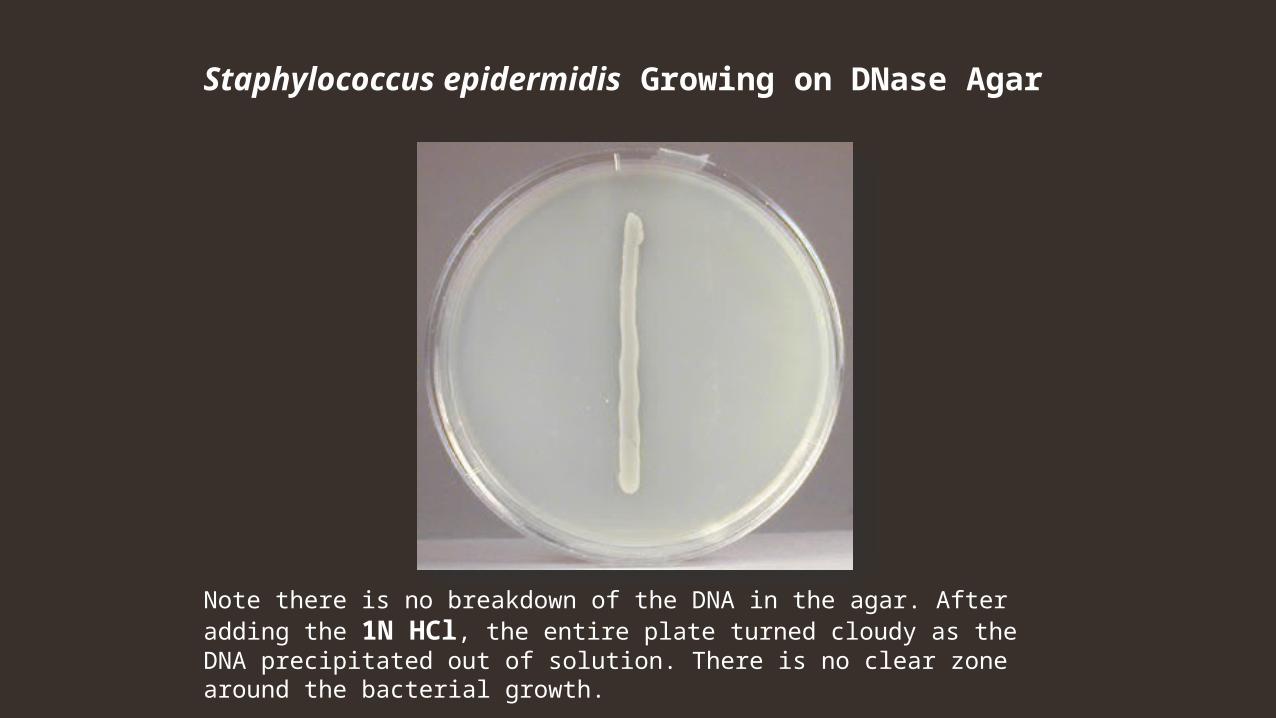
Staphylococcus epidermidis Growing on DNase Agar
Note there is no breakdown of the DNA in the agar. After adding the 1N HCl, the entire plate turned cloudy as the DNA precipitated out of solution. There is no clear zone around the bacterial growth.
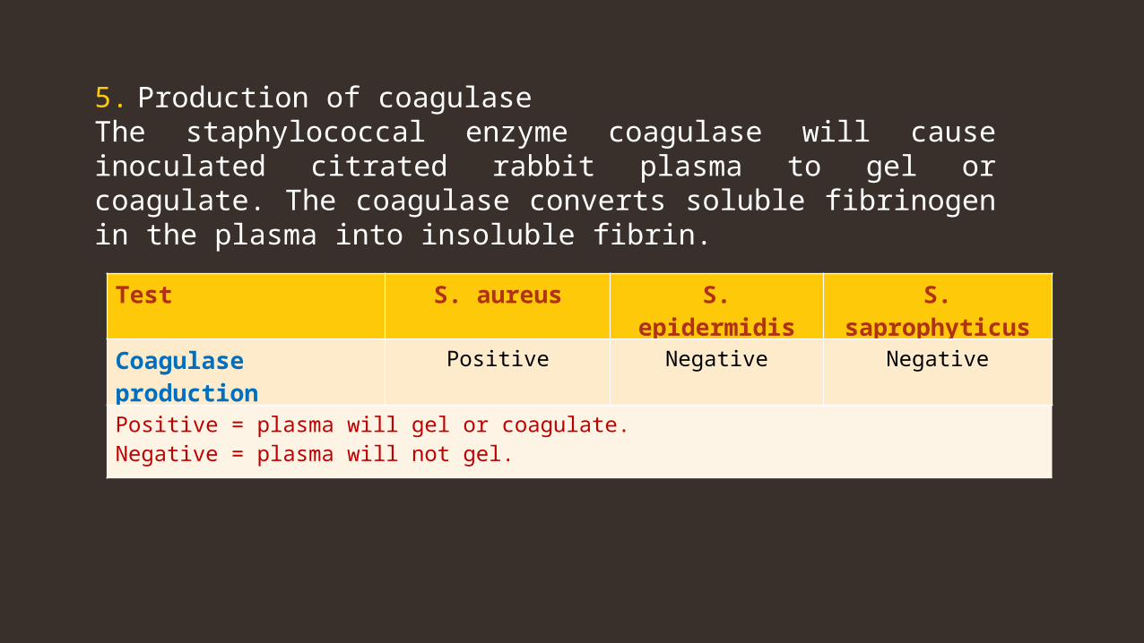
5. Production of coagulase The staphylococcal enzyme coagulase will cause inoculated citrated rabbit plasma to gel or coagulate. The coagulase converts soluble fibrinogen in the plasma into insoluble fibrin.
Test S. aureus S. epidermidis S. saprophyticus
Coagulase production Positive Negative Negative
Positive = plasma will gel or coagulate. Negative = plasma will not gel.
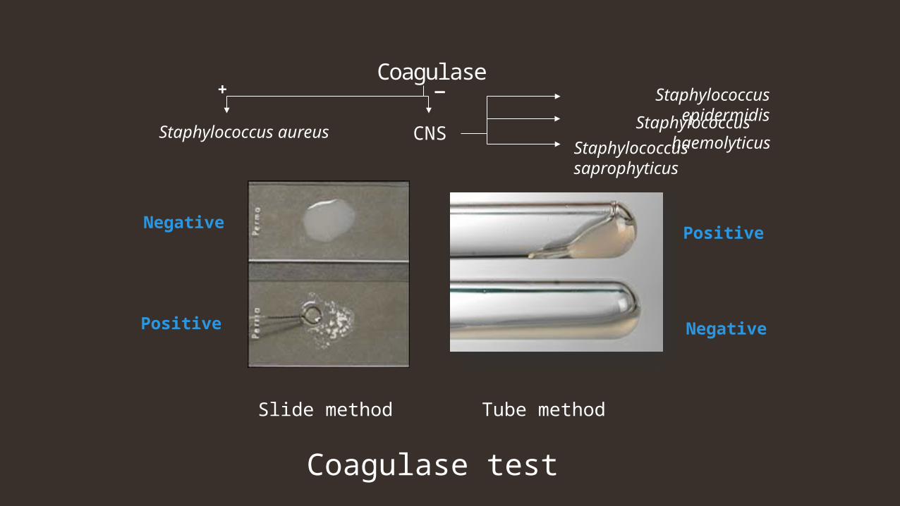
Coagulase test
Negative
Positive
Slide method
Negative
Positive
Tube method
Coagulase
Staphylococcus aureus CNS
Staphylococcus epidermidis
Staphylococcus saprophyticus
+ _
Staphylococcus haemolyticus
32
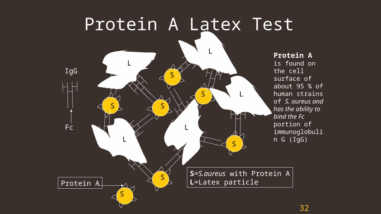
Protein A Latex Test
S
S
SS
S
L
L
L
L
L
IgG
S=S.aureus with Protein AL=Latex particleProtein A
S
Fc
S
Protein A is found on the cellsurface of about 95 % of human strains of S. aureus and has the ability to bind the Fc portion of immunoglobulin G (IgG)
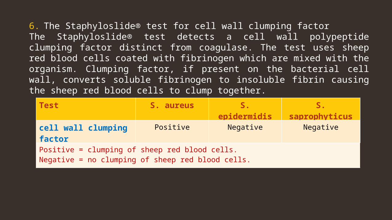
6. The Staphyloslide® test for cell wall clumping factor The Staphyloslide® test detects a cell wall polypeptide clumping factor distinct from coagulase. The test uses sheep red blood cells coated with fibrinogen which are mixed with the organism. Clumping factor, if present on the bacterial cell wall, converts soluble fibrinogen to insoluble fibrin causing the sheep red blood cells to clump together.
Test S. aureus S. epidermidis S. saprophyticus
cell wall clumping factor Positive Negative Negative
Positive = clumping of sheep red blood cells.Negative = no clumping of sheep red blood cells.
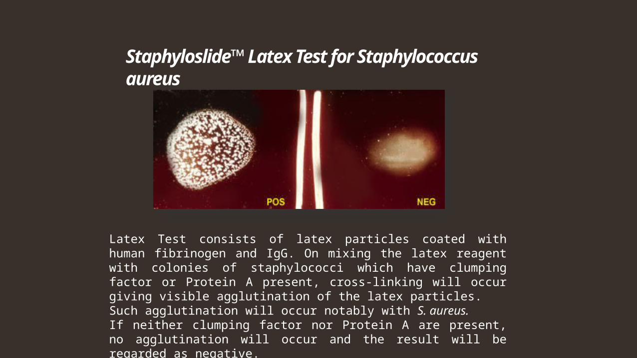
Staphyloslide™ Latex Test for Staphylococcus aureus
Latex Test consists of latex particles coated with human fibrinogen and IgG. On mixing the latex reagent with colonies of staphylococci which have clumping factor or Protein A present, cross-linking will occur giving visible agglutination of the latex particles. Such agglutination will occur notably with S. aureus.If neither clumping factor nor Protein A are present, no agglutination will occur and the result will be regarded as negative.