ENDOMETRIAL RESEPTIVITE DEĞERLENDİRMESİ
78
ENDOMETRIAL RESEPTIVITE DEĞERLENDİRMESİ PROF.DR.MEHMET ÇOLAKOĞLU NEU MERAM TIP FAKULTESİ KADIN HST VE DOĞUM AD UREME ENDOKRINOLOJISI VE INFERTILITE BD KONYA
description
ENDOMETRIAL RESEPTIVITE DEĞERLENDİRMESİ. PROF.DR.MEHMET ÇOLAKOĞLU NEU MERAM TIP FAKULTESİ KADIN HST VE DOĞUM AD UREME ENDOKRINOLOJISI VE INFERTILITE BD KONYA. Embryo implantasyonu için geçici olarak endometriumun uygun hale gelmesine denir. RESEPTİVİTE TARİF. IMPLANTATION WINDOW. - PowerPoint PPT Presentation
Transcript of ENDOMETRIAL RESEPTIVITE DEĞERLENDİRMESİ

ENDOMETRIAL RESEPTIVITE DEĞERLENDİRMESİ
PROF.DR.MEHMET ÇOLAKOĞLUNEU MERAM TIP FAKULTESİKADIN HST VE DOĞUM AD
UREME ENDOKRINOLOJISI VE INFERTILITE BDKONYA
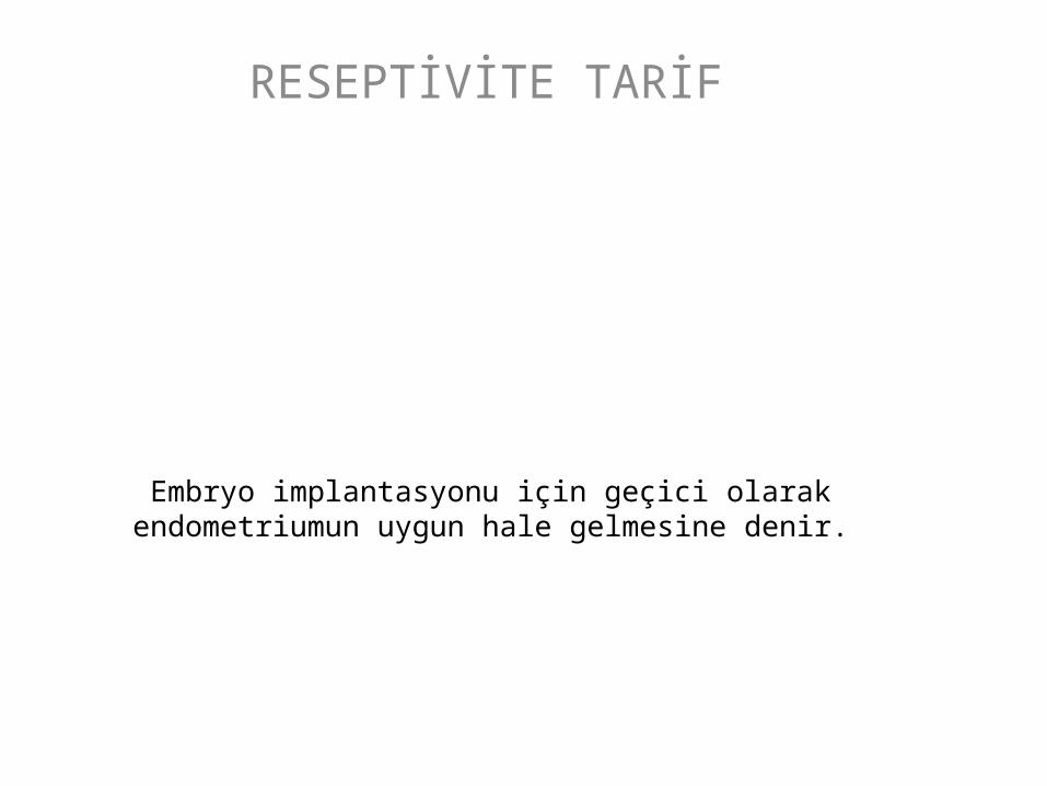
Embryo implantasyonu için geçici olarak endometriumun uygun hale gelmesine denir.
RESEPTİVİTE TARİF
IMPLANTATION WINDOW
• Postovulatuar 6-10 günlerde açılır, bunun dışında non-reseptiftir. Siklus boyunca optimal hazırlıkların yapıldığı devredir. External hormonlarla bu günlerde oynama yapılabilir.


Implantation Process
• Human embryo implantation üç aşamalı işlemdir (apposition, adhesion and invasion) functional blastocyst ile reseptive endometrium arasında olur. Bu ovarian steroid bağımlı bir olaydır.


The selectin adhesion system is well established at the maternal–fetal interface
• On the blastocyst side, strong L-selectin staining has been observed over the entire embryo surface (Genbacev et al., 2003 ). On the maternal side, the expression of selectin
oligosaccharide-based ligands, such as MECA-79 or HECA-452, is up-regulated during the window of implantation (Genbacev et al., 2003 ).

Human embryo implantation in the uterus. (A) Endometrium proliferates under estrogen enhancement. (B) Progesterone from luteinized follicles leads to endometrial differentiation. (C) The blastocyst enters the uterus through the ostia and
rolls freely over the endometrium under signals by L-selectin. (D) Mucin-1 (MUC-1) repels the blastocyst and prevents its adhesion to endometrial areas with poor chances of implantation. (E) Chemokines and cytokines attract the blastocyst to the optimal implantation spot. (F) Adhesion molecules (e.g. integrins and cadherins) firmly attach the blastocyst to
the endometrial pinopods to ensure further successful implantation.

ENDOMETRIAL STEROID RECEPTORLERİ
• İmplantasyonda Estrogen ve Progesteron reseptorleri önemlidir.
• Luteal fazda , progesteron glandular epitelde PR downregulasyonu yapar. PR downregulation ve pre-implantation integrin expresyonu arasında sıkı ilişki vardır.

ENDOMETRİAL RESEPTİVİTEYİ ETKİLEYEN GENETİK FAKTÖRLER
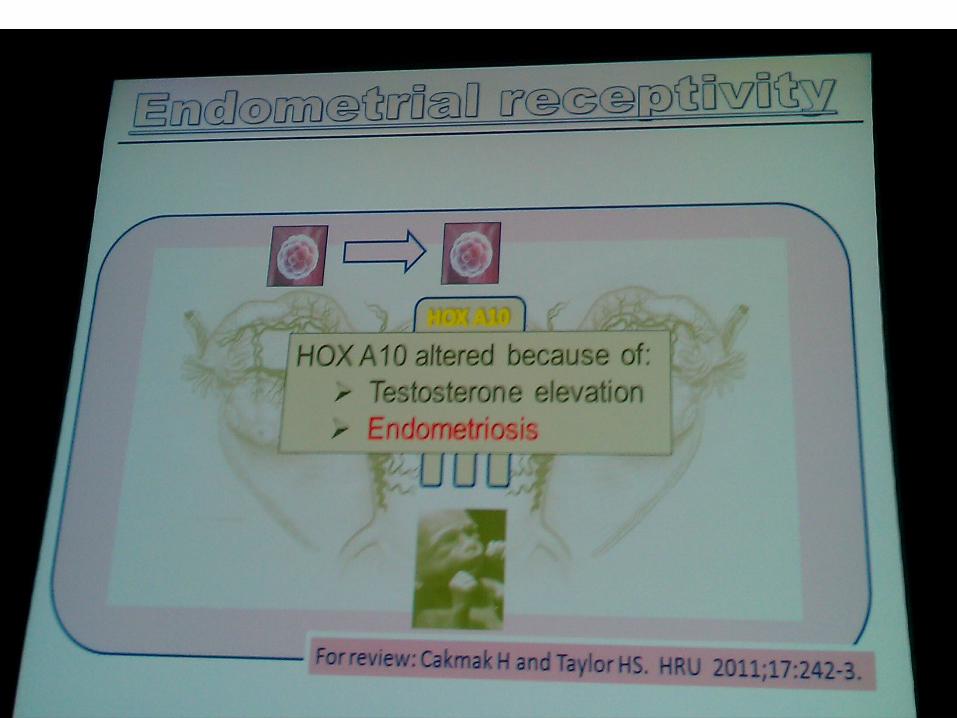
• Hoxa 10 expressionu

• Pinopods appear progesterone dependant. Association between mid-luteal increase of progesterone level and the first appearance of pinopods throughout the menstrual cycle was noted (Stavreus-Evers et al., 2001 ; Usadi et al., 2003 ). Moreover, HOXA-10, a homeobox gene whose expression is necessary for endometrial receptivity to blastocyst implantation, has an essential role in pinopod
development. Indeed, blocking HOXA-10 expression dramatically decreases the number of pinopods. HOXA-10 illustrates a dual role in the endometrium by regulating both endometrial stromal cell (ESC) proliferation and epithelial cell morphogenesis (Bagot et al., 2001 ).
ENDOMETRİAL RESEPTİVİTEYİ ETKİLEYEN GENETİK FAKTÖRLER
• Uterine sensitization-associated gene-1 (USAG-1),
ENDOMETRİAL RESEPTİVİTEYİ ETKİLEYEN GENETİK FAKTÖRLER
• Endometrial bleeding associated factor (EBAF)• found to be expressed in the late secretory
and menstrual phase of the endometrium.

ENDOMETRİAL RESEPTİVİTEYİ ETKİLEYEN HORMONLAR
• COH ESNASINDAKİ YÜKSEK ESTROGEN RESEPTİVİTEYİ KÖTÜ ETKİLER

ENDOMETRİAL RESEPTİVİTEYİ ETKİLEYEN HORMONLAR
Estrogen and progesterone
• At 527 cycles in subfertile patients, it was• found that significantly more viable pregnancies• occurred among patients with an estrogen to• progesterone ratio in the range of 7.36 to 12.22• (calculated as estrogen in pmol/L divided by• progesterone in nmol/L).
• Yang et al.
Gonadotropin Hormonlar
• Periimplantasyon peryodunda LH reseptor sayısı ve LH tarafından işgalleri artar. Bu LH ın implantasyon ve desidualizasyonda önemini gösterir
• Bonnamy et al.

GnRh agonist and GnRh antagonist
• Agonist ve antagonist tedavilerden sonra düşük LH seviyesi gözlenir. Bu durum korpus luteum fonksion bozukluğu ve kısa luteal fazla beraberdir.
• Tavaniotou A, Smitz J, Bourgain C, Devroey P. Ovulation• induction disrupts luteal phase function. Ann N Y Acad Sci• 2001;943:55-63

GnRh agonist and GnRh antagonist
• In GnRh-agonist cycles, mid-luteal biopsies has• revealed increased glandulo-stromal dyssynchrony• and delay in endometrial development, strong• positivity of endometrial glands for progesterone• receptors, decreased cell adhesion molecule profiles• with early appearance of pinopodes. These changes• suggest a shift forwards of implantation window.• Progesterone supplementation improves endometrial• histology, and its necessity has been established, at• least in cycles, using GnRh agonists • Soliman S, Daya S, Graham RA, Seif MW, Cook ID. The• role of luteal phase support in infertility treatment: a metaanalysis• of randomized trials. Fertil Steril 1994;61:1068-76
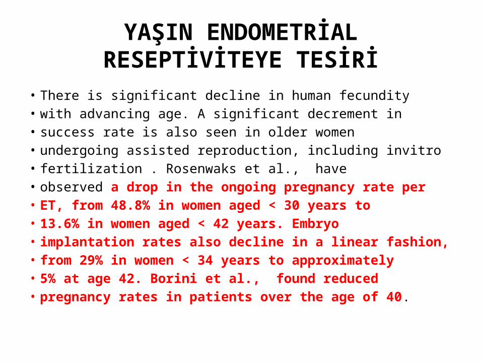
YAŞIN ENDOMETRİAL RESEPTİVİTEYE TESİRİ
• There is significant decline in human fecundity• with advancing age. A significant decrement in• success rate is also seen in older women• undergoing assisted reproduction, including invitro• fertilization . Rosenwaks et al., have• observed a drop in the ongoing pregnancy rate per• ET, from 48.8% in women aged < 30 years to• 13.6% in women aged < 42 years. Embryo• implantation rates also decline in a linear fashion,• from 29% in women < 34 years to approximately• 5% at age 42. Borini et al., found reduced• pregnancy rates in patients over the age of 40.
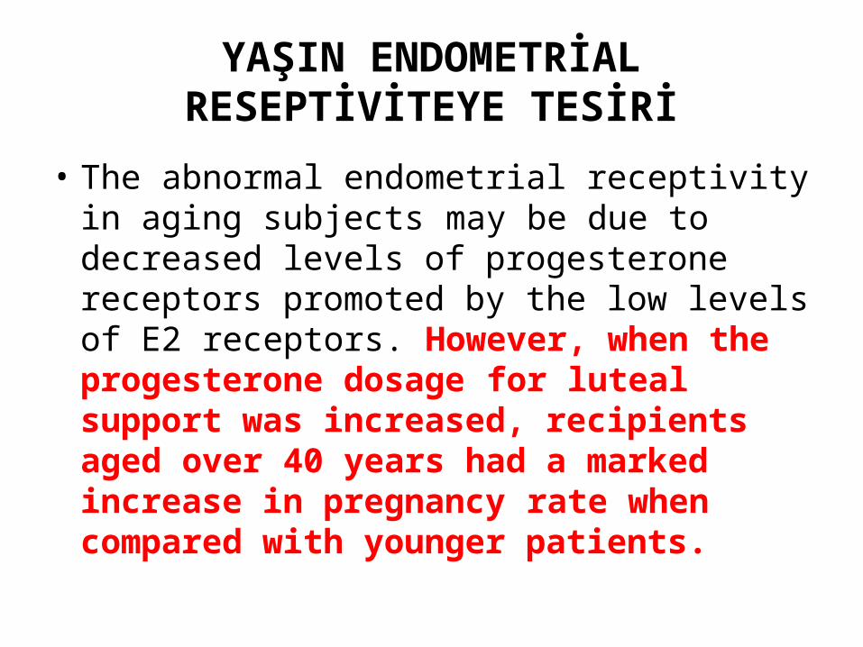
YAŞIN ENDOMETRİAL RESEPTİVİTEYE TESİRİ
• The abnormal endometrial receptivity in aging subjects may be due to decreased levels of progesterone receptors promoted by the low levels of E2 receptors. However, when the progesterone dosage for luteal support was increased, recipients aged over 40 years had a marked increase in pregnancy rate when compared with younger patients.

YAŞIN ENDOMETRİAL RESEPTİVİTEYE TESİRİ
• Oosit tükenmesi endometrial reseptiviteyi de bozar. Donor oosit çalışmalarında bu gösterilmiştir.
• Navot D, Bergh PA, Williams AM, John Garrisi G, Guzman• I, Sandler B, Rabinowitz R, Birkenfeld A. Poor oocyte• quality rather than implantation failure as a cause of agerelated• decline in female fertility. Lancet 1991;337:1375-7

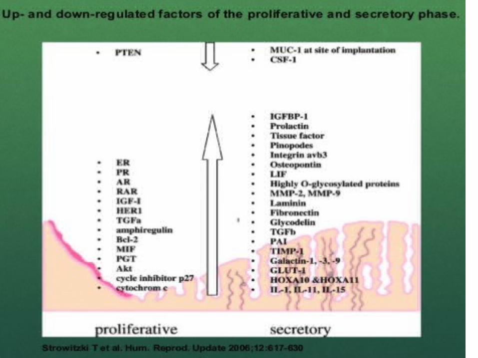
ENDOMETRİAL RESEPTİVİTENİN POTANSİEL MARKERLARI
• PROLİFERASYON,DİFERANSİASYON VE SEKRESYON GİBİ BİRÇOK HÜCRE FONKSİONU GROWTH FAKTÖRLER VE SİTOKİNLER TARAFINDAN YÖNETİLİR

Endometrial adhesion molekulleri
• 4 Ana grup:• integrins,• cadherins, • selectins • immunoglobulin• Springer TA. Adhesion receptors of the immune system.• Nature 1990;346:425-34.

Endometrial adhesion molecules• Integrins are cell adhesion molecules involved in cell-cell and cell-matrix
interactions and contributing to cell migration and signal
• transduction . Integrins are a family of• transmembrane glycoproteins that act as a receptor• for extracellular matrix ligand, osteopontin (OPN).• Three integrins are expressed by the endometrium• with a pattern that coincides well with the window• of implantation: α1β1, α4β1, and αvβ3 are• coexpressed on glandular epithelium only during• cycle days 20 to 24 corresponding the putative• window of implantation. They have been proposed• as the best of the immunohistochemical markers of• endometrial receptivity during implantation window• . Lessey BA. Endometrial integrins and the establishment of• uterine receptivity. Hum Reprod 1998;13:247-61

• RESEPTİVİTE BOZULMASINDA• Düşenler• Failure of appearance of a specific integrin – V 3 in the endometrium
at the time of implantation was suggested as a cause of implantation failure (Tei et al., 2003 ; Thomas et al., 2003 ).
• Yükselenler• High levels of aromatase p450 mRNA (Brosens et al., 2004 ), changes in pinopode
expression (Pantos et al., 2004 ) and high matrix metalloproteinases (Inagaki et al., 2003 ) have been suggested to be associated with RIF.

Endometrial adhesion molekulleri
• Mid-luteal integrinler naturale gore indukte sikluslarda dusuk bulunurlar buna "type I defect`` denir. Bu hastalarin cogu progesteronla tedavide hem endometrial histoloji hemde αv β3 integrinde duzelme olur
• Histolojik olarak normal ama αv β3• Integrin eksik olursa "type II defect", denir
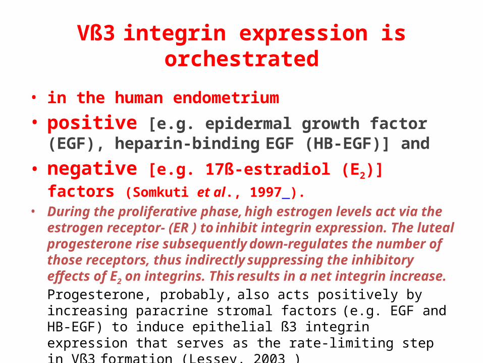
Vß3 integrin expression is orchestrated
• in the human endometrium
• positive [e.g. epidermal growth factor (EGF), heparin-binding EGF (HB-EGF)] and
• negative [e.g. 17ß-estradiol (E2)] factors (Somkuti et al., 1997 ). • During the proliferative phase, high estrogen levels act via the estrogen
receptor- (ER ) to inhibit integrin expression. The luteal progesterone rise subsequently down-regulates the number of those receptors, thus indirectly suppressing the inhibitory effects of E2 on integrins. This results in a net integrin increase. Progesterone, probably, also acts positively by increasing paracrine stromal factors (e.g. EGF and HB-EGF) to induce epithelial ß3 integrin expression that serves as the rate-limiting step in Vß3
formation (Lessey, 2003 )


Aberrant Vß3 integrin expression pattern has been associated with
• unexplained infertility (Klentzeris et al., 1993 ; Lessey et al., 1995 ; Tei et al., 2003 ),
• endometriosis (Lessey et al., 1994b ), • hydrosalpinx (Meyer et al., 1997 ),• luteal phase deficiency (LPD; Lessey et al., 1992 ) and,
more recently,
• polycystic ovarian syndrome (PCOS; Apparao et al., 2002 ). Other investigators could not, however, demonstrate different integrin pattern in endometriosis (Creus et al., 1998 ).

Endometrial anti-adhesion molecules
• As the attaching embryo approaches the• luminal epithelial surface of the uterus, it• encounters a mucinous layer, the glycocalyx .• The mucins in this layer are a group of antiadhesive• molecules, the most important of which is• mucin 1(MUC-1). Mucins are a family of• glycoprotein present on the surface of human• epithelial cells. In human its expression is high• during periimplantation period . It is possible that• the high periimplantation levels of MUC1 could play• a role in "shielding" the implanting blastocyst from• other inhibitory factors on the epithelial surface.• Tabibzadeh S. Molecular control of the implantation• window. Hum Reprod Update 1998;4:465-71

Human embryo implantation in the uterus. (A) Endometrium proliferates under estrogen enhancement. (B) Progesterone from luteinized follicles leads to endometrial differentiation. (C) The blastocyst enters the uterus through the ostia and
rolls freely over the endometrium under signals by L-selectin. (D) Mucin-1 (MUC-1) repels the blastocyst and prevents its adhesion to endometrial areas with poor chances of implantation. (E) Chemokines and cytokines attract the blastocyst to the optimal implantation spot. (F) Adhesion molecules (e.g. integrins and cadherins) firmly attach the blastocyst to
the endometrial pinopods to ensure further successful implantation.

Endometrial anti-adhesion molecules
• Alternatively, it could carry a specific recognition• structure for the embryo. In women who
suffer recurrent miscarriage there is evidence for reduced levels of MUC1 suggesting that these molecules play a significant role in the establishment and maintenance of early pregnancy .

Endometrial cytokines• Although many• cytokines may play a part in implantation, a vital role has been clarified in
four namely: Leukaemia inhibitory factor, interleukin-1, interleukin-11 and colony-stimulating factor . Leukaemia
• inhibitory factor (LIF) is produced by the receptive• phase endometrium . Danielsson et al.• showed reduced immunostaining for LIF after• treatment with the antiprogestin, mifepristone.• These circumstantial evidences suggest that LIF• plays a role in endometrial receptivity, but its exact• role is currently unclear.• Sharkey A. Cytokines and implantation. Rev Reprod• 1998;3:52-61

Endometrial pinopods’ development is associated with the mid-luteal phase increased expression of
• leukaemia inhibitory factor (LIF) and its receptor
(Aghajanova et al., 2003 ), progesterone (Stavreus-Evers et al., 2001 ) and integrin Vß3 (Lessey et al., 1992 ). The detection of pinopods during the mid-secretory phase may be extremely useful for the assessment of endometrial receptivity to optimize implantation rates.
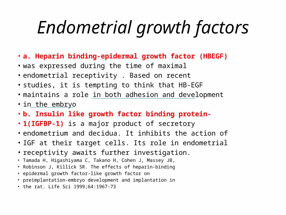
Endometrial growth factors• a. Heparin binding-epidermal growth factor (HBEGF)• was expressed during the time of maximal• endometrial receptivity . Based on recent• studies, it is tempting to think that HB-EGF• maintains a role in both adhesion and development• in the embryo • b. Insulin like growth factor binding protein-• 1(IGFBP-1) is a major product of secretory• endometrium and decidua. It inhibits the action of• IGF at their target cells. Its role in endometrial• receptivity awaits further investigation.• Tamada H, Higashiyama C, Takano H, Cohen J, Massey JB,• Robinson J, Killick SR. The effects of heparin-binding• epidermal growth factor-like growth factor on• preimplantation-embryo development and implantation in• the rat. Life Sci 1999;64:1967-73

Endometrial immune markers• The endometrium has a large population of• lympho-myeloid cells that undoubtedly play a• variety of roles in the implantation process. It• has been reported that women with unexplained• infertility have significant lower levels of• endometrial CD8+ (T suppressor/cytotoxic) and• CD56+ (natural killer) cells, and higher levels of• CD4+ (T helper/inducer) cells, than fertile control
• Klentzeris LD, Bulmer JN, Warren MA, Morrision L, Li• TC, Cooke ID. Lymphoid tissue in the endometrium of• women with unexplaned infertility: Morphometric and• Immunohistochemical Aspects. Hum Reprod 1994;9:646

Mouse Ascites Golgi (MAG)• The MAG test, done during an• endometrial biopsy measures sticky mucinous• substances secreted by endometrial glands before• implantation and is considered as an endometrial• function test (EFT). Over 85% of the• endometrial biopsies from normal, fertile women• express higher levels of MAG between days 5 and• 18 of the menstrual cycle with no expression after• day 19.• kliman H, Feinberg R, Schwartz L, Feinman M, Laui E,• Meawough E. A mucin like glycoprotein identified by MAG• (mouse ascites Golgi) antibodies. Menstrual cycle dependent• localization in human endometrium. Am J Pathol• 1995;146:166-81.
• Dubowy et al, developed an EFT• based on the endometrial expression of cyclin E• and p27 . This test allows dating of the• endometrium and differentiating between normally• and abnormally developing endometrium. Cyclin E• progressed from the basal to the lateral cytoplasm• (midproliferative phase) to the nuclus (day 18 to• 19) and was absent in biopsies after day 20. First• appearing on days 17 to 19, p27 was found only in• the nuclei.• DuboyL, Feinberg F, Keefe D et al. Improved endometrial• assessment using cyclin E and p27. Fertil Steril
• 2003;80:146-56.


Matrix Proteinler
• Laminin,• fibronectin , collagen IV are found in secretory endometrium but are absent in the• endometrium of patients with unexplained• infertility . These suggest that these matrix• proteins are likely to be required for implantation.• Bilalis D, Klentzeris L, Fleming S. Immunohistochemical• localization of extracellular matrix proteins in luteal phase• endometrium of fertile and infertile patients. Hum Reprod• 1996;11;271-18

• Another endometrial protein, glycodelin, has a proposed immunomodulatory role during
• implantation. This protein is present in the• endometrium under the control of
progesterone and• antiprogestins • Muller M, Vinge J, Vaisse C, Taylor R. Glycodelin: a pane• in the implantation window. Semin Reprod Med• 2000;18:289-98.

Human embryo implantation in the uterus. (A) Endometrium proliferates under estrogen enhancement. (B) Progesterone from luteinized follicles leads to endometrial differentiation. (C) The blastocyst enters the uterus through the ostia and
rolls freely over the endometrium under signals by L-selectin. (D) Mucin-1 (MUC-1) repels the blastocyst and prevents its adhesion to endometrial areas with poor chances of implantation. (E) Chemokines and cytokines attract the blastocyst to the optimal implantation spot. (F) Adhesion molecules (e.g. integrins and cadherins) firmly attach the blastocyst to
the endometrial pinopods to ensure further successful implantation.


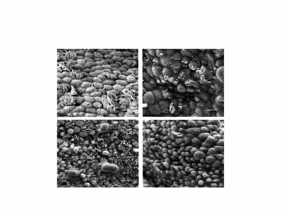
Morphological Markers
Pinopodes
• The endometrium undergoes a well-establishedseries of histological and ultrastructural changes under the influence of estrogen and progesterone
• during the menstrual cycle . Morphological• changes include characteristic histological• transformations, such as reduced mitotic activity,• glandular secretion, and stromal edema, that are• often accompanied by the presence of globular• protrusions in the surface membrane of epithelial• cells, named pinopodes .• 70. Noyes RW, Hertig and Rock J. Dating the endometrial• biopsy. Fertil Steril 1950;1: 3-25• 71. Sarantis L, Roche D, and Psychoyos A. Displacement of• receptivity for nidation in the rat by the progesterone• antagonist RU 486: a scanning electron microscopy study.• Hum Reprod 1988;3:251-5

Apoptosis
• Apoptosis is a usual phenomenon throughoutthe menstrual cycle, peaking at menses, but locallyregulated apoptosis is also vital for successfulimplantation. Recent evidence suggests thatregulated apoptosis is important during the windowof receptivity . On days 19-20, apoptosis isdetectable in the glands of the basal layer,subsequently extending to the functional layer. Thesignificance of this finding in relation to openingof the implantation window is under investigation.
• Galan A, O'Connor E, Valbuena D, Herrer R, Remohi J. The• human blastocyst regulates endometrial epithelial apoptosis
• in embryonic adhesion. Biol Reprod 2000;63:430-9.

ASSESSMENT OFENDOMETRIAL RECEPTIVITY
• A good correlation between endometrial• thickness and the prevalence of conception has• been found . On the other hand other• studies do not support this view .• However, a very thin endometrium (<7mm)• seems to be accepted as a reliable sign of• suboptimal implantation potential .• Implantation and pregnancy rates are• significantly reduced if the endometrial• thickness is increased (>14 mm) . This• finding was not proved by other authors • Endometrial thickness has a significant positive• spontaneous and stimulated cycles. Hum Reprod 1990;5:377• . Leibovitz Z, Grinin V, Rabia R, Degani S, Shapiro I, Tal J,• Eibschitz I, Harari O, Paltieli Y, Aharoni A, Zeevi J, Ohel G.• Assessment of endometrial receptivity for gestation in• patients undergoing in vitro fertilization, using endometrial• thickness and the endometrium-myometrium relative• echogenicity coefficient. Ultrasound Obstet Gynecol• 1999;143:194-9Elnashar A, Afifi A, Donia O. Endometrial thickness and• pregnancy rates in infertile couples undergoing AIH. Benha• M J 1995;12:1-9.

ASSESSMENT OFENDOMETRIAL RECEPTIVITY
• It was found that the multilayered echogenic• pattern, the so-called triple line appearance,
was predictive of pregnancy. However,• pregnancies can occur in absence of this• pattern, albeit at a lower frequency.• Failure to establish a homogenous• hyperechogenic pattern by the midluteal phase• is associated with lower pregnancy rates .
ASSESSMENT OFENDOMETRIAL RECEPTIVITY
• Uterine artery Doppler measurements are not representative of
• endometrial receptivity since they are based on
• flow to the entire uterus. Also spiral artery Doppler
• pulsatility index failed to predict implantation.

ASSESSMENT OFENDOMETRIAL RECEPTIVITY
• Raga et al. performed three-dimensional• volumetry of the endometrum at the time of ET to• assess its value in predicting endometrial• receptivity. The investigator found that a minimum• volume of 2ml was a prerequisite for a receptive• endometrium and that no pregnancy was achieved• when endometrial volume measured <1ml. Beyond• endometrial volume of 2ml, no relationship was• apparent in terms of endometrial receptivity• increasing if endometrial volume increased from 2-• 4 ml to > 4 ml.

ASSESSMENT OFENDOMETRIAL RECEPTIVITY
• Kupesic et al. performed• three-dimensional power Doppler ultrasonography• of the endometrium on the day of embryo transfer,• they concluded that endometrial thickness and• volume, endometrial morphology and subendometrial
perfusion can not predict endometrial receptivity. Use of subendometrial vascularization index was superior in predicting the pregnancy rate
• of IVF to using endometrial volume .
STRATEGIES FOR IMPROVINGENDOMETRIAL RECEPTIVITY
• To develop ovarian stimulation protocols that• cause a minimum reduction in endometrial• receptivity or may even increase it. • Improving endometrial receptivity by• decreasing estradiol levels, during the• preimplantation period in high responders, with• use of FSH step-down regimen. Controlled ovarian• hyperstimulation is associated with• supraphysiologic hormone levels compared with• natural cycles . High E2 levels, which are• known to be interceptive and altered• E2/progesterone ratios which are also associated• with impairment of endometrial receptivity, are the• main factors affecting receptivity in high• responders

STRATEGIES FOR IMPROVINGENDOMETRIAL RECEPTIVITY
• The early luteal phase of cycles undergoing• controlled ovarian hyperstimulation is• characterized by markedly elevated serum• progesterone levels during the periovulatory• period, advanced endometrial histological features,• and an absence of endometrial pinopodes at the• time of embryo implantation. Early progesterone• rise has a negative impact on endometrial• receptivity, but not on oocyte-embryo quality• . These cause premature endometrial• luteinization and premature appearance of• implantation window,

STRATEGIES FOR IMPROVINGENDOMETRIAL RECEPTIVITY
• To improve uterine vascularization:• 1. Low dose aspirin• 2. L-arginine (Nitric oxide donor): L-arginine• supplementation improves the uterine blood
flow, endometrial receptivity, implantation

STRATEGIES FOR IMPROVINGENDOMETRIAL RECEPTIVITY
• To treat the pathological conditions:• 1. Luteal phase defect:• 2. Fibroids distorting the uterine cavity • 3. Intrauterine adhesions: • 4. Uterine septum:• 5. Hydrosalpinx• 6. Endometriosis• 7. Autoimmune conditions: Women with unexplained recurrent• abortion and infertile women in whom multiple• attempts at embryo transfer have failed, show• elevated levels of peripheral and endometrial• CD56+ CD16+NK-cells.


• Anatomic abnormalities are lesions inside the uterus that mechanically inhibit implantation. These anatomic abnormalities act like an intrauterine device to prevent implantation of the embryo


SUBMÜKÖZ MYOM


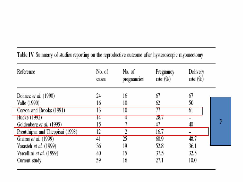
Myomectomy•
The favourable PRs obtained after myomectomy lead many clinicians to believe that removal of myomas increases pregnancy and live-birth rates (review Donnez and Jadoul, 2002 ). However, no appropriate prospective studies have been performed. Furthermore, no information on the value and complications of myomectomy in RIF is available,
although most clinicians recommend hysteroscopic removal of submucous fibroids distorting the uterine cavity.
?

ENDOMETRİAL POLİP HS GÖRÜNTÜ

HİSTEROSKOPİK ASHERMAN GÖRÜNTÜ