ENDOMETRIAL HYPERPLASIA - PESIMSRpesimsr.pes.edu/obgyan/pdf/endometrial hyperplasia final.pdf ·...
24
ENDOMETRIAL HYPERPLASIA DR.ANBU SUBBIAN
Transcript of ENDOMETRIAL HYPERPLASIA - PESIMSRpesimsr.pes.edu/obgyan/pdf/endometrial hyperplasia final.pdf ·...

ENDOMETRIAL HYPERPLASIA
DR.ANBU SUBBIAN
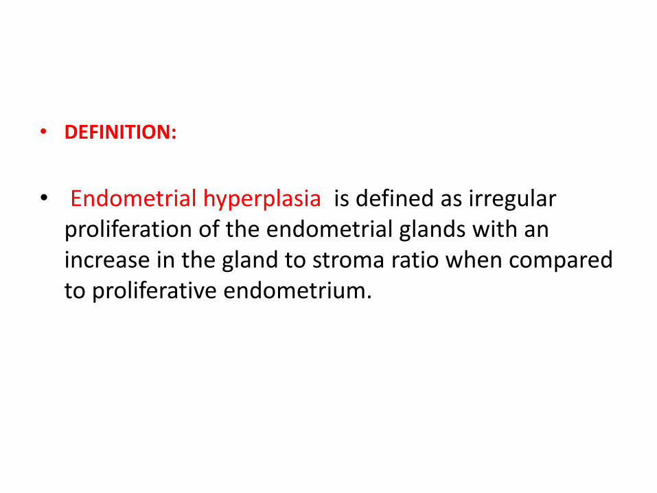
• DEFINITION:
• Endometrial hyperplasia is defined as irregular
proliferation of the endometrial glands with an
increase in the gland to stroma ratio when compared
to proliferative endometrium.
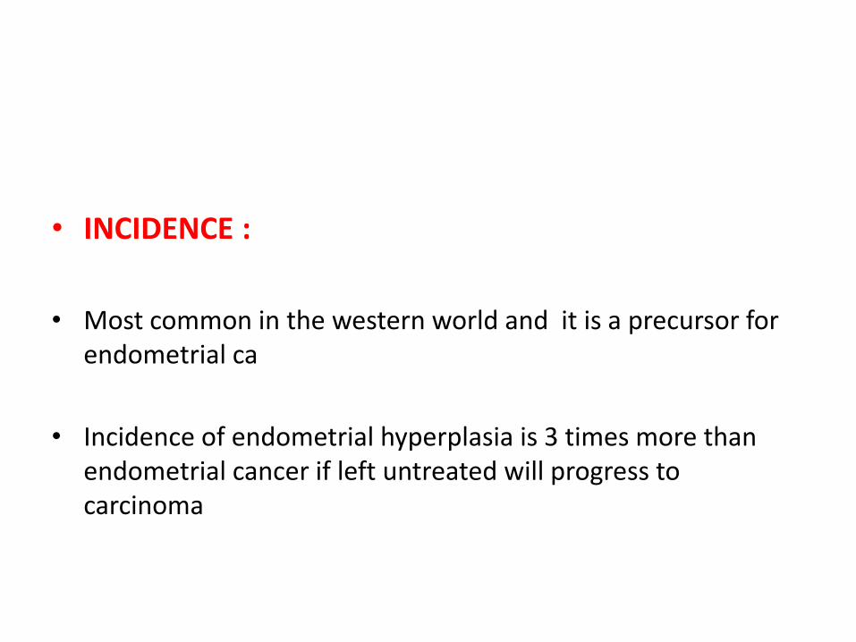
• INCIDENCE :
• Most common in the western world and it is a precursor for
endometrial ca
• Incidence of endometrial hyperplasia is 3 times more than
endometrial cancer if left untreated will progress to
carcinoma
• PRESENTATION:
• Most common - abnormal uterine bleeding.
• This includes -heavy menstrual bleeding
- inter menstrual bleeding
- irregular bleeding
- unscheduled bleeding on hormone
replacement therapy
- postmenopausal bleeding.
• RISK FACTORS :
• Increased body mass index (BMI)
• Polycystic ovary syndrome (PCOS)
• Estrogen-secreting ovarian tumours e.g. granulosa cell tumours.
• Drug-induced endometrial stimulation e.g. the use of estrogen replacement therapy (ERT) or those on long-term tamoxifen.
• Cochrane meta-analysis found that unopposed ERT is associated with an increased incidence of hyperplasia at all doses and is not recommended for use in women with a uterus
• Immunosuppression and infection.
• CLASSIFICATION: (Revised 2014 WHO classification)
• (i) Hyperplasia without atypia
• (ii) hyperplasia with atypia
• The complexity of architecture is no longer part of the
classification.
DIAGNOSIS AND ENDOMETRIAL
SURVEILLANCE :
• Diagnosis of endometrial hyperplasia requires histological
examination of the endometrial tissue.
• Endometrial surveillance should include endometrial sampling
by an outpatient endometrial biopsy.
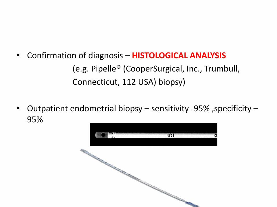
• Confirmation of diagnosis – HISTOLOGICAL ANALYSIS
(e.g. Pipelle® (CooperSurgical, Inc., Trumbull,
Connecticut, 112 USA) biopsy)
• Outpatient endometrial biopsy – sensitivity -95% ,specificity –
95%
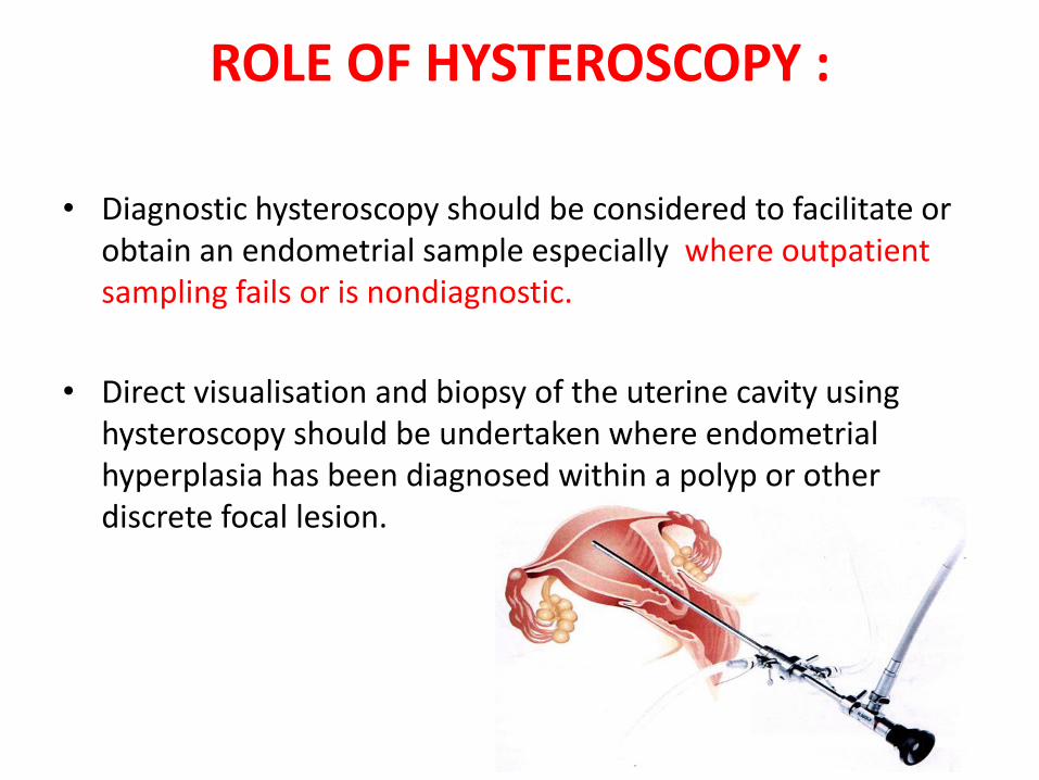
ROLE OF HYSTEROSCOPY :
• Diagnostic hysteroscopy should be considered to facilitate or
obtain an endometrial sample especially where outpatient
sampling fails or is nondiagnostic.
• Direct visualisation and biopsy of the uterine cavity using
hysteroscopy should be undertaken where endometrial
hyperplasia has been diagnosed within a polyp or other
discrete focal lesion.
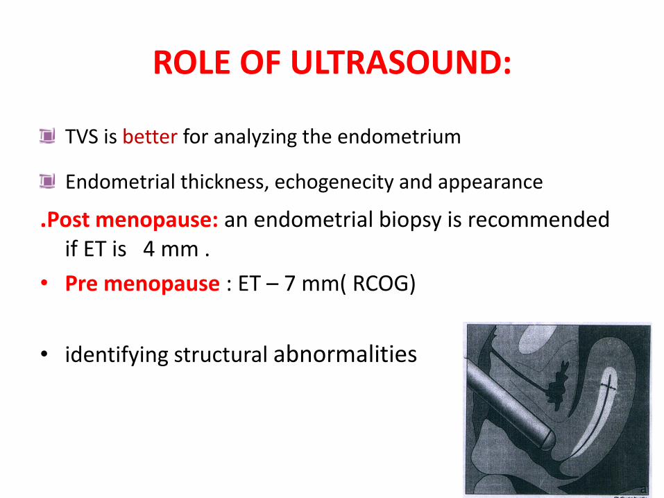
ROLE OF ULTRASOUND:
TVS is better for analyzing the endometrium
Endometrial thickness, echogenecity and appearance
.Post menopause: an endometrial biopsy is recommended
if ET is 4 mm .
• Pre menopause : ET – 7 mm( RCOG)
• identifying structural abnormalities
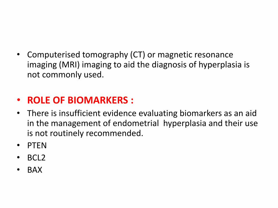
• Computerised tomography (CT) or magnetic resonance imaging (MRI) imaging to aid the diagnosis of hyperplasia is not commonly used.
• ROLE OF BIOMARKERS :
• There is insufficient evidence evaluating biomarkers as an aid in the management of endometrial hyperplasia and their use is not routinely recommended.
• PTEN
• BCL2
• BAX
MANAGEMENT
• ENDOMETRIAL HYPERPLASIA WITHOUT ATYPIA :
• Women should be informed that the risk of endometrial
hyperplasia without atypia progressing to endometrial cancer
is less than 5% over 20 years.
• Reversible risk factors such as obesity and the use of HRT
should be identified and treated.
• Observation alone with follow-up endometrial biopsies is
helpful when identifiable risk factors can be reversed.
• Treatment with progesterone has a higher disease regression
rate compared with observation alone.
• Progesterone treatment is indicated
(a) in women who fail to regress following observation
alone
(b) symptomatic women with abnormal uterine bleeding.
FIRST LINE OF MEDICAL
MANAGEMENT
• Both continuous oral and local intrauterine progestogens
[LNG-IUS]) are effective
• LNG-IUS is preferred .
• Continuous progestogens (medroxy progesterone 10–20
mg/day or norethisterone 10–15 mg/day) for women who
decline the LNG-IUS.
• Cyclical progestogens should not be used because they are
less effective

• DURATION OF TREATMENT:
• Minimum of 6 months.
• In the absence of side effects or need for fertility, women are
advised to retain the LNG-IUS for the duration of its licensed
use as this may prevent relapse of endometrial hyperplasia
without atypia.
• Endometrial surveillance 6-months once
• At least two consecutive 6-monthly negative biopsies should
be obtained prior to discharge.
• RELAPSE:
- vaginal bleeding
- obesity has high risk
- patient should be referred
- obese patients should undergo annual follow up after
two consecutive negative biopsies , inorder to detect
relapse.
ROLE OF SURGERY:
• Second line of management for endometrial hyperplasia without atypia
• First line of treatment for atypical hyperplasia
• indicated in women NOT wanting to preserve their fertility when:
• (i) progression to atypical hyperplasia occurs during follow-up;
or
• (ii) there is lack of histological regression of hyperplasia within 12 months of treatment;
or
• (iii) there is relapse of endometrial hyperplasia after completing progestogen treatment;
or
• (iv) there is persistence of bleeding symptoms;
or
• (v) the woman declines to undergo endometrial surveillance and comply with medical treatment.
• Postmenopausal women requiring surgical management for endometrial
hyperplasia without atypia should undergo TAH with BSO.
• For premenopausal women, the decision to remove the ovaries should be
individualised, but a bilateral salpingectomy can be considered as this
may reduce the risk of a future ovarian malignancy.
• Endometrial ablation is not recommended for the treatment of
endometrial hyperplasia because complete and persistent endometrial
destruction cannot be ensured and intrauterine adhesion formation may
preclude future endometrial histological surveillance.
MANAGEMENT OF ATYPICAL HYPERPLASIA
• FIRST LINE OF TREATMENT:
• POSTMENOPAUSE: Total hysterectomy with bilateral salpingooopherectomy
• PREMENOPAUSE: Total hysterectomy , the decision to remove the ovaries should be individualised, but a bilateral salpingectomy can be considered as this may reduce the risk of a future ovarian malignancy.
• There is no benefit from intraoperative frozen section analysis of the endometrium or routine lymphadenectomy.
• Women wishing to retain their fertility should be counselled about the risks of underlying malignancy and subsequent progression to endometrial cancer.
• Pretreatment investigations should aim to rule out invasive endometrial cancer or co-existing ovarian cancer.
• Hormone receptor (ER and PR )studies should be done prior to medical management.
• Histology, imaging and tumour marker results should be reviewed in a multidisciplinary meeting and a plan for management and ongoing endometrial surveillance formulated.
• First-line treatment with the LNG-IUS should be
recommended and oral progestogens as a second- best
alternative.
• Once fertility is no longer required, hysterectomy should be
offered in view of the high risk of disease relapse.
• ENDOMETRIAL SURVEILLANCE:
-endometrial biopsy.
-every 3–6 months until 2
consecutive negative biopsies are obtained.
• In asymptomatic women with a uterus :
after 2 consecutive negative endometrial
biopsies, long-term follow-up every 6–12 months is
recommended until a hysterectomy is performed.
MANAGEMENT OF ENDOMETRIAL HYPERPLASIA IN
WOMEN WISHING TO CONCEIVE:
• Disease regression should be achieved on at least one
negative endometrial sample before women attempt to
conceive.
• Assisted reproduction can be considered over natural
conception.


![Endometrium presentation - Dr Wright[1] · Endometrial Hyperplasia Simple hyperplasia Complex hyperplasia (adenomatous) Simple atypical hyperplasia ... Progression of Hyperplasia](https://static.fdocuments.in/doc/165x107/5b8a421e7f8b9a50388bc13d/endometrium-presentation-dr-wright1-endometrial-hyperplasia-simple-hyperplasia.jpg)