Diagnosis and Molecular Epidemiology of Bovine Rotavirus and
Ecology and Epidemiology of Bovine-Related
Coagulase-Negative Staphylococcus Species
Anneleen De Visscher
Merelbeke, 2016


Our greatest weakness lies in giving up. The most certain way to
succeed is always to try just one more time.
Thomas A. Edison
Ecology and Epidemiology of Bovine-Related Coagulase-Negative
Staphylococcus Species
Anneleen De Visscher
Cover photos: Jordy Haverbeke
Cover cows: Dirk Van Eetvelde
Cover editing: Arne Vanheuverbeke
Printing: University Press, Zelzate
Printing of this thesis was financially supported by MSD and Boehringer Ingelheim.
This research was financed by a PhD grant (n° 111588) by the Agency for Innovation
by Science and Technology in Flanders (IWT Vlaanderen) and performed at Ghent
University.
Ecology and Epidemiology of Bovine-Related Coagulase-Negative
Staphylococcus Species
Anneleen De Visscher
Department of Reproduction, Obstetrics, and Herd Health
Faculty of Veterinary Medicine
Ghent University
Dissertation submitted in the fulfillment of the requirements for the degree of Doctor
in Veterinary Sciences, Faculty of Veterinary Medicine, Ghent University, February 5,
2016
Ecologie en Epidemiologie van Boviene Coagulase-Negatieve
Staphylococcus Species
Anneleen De Visscher
Vakgroep Voortplanting, Verloskunde en Bedrijfsdiergeneeskunde
Faculteit Diergeneeskunde
Universiteit Gent
Proefschrift voorgedragen tot het behalen van de graad van Doctor in de
Diergeneeskundige Wetenschappen aan de Faculteit Diergeneeskunde, Universiteit
Gent, 5 februari 2016

Promoters
Prof. dr. Sarne De Vliegher
Faculty of Veterinary Medicine, Ghent University, Belgium
Prof. dr. Freddy Haesebrouck
Faculty of Veterinary Medicine, Ghent University, Belgium
Dr. Sofie Piepers
Faculty of Veterinary Medicine, Ghent University, Belgium
Members of the Examination Committee
Prof. em. dr. dr.h.c. Aart de Kruif
Chairman - Ghent University, Belgium
Prof. dr. Herman Barkema
University of Calgary, Canada - Ghent University, Belgium
Prof. dr. ir. Frédéric Leroy
University of Brussels, Belgium
Prof. dr. Evelyne Meyer
Ghent University, Belgium
Dr. Karlien Supré
Milk Control Centre Flanders (MCC), Belgium
Dr. ir. Els Van Coillie
Institute for Agricultural and Fisheries Research (ILVO), Belgium
Prof. dr. Filip Van Immerseel
Ghent University, Belgium

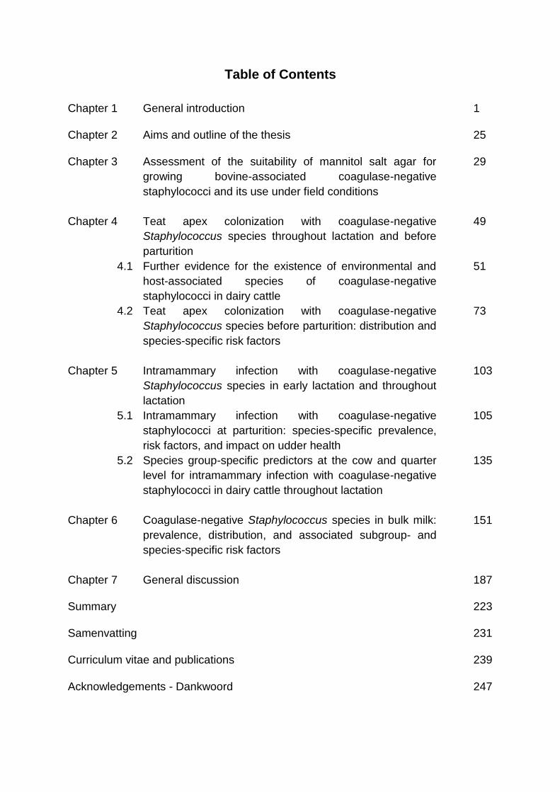
Table of Contents
Chapter 1 General introduction 1
Chapter 2 Aims and outline of the thesis 25
Chapter 3 Assessment of the suitability of mannitol salt agar for
growing bovine-associated coagulase-negative
staphylococci and its use under field conditions
29
Chapter 4 Teat apex colonization with coagulase-negative
Staphylococcus species throughout lactation and before
parturition
49
4.1 Further evidence for the existence of environmental and
host-associated species of coagulase-negative
staphylococci in dairy cattle
51
4.2 Teat apex colonization with coagulase-negative
Staphylococcus species before parturition: distribution and
species-specific risk factors
73
Chapter 5 Intramammary infection with coagulase-negative
Staphylococcus species in early lactation and throughout
lactation
103
5.1 Intramammary infection with coagulase-negative
staphylococci at parturition: species-specific prevalence,
risk factors, and impact on udder health
105
5.2 Species group-specific predictors at the cow and quarter
level for intramammary infection with coagulase-negative
staphylococci in dairy cattle throughout lactation
135
Chapter 6 Coagulase-negative Staphylococcus species in bulk milk:
prevalence, distribution, and associated subgroup- and
species-specific risk factors
151
Chapter 7 General discussion 187
Summary 223
Samenvatting 231
Curriculum vitae and publications
239
Acknowledgements - Dankwoord 247

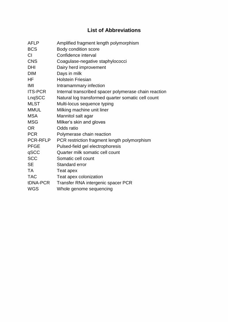
List of Abbreviations
AFLP Amplified fragment length polymorphism
BCS Body condition score
CI Confidence interval
CNS Coagulase-negative staphylococci
DHI Dairy herd improvement
DIM Days in milk
HF Holstein Friesian
IMI Intramammary infection
ITS-PCR Internal transcribed spacer polymerase chain reaction
LnqSCC Natural log transformed quarter somatic cell count
MLST Multi-locus sequence typing
MMUL Milking machine unit liner
MSA Mannitol salt agar
MSG Milker’s skin and gloves
OR Odds ratio
PCR Polymerase chain reaction
PCR-RFLP PCR restriction fragment length polymorphism
PFGE Pulsed-field gel electrophoresis
qSCC Quarter milk somatic cell count
SCC Somatic cell count
SE Standard error
TA Teat apex
TAC Teat apex colonization
tDNA-PCR Transfer RNA intergenic spacer PCR
WGS Whole genome sequencing

Chapter 1
General Introduction
A. De Visscher
Department of Reproduction, Obstetrics, and Herd Health
Faculty of Veterinary Medicine
Ghent University, Merelbeke, Belgium

Chapter 1 General introduction
3
Bovine mastitis
Mastitis, an inflammation of the mammary gland, is one of the most prevalent and
costly diseases in the dairy industry worldwide. Economic losses are due to factors
such as milk production losses, additional treatment, and increased culling (Halasa
et al., 2007) and vary between €65 and €182 per cow in the herd per year (Huijps et
al., 2008). The disease also affects milk quality and animal welfare (Heringstad et al.,
2000) and is associated with the highest antimicrobial drug use on dairy farms (Pol
and Ruegg, 2007; Barlow, 2011). Growing concern rises over the use of and
resistance to antimicrobials in both human and animal health (Barlow, 2011; WHO,
2014), indicating the need for good prevention rather than treatment programs. Herd-
management practices and cow- and quarter-level risk factors, helping to effectively
control and especially prevent mastitis are thus necessary (Gruet et al., 2001;
Halasa et al., 2007; Piepers et al., 2007; Supré et al., 2011), though depend on the
causative agents involved (Barkema et al., 1999b; Smith and Hogan, 2001; Zadoks
et al., 2001; Piepers et al., 2011).
Mastitis typically results from bacterial intramammary infection (IMI) and is called
clinical or subclinical in the presence or absence of visible symptoms, respectively
(Kemp et al., 2008). The causative bacteria can be grouped according to their
ecological nature (i.e. habitat) in so-called host(dairy cow)-adapted (e.g.
Staphylococcus aureus) and environmental pathogens (e.g. Streptococcus uberis)
(Gruet et al., 2001; Pyörälä and Taponen, 2009; Blowey and Edmonson, 2010).
Also, their epidemiology differentiates bacteria in contagious and opportunistic
subgroups (Pyörälä and Taponen, 2009). Contagious pathogens (e.g. S. aureus) are
characterized by a single origin, multiple infected animals in a herd, and a cow-to-
cow-transmission through a vector (e.g. the milking machine), whereas opportunistic
bacteria (e.g. Escherichia coli) have a range of different origins, are not spread
among animals and only cause IMI under favoring conditions (e.g. lack of hygiene).
This classification, however, is not always straightforward and strain-specific
differences exist within bacterial species (Zadoks et al., 2003). More than 100
species and subspecies are able to cause bovine mastitis (Smith and Hogan, 2001).
In Flanders, the predominant major pathogen causing clinical mastitis is S. uberis,
followed by E. coli, S. aureus and Streptococcus dysgalactiae (Verbeke et al., 2012).
Subclinical mastitis is, on the other hand, mainly caused by S. aureus and esculine-
Chapter 1 General introduction
4
positive cocci (Piepers et al., 2007). The latter pathogen distributions are in
accordance with those described in other countries and regions (Pitkälä et al., 2004;
Tenhagen et al., 2006; Bradley et al., 2007). Yet, the coagulase-negative
staphylococci (CNS) are currently the overall most frequently isolated bovine mastitis
pathogens in Flanders (Piepers et al., 2007) and other regions and countries
(Pyörälä and Taponen, 2009).
Coagulase-negative staphylococci
The heterogeneous group of CNS includes 50 species (www.bacterio.net, 2015).
Several commercially available phenotypic identification methods, such as API Staph
ID 20 (Zadoks and Watts, 2009; Park et al., 2011), API Staph ID 32 (Thorberg and
Brändström, 2000; Taponen et al., 2006; Ruegg, 2009; Sampimon et al., 2009b;
Zadoks and Watts, 2009), API Staph Trac-System (Matthews et al., 1990b), BBL
Crystal Gram-Positive System (Ruegg, 2009; Zadoks and Watts, 2009), STAPHase,
Staph-Ident (Watts et al., 1984), Staph-Zym (Thorberg and Brändström, 2000;
Capurro et al., 2009; Sampimon et al., 2009b; Zadoks and Watts, 2009), VITEK 2
(Bal et al., 2010), and VITEK Gram-Positive Identification Card (Matthews et al.,
1990b), have been used and described before. These phenotypic systems rely on
the evaluation of the expression of several characteristics of the bacterial species,
though suffer from a subjective interpretation of the tests, a variable expression, and
they are mainly validated for human CNS species (Ruegg, 2009; Zadoks and Watts,
2009). Comparison of those phenotypic methods with the so-called gold standard,
i.e. gene sequencing, revealed the necessity of a genotypic method, based on the
detection of the bacterial DNA, when identifying CNS species in order to obtain a
sufficient accuracy, reproducebility and typeability (Sampimon et al., 2009b; Zadoks
and Watts, 2009; Park et al., 2011; Vanderhaeghen et al., 2015). Validation of
accurate identification methods was thus recommended (Taponen et al., 2006;
Ruegg, 2009; Sampimon et al., 2009b) and performed (Bes et al., 2000; Santos et
al., 2008; Capurro et al., 2009; Supré et al., 2009; Piessens et al., 2010; Braem et
al., 2011; Park et al., 2011). The implementation of those techniques revealed
species-specific traits and characteristics.
As a group, CNS are considered as minor pathogens for udder health (Schukken
et al., 2009) though contrasting findings have been reported and differences among
Chapter 1 General introduction
5
species have been revealed in their impact on udder health. Coagulase-negative
staphylococci have also been isolated from mild cases of clinical mastitis (Trinidad et
al., 1990; Bradley et al., 2007; Compton et al., 2007; Olde Riekerink et al., 2007;
Taponen et al., 2007; Gillespie et al., 2009; Persson Waller et al., 2011; Piessens et
al., 2011; Simojoki et al., 2011), but seem to lack the ability to cause severe clinical
mastitis (Taponen et al., 2007). However, the latter hypothesis was also countered
as a higher body temperature and disturbed clinical condition was occasionally
caused by CNS (Jarp, 1991). The impact of CNS IMI on milk production has also
been discussed and questioned. Either no impact was observed (Kirk et al., 1996) or
even a lower milk production was detected in some studies (Pyörälä and Taponen,
2009; Thorberg et al., 2009; Simojoki et al., 2011), whereas others showed a higher
milk production (Compton et al., 2007; Schukken et al., 2009; Piepers et al., 2010).
Besides, potential protective effects of CNS have also been explored. For teat
apices colonized with whatever CNS species (Piepers et al., 2011) or with
Staphylococcus chromogenes (De Vliegher et al., 2003) the protective assumption
could be substantiated. Different hypotheses have been suggested to explain the
observed findings such as competitive exclusion or an already activated immune
response (Rainard and Poutrel, 1988; Matthews et al., 1990a; Piepers et al., 2009b).
The production of inhibitory substances (e.g. bacteriocins) has been proposed for S.
chromogenes (De Vliegher et al., 2004) and noticed for several other staphylococci
(Allgaier et al., 1985; Sashihara et al., 2000; Nascimento et al., 2005; Coelho et al.,
2007; Wilaipun et al., 2008; Ceotto et al., 2010; Fagundes et al., 2011; Braem et al.,
2014).
Prevalence and distribution of coagulase-negative staphylococci
Coagulase-negative staphylococci are the predominant mastitis pathogens in
prevalence studies performed in recent years (Barkema et al., 1999a; Pitkälä et al.,
2004; Tenhagen et al., 2006; Bradley et al., 2007; Piepers et al., 2007; Sampimon et
al., 2009a; Schukken et al., 2009; Reyher et al., 2011). At parturition, CNS have also
commonly been observed in milk samples (Kirk et al., 1996; Rajala-Schultz et al.,
2005, Parker et al., 2007; Piepers et al., 2011; Rajala-Schultz et al., 2009). A fast
decrease in the number of CNS-infected quarters and animals after calving has been
Chapter 1 General introduction
6
recorded (Myllys, 1995; Piepers et al., 2009a), although differences among CNS
species have been noticed (Aarestrup and Jensen, 1997; Taponen et al., 2006).
Accurate studies relying on genotypic identification, as recommended for CNS
research, revealed approximately 25 CNS species causing bovine IMI (Santos et al.,
2008; Sampimon et al., 2009b; Park et al., 2011; Persson Waller et al., 2011;
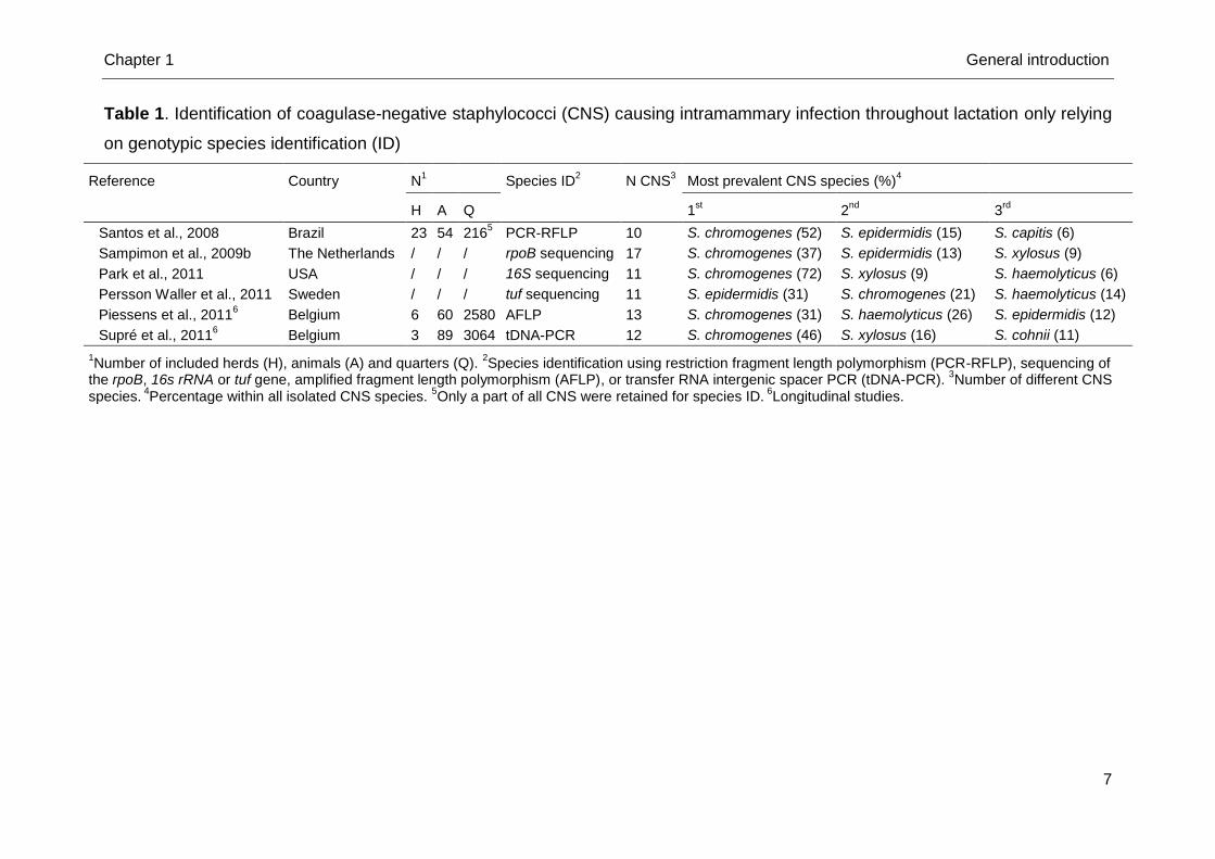
Piessens et al., 2011; Supré et al., 2011) (Table 1). Staphylococcus chromogenes
was the predominant species in all except one study (Persson Waller et al., 2011).
Staphylococcus haemolyticus and S. epidermidis were also frequently isolated in all
studies. Species other than the aforementioned ones isolated in most studies are S.
cohnii, S. hominis, S. hyicus, S. sciuri, S. simulans, and S. xylosus. Large
discrepancies exist between studies in reporting the presence of S. arlettae, S.
auricularis, S. capitis, S. caprae, S. caseolyticus, S. devriesei, S. equorum, S.
fleurettii, S. gallinarum, S. lentus, S. nepalensis, S. pasteuri, S. pseudintermedius, S.
saprophyticus, S. succinus, and S. warneri. As some species were only recently
described (e.g. S. devriesei, Supré et al., 2010) this is not unexpected. Furthermore,
a herd- or even country-specific microbiota might exist (Piepers et al., 2007; Gillespie
et al., 2009; Piessens et al., 2011; Supré et al., 2011) as well as the applied
identification method and study design could influence the CNS prevalence and
distribution.
The CNS distribution has been unraveled throughout lactation in the
aforementioned studies, but the species-specific distribution at parturition needs
further exploration.
Relevance of coagulase-negative staphylococci for bovine udder health
The relevance of CNS for bovine udder health has been studied for many years
and concerns topics such as their impact on the (quarter milk) somatic cell count and
their potential to cause persistent IMI.
A mild to moderate increase in SCC in CNS-infected cows and quarters, as
opposed to uninfected cows and quarters, has been observed (Lam et al., 1997;
Barkema et al., 1999a; Taponen et al., 2007; Gillespie et al., 2009; Thorberg et al.,
2009; Sampimon et al., 2009a; Schukken et al., 2009; Piepers et al., 2010;
Sampimon et al., 2010).
Chapter 1 General introduction
7
Table 1. Identification of coagulase-negative staphylococci (CNS) causing intramammary infection throughout lactation only relying
on genotypic species identification (ID)
Reference Country N1 Species ID
2 N CNS
3 Most prevalent CNS species (%)
4
H A Q 1st
2nd
3rd
Santos et al., 2008 Brazil 23 54 2165 PCR-RFLP 10 S. chromogenes (52) S. epidermidis (15) S. capitis (6)
Sampimon et al., 2009b The Netherlands / / / rpoB sequencing 17 S. chromogenes (37) S. epidermidis (13) S. xylosus (9)
Park et al., 2011 USA / / / 16S sequencing 11 S. chromogenes (72) S. xylosus (9) S. haemolyticus (6)
Persson Waller et al., 2011 Sweden / / / tuf sequencing 11 S. epidermidis (31) S. chromogenes (21) S. haemolyticus (14)
Piessens et al., 20116 Belgium 6 60 2580 AFLP 13 S. chromogenes (31) S. haemolyticus (26) S. epidermidis (12)
Supré et al., 20116 Belgium 3 89 3064 tDNA-PCR 12 S. chromogenes (46) S. xylosus (16) S. cohnii (11)
1Number of included herds (H), animals (A) and quarters (Q).
2Species identification using restriction fragment length polymorphism (PCR-RFLP), sequencing of
the rpoB, 16s rRNA or tuf gene, amplified fragment length polymorphism (AFLP), or transfer RNA intergenic spacer PCR (tDNA-PCR). 3Number of different CNS
species. 4Percentage within all isolated CNS species.
5Only a part of all CNS were retained for species ID.
6Longitudinal studies.
Chapter 1 General introduction
8
Especially on low bulk milk SCC herds, CNS have an important contribution to the
total number of somatic cells in the bulk milk (Rainard et al., 1990; Piepers et al.,
2009a; Pyörälä and Taponen et al., 2009; Schukken et al., 2009; Sampimon et al.,
2010). In contrast, some studies did not detect an impact on the SCC (Kirk et al.,
1996; Compton et al., 2007) although the latter solely concerned heifers.
Species-specific analysis actually revealed differences among CNS species
(Sampimon et al., 2009a; Thorberg et al., 2009; Simojoki et al., 2011; Supré et al.,
2011): S. chromogenes, S. simulans and S. xylosus cause a statistically significant
elevation in SCC not different from S. aureus in quarters from lactating dairy cows
and heifers (Supré et al., 2011), suggesting that those species are more relevant for
udder health than others. The latter finding needs further investigation and should be
substantiated for IMI at parturition as well. It suggests that prevention of CNS IMI on
well-managed dairy farms should at least target these “more relevant” species.
Coagulase-negative staphylococci are able to cause persistent IMI (Rainard et al.,
1990) even through the dry period (Rajala-Schultz et al., 2009; Supré et al., 2011)
despite a high spontaneous cure rate (Taponen et al., 2006). Differences among
species have been observed (Aarestrup and Jensen, 1997; Gillespie et al., 2009;
Rajala-Schultz et al., 2009; Thorberg et al., 2009; Piessens et al., 2011), although
also similar strains have been isolated from both persistent and transient IMI
(Taponen et al., 2007). The latter suggests not only the bacterium plays a role, but
also the host-response to IMI.
Different cow-associated habitats harboring coagulase-negative staphylococci
Coagulase-negative staphylococci as a group are considered as opportunistic
pathogens, part of the normal skin microbiota (Devriese and De Keyser, 1980;
Schukken et al., 2011). Differences in CNS prevalence are observed when exploring
various cow-related habitats (e.g. teat skin, teat canal, udder skin, perineum…)
(White et al., 1989; Trinidad et al., 1990; Taponen et al., 2008). Research picturing
CNS distribution on teat apices is only suggestive and incomplete as species
identification lacked accuracy by the use of phenotypic tests in one study (White et
al., 1989) and only lactating dairy cows from one herd were included in another study
(Taponen et al., 2008). Large studies relying on molecular identification are required
to unravel CNS teat apex distribution and to study potential infection sources from
Chapter 1 General introduction
9
different habitats. We focus on the 2 most important ones: teat apices and the
environment.
A higher risk of developing a S. aureus IMI at parturition when teat apices were
colonized with S. aureus was suggested more than 2 decades ago (Roberson et al.,
1994). This was not substantiated in a more recent study as different S. aureus
strains were isolated from IMI and teat skin from lactating dairy cows (Zadoks et al.,
2002). Analogous to S. aureus, teat skin might act as a reservoir for bovine CNS.
Teat apices colonized with CNS as a group or solely S. chromogenes, only
phenotypically identified, were not associated with IMI in quarters of heifers at
parturition (De Vliegher et al., 2003; Piepers et al., 2011). In contrast, highly similar
S. chromogenes strains were detected in milk and on udder skin of lactating cows
(Taponen et al., 2008). Genotypic peri-partum studies are needed to bring further
insights.
The cows’ environment acts as a potential source for bovine CNS IMI (Matos et
al., 1991; Piessens et al., 2011). Staphylococcus equorum, S. cohnii, and S. sciuri
are considered as environmental species as they were frequently observed in the
cows’ environment, i.e. air, slatted floors, sawdust of the cubicles and sawdust of the
stock (Piessens et al., 2011), and were hardly isolated from bovine IMI. On the other
hand, S. chromogenes and S. epidermidis were rarely present in the bovine
environment, although being a predominant cause of bovine IMI. Staphylococcus
haemolyticus and S. simulans were both present in the environment and causing
bovine IMI (Santos et al., 2008; Sampimon et al., 2009b; Park et al., 2011; Persson
Waller et al., 2011; Piessens et al., 2011; Supré et al., 2011) and therefore deemed
opportunistic in nature. Piessens et al. (2011) found S. xylosus only in environmental
samples whereas all other aforementioned genotypic studies detected S. xylosus in
bovine milk samples. The ecological nature of the different CNS species should be
further elucidated.
Challenges related to coagulase-negative staphylococcal research
Research relying on genotypic identification demonstrated the abundant presence
of diverse CNS species in different bovine habitats, such as the cows’ environment
(Piessens et al., 2011), milk samples (Santos et al., 2008; Sampimon et al., 2009b;
Park et al., 2011; Persson Waller et al., 2011; Piessens et al., 2011; Supré et al.,
Chapter 1 General introduction
10
2011), and other udder-related habitats (Taponen et al., 2008) indicating their
ubiquitous nature. Picturing CNS distribution in those different habitats and
investigating potential transmission routes is needed in order to understand variation
in epidemiological and ecological nature among species. Identification of associated
factors seems a logical next step (Pyörälä and Taponen, 2009).
Species-specific research, however, requires extensive studies including a large
number of herds, cows and/or quarters in order to obtain a sufficient number of
isolated CNS species. Bulk milk samples have previously been used (Jayarao et al.,
2004; Virgin et al., 2009; Olde Riekerink et al., 2010) and allow for including a large
number of herds. Yet, numerous bacteria other than CNS are present in bulk milk
samples impeding CNS isolation and indicating that bacteria in bulk milk are not
solely originating from infected quarters.
Another major challenge turns up when using swabs to explore different habitats
(e.g. teat apices) as again mixed cultures are observed, potentially suppressing
isolation of CNS (De Vliegher et al., 2003; Taponen et al., 2008).
The use of selective media offers advantages and helps to circumvent the
inconveniences of overgrowth and hampered isolation, when using bulk milk
samples or swabs (Finegold and Sweeney, 1961). Various media have already been
used for the recovery of S. aureus and CNS, e. g. Baird Parker agar (Jayarao et al.,
2004), Vogel-Johnson agar (Olde Riekerink et al., 2006), DNA-toluidine blue agar
(Jarp, 1991; Chapin and Lauderdale, 2007), and different modifications of
CHROMagar (Chapin and Lauderdale, 2007; Han et al., 2007; Virgin et al., 2009).
These media are mainly recommended for the selective recovery of S. aureus
though growth of other bacteria was not completely avoided. Chapman (1945)
attempted to block the growth of bacteria other than staphylococci by developing a
selective medium with a high sodium chloride concentration, i.e. mannitol salt agar
(MSA). Nowadays, MSA is commercially available and offers advantages in the
recovery of CNS (Kateete et al., 2010). This medium has already been used both in
human (Jayaratne and Rutherford, 1999; Shittu et al., 2004; Shittu et al., 2006; Han
et al., 2007) and veterinary medicine (Cheng et al., 2010; Addis et al., 2011;
Piessens et al., 2011) though no studies have tested the growth of bovine-
associated CNS species on this medium and its reliability. The latter should be
examined as a prerequisite of any selective medium is the growth of the genera
and/or species of interest.
Chapter 1 General introduction
11
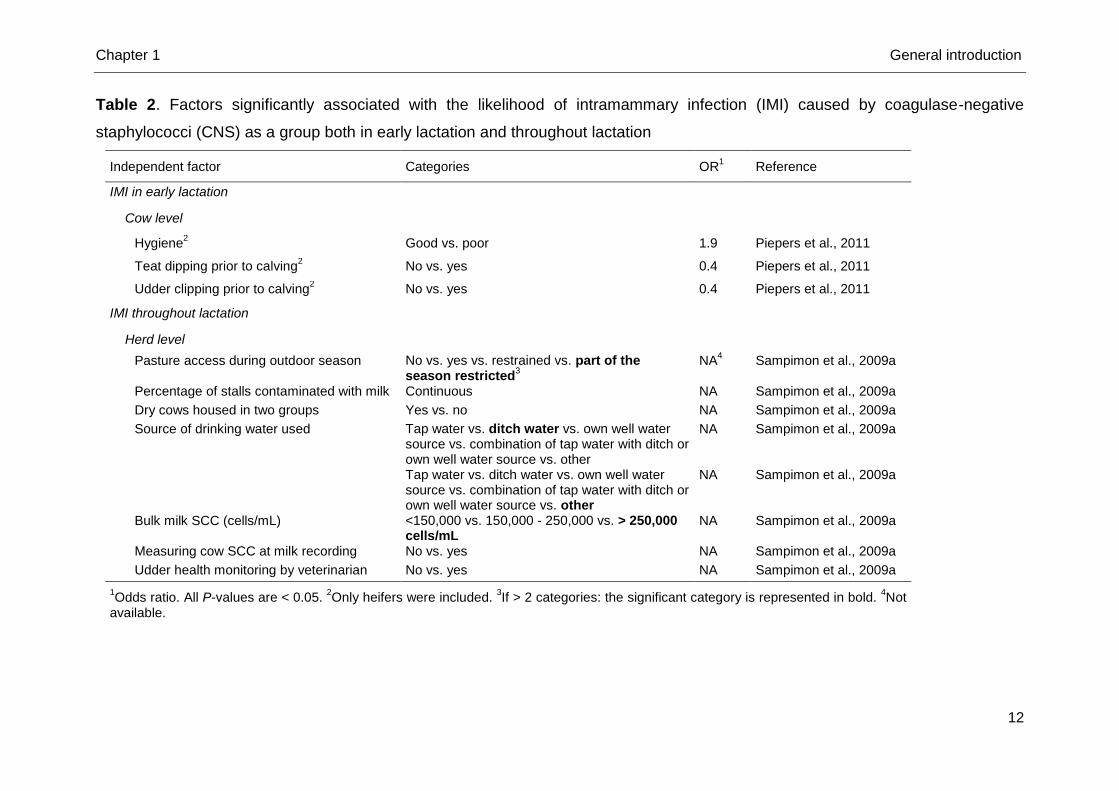
Risk factors for the presence of coagulase-negative staphylococci in different
habitats
As prevention and control measures differ among mastitis pathogens (Barkema et
al., 1999b; Smith and Hogan, 2001; Zadoks et al., 2001; Piepers et al., 2011), risk
factor studies should be conducted for all mastitis-associated bacteria including CNS
(Pyörälä and Taponen, 2009).
Several risk factors such as source of drinking water, housing of dry cows, pasture
access, monthly measuring of SCC, milk leakage, bulk milk SCC and udder health
monitoring by a veterinarian, are associated with IMI caused by CNS as a group
(Sampimon et al., 2009a) (Table 2). Poor hygiene, non-clipped udders and no
application of teat disinfectants prior to calving were found significant risk factors for
CNS-group IMI in heifers (Piepers et al., 2011) (Table 2). An environmental nature of
the involved CNS species, which were not differentiated, was assumed in the latter
study based on the risk factors that were identified, yet not substantiated.
Additionally, differences between cows and quarters in susceptibility for IMI have
been observed (Zadoks et al., 2001; Piessens et al., 2011; Supré et al., 2011) and a
herd-dependent CNS microbiota has been reported (Piepers et al., 2007; Gillespie et
al., 2009; Piessens et al., 2011; Supré et al., 2011). Research identifying risk factors
associated with CNS IMI both throughout lactation and at parturition is required and
should therefore be conducted at the quarter and cow level as well as at the herd
level.
Conclusion
Many efforts are ongoing to enlarge our knowledge of the various CNS species
belonging to the heterogeneous group of bacteria. The species-specific prevalence
and distribution of several habitats need to be further explored using adequate semi-
selective media and should rely on genotypic identification methods. The impact of
the different species should also be investigated and substantiated. Furthermore,
risk factor studies at the species or subgroup level should be conducted in order to
gain more insight in the ecology and epidemiology of bovine-related CNS species.
Chapter 1 General introduction
12
Table 2. Factors significantly associated with the likelihood of intramammary infection (IMI) caused by coagulase-negative
staphylococci (CNS) as a group both in early lactation and throughout lactation
Independent factor Categories OR1 Reference
IMI in early lactation
Cow level
Hygiene2 Good vs. poor 1.9 Piepers et al., 2011
Teat dipping prior to calving2 No vs. yes 0.4 Piepers et al., 2011
Udder clipping prior to calving2 No vs. yes 0.4 Piepers et al., 2011
IMI throughout lactation
Herd level
Pasture access during outdoor season No vs. yes vs. restrained vs. part of the season restricted
3
NA4 Sampimon et al., 2009a
Percentage of stalls contaminated with milk Continuous NA Sampimon et al., 2009a
Dry cows housed in two groups Yes vs. no NA Sampimon et al., 2009a
Source of drinking water used Tap water vs. ditch water vs. own well water source vs. combination of tap water with ditch or own well water source vs. other
NA Sampimon et al., 2009a
Tap water vs. ditch water vs. own well water source vs. combination of tap water with ditch or own well water source vs. other
NA Sampimon et al., 2009a
Bulk milk SCC (cells/mL) <150,000 vs. 150,000 - 250,000 vs. > 250,000 cells/mL
NA Sampimon et al., 2009a
Measuring cow SCC at milk recording No vs. yes NA Sampimon et al., 2009a
Udder health monitoring by veterinarian No vs. yes NA Sampimon et al., 2009a
1Odds ratio. All P-values are < 0.05.
2Only heifers were included.
3If > 2 categories: the significant category is represented in bold.
4Not
available.
Chapter 1 General introduction
13
References
Aarestrup, F. M., and N. E. Jensen. 1997. Prevalence and duration of intramammary
infection in Danish heifers during the peripartum period. J. Dairy Sci. 80:307-312.
Addis, M., P. Mahindra, and N. K. Moses. 2011. Isolation and identification of
Staphylococcus species from raw bovine milk in Debre Zeit, Ethiopia. Vet. Res. 4,
45-49.
Allgaier, H., G. Jung, R. G. Werner, U. Schneider, and H. Zähner. 1985. Elucidation
of the structure of epidermin, a ribosomally synthesized, tetracyclic heterodetic
polypeptide antibiotic. Angew. Chem. Int. Edit. 24:1051-1053.
Bal, E. B. B., M. A. Bal, T. Isevi and E. Yula. 2010. Application of PCR-RFLP of gap
gene method as a molecular typing tool for coagulase-negative staphylococci from
bovine and human origin identified with VITEK 2. Afr. J. Microbiol. Res. 4:775-782.
Barkema, H. W., H. A. Deluyker, Y. H. Schukken, and T. J. G. M. Lam. 1999a.
Quarter-milk somatic cell count at calving and at the first six milkings after calving.
Prev. Vet. Med. 38:1-9.
Barkema, H. W., Y. H. Schukken, T. J. G. M. Lam, M. L. Beiboer, G. Benedictus, and
A. Brand. 1999b. Management practices associated with the incidence rate of
clinical mastitis. J. Dairy Sci. 82:1643-1654.
Barlow, J. 2011. Mastitis therapy and antimicrobial susceptibility: A multispecies
review with a focus on antibiotic treatment of mastitis in dairy cattle. J. Mammary
Gland. Biol. 16:383-407.
Bes, M., V. Guérin-Faublée, H. Meugnier, J. Etienne, and J. Freney. 2000.
Improvement of the identification of staphylococci isolated from bovine mammary
infections using molecular methods. Vet. Microbiol. 71:287-294.
Blowey, R. and P. Edmonson. 2010. Mastitis - Causes, epidemiology and control. In:
Blowey, R. and P. Edmonson, Mastitis control in dairy herds. 2nd edition, CAB
International, London, UK, pp. 27-45.
Bradley, A. J., K. A. Leach, J. E. Breen, L. E. Green, and M. J. Green. 2007. Survey
of the incidence and aetiology of mastitis on dairy farms in England and Wales.
Vet. Rec. 160:253-258.
Braem, G., S. De Vliegher, K. Supré, F. Haesebrouck, F. Leroy and L. De Vuyst.
2011. (GTG)5-PCR fingerprinting for the classification and identification of
Chapter 1 General introduction
14
coagulase-negative Staphylococcus species from bovine milk and teat apices: A
comparison of type strains and field isolates. Vet. Microbiol. 147:67-74.
Braem, G., B. Stijlemans, W. Van Haken, S. De Vliegher, L. De Vuyst, and F. Leroy.
2014. Antibacterial activities of coagulase-negative staphylococci from bovine teat
apex skin and their inhibitory effect on mastitis-related pathogens. J. App.
Microbiol. 116:1084-1093.
Capurro, A., K. Artursson, K. Persson Waller, B. Bengtsson, H. Ericsson-Unnerstad,
and A. Aspán. 2009. Comparison of a commercialized phenotyping system,
antimicrobial susceptibility testing, and tuf gene sequences-based genotyping for
species-level identification of coagulase-negative staphylococci isolated from
cases of bovine mastitis. Vet. Microbiol. 134:327-333.
Ceotto, H., H. Holo, K. F. S. Costa, J. S. Nascimento, A. Salehian, I. F. Nes, and M.
C. F. Bastos. 2010. Nukacin 3299, a lantibiotic produced by Staphylococcus
simulans 3299 identical to nukacin ISK-1. Vet. Microbiol. 146:124-131.
Chapin, K. C., and T.-L. Lauderdale. 2007. Reagents, stains, and media:
bacteriology. In: Murray, P. R., Manual of Clinical Microbiology. 9th edition, ASM
Press, Washington DC, USA, pp. 334-411.
Chapman, G. H. 1945. The significance of sodium chloride in studies of
staphylococci. J. Bacteriol. 50:201-203.
Cheng, D. R., S. Y. Zhu, Z. H. Yin, W. W. Ding, Z. X. Mu, Z. R. Su, and H. C. Sun.
2010. Prevalence of bacterial infection responsible for bovine mastitis. Afr. J.
Microbiol. Res. 4:1110-1116.
Coelho, M. L. V., J. S. Nascimento, P. C. Fagundes, D. J. Madureira, S. S. Oliveira,
M. A. V. P. Brito, and M. C. F. Bastos. 2007. Activity of staphylococcal
bactericocins against Staphylococcus aureus and Streptococcus agalactiae
involved in bovine mastitis. Res. Microbiol. 158:625-630.
Compton, C. W. R., C. Heuer, K. Parker, and S. McDougall. 2007. Epidemiology of
mastitis in pasture-grazed peripartum dairy heifers and its effects on productivity.
J. Dairy Sci. 90:4157-4170.
De Vliegher, S., H. Laevens, L. A. Devriese, G. Opsomer, J. L. M. Leroy, H. W.
Barkema, and A. de Kruif. 2003. Prepartum teat apex colonization with
Staphylococcus chromogenes in dairy heifers is associated with low somatic cell
count in early lactation. Vet. Microbiol. 92:245-252.
Chapter 1 General introduction
15
De Vliegher, S., G. Opsomer, A. Vanrolleghem, L. A. Devriese, O. C. Sampimon, J.
Sol, H. W. Barkema, F. Haesebrouck, and A. de Kruif. 2004. In vitro growth
inhibition of major mastitis pathogens by Staphylococcus chromogenes originating
from teat apices of dairy heifers. Vet. Microbiol. 101:215-221.
Devriese, L. A. , and H. De Keyser. 1980. Prevalence of different species of
coagulase-negative staphylococci on teats and in milk samples from dairy cows.
J. Dairy Res. 47:155-158.
Fagundes, P. C., H. Ceotto, A. Potter, M. A. V. P. Brito, D. Brede, I. F. Nes, and M.
C. F. Bastos. 2011. Hyicin 3682, a bioactive peptide produced by Staphylococcus
hyicus 3682 with potential applications for food preservation. Res. Microbiol.
162:1052-1059.
Finegold, S. M., and E. E. Sweeney. 1961. New selective and differential medium for
coagulase-positive staphylococci allowing rapid growth and strain differentiation.
J. Bacteriol. 81:636-641.
Gillespie, B. E., S. I. Headrick, S. Boonyayatra, and S. P. Oliver. 2009. Prevalence
and persistence of coagulase-negative Staphylococcus species in three dairy
research herds. Vet. Microbiol. 134: 65-72.
Gruet, P., P. Maincent, X. Berthelot, and V. Kaltsatos. 2001. Bovine mastitis and
intramammary drug delivery: Review and perspectives. Adv. Drug Deliver. Rev.
50:245-259.
Halasa, T., K. Huijps, O. Østerås, and H. Hogeveen. 2007. Economic effects of
bovine mastitis and mastitis management. A review. Vet. Quart. 29:18-31.
Han, Z., E. Lautenbach, N. Fishman, and I. Nachamkin. 2007. Evaluation of mannitol
salt agar, CHROMagar Staph. aureus and CHROMagar MRSA for detection of
methicillin-resistant Staphylococcus aureus from nasal swab specimen. J. Med.
Microbiol. 46:43-46.
Heringstad, B., G. Klemetsdal, and J. Ruane. 2000. Selection for mastitis resistance
in dairy cattle: a review with focus on the situation in the Nordic countries. Livest.
Prod. Sci. 64:95-106.
Huijps, K., T. J. G. M. Lam, and H. Hogeveen. 2008. Costs of mastitis: facts and
perception. J. Dairy Res. 75:113-120.
Jarp, J. 1991. Classification of coagulase-negative staphylococci isolated from
bovine clinical and subclinical mastitis. Vet. Microbiol. 27:151-158.
Chapter 1 General introduction
16
Jayarao, B. M., S. R. Pillai, A. A. Sawant, D. R. Wolfgang, and N. V. Hegde. 2004.
Guidelines for monitoring bulk milk somatic cell count an bacterial counts. J. Dairy
Sci. 87:3561-3573.
Jayaratne, P., and C. Rutherford. 1999. Detection of methicillin-resistant
Staphylococcus aureus (MRSA) from growth on mannitol salt oxacillin agar using
PCR for nosocomial surveillance. Diagn. Microbiol. Infect. Dis. 35:13-18.
Kateete, D. P., C. N. Kimani, F. A. Katabazi, A. Okeng, M. S. Okee, A. Nanteza, M.
L. Joloba, and F. C. Najjuka. 2010. Identification of Staphylococcus aureus:
DNase and mannitol salt agar improve the efficiency of the tube coagulase test.
Ann. Clin. Microbiol. Antimicrob. 9:23.
Kemp, M. H., A. M. Nolan, P. J. Cripps, and J. L. Fitzpatrick. 2008. Animal-based
measurements of the severity of mastitis in dairy cows. Vet. Rec. 163:175-179.
Kirk, J. H., J. C. Wright, S. L. Berry, J. P. Reynolds, J. P. Maas, and A. Ahmadi.
1996. Relationships of milk culture status at calving with somatic cell counts and
milk production of dairy heifers during early lactation on a Californian dairy. Prev.
Vet. Med. 28:187-198.
Lam, T. J., Y. H. Schukken, J. H. van Vliet, F. J. Grommers, M. J. Tielen, and A.
Brand. 1997. Effect of natural infection with minor pathogens on susceptibility to
natural infection with major pathogens in the bovine mammary gland. Am. J. Vet.
Res. 58:17-22.
Matos, J. S. , D. G. White, R. J. Harmon, and B. E. Langlois. 1991. Isolation of
Staphylococcus aureus from sites other than the lactating mammary gland. J.
Dairy Sci. 74:1544-1549.
Matthews, K. R., R. J. Harmon, and B. A. Smith. 1990a. Protective effect of
Staphylococcus chromogenes infection against Staphylococcus aureus infection
in the lactating bovine mammary gland. J. Dairy Sci. 73:3457-3462.
Matthews, K. R., S. P. Oliver, and S. H. King. 1990b. Comparison of Vitek Gram-
positive identification system with API Staph-Trac system for species identification
of staphylococci of bovine origin. J. Clin. Microbiol. 28:1649-1651.
Myllys, V. 1995. Staphylococci in heifer mastitis before and after parturition. J. Dairy
Res. 62:51-60.
Nascimento, J. S., P. C. Fagundes, M. A. V. P. Brito, K. R. N. dos Santos, and M. C.
F. Bastos. 2005. Production of bacteriocins by coagulase-negative staphylococci
involved in bovine mastitis. Vet. Microbiol. 106:61-71.
Chapter 1 General introduction
17
Olde Riekerink, R. G. M., H. W. Barkema, S. Veenstra, D. E. Poole, R. T. Dingwell,
and G. P. Keefe. 2006. Prevalence of contagious mastitis pathogens in bulk tank
milk in Prince Edward Island. Can. Vet. J. 47:567-572.
Olde Riekerink, R. G. M., H. W. Barkema, and H. Stryhn. 2007. The effect of season
on somatic cell count and the incidence of clinical mastitis. J. Dairy Sci. 90:1704-
1715.
Olde Riekerink, R. G. M., H. W. Barkema, D. T. Scholl, D. E. Poole, and D. F. Kelton.
2010. Management practices associated with the bulk-milk prevalence of
Staphylococcus aureus in Canadian dairy farms. Prev. Vet. Med. 97:20-28.
Park, J. Y., L. K. Fox, K. S. Seo, M. A. McGuire, Y. H. Park, F. R. Rurangirwa, W. M.
Sischo, and G. A. Bohach. 2011. Comparison of phenotypic and genotypic
methods for the species identification of coagulase-negative staphylococcal
isolates from bovine intramammary infections. Vet. Microbiol. 2011:142-148.
Parker, K. I., C. Compton, F. M. Anniss, A. Weir, C. Heuer, and S. McDougall. 2007.
Subclinical and clinical mastitis in heifers following the use of a teat sealant
precalving. J. Dairy Sci. 90:207-218.
Persson Waller, K., A. Aspán, A. Nyman, Y. Persson, and U. Grönlund Andersson.
2011. CNS species and antimicrobial resistance in clinical and subclinical bovine
mastitis. Vet. Microbiol. 152:112-116.
Piepers, S., L. De Meulemeester, A. de Kruif, G. Opsomer, H. W. Barkema, and S.
De Vliegher. 2007. Prevalence and distribution of mastitis pathogens in
subclinically infected dairy cows in Flanders, Belgium. J. Dairy Res. 74:478-483.
Piepers, S., S. De Vliegher, A. de Kruif, G. Opsomer, and H. W. Barkema. 2009a.
Impact of intramammary infections in dairy heifers on future udder health, milk
production, and culling. Vet. Microbiol. 134:113-121.
Piepers, S., G. Opsomer, E. Meyer, K. Demeyere, H. W. Barkema, A. de Kruif, and
S. De Vliegher. 2009b. Heifer and quarter characteristics associated with
periparturient blood and milk neutrophil apoptosis in healthy heifers and in heifers
with subclinical mastitis. J. Dairy Sci. 92:4330-4339.
Piepers, S., G. Opsomer, H. W. Barkema, A. de Kruif, and S. De Vliegher. 2010.
Heifers infected with coagulase-negative staphylococi in early lactation have fewer
cases of clinical mastitis and a higher milk production in their first lactation than
non-infected heifers. J. Dairy Sci. 93:2014-2024.
Chapter 1 General introduction
18
Piepers, S., K. Peeters, G. Opsomer, H. W. Barkema, K. Frankena, and S. De
Vliegher. 2011. Pathogen group specific risk factors at herd, heifer and quarter
levels for intramammary infections in early lactating dairy heifers. Prev. Vet. Med.
99:91-101.
Piessens, V., K. Supré, M. Heyndrickx, F. Haesebrouck, S. De Vliegher, and E. Van
Coillie. 2010. Validation of amplified fragment length polymorphism genotyping for
identification of bovine associated coagulase-negative Staphylococcus species. J.
Microbiol. Meth. 80:287-294.
Piessens, V., E. Van Coillie, B. Verbist, K. Supré, G. Braem, A. Van Nuffel, L. De
Vuyst, M. Heyndrickx, and S. De Vliegher. 2011. Distribution of coagulase-
negative Staphylococcus species from cows’s milk and environment differs
between herds. J. Dairy Sci. 94:2933-2944.
Pitkälä, A., M. Haveri, S. Pyörälä, V. Myllys, and T. Honkanen-Buzalski. 2004.
Bovine mastitis in Finland 2001 - Prevalence, distribution of bacteria, and
antimicrobial resistance. J. Dairy Sci. 87:2433-2441.
Pol, M., and P. L. Ruegg. 2007. Treatment practices and quantification of
antimicrobial drug usage in conventional and organic dairy farms in Wisconsin. J.
Dairy Sci. 90:249-261.
Pyörälä S., and S. Taponen. 2009. Coagulase-negative staphylococci - Emerging
mastitis pathogens. Vet. Microbiol. 134:3-8.
Rainard, P., and B. Poutrel. 1988. Effect of naturally occurring intramammary
infections by minor pathogens on new infections by major pathogens in cattle. Am.
J. Vet. Res. 49:327-329.
Rainard, P., M. Ducelliez, and B. Poutrel. 1990. The contribution of mammary
infections by coagulase-negative staphylococci to the herd bulk milk somatic cell
count. Vet. Res. Commun. 14:193-198.
Rajala-Schultz, P. J., J. S. Hogan, and K. L. Smith. 2005. Short communication:
Association between milk yield at dry-off and probability of intramammary
infections at calving. J. Dairy Sci. 88:577-579.
Rajala-Schultz, P. J., A. H. Torres, F. J. DeGraves, W. A. Gebreyes, and P.
Patchanee. 2009. Antimicrobial resistance and genotypic characterization of
coagulase-negative staphylococci over the dry period. Vet. Microbiol. 134:55-64.
Reyher, K. K., S. Dufour, H. W. Barkema, L. Des Côteaux, T. J. DeVries, I. R Dohoo,
G. P. Keefe, J.-P. Roy, and D. T. Scholl. 2011. The national cohort of dairy farms -
Chapter 1 General introduction
19
A data collection platform for mastitis research in Canada. J. Dairy Sci. 94:1616-
1626.
Roberson, J. R. , L. K. Fox, D. D. Hancock, and J. M. Gay. 1994. Ecology of
Staphylococcus aureus isolated from various sites on dairy farms. J. Dairy Sci.
77:3354-3364.
Ruegg, P. 2009. The quest for the perfect test: Phenotypic versus genotypic
identification of coagulase-negative staphylococci associated with bovine mastitis.
Vet. Microbiol. 134:15-19.
Sampimon, O. C., H. W. Barkema, I. M. G. A. Berends, J. Sol, and T. J. G. M. Lam.
2009a. Prevalence and herd-level risk factors for intramammary infection with
coagulase-negative staphylococci in Dutch dairy herds. Vet. Microbiol. 134:37-44.
Sampimon, O. C., R. N. Zadoks, S. De Vliegher, K. Supré, F. Haesebrouck, H. W.
Barkema, J. Sol, and T. J. G. M. Lam. 2009b. Performance of API Staph ID 32
and Staph-Zym for identification of coagulase-negative staphylococci isolated
from bovine milk samples. Vet. Microbiol. 136:300-305.
Sampimon, O. C., B. H. P. van den Borne, I. Santman-Berends, H. W. Barkema, and
T. J. G. M. Lam. 2010. Effect of coagulase-negative staphylococci on somatic cell
count in Dutch dairy herds. J. Dairy Res. 77:318-324.
Santos, O. C. S., E. M. Barros, M. A. V. P. Brito, M. C. F. Bastos, K. R. N. Santos,
and M. Giambiagi-deMarval. 2008. Identification of coagulase-negative
staphylococci from bovine mastitis using RFLP-PCR of the groEL gene. Vet.
Microbiol. 130:134-140.
Sashihara, T., H. Kimura, T. Higuchi, A. Adachi, H. Matsusaki, K. Sonomoto, and A.
Ishizaki. 2000. A novel lantibiotic, nukacin ISK-1, of Staphylococcus warneri ISK-
1: Cloning of the structural gene and identification of the structure. Biosci.
Biotechnol. Biochem. 64:2420-2428.
Schukken, Y. H., R. N. González, L. L. Tikofsky, H. F. Schulte, C. G. Santisteban, F.
L. Welcome, G. J. Bennett, M. J. Zurakowski, and R. N. Zadoks. 2009. CNS
mastitis: Nothing to worry about? Vet. Microbiol. 134:9-14.
Shittu, A., J. Lin, D. Morrsion, and D. Kolawole. 2004. Isolation and molecular
characterization of multiresistant Staphylococcus sciuri and Staphylococcus
haemolyticus associated with skin and soft-tissue infections. J. Med. Microbiol.
53:51-55.
Chapter 1 General introduction
20
Shittu, A., J. Lin, D. Morrison, and D. Kolawole. 2006. Identification and molecular
characterization of mannitol salt positive, coagulase-negative staphylococci from
nasal samples of medical personnel and students. J. Med. Microbiol. 55:317-324.
Simojoki, H., T. Salomäki, S. Taponen, A. Iivanainen, and S. Pyörälä. 2011. Innate
immune response in experimentally induced bovine intrammmary infection with
Staphylococcus simulans and S. epidermidis. Vet. Res. 42:49.
Smith, K. L., and J. S. Hogan. 2001. The world of mastitis. In: Proceedings of the
Second International Symposium Mastitis and Milk Quality, Vancouver, BC,
Canada, September 13-15, pp. 1-12.
Supré, K., S. De Vliegher, O. C. Sampimon, R. N. Zadoks, M. Vaneechoutte, M.
Baele, E. De Graef, S. Piepers, and F. Haesebrouck. 2009. Technical note: Use of
tRNA-intergenic spacer PCR combined with capillary electrophoresis to identify
coagulase-negative Staphylcococcus species originating from bovine milk and
teat apices. J. Dairy Sci. 92:3204-3210.
Supré, K., S. De Vliegher, I. Cleenwerck, K. Engelbeen, S. Van Trappen, S. Piepers,
O. C. Sampimon, R. N. Zadoks, P. De Vos, and F. Haesebrouck. 2010.
Staphylococcus devriesei sp. nov., isolated from teat apices and milk of dairy
cows. Int. J. Syst. Evol. Microbiol. 60:2739-2744.
Supré, K., F. Haesebrouck, R. N. Zadoks, M. Vaneechoutte, S. Piepers, and S. De
Vliegher. 2011. Some coagulase-negative Staphylococcus species affect udder
health more than others. J. Dairy Sci. 94:2329-2340.
Taponen, S., H. Simojoki, M. Haveri, H. D. Larsen, and S. Pyörälä. 2006. Clinical
characteristics and persistence of bovine mastitis caused by different species of
coagulase-negative staphylococci identified with API or AFLP. Vet. Microbiol.
115:199-207.
Taponen, S., J. Koort, J. Björkroth, H. Saloniemi, and S. Pyörälä. 2007. Bovine
intramammary infections caused by coagulase-negative staphylococci may persist
throughout lactation according to amplified fragment length polymorphism-based
analysis. J. Dairy Sci. 90:3301-3307.
Taponen, S., J. Björkroth, and S. Pyörälä. 2008. Coagulase-negative staphylococci
isolated from bovine extramammary sites and intramammary infections in a single
dairy herd. J. Dairy Res. 75:422-429.
Chapter 1 General introduction
21
Tenhagen, B.-A., G. Köster, J. Wallmann, and W. Heuwieser. 2006. Prevalence of
mastitis pathogens and their resistance against antimicrobial agents in dairy cows
in Brandenburg, Germany. J. Dairy Sci. 89:2542-2551.
Thorberg, B.-M., and B. Brändström. 2000. Evaluation of two commercial systems
and a new identification scheme based on solid substrates for identifying
coagulase-negative staphylococci from bovine mastitis. J. Vet. Med. B 47:683-
691.
Thorberg, B.-M., M.-L. Danielsson-Tham, U. Emanuelson, and K. Persson Waller.
2009. Bovine subclinical mastitis caused by different types of coagulase-negative
staphylococci. J. Dairy Sci. 92:4962-4970.
Trinidad, P., S. C. Nickerson, and T. K. Alley. 1990. Prevalence of intramammary
infection and teat canal colonization in unbred and primigravid dairy heifers. J.
Dairy Sci. 73:107-114.
Vanderhaeghen, W., S. Piepers, F. Leroy, E. Van Coillie, F. Haesebrouck, and S. De
Vliegher. 2015. Identification, typing, ecology and epidemiology of coagulase-
negative staphylococci associated with ruminants. Vet. J. 203:44-51.
Verbeke, J., S. Piepers, K. Supré, and S. De Vliegher. 2012. Pathogen-specific
incidence rate of clinical mastitis in Flemish dairy herds, severity, and association
with herd hygiene. J. Dairy Sci. 97:6926-6934.
Virgin, J. E., T. M. Van Slyke, J. E. Lombard, and R. N. Zadoks. 2009. Short
communication: methicillin-resistant Staphylococcus aureus detection in US bulk
tank milk. J. Dairy Sci. 92:4988-4991.
Watts, J. L., J. W. Pankey, and S. C. Nickerson. 1984. Evaluation of the Staph-Ident
and STAPHase systems for identification of staphylococci from bovine
intramammary infections. J. Clin. Microbiol. 20:448-452.
White, D. G., R. J. Harmon, J. E. S. Matos, and B. E. Langlois. 1989. Isolation and
identification of coagulase-negative Staphylococcus species from bovine body
sites and streak canals of nulliparous heifers. J. Dairy Sci. 72:1886-1892.
WHO, 2014. Antimicrobial resistance. Global report on surveillance 2014. World
Health Organization. Online: http://
apps.who.int/iris/bitstream/10665/112642/1/9789241564748 _eng.pdf?ua=1.
Wilaipun, P., T. Zendo, K. Okuda, J. Nakayama, and K. Sonomoto. 2008.
Identification of the nukacin KQU-131, a new type-A(II) lantibiotic produced by
Chapter 1 General introduction
22
Staphylococcus hominis KQU-131 isolated from Thai fermented fish product (Pla-
ra). Biosci. Biotechnol. Biochem. 72:2232-2235.
Zadoks, R. N., H. G. Allore, H. W. Barkema, O. C. Sampimon, G. J. Wellenberg, Y.
T. Gröhn, and Y. H. Schukken. 2001. Cow- and quarter-level risk factors for
Streptococcus uberis and Staphylococcus aureus mastitis. J. Dairy Sci. 84: 2649-
2663.
Zadoks, R. N., W. B. van Leeuwen, D. Kreft, L. K. Fox, H. W. Barkema, Y. H.
Schukken, and A. van Belkum. 2002. Comparison of Staphylococcus aureus
isolates from bovine and human skin, milking equipment, and bovine milk by
phage typing, pulsed-field gel electrophoresis, and binary typing. J. Clin. Microbiol.
40:3894-3902.
Zadoks, R. N., B. E. Gillespie, H. W. Barkema, O. C. Sampimon, S. P. Oliver, and Y.
H. Schukken. 2003. Clinical, epidemiological and molecular characteristics of
Streptococcus uberis infections in dairy herds. Epidemiol. Infect. 130:335-349.
Zadoks, R. N., and J. L. Watts. 2009. Species identification of coagulase-negative
staphylococci: Genotyping is superior to phenotyping. Vet. Microbiol. 134:20-28.


Chapter 2
Aims and Outline of the Thesis
A. De Visscher
Department of Reproduction, Obstetrics, and Herd Health
Faculty of Veterinary Medicine
Ghent University, Merelbeke, Belgium

Chapter 2 Aims of the thesis
27
The general aim of this thesis was to broaden our knowledge of the ecological
nature and the epidemiological behavior of bovine-related coagulase-negative
Staphylococcus species (CNS). We also aimed to obtain better insights in the
significance for udder health of the different species belonging to this heterogeneous
group of bacteria.
The specific aims of this thesis were:
To evaluate mannitol salt agar as a convenient medium for bovine CNS
recovery, i.e. being able to circumvent overgrowth and hampered CNS isolation
from overcrowded habitats, such as teat apices and bulk milk (Chapter 3).
To describe the prevalence and the distribution of bovine-related CNS species in
different habitats, i.e.:
- teat apices of lactating dairy cows and heifers and extramammary habitats
close to the udder (Chapter 4.1)
- teat apices of non-lactating dairy cows and end-term heifers prior to calving
(Chapter 4.2)
- quarter milk samples of fresh dairy cows and heifers (Chapter 5.1)
- bulk milk samples (Chapter 6).
To identify risk factors associated with the presence of bovine-related CNS
species in different habitats,i.e.:
- teat apices of dry dairy cows and end-term heifers prior to calving (Chapter
4.2)
- quarter milk samples of fresh dairy cows and heifers (Chapter 5.1)
- quarter milk samples of lactating dairy cows and heifers (Chapter 5.2)
- bulk milk samples (Chapter 6).
To examine the impact of different bovine-related CNS species on udder health,
i.e.:
- the effect on the quarter SCC in early lactation (Chapter 5.1)
- the effect on the quarter SCC throughout lactation (Chapter 5.2).

Chapter 3
Assessment of the Suitability of Mannitol Salt
Agar for Growing Bovine-Associated
Coagulase-Negative Staphylococci and its Use
under Field Conditions
A. De Visscher1, F. Haesebrouck2, S. Piepers1, W. Vanderhaeghen1,
K. Supré3, F. Leroy4, E. Van Coillie5, and S. De Vliegher1
1Department of Reproduction, Obstetrics, and Herd Health, and
2Department of Pathology, Bacteriology, and Avian Diseases,
Faculty of Veterinary Medicine, Ghent University, Merelbeke, Belgium
3Milk Control Centre Flanders, Lier, Belgium
4Research Group of Industrial Microbiology and Food Biotechnology, Faculty of
Sciences and Bioengineering Sciences, University of Brussels, Brussels, Belgium
5Institute for Agricultural and Fisheries Research (ILVO), Technology and Food
Science Unit, Melle, Belgium
Research in Veterinary Science, 2013, 95:347-351

Chapter 3 Mannitol salt agar
31
Abstract
This study aimed at testing the applicability of mannitol salt agar (MSA), a medium
generally used in human medicine for differentiating Staphylococcus aureus from
coagulase-negative staphylococci (CNS), for culturing bovine-associated CNS
species. All test isolates from a comprehensive collection of well-identified CNS
species, including both reference strains and field isolates, were able to grow.
Subsequently, bulk milk samples and teat apex swabs were used to examine the
capability of MSA for yielding CNS under field conditions. Sixty-nine and 47
phenotypically different colonies were retrieved from bulk milk and teat apices,
respectively. The majority of isolates from teat apices were staphylococci, whereas
in bulk milk, staphylococci formed a minority. After 24h of growth, recovery of
separate colonies of CNS was much more convenient on MSA compared to a non-
selective blood agar. The results of this study indicate that MSA is a suitable medium
for both growth and recovery of bovine-associated CNS.
Key words
Coagulase-negative Staphylococcus species, CNS, Mannitol salt agar, Dairy cows,
Bulk milk
Chapter 3 Mannitol salt agar
32
Introduction
In many countries, coagulase-negative staphylococci (CNS) have become the
most common cause of subclinical mastitis in dairy cows (e.g. Piepers et al., 2007;
Sampimon et al., 2009a; Schukken et al., 2009). In addition, CNS are abundantly
present both in the cows’ environment (Taponen et al., 2008; Piessens et al., 2011)
and on their teat apices (De Vliegher et al., 2003; Braem et al., 2013). Increasing
evidence exists that the different CNS species vary in virulence and epidemiological
behavior (Taponen et al., 2006; Park et al., 2011; Piessens et al., 2011, 2012a;
Sampimon et al., 2011). More research is needed to understand the herd-specific
distribution of CNS species, to identify species-specific infection sources and
transmission routes and to picture differences in virulence characteristics (Pyöräla
and Taponen, 2009; Supré et al., 2011).
Current microbiological techniques in mastitis research mostly rely on differential
non-selective (blood) isolation media, also for recovery of CNS (Rajala-Schultz et al.,
2009; Persson Waller et al., 2011). Studies focusing on CNS might benefit from
adequate selective isolation media as a range of bacteria other than CNS could be
cultured from different bovine-related niches (Piessens et al., 2011; Braem et al.,
2013). Recent studies have made use of mannitol salt agar (MSA) to grow CNS
(Piessens et al., 2011; Quirk et al., 2012; Braem et al., 2013), although it has not
been shown that all bovine-associated CNS grow on this medium. MSA was
developed in 1945 (Chapman, 1945). Most bacteria other than staphylococci are not
able to grow on this agar due to the high sodium chloride concentration (7.5%)
(Chapman, 1945; Finegold and Sweeney, 1961; Shittu et al., 2006). Currently, MSA
is commercially available and recommended for the recovery of staphylococci as the
mannitol fermentation offers advantages in the differentiation of Staphylococcus
aureus and CNS species (Kateete et al.; 2010). Mannitol salt agar has so far mainly
been used for selective growth of human S. aureus and human CNS (Shittu et al.,
2004, 2006; Han et al., 2007), but also to follow desirable growth of CNS during
meat fermentations (Janssens et al., 2012). Obviously, a prerequisite for any kind of
selective medium is that the ability to grow the desired species is proven both from
storage (e.g. -80°C) and from the source material. As phenotypic identification
techniques developed for human CNS isolates have been shown to lack accuracy for
bovine- (Sampimon et al., 2009b; Zadoks and Watts, 2009) and caprine- (Koop et
Chapter 3 Mannitol salt agar
33
al., 2012) associated CNS, differences in growth capabilities between human and
the most common bovine-associated CNS on MSA should be anticipated. This study
aimed at evaluating the growth capabilities of the most common CNS species, using
both reference strains and field isolates, originating from dairy cows and their
environment on MSA. In addition, the applicability of MSA for routine recovery of
CNS species was assessed, using bulk milk samples and swabs of teat apices.
Materials and methods
Assessment of growth of CNS species on MSA
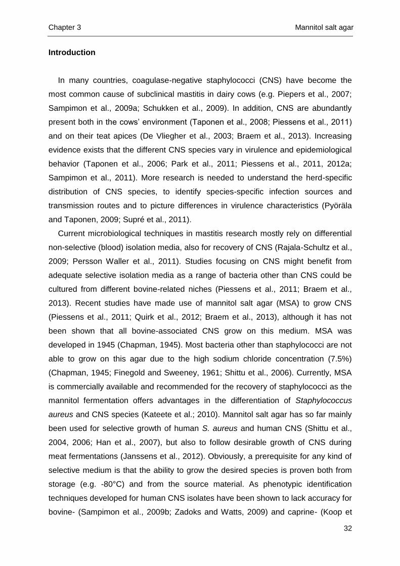
In a first experiment, the 25 most frequently isolated CNS species from bovine
intra- and extra-mammary sites were assembled (Table 1). Of each species, both a
reference strain and a field isolate were included (ntotal = 50). All field isolates were
previously described in published studies and were identified to the species level
using either transfer RNA intergenic spacer PCR (tDNA-PCR) and/or rpoB
sequencing (n = 18) (Supré et al., 2009, 2011 and unpublished data), amplified
fragment length polymorphism (AFLP) genotyping (n = 5) (Piessens et al., 2010,
2011), (GTG)5-PCR fingerprinting (n = 1) (Braem et al., 2011, 2013) or rpoB, tuf and
16S rRNA sequencing (n = 1) (Taponen et al., 2006, 2012). All 50 isolates were
stored at -80°C in either Microbanks (MicrobankTM, Pro-lab diagnostics,
Bromborough, UK) or brain heart infusion with 15% (w/v) glycerol (Oxoid,
Basingstoke, UK). For performing of the test, the isolates were plated directly (one
quadrant per isolate) on MSA (Chapman medium, Oxoid) and on Columbia agar with
sheep blood (Oxoid), the latter being used as a non-selective medium. Plates were
aerobically incubated at 37°C. Growth was examined after 24h and 48h, after which
incubation for another 24h at room temperature was initiated. If no growth could be
detected on MSA in this first attempt, the procedure was repeated on both agars.
Subsequently, in a second experiment, the same procedure was performed for 10
isolates of the six and four CNS species that have most frequently been isolated at
our laboratory from milk and teat apices, respectively (ntotal = 100) (Table 2). All
these isolates were previously collected as part of different field studies (Piessens et
al., 2011; Supré et al., 2011; Braem et al., 2013) and were identified to the species
level using tDNA-PCR (all Staphylococcus chromogenes, S. cohnii, S. equorum, S.
haemolyticus, S. simulans and S. xylosus), AFLP genotyping (all S. epidermidis) or
Chapter 3 Mannitol salt agar
34
(GTG)5-PCR fingerprinting (all S. saprophyticus). All 100 isolates were stored at -80
°C in either Microbanks or brain heart infusion with 15% (w/v) glycerol. The test was
performed as described above. If no growth could be detected on MSA the
procedure was repeated once on both agars.
Suitability of MSA for CNS recovery in field applications
A bulk milk sample was collected on 20 randomly selected Flemish dairy herds by
the Milk Control Centre (MCC, Lier, Flanders). Each milk sample (10 µl) was plated
on MSA, and Columbia agar with sheep blood (Oxoid). The resulting 40 plates were
aerobically incubated at 37°C and read after 24h and 48h. All colonies were
phenotypically assessed by size, shape, smoothness, opacity, color, butyrous
consistency, mannitol fermentation, hemolysis and lustre of the colonies. From all
colony types represented by at least two colonies, one colony was picked up and
subcultured on esculin blood agar (Oxoid) (one quadrant per isolate) to obtain pure
cultures. All purified isolates were subjected to a catalase test and all catalase-
positive isolates were examined using Gram-staining. Gram-positive cocci were then
subjected in parallel to DNase and coagulase tests. For all isolates that were DNase-
positive and coagulase-positive, mannitol fermentation was tested and a polymyxin
test was performed, in order to further distinguish S. aureus (coagulase-positive,
DNase-positive, mannitol salt-positive and polymyxin-resistant) and non-S. aureus
staphylococci (coagulase-negative and/or DNase-negative, mannitol salt-positive or -
negative and polymyxin-sensitive) (Finegold and Sweeney, 1961; Hogan et al., 1999;
Kateete et al., 2010). All coagulase- or DNase-negative bacteria were considered to
be non-S. aureus staphylococci and were further referred to as CNS, despite a
minority being coagulase-positive. CNS were identified to the species level using
tDNA-PCR followed by capillary electrophoresis as described by Supré et al. (2009).
If no identification could be obtained, the isolates were subjected to sequencing of
the rpoB gene. Sequencing of the 16S rRNA gene was performed if amplification of
the rpoB gene failed.
In addition, swabs were collected from teat apices of three dry cows and two
heifers one month before parturition as described by De Vliegher et al. (2003). All 20
swabs were plated on MSA and Columbia agar with sheep blood (half a plate per
swab). Plates were examined and isolates were subjected to further analysis as
described above.

Chapter 3 Mannitol salt agar
35
Results
Assessment of growth of CNS species on MSA
Results of the growth assessment experiments are shown in Tables 1 and 2. In
the first experiment, only the S. equorum reference strain and the S. hominis and S.
lugdunensis field isolates showed delayed growth (after 24h) on MSA in comparison
with Columbia agar with sheep blood on which growth was detected within 24h. The
S. devriesei reference strain grew within 24h on MSA but only after a second
attempt. In contrast, on Columbia agar with sheep blood, growth was detected within
24h after the first attempt. In the second experiment, only a single S. chromogenes
isolate and two S. haemolyticus isolates originating from milk showed delayed
growth (after 24h) in comparison with Columbia agar with sheep blood (within 24h).
A single S. xylosus isolate from milk grew within 24h on MSA but only after a second
attempt. On Columbia agar with sheep blood, that isolate grew within 24h after the
first attempt. Among the isolates originating from teat apices, only a single S.
haemolyticus isolate failed to grow on MSA, even when the test was repeated.
However, when that S. haemolyticus isolate was picked up from Columbia agar with
sheep blood after 24h incubation, and then plated on MSA, growth was also
detected on MSA within 24h.
Suitability of MSA for CNS recovery in field applications
Concerning the bulk milk samples, no further analysis was done of colonies
growing on Columbia agar with sheep blood, as colonies could not be conveniently
picked up, due to overgrowth or a too dense configuration after 24h incubation. On
all MSA plates, in total 69 phenotypically different colony types were recovered after
48h. From all these types at least two representative colonies were apparent. Per
plate, one to five different types were present.
Chapter 3 Mannitol salt agar
36
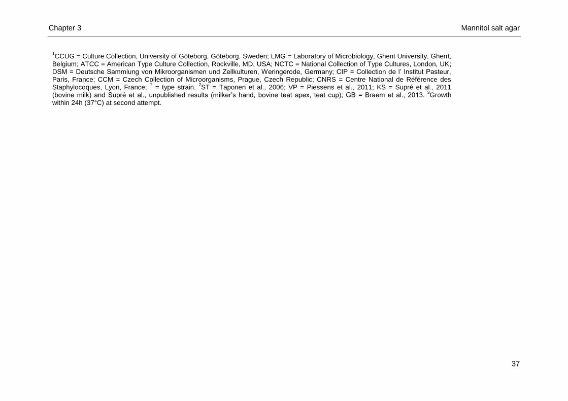
Table 1. Origin and growth of reference strains and field isolates, respectively, of 25 coagulase-negative Staphylococcus species
frequently isolated from cows’ milk or environment, on mannitol salt agar (aerobic incubation at 37°C)
CNS species Reference strain Field isolate
Origin Strain number1 Growth Origin Strain number
2 Growth
S. agnetis Bovine milk CCUG 59809T 24h Bovine milk ST 1 24h
S. arlettae Poultry skin LMG 19114T 24h Sawdust VP 0428 24h
S. auricularis Human ear ATCC 33753T 24h Milker’s hand KS 536 24h
S. capitis Human skin ATCC 49326T 24h Milker’s hand KS 547 24h
S. chromogenes Bovine skin NCTC 10530T 24h Bovine teat apex KS 81 24h
S. cohnii Human skin DSM 20260T 24h Bovine teat apex KS 567 24h
S. devriesei Bovine teat apex CIP 110234T 24h
3 Teat cup KS 447 24h
S. epidermidis Human nose LMG 10474T 24h Milker’s hand KS 414 24h
S. equorum Surface of cheese DSM 15097 T
24h-48h Teat cup KS 402 24h
S. fleurettii Caprine cheese CCM 4922T 24h Teat cup KS 339 24h
S. gallinarum Poultry skin CCM 3572T 24h Stable floor VP 0303 24h
S. haemolyticus Human skin CCM 2737T 24h Bovine teat apex KS 516 24h
S. hominis Human skin CCM 2732 24h Teat cup KS 338 24h-48h
S. hyicus Porcine skin NCTC 10350T 24h Bovine milk KS 167 24h
S. kloossii Skin squirrel DSM 20676 T
24h Bovine teat apex GB G057 24h
S. lentus Caprine udder ATCC 29070T 24h Stable floor VP 273 24h
S. lugdunensis Human axillary lymph node ATCC 43809T 24h Teat cup KS 699 24h-48h
S. nepalensis Caprine nasal mucosa DSM 15150 T
24h Milker’s hand KS 774 24h
S. pasteuri Human vomit ATCC 51129T 24h Bovine milk KS 219 24h
S. sciuri Skin eastern gray squirrel ATCC 29062T 24h Teat cup KS 491 24h
S. simulans Human skin CCM 2705T 24h Bovine teat apex KS 129 24h
S. succinus Dominican amber LMG 22185T 24h Stable floor VP 613 24h
S. vitulinus Ground lamb CCM 4511T 24h Stable floor VP 105 24h
S. warneri Human skin ATCC 27836T 24h Milker’s hand KS 412 24h
S. xylosus Human skin CNRS N850267 24h Bovine milk KS 13 24h
Chapter 3 Mannitol salt agar
37
1CCUG = Culture Collection, University of Göteborg, Göteborg, Sweden; LMG = Laboratory of Microbiology, Ghent University, Ghent,
Belgium; ATCC = American Type Culture Collection, Rockville, MD, USA; NCTC = National Collection of Type Cultures, London, UK; DSM = Deutsche Sammlung von Mikroorganismen und Zellkulturen, Weringerode, Germany; CIP = Collection de l’ Institut Pasteur, Paris, France; CCM = Czech Collection of Microorganisms, Prague, Czech Republic; CNRS = Centre National de Référence des Staphylocoques, Lyon, France;
T = type strain.
2ST = Taponen et al., 2006; VP = Piessens et al., 2011; KS = Supré et al., 2011
(bovine milk) and Supré et al., unpublished results (milker’s hand, bovine teat apex, teat cup); GB = Braem et al., 2013. 3
Growth within 24h (37°C) at second attempt.
Chapter 3 Mannitol salt agar
38
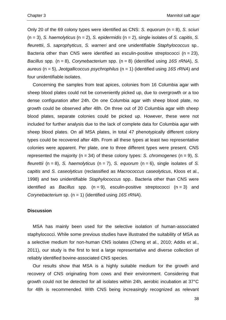
Only 20 of the 69 colony types were identified as CNS: S. equorum (n = 8), S. sciuri
(n = 3), S. haemolyticus (n = 2), S. epidermidis (n = 2), single isolates of S. capitis, S.
fleurettii, S. saprophyticus, S. warneri and one unidentifiable Staphylococcus sp..
Bacteria other than CNS were identified as esculin-positive streptococci (n = 23),
Bacillus spp. (n = 8), Corynebacterium spp. (n = 8) (identified using 16S rRNA), S.
aureus (n = 5), Jeotgallicoccus psychrophilus (n = 1) (identified using 16S rRNA) and
four unidentifiable isolates.
Concerning the samples from teat apices, colonies from 16 Columbia agar with
sheep blood plates could not be conveniently picked up, due to overgrowth or a too
dense configuration after 24h. On one Columbia agar with sheep blood plate, no
growth could be observed after 48h. On three out of 20 Columbia agar with sheep
blood plates, separate colonies could be picked up. However, these were not
included for further analysis due to the lack of complete data for Columbia agar with
sheep blood plates. On all MSA plates, in total 47 phenotypically different colony
types could be recovered after 48h. From all these types at least two representative
colonies were apparent. Per plate, one to three different types were present. CNS
represented the majority (n = 34) of these colony types: S. chromogenes (n = 9), S.
fleurettii (n = 8), S. haemolyticus (n = 7), S. equorum (n = 6), single isolates of S.
capitis and S. caseolyticus (reclassified as Macrococcus caseolyticus, Kloos et al.,
1998) and two unidentifiable Staphylococcus spp.. Bacteria other than CNS were
identified as Bacillus spp. (n = 9), esculin-positive streptococci (n = 3) and
Corynebacterium sp. (n = 1) (identified using 16S rRNA).
Discussion
MSA has mainly been used for the selective isolation of human-associated
staphylococci. While some previous studies have illustrated the suitability of MSA as
a selective medium for non-human CNS isolates (Cheng et al., 2010; Addis et al.,
2011), our study is the first to test a large representative and diverse collection of
reliably identified bovine-associated CNS species.
Our results show that MSA is a highly suitable medium for the growth and
recovery of CNS originating from cows and their environment. Considering that
growth could not be detected for all isolates within 24h, aerobic incubation at 37°C
for 48h is recommended. With CNS being increasingly recognized as relevant
Chapter 3 Mannitol salt agar
39
bacteria for udder health and recent efforts directed at unraveling the species- and
strain-specific epidemiology, these results are highly valuable for future studies.
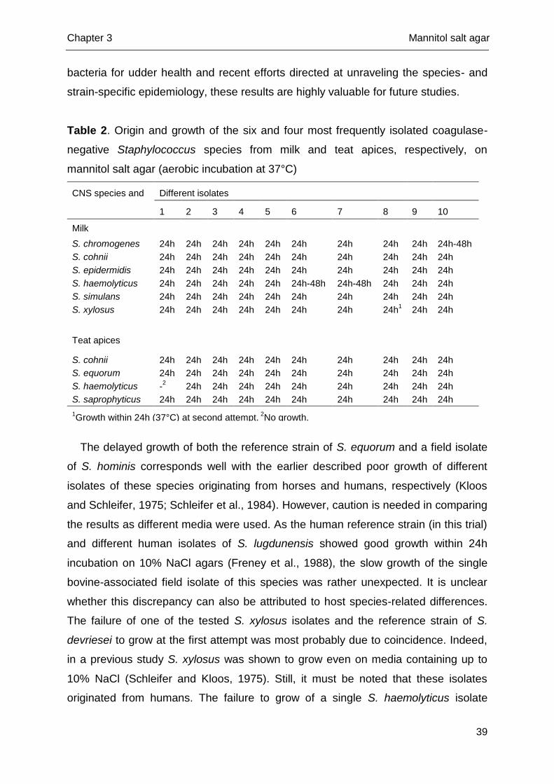
Table 2. Origin and growth of the six and four most frequently isolated coagulase-
negative Staphylococcus species from milk and teat apices, respectively, on
mannitol salt agar (aerobic incubation at 37°C)
CNS species and origin
Different isolates
1 2 3 4 5 6 7 8 9 10
Milk
S. chromogenes 24h 24h 24h 24h 24h 24h 24h 24h 24h 24h-48h
S. cohnii 24h 24h 24h 24h 24h 24h 24h 24h 24h 24h
S. epidermidis 24h 24h 24h 24h 24h 24h 24h 24h 24h 24h
S. haemolyticus 24h 24h 24h 24h 24h 24h-48h 24h-48h 24h 24h 24h
S. simulans 24h 24h 24h 24h 24h 24h 24h 24h 24h 24h
S. xylosus 24h 24h 24h 24h 24h 24h 24h 24h1 24h 24h
Teat apices
S. cohnii 24h 24h 24h 24h 24h 24h 24h 24h 24h 24h
S. equorum 24h 24h 24h 24h 24h 24h 24h 24h 24h 24h
S. haemolyticus -2 24h 24h 24h 24h 24h 24h 24h 24h 24h
S. saprophyticus 24h 24h 24h 24h 24h 24h 24h 24h 24h 24h
1Growth within 24h (37°C) at second attempt.
2No growth.
The delayed growth of both the reference strain of S. equorum and a field isolate
of S. hominis corresponds well with the earlier described poor growth of different
isolates of these species originating from horses and humans, respectively (Kloos
and Schleifer, 1975; Schleifer et al., 1984). However, caution is needed in comparing
the results as different media were used. As the human reference strain (in this trial)
and different human isolates of S. lugdunensis showed good growth within 24h
incubation on 10% NaCl agars (Freney et al., 1988), the slow growth of the single
bovine-associated field isolate of this species was rather unexpected. It is unclear
whether this discrepancy can also be attributed to host species-related differences.
The failure of one of the tested S. xylosus isolates and the reference strain of S.
devriesei to grow at the first attempt was most probably due to coincidence. Indeed,
in a previous study S. xylosus was shown to grow even on media containing up to
10% NaCl (Schleifer and Kloos, 1975). Still, it must be noted that these isolates
originated from humans. The failure to grow of a single S. haemolyticus isolate
Chapter 3 Mannitol salt agar
40
originating from a teat apex should not be a reason for concern, as all other isolates
grew very well and among bovine-associated S. haemolyticus isolates, a high
genetic diversity has been observed (Piessens et al., 2012b).
In the field application study, analysis was performed on one representative
colony per phenotypically distinguished colony type. The possibility exists that in this
way colonies with similar phenotypic characteristics actually were different CNS
species, leading to an underestimation of the true CNS diversity in the samples.
Therefore, not having investigated all grown colonies is a limitation of the current
study. However, as the purpose was to test the practical use and applicability of
MSA for recovery of the most common bovine-associated CNS species, picking up
all present colonies was not feasible. Obviously, having to do so would also be highly
unfeasible in routine practice or in future research studying CNS in bulk milk, on teat
apices, or other niches. Yet, in future work this inconvenience could be overcome at
least partially by repeated sampling of the niche being studied. On the other hand, it
was observed that different colony types sometimes belonged to the same species.
Also, differences in biochemical characteristics (e.g. mannitol fermentation) between
the reference strain and field isolates and among field isolates of the same species
(see Supplementary Table S1), were noted, suggesting strain differences within
species. Another drawback of our study is the lack of a “gold standard” reference
medium in the field application study. This means we are not able to conclude that
MSA grew the full range of CNS species potentially present in the samples. In this
study, Columbia agar with sheep blood, a medium frequently used in daily practice,
was mainly included to show the applicability and advantages of MSA in field
applications compared to this non-selective medium. Columbia agar was not
intended to be the “gold standard” and could not be used as such due to overgrowth.
The use of serial dilutions as was done by Jayarao et al. (2004) could have been a
partial solution, but this holds the risk that species that are present in very low
numbers are disregarded through such an approach. Overall, it must be
acknowledged that a straightforward method for detecting all species present in any
kind of sample is still lacking. This might be a relevant topic for future research.
Nonetheless, our results illustrate that MSA is far better suited for detection of
bovine-associated CNS.
It was not surprising that bacteria other than staphylococci grew on MSA in the
performed studies as also in previous studies growth of Corynebacterium spp.
Chapter 3 Mannitol salt agar
41
(Braem et al., 2013), esculin-positive streptococci (Chapman, 1945; Braem et al.,
2013) and Bacillus spp. (Braem et al., 2013; Han et al., 2007) has been observed.
Yet, this appeared not to be an inconvenience for the isolation of separate CNS
colonies and should therefore not hamper the use of MSA as an isolation medium for
bovine-associated CNS.
Conclusions
The most common CNS species from dairy cows and dairy cows’ environment
were illustrated to grow well on MSA. As phenotypically different colonies on MSA
could be picked up easily, MSA clearly offers advantages in comparison with non-
selective media. This study shows that MSA is a practical and appropriate medium to
further unravel the epidemiology and behavior of the different bovine-associated
CNS species from different intra- and extra-mammary niches.
Chapter 3 Mannitol salt agar
42
Appendix A. supplementary data
Supplementary data associated with this article can be found, in the online
version, at http://dx.doi.org/10.1016/j.rvsc.2013.05.015.
References
Addis, A., P. Mahindra, and N. K. Moses. 2011. Isolation and identification of
Staphylococcus species from raw bovine milk in Debre Zeit, Ethiopia. Vet. Res.
4:45-49.
Braem, G., S. De Vliegher, K. Supré, F. Haesebrouck, F. Leroy, and L. De Vuyst.
2011. (GTG)5-PCR fingerprinting for the classification and identification of
coagulase-negative Staphylococcus species from bovine milk and teat apices. A
comparison of type strains and field isolates. Vet. Microbiol. 147:67-74.
Braem, G., S. De Vliegher, B. Verbist, V. Piessens, E. Van Coillie, L. De Vuyst, and
F. Leroy. 2013. Unraveling the microbiota of teat apices of clinically healthy
lactating dairy cows, with special emphasis on coagulase-negative staphylococci.
J. Dairy Sci. 96:1499-1510.
Chapman, G. H. 1945. The significance of sodium chloride in studies of
staphylococci. J. Bacteriol. 50:201-203.
Cheng, D. R., S. Y. Zhu, Z. H. Yin, W. W. Ding, Z. X. Mu, Z. R. Su, and H. C. Sun.
2010. Prevalence of bacterial infection responsible for bovine mastitis. Afr. J.
Microbiol. Res. 4:1110-1116.
De Vliegher, S., H. Laevens, L. A. Devriese, G. Opsomer, J. L. Leroy, H. W.
Barkema, and A. de Kruif. 2003. Pre-partum teat apex colonization with
Staphylococcus chromogenes in dairy heifers is associated with low somatic cell
count in early lactation. Vet. Microbiol. 92:245-252.
Finegold, S. M., and E. E. Sweeney. 1961. New selective and differential medium for
coagulase-positive staphylococci allowing rapid growth and strain differentiation.
J. Bacteriol. 81:636-641.
Freney, J., Y. Brun, M. Bes, H. Meugnier, F. Grimont, P. A. D. Grimont, C. Nervi, and
J. Fleurette. 1988. Staphylococcus lugdunensis sp. nov. and Staphylococcus
schleiferi sp. nov., two species from human clinical specimens. Int. J. Syst.
Bacteriol. 38:168-172.
Chapter 3 Mannitol salt agar
43
Han, Z. L., E. Lautenbach, N. Fishman, and I. Nachamkin. 2007. Evaluation of
mannitol salt agar, CHROMagar Staph. aureus and CHROMagar MRSA for
detecton of meticillin-resistant Staphylococcus aureus from nasal swab
specimens. J. Med. Microbiol. 56:43-46.
Hogan, J. S., R. N. Gonzáles, R. J. Harmon, S. C. Nickerson, S. P. Oliver, J. W.
Pankey, and K. L. Smith. 1999. Laboratory Handbook on Bovine Mastitis. Rev.
Ed. National Mastitis Council, Madison, WI.
Janssens, M., N. Myter, L. De Vuyst, and F. Leroy. 2012. Species diversity and
metabolic impact of the microbiota are low in spontaneously acidified Belgian
sausages with an added starter culture of Staphylococcus carnosus. Food
Microbiol. 29:167-177.
Jayarao, B. M., S. R. Pillai, A. A. Sawant, D. R. Wolfgang, and N. V. Hegde. 2004.
Guidelines for monitoring bulk tank milk somatic cell and bacterial counts. J. Dairy
Sci. 87, 3561-3573.
Kateete, D. P., C. N. Kimani, F. A. Katabazi, A. Okeng, M. S. Okee, A. Nanteza, M.
L. Joloba, and F. C. Najjuka. 2010. Identification of Staphylococcus aureus:
DNase and mannitol salt agar improve the efficiency of the tube coagulase test.
Ann. Clin. Microbiol. Antimicrob. 9:23-29.
Kloos, W. E., and K. H. Schleifer. 1975. Isolation and characterization of
staphylococci from human skin II. Descriptions of four new species:
Staphylococcus warneri, Staphylococcus capitis, Staphylococcus hominis and
Staphylococcus simulans. Int. J. Syst. Bacteriol. 25:62-79.
Kloos, W. E., D. N. Ballard, C. G. George, J. A. Webster, R. J. Hubner, W. Ludwig,
K. H. Schleifer, F. Fiedler, and K. Schubert. 1998. Delimiting the genus
Staphylococcus through description of Macrococcus caseolyticus gen. nov.,
comb. nov. and Macrococcus equipercicus sp. nov., Macrococcus bovicus sp.
nov. and Macrococcus carouselicus sp. nov.. Int. J. Syst. Bacteriol. 48:859-877.
Koop, G., A. De Visscher, C. A. Collar, D. A. Bacon, E. A. Maga, J. D. Murray, K.
Supré, S. De Vliegher, F. Haesebrouck, J. D. Rowe, M. Nielen, and T. van
Werven. 2012. Short communication: Identification of coagulase-negative
Staphylococcus species from goat milk with the API Staph identification test and
with transfer RNA-intergenic spacer PCR combined with capillary electrophoresis.
J. Dairy Sci. 95:7200-7205.
Chapter 3 Mannitol salt agar
44
Park, J. Y., L. K. Fox, K. S. Seo, M. A. McGuire, Y. H. Park, F. R. Rurangirwa, W. M.
Sischo, and G. A. Bohach. 2011. Detection of classical and newly described
staphylococcal superantigen genes in coagulase-negative staphylococci isolated
from bovine intramammary infections. Vet. Microbiol. 147:149-154.
Persson Waller, K., A. Aspán, A. Nyman, Y. Persson, and U. Grönlund Andersson.
2011. CNS species and antimicrobial resistance in clinical and subclinical bovine
mastitis. Vet. Microbiol. 152:112-116.
Piepers, S., L. De Meulemeester, A. de Kruif, G. Opsomer, H. W. Barkema, and S.
De Vliegher. 2007. Prevalence and distribution of mastitis pathogens in
subclinically infected dairy cows in Flanders, Belgium. J. Dairy Res. 74:478-483.
Piessens, V., K. Supré, M. Heyndrickx, F. Haesebrouck, S. De Vliegher, and E. Van
Coillie. 2010. Validation of amplified fragment length polymorphism genotyping for
species identification of bovine associated coagulase-negative staphylococci. J.
Microbiol.Meth. 80:287-294.
Piessens, V., E. Van Coillie, B. Verbist, K. Supré, G. Braem, A. Van Nuffel, L. De
Vuyst, M. Heyndrickx, and S. De Vliegher. 2011. Distribution of coagulase-
negative Staphylococcus species from cows’ milk and environment differs
between herds. J. Dairy Sci. 94:2933-2944.
Piessens, V., S. De Vliegher, B. Verbist, G. Braem, A. Van Nuffel, L. De Vuyst, M.
Heyndrinckx, and E. Van Coillie. 2012a. Characterizatoin of coagulase-negative
Staphylococcus species from cows’ milk and environment based on bap, icaA,
and mecA genes and phenotypic susceptibility to antimicrobials and teat dips. J.
Dairy Sci. 95:7027-7038.
Piessens, V., S. De Vliegher, B. Verbist, K. Supré, G. Braem, A. Van Nuffel, L. De
Vuyst, M. Heyndrickx, and E. Van Coillie. 2012b. Intra-species diversity and
epidemiology varies among different coagulase-negative Staphylococcus species
causing bovine intramammary infections. Vet. Microbiol. 155:62-71.
Pyörälä, S., and S. Taponen. 2009. Coagulase-negative staphylococci. Emerging
mastitis pathogens. Vet. Microbiol. 134:3-8.
Quirk, T., L. K. Fox, D. D. Hancock, J. Capper, J. Wenz, and J. Park. 2012.
Intramammary infections and teat canal colonization with coagulase-negative
staphylococci after postmilking teat disinfection. Species-specific responses. J.
Dairy Sci. 95:1906-1912.
Chapter 3 Mannitol salt agar
45
Rajala-Schultz, P. J., A. H. Torres, F. J. DeGraves, W. A. Gebreyes, and P.
Patchanee. 2009. Antimicrobial resistance and genotypic characterization of
coagulase-negative staphylococci over the dry period. Vet. Microbiol. 134:55-64.
Sampimon, O. C., H. W. Barkema, I. M. G. A. Berends, J. Sol, and T. J. G. M. Lam.
2009a. Prevalence and herd-level risk factors for intramammary infection with
coagulase-negative staphylococci in Dutch dairy herds. Vet. Microbiol. 134:37-44.
Sampimon, O. C., R. N. Zadoks, S. De Vliegher, K. Supré, F. Haesebrouck, H. W.
Barkema, J. Sol, and T. J. G. M. Lam. 2009b. Performance of API Staph ID 32
and Staph-Zym for identification of coagulase-negative staphylococci isolated
from bovine milk samples. Vet. Microbiol. 136:300-305.
Sampimon, O. C., T. J. G. M. Lam, D. J. Mevius, Y. H. Schukken, and R. N. Zadoks.
2011. Antimicrobial susceptibility of coagulase-negative staphylococci isolated
from bovine milk samples. Vet. Microbiol. 150:173-179.
Schleifer, K. H., and W. E. Kloos. 1975. Isolation and characterization of
staphylococci from human skin I. Amended descriptions of Staphylococcus
epidermidis and Staphylococcus saprophyticus and descriptions of three new
species: Staphylococcus cohnii, Staphylococcus haemolyticus and
Staphylococcus xylosus. Int. J. Syst. Bacteriol. 25:50-61.
Schleifer, K. H., R. Kilpperbalz, and L. A. Devriese. 1984. Staphylococcus arlettae
sp. nov., Staphylococcus equorum sp. nov. and Staphylococcus kloossii sp. nov..
Three new coagulase-negative, novobiocin-resistant species from animals. Syst.
Appl. Microbiol. 5:501-509.
Schukken, Y. H., R. N. Gonzalez, L. L. Tikofsky, H. F. Schulte, C. G. Santisteban, F.
L. Welcome, G. J. Bennett, M. J. Zurakowski, and R. N. Zadoks. 2009. CNS
mastitis. Nothing to worry about? Vet. Microbiol. 134:9-14.
Shittu, A., J. Lin, D. Morrison, and D. Kolawole. 2004. Isolation and molecular
characterization of multiresistant Staphylococcus sciuri and Staphylococcus
haemolyticus associated with skin and soft-tissue infections. J. Med. Microbiol.
53:51-55.
Shittu, A., J. Lin, D. Morrison, and D. Kolawole. 2006. Identification and molecular
characterization of mannitol salt positive, coagulase-negative staphylococci from
nasal samples of medical personnel and students. J. Med. Microbiol. 55:317-324.
Supré, K., S. De Vliegher, O. C. Sampimon, R. N. Zadoks, M. Vaneechoutte, M.
Baele, E. De Graef, S. Piepers, and F. Haesebrouck. 2009. Technical note. The
Chapter 3 Mannitol salt agar
46
use of transfer RNA-intergenic spacer PCR combined with capillary
electrophoresis to identify coagulase-negative Staphylococcus species originating
from bovine milk and teat apices. J. Dairy Sci. 92:3204-3210.
Supré, K., F. Haesebrouck, R. N. Zadoks, M. Vaneechoutte, S. Piepers, and S. De
Vliegher. 2011. Some coagulase-negative Staphylococcus species affect udder
health more than others. J. Dairy Sci. 94:2329-2340.
Taponen, S., H. Simojoki, M. Haveri, H. D. Larsen, and S. Pyörälä. 2006. Clinical
characteristics and persistence of bovine mastitis caused by different species of
coagulase-negative staphylococci identified with API of AFLP. Vet. Microbiol.
115:199-207.
Taponen, S., J. Bjorkroth, and S. Pyörälä. 2008. Coagulase-negative staphylococci
isolated from bovine extramammary sites and intramammary infections in a single
dairy herd. J. Dairy Res. 75:422-429.
Taponen, S., K. Supré, V. Piessens, E. Van Coillie, S. De Vliegher, and J. M. K.
Koort. 2012. Staphylococcus agnetis sp. nov., a coagulase-variable species from
bovine subclinical and milk clinical mastitis. Int. J. Syst. Evol. Microbiol. 62:61-65.
Zadoks, R. N., and J. L. Watts. 2009. Species identification of coagulase-negative
staphylococci. Genotyping is superior to phenotyping. Vet. Microbiol. 134:20-28.


Chapter 4
Teat Apex Colonization with Coagulase-
Negative Staphylococcus Species throughout
Lactation and before Parturition

Chapter 4.1
Further Evidence for the Existence of
Environmental and Host-Associated Species of
Coagulase-Negative Staphylococci in Dairy
Cattle
A. De Visscher1, K. Supré2, F. Haesebrouck3, R. N. Zadoks4,5, V.
Piessens6, E. Van Coillie7, S. Piepers1, and S. De Vliegher1
1Department of Reproduction, Obstetrics, and Herd Health, Faculty of Veterinary
Medicine, Ghent University, Merelbeke, Belgium
2Milk Control Centre Flanders, Lier, Belgium
3Department of Pathology, Bacteriology, and Avian Diseases, Faculty of Veterinary
Medicine, Ghent University, Merelbeke, Belgium
4Moredun Research Institute, Pentlands Science Park, Penicuik, Scotland UK
5Institute of Biodiversity, Animal Health and Comparative Medicine, College of
Medical, Veterinary, and Life Sciences, University of Glasgow, Glasgow, UK
6Laboratory of Microbiology, Department of Biochemistry and Microbiology, Faculty
of Science, Ghent University, Ghent, Belgium
7Institute for Agricultural and Fisheries Research (ILVO), Technology and Food
Science Unit, Melle, Belgium
Veterinary Microbiology, 2014, 172:466-474

Chapter 4.1 Teat apex colonization throughout lactation
53
Abstract
Coagulase-negative staphylococci (CNS) are abundantly present in the dairy farm
environment and on bovine skin and mucosae. They are also the most prevalent
bacteria causing bovine intramammary infections (IMI). Reservoirs and transmission
routes of CNS are not yet fully unraveled. The objectives of this study were to
explore the distribution of CNS in parlor-related extramammary niches and to
compare it to the distributions of CNS causing IMI in those herds. Niches that were
targeted in this study were cows’ teat apices, milking machine unit liners, and
milker’s skin or gloves. Each of the three herds had its own CNS microbiota in those
niches. The most prevalent species in the parlor-related extramammary niches were
Staphylococcus cohnii, S. fleurettii, and S. equorum in the first, second, and third
herd, respectively, whereas S. haemolyticus and S. sciuri were found in all herds.
Staphylococcus cohnii and S. fleurettii, as well as S. haemolyticus, which was
present in each herd, were also frequently found in milk samples. By contrast, S.
chromogenes, S. simulans, and S. xylosus favored the mammary gland, whereas S.
equorum was more common in the parlor-associated niches. Within each herd,
species distribution was similar between teat apices and milking machine unit liners.
In conclusion, some of the extramammary niches related to the milking process
might act as infection sources for IMI-causing CNS. This study provides further
evidence that the group of CNS species is comprised of environmental, opportunistic
and host-adapted species which differ in ecology.
Key words
Coagulase-negative Staphylococcus species, Dairy cow, Intramammary infection,
Parlor-associated extramammary niche
Chapter 4.1 Teat apex colonization throughout lactation
54
Introduction
Interest in coagulase-negative staphylococci (CNS) increased over the past years
both in human (Rasmussen et al., 2000; Dekio et al., 2005) and in veterinary
medicine (Taponen et al., 2007; Gillespie et al., 2009). Effort is ongoing to unravel
the characteristics of CNS as a group (Sampimon et al., 2009; Piepers et al., 2010),
and the specific characteristics of the different species (Simojoki et al., 2011; Supré
et al., 2011). Recently, we reported that several CNS species were abundantly
present in the cows’ environment (air, slatted floor, and sawdust), but rarely
associated with intramammary infections (IMI) (Piessens et al., 2011). These
“environmental CNS” include Staphylococcus equorum, S. fleurettii, and S. sciuri.
Other species, such as S. haemolyticus and S. simulans, were commonly found in
the barn environment and were also isolated from quarters with IMI. These species
are considered to be “opportunistic pathogens” (Piessens et al., 2011).
Staphylococcus chromogenes was commonly associated with IMI, but rarely found in
the barn environment, implying that other reservoirs potentially play a role in its
epidemiology (Piessens et al., 2011). Staphylococcus cohnii and S. xylosus are also
frequently isolated from infected quarters in some herds (Supré et al., 2011), but
their sources are as yet unidentified.
The udder contains a natural barrier mechanism to separate and protect the
mammary gland from the environment. Potential extramammary sources of infection
that have not been examined in previous studies include the cows’ teat apices (TA),
the milking machine unit liners (MMUL) and milkers’ skin. Previous work suggests
that the presence of Staphylococcus aureus on teat skin can be associated with the
risk of infection in early lactation (Roberson et al., 1994), while milking machine unit
liners also play a role in S. aureus transmission (Zadoks et al., 2002). Milkers’ skin
can carry S. epidermidis, which may cause IMI in cows (Thorberg et al., 2006). By
contrast, presence of CNS (Piepers et al., 2011) and S. chromogenes (De Vliegher
et al., 2003) on TA pre-partum was not associated with IMI (Piepers et al., 2011) and
even associated with a decreased risk of S. chromogenes IMI in heifers in early
lactation (De Vliegher et al., 2003). However, little is known about the distribution of
CNS species on TA, MMUL, and milkers’ skin or gloves (MSG) and their possible
role as source of CNS IMI.
Chapter 4.1 Teat apex colonization throughout lactation
55
The aims of this study were (1) to map the distribution of the parlor-associated
CNS species, based on a molecular identification technique, (2) to compare the
distribution of parlor-associated species and CNS species causing IMI in these
herds, and (3) to explore whether TA, MMUL, and the milker could play a role in
CNS transmission in these herds.
Material and methods
Herds, animals, and teat apices
A single cross-sectional sampling was performed in 3 commercial Flemish dairy
herds in February 2008. The herds were part of a longitudinal study (starting in
September 2007, ending in January 2009) to determine the impact of CNS species-
specific IMI on quarter milk somatic cell count (Supré et al., 2011). Herd
characteristics and udder health practices are described in Table 1.
Per herd, 25 animals were included in the longitudinal study on IMI and 6 of the 25
animals were randomly selected for the current study, with stratification by parity, i.e.
2 animals in their first, second, or higher lactation, respectively. Teat apices (n = 72)
were sampled both pre- and post-milking, with the exception of one animal in herd 3
that was only sampled pre-milking (nTA = 140). Sterile cotton swabs were used
(Copan Diagnostics Inc., CA, USA). The sampling protocol was as follows: cleansing
of the teat with an individual dry paper cloth, pre-milking swabbing of the teat apex,
disinfection of the teats with alcohol, attachment of the milking cluster, milking,
removal of the milking cluster, post-milking swabbing of the teat apex, and finally
post-milking teat dipping. Contact with milk was avoided during swabbing. After
sampling, swabs were placed in sterile tubes and immediately stored at 4°C.
Extramammary niches
All MMUL (40 on farms 1 and 2, 48 on farm 3) (n = 128) were sampled before the
milking session started and, with the exception of one unit on farm 3, again after
milking of all cows was finished, prior to cleaning of the milking machine (nMMUL =
252). The inside of all liners was sampled by rubbing and turning the swab and again
contact with milk was avoided.
Chapter 4.1 Teat apex colonization throughout lactation
56
Table 1. Overview of herd characteristics and udder health management practices of
the three participating herds
Herd 1 Herd 2 Herd 3
Herd characteristics1
Number of lactating cows 43.5
(39 - 48)2
48.9 (43 - 53)
65.7 (61 - 70)
Mean milk yield (kg/day) 30.9 (28.5 - 32.4)
32.3 (30.1 - 34.3)
31 (29.4 - 31.8)
Mean 305d-yield (in kg) 9,921.6 (9,551 - 10,189)
10,194.7 (9,975 - 10,396)
10,180.2 (9,978 - 10,367)
Herd SCC (x 1000 cells/mL)3 152.9
(98 - 220) 195.9 (133 - 323)
186.7 (103 - 298)
Udder health management
Gloves during milking 1 of 2 milkers
All milkers 1 of 2 milkers
Iodine post-milking teat desinfection Dip
Dip Spray
Dry-off management
Long-acting antimicrobial Cloxacillin Cefquinome Nafcillin / penicillin / streptomycin
Internal teat sealer Yes
Yes Yes
Routine culling of chronically infected cows No
Yes No
Calving pen
Separate straw box Yes
Yes Yes
Used for sick cows Yes
No Yes
Bedding material Sawdust
Sawdust Sawdust
Cleaning of slatted floor 2x per day Not regularly 2x per day
1Measured through the Dairy Herd Improvement (DHI) program.
2(Minimum - maximum).
3Average of the
monthly herd somatic cell count (SCC), measured through the DHI program.
Gloves or bare hands of the milkers (2 on farms 1 and 3, 1 on farm 2) and the
bends of their elbows were sampled both before and after milking, after wiping them
with a dry paper cloth as described before (Thorberg et al., 2006). Thus, the group of
MSG samples comprised of 20 samples from hands or gloves and 20 samples from
elbows (nMSG = 40).
Identification of CNS species
All swabs were streaked onto a Columbia agar with 5 % ovine blood (Oxoid,
Basingstoke, UK). After aerobic incubation at 37 °C for 24h, plates were examined.
CNS were presumptively identified following NMC procedures (Hogan et al., 1999).
All colonies per morphotype were counted. One colony was picked up from every
Chapter 4.1 Teat apex colonization throughout lactation
57
morphotype with ≥ 5 colonies, allowing for isolation of more than one morphotype
per swab, and was streaked onto Columbia blood agar (Oxoid) to obtain pure
cultures. After 18 - 24h aerobic incubation at 37°C, individual colonies were checked
for purity and stored at -80°C until further analysis. DNA was prepared according to
Unal et al. (1992). All CNS were identified to the species level using transfer RNA-
intergenic spacer PCR (tDNA-PCR) (Supré et al., 2009). If no identification could be
obtained, isolates were subjected to sequencing of the rpoB-housekeeping gene as
described before (Supré et al., 2009). Results were recorded as presence or
absence of a species in a sample, regardless of the number of times this particular
species was isolated in that sample.
Descriptive and statistical analysis
The distribution of each identified CNS species in the parlor-associated
extramammary niches was described and counted within each herd. The distribution
of CNS species in the extramammary niches and the distribution of CNS species
from IMI in the same herds, based on culture results from 3 consecutive samplings,
were displayed next to each other (Supré et al., 2011). The latter was done to
explore whether the parlor-associated extramammary niches could act as infection
source or fomite for IMI in these herds.
McNemar’s Tests were performed in SPSS (SPSS Statistics 22, SPSS Inc.,
Chicago, IL, USA) to determine associations between presence of CNS before and
after milking from TA and in MMUL, respectively. Associations between presence of
CNS on hands or gloves and in elbow bends were checked using a Fisher Exact
Test (SPSS Statistics 22, SPSS Inc., Chicago, IL, USA).
Results and discussion
In previous studies, the distribution of CNS species in milk samples and the barn
environment had been described (Piessens et al., 2011; Supré et al., 2011), but
potential niches of CNS in the milking parlor, which plays a crucial role in mastitis
epidemiology and control, had not been explored in depth. In this study, we focused
on niches related to the milking process in order to broaden our knowledge of the
epidemiology of CNS species.
Chapter 4.1 Teat apex colonization throughout lactation
58
For precise identification of CNS species, we used a molecular identification
technique, tRNA-intergenic spacer PCR (Supré et al., 2009). This method showed
high typeability for CNS from extramammary sites in this study. Only 1.4% of 288
CNS, isolated from TA, in MMUL, and on MSG, could not be identified to the species
level using this method. The remaining isolates were subjected to sequencing of the
rpoB gene. To our knowledge, only one other molecular study compared CNS
isolated from parlor-associated niches and from infected quarters (Taponen et al.,
2008).
In total, 432 extramammary samples were collected. At least one CNS species
was found in 46.8% of those samples and 19 different CNS species were isolated in
this cross-sectional study. The most common species were S. cohnii (23.3% of all
extramammary CNS isolates), S. haemolyticus (17.4%), S. equorum (16.0%), S.
fleurettii (15.3%) and S. sciuri (10.4%). The three herds had distinct extramammary
microbiota (Figure 1A). Staphylococcus cohnii was the dominant species in herd 1
(46.8% of all extramammary CNS isolates in that herd), S. fleurettii in herd 2 (68.3%
of all extramammary CNS isolates in that herd), and S. equorum in herd 3 (47.6% of
all extramammary CNS isolates in that herd). Herd-specific species distribution was
also described for CNS isolated from sawdust, floors, and air, in particular for S.
fleurettii but not for S. cohnii and S. equorum as those species were isolated in all
herds in that study (Piessens et al., 2011). In contrast, S. haemolyticus and S. sciuri
were present in the extramammary niches in all herds in the present study (Figure
1), which corresponds well with the findings for those species in environmental
samples in the study of Piessens et al. (2011).
High numbers of CNS isolates were recovered from teat apices. Of the 140 TA
samples 59.3% were CNS-positive. All cows had at least one TA that harbored CNS.
The number of different species isolated per TA ranged from 1 to 4, while up to 6
were found per cow. Staphylococcus cohnii was found in high proportion on TA in
herd 1 (50.0% of the TA isolates in that herd) but it was absent from TA in the other
herds (Figure 1). Staphylococcus fleurettii was frequently and only isolated from TA
in herd 2 (72.4% of the TA isolates in that herd). Staphylococcus equorum was
commonly found in herd 3 (50.0% of the TA isolates in that herd). Staphylococcus
haemolyticus and S. sciuri were the only species isolated from TA in all herds
(Figure 1).
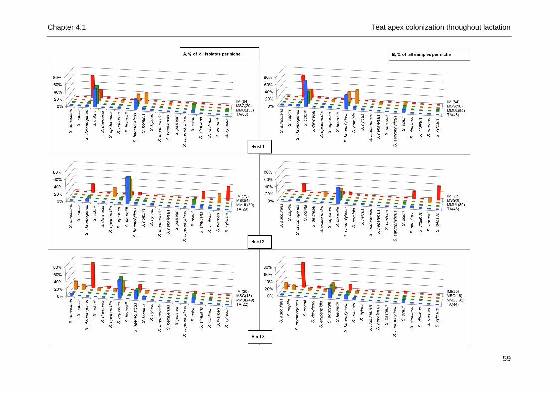
Chapter 4.1 Teat apex colonization throughout lactation
59
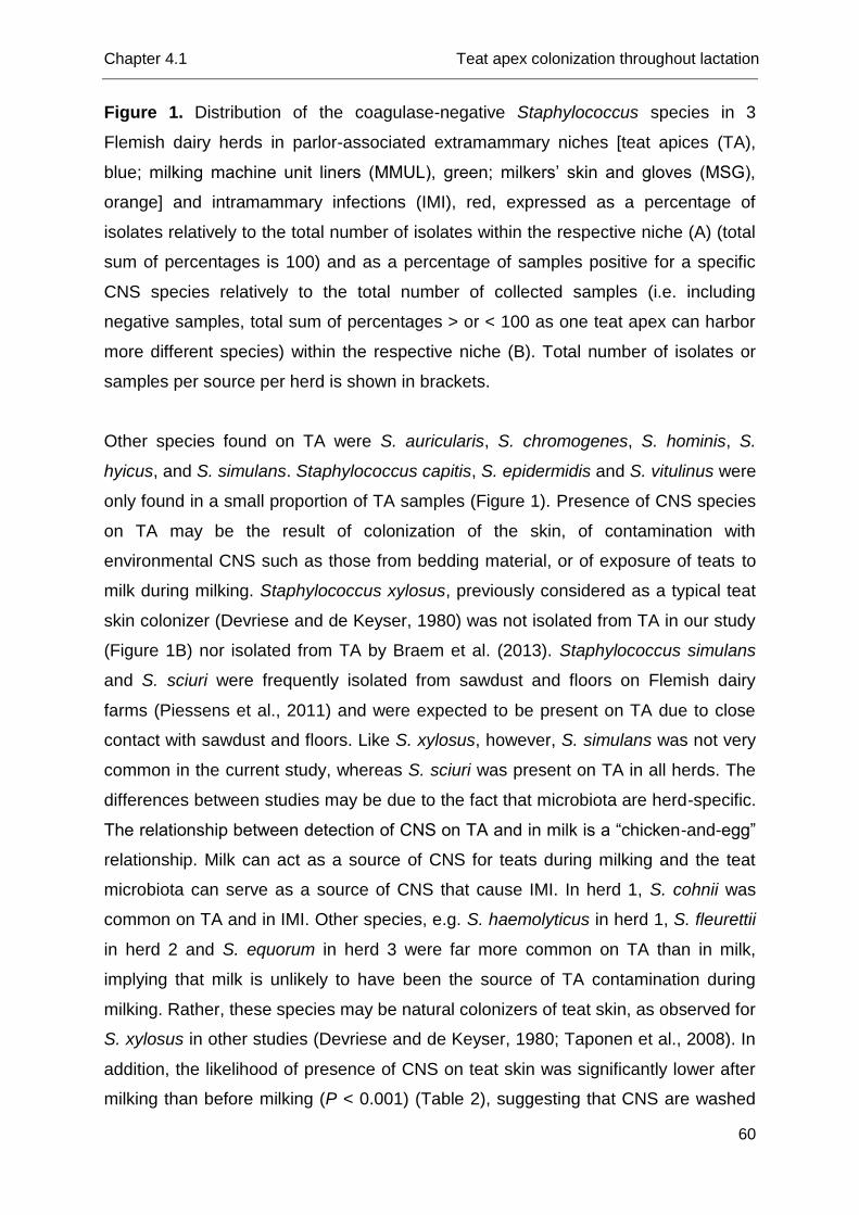
Chapter 4.1 Teat apex colonization throughout lactation
60
Figure 1. Distribution of the coagulase-negative Staphylococcus species in 3
Flemish dairy herds in parlor-associated extramammary niches [teat apices (TA),
blue; milking machine unit liners (MMUL), green; milkers’ skin and gloves (MSG),
orange] and intramammary infections (IMI), red, expressed as a percentage of
isolates relatively to the total number of isolates within the respective niche (A) (total
sum of percentages is 100) and as a percentage of samples positive for a specific
CNS species relatively to the total number of collected samples (i.e. including
negative samples, total sum of percentages > or < 100 as one teat apex can harbor
more different species) within the respective niche (B). Total number of isolates or
samples per source per herd is shown in brackets.
Other species found on TA were S. auricularis, S. chromogenes, S. hominis, S.
hyicus, and S. simulans. Staphylococcus capitis, S. epidermidis and S. vitulinus were
only found in a small proportion of TA samples (Figure 1). Presence of CNS species
on TA may be the result of colonization of the skin, of contamination with
environmental CNS such as those from bedding material, or of exposure of teats to
milk during milking. Staphylococcus xylosus, previously considered as a typical teat
skin colonizer (Devriese and de Keyser, 1980) was not isolated from TA in our study
(Figure 1B) nor isolated from TA by Braem et al. (2013). Staphylococcus simulans
and S. sciuri were frequently isolated from sawdust and floors on Flemish dairy
farms (Piessens et al., 2011) and were expected to be present on TA due to close
contact with sawdust and floors. Like S. xylosus, however, S. simulans was not very
common in the current study, whereas S. sciuri was present on TA in all herds. The
differences between studies may be due to the fact that microbiota are herd-specific.
The relationship between detection of CNS on TA and in milk is a “chicken-and-egg”
relationship. Milk can act as a source of CNS for teats during milking and the teat
microbiota can serve as a source of CNS that cause IMI. In herd 1, S. cohnii was
common on TA and in IMI. Other species, e.g. S. haemolyticus in herd 1, S. fleurettii
in herd 2 and S. equorum in herd 3 were far more common on TA than in milk,
implying that milk is unlikely to have been the source of TA contamination during
milking. Rather, these species may be natural colonizers of teat skin, as observed for
S. xylosus in other studies (Devriese and de Keyser, 1980; Taponen et al., 2008). In
addition, the likelihood of presence of CNS on teat skin was significantly lower after
milking than before milking (P < 0.001) (Table 2), suggesting that CNS are washed
Chapter 4.1 Teat apex colonization throughout lactation
61
away from TA rather than deposited on TA during milking. Finally, S. chromogenes,
the most frequently isolated species from infected quarters, was only occasionally
isolated from TA (2.9% of all TA swabs), providing further support to the notion that
milk is not a frequent source of teat skin contamination. Staphylococcus
chromogenes could not be isolated from teat apices by Braem et al. (2013) either. In
other studies, isolation of S. chromogenes from teat apices or udder skin was more
common than in the current study (De Vliegher et al., 2003; Taponen et al. 2008;
Bexiga et al., 2014). Again, herd differences in TA colonization by different CNS
species may explain the discrepancies between studies.
Of the 252 MMUL samples, 36.5% were found CNS-positive. In herd 1, 46.3% of
the MMUL were CNS-positive, 31.3% in herd 2 and 32.6% in herd 3. An efficient
cleaning procedure of the milking machine in between milkings was evidenced by
the absence of CNS in all but one pre-milking MMUL swabs (Table 2), from which S.
xylosus was recovered. Taponen et al. (2008) reported a low number of CNS-
positive samples after washing of the MMUL as well. After the milking process, a
significantly higher proportion of MMUL (73.4%) were CNS-positive (Table 2). Within
each herd, the CNS species distribution in MMUL samples was very similar to the
distribution in TA samples (Figure 1A). The latter finding can be explained by direct
contact between teat skin and liners. For example, as S. equorum and S. fleurettii
are not frequently isolated from milk in herd 3 and herd 2, respectively (Figure 1A),
contact with contaminated milk is an unlikely explanation for the presence of CNS in
the MMUL. The likelihood of presence of CNS on teat apices was significantly lower
after milking than before milking (P < 0.001), while the reverse was true for MMUL
(Table 2) (P < 0.001). This suggests that CNS are washed off the teats during
milking and transferred to MMUL. This could increase the risk of exposure to CNS
for the cows being milked last as compared to those that are milked first.
Of the 40 collected samples from MSG, 67.5% were positive. All milkers were
CNS-positive on at least one of the sampled sites, both pre- and/or post-milking.
Eleven species were isolated from MSG, with S. auricularis, S. cohnii, S. equorum,
S. haemolyticus, and S. hominis the predominant species (Figure 1 and Table 3).
Staphylococcus capitis, S. epidermidis, S. fleurettii, S. nepalensis, S. sciuri, and S.
warneri were isolated less frequently.
Chapter 4.1 Teat apex colonization throughout lactation
62
Table 2. Number of coagulase-negative Staphylococcus isolates recovered before (b) and after (a) milking from the parlor-
associated niches (teat apices, milking machine unit liners, and milkers’ skin and gloves) in 3 Flemish dairy herds
Species Teat apices Milking machine unit liners Milkers’ skin and gloves Total
Herd 1
Herd 2
Herd 3
S1 Herd
1 Herd 2
Herd 3
S Herd 1
Herd 2
Herd 3
S n %
b nc
2 a nc b nc a nc b nc a n
c b ncl
3 a ncl b ncl a ncl b ncl a ncl b a b a b a
S. auricularis 1 1 1 1 2 1 1 1 1 2 3 6 2.1
S. capitis 1 1 1 0 2
2 3 1.0
S. chromogenes 1 1 1 1 1 1 1 1 4 2 2 2 2 1 1 5 0 9 3.1
S. cohnii 19 6 15 6 34 27 10 27 1 4 1 6 67 23.3
S. devriesei 0 1 1 1 0 1 0.3
S. epidermidis 1 1 1 1 1 1 1
1 2 4 1.4
S. equorum 1 1 1 1 10 6 1 1 13 1 1 2 2 25 10 28 1
2 2 5 46 16.0
S. fleurettii 19 6 2 2 21 20 8 1 1 21 1 1
2 44 15.3
S. haemolyticus 13 6 6 4 1 1 4 4 1 2 25 8 5 1 1 10 6 19 1 4 1 6 50 17.4
S. hominis 1 1 2 2 2 2 5 1 1 1 3 3
1 7 13 4.5
S. hyicus 1 1 1 1 2 0 0 2 0.7
S. lugdunensis 0 1 1 1 0 1 0.3
S. nepalensis 0 0 1 1 1 0.3
S. saphrophyticus 0 1 1 1
0 1 0.3
S. sciuri 2 2 2 2 1 1 3 3 8 8 7 4 3 8 6 20 2 2 30 10.4
S. simulans 1 1 1 1 2 1 1 1 0 3 1.0
S. vitulinus 1 1 1 2 2 2 0 3 1.0
S. warneri 0 0 1
1 1 0.3
S. xylosus 0 1 1 2 2 3 0 3 1.0
Total 40 28 25 4 20 2 119 1 52 0 30 0 49 132 7 13 2 2 8 5 37 288 100.0
1Subtotal.
2Number of cows.
3Number of clusters.
Chapter 4.1 Teat apex colonization throughout lactation
63
Unlike MMUL, hands or gloves and elbow bends had similar levels of CNS detection
before and after milking (60% and 70%, respectively for hands or gloves, and 70%
for elbow bends regardless of sampling time). The distribution of CNS species on
MSG differed among the three herds (Figure 1 and Table 3). Three species, S.
hominis (herd 1 and 3), S. epidermidis (herd 2 and 3), and S. capitis (herd 3) were
solely isolated from elbow bends. For S. hominis, the association with isolation from
elbows as opposed to hands was positively significant (P = 0.033) whereas the
number of isolates from the other species was too low for meaningful significance
testing. According to the interpretation of Thorberg et al. (2006), species that are
predominantly found in the elbow bend belong to the human microbiota. The
association between those species and human skin is not exclusive, because the
same species were detected on TA or in MMUL, albeit at low prevalence (1.0 - 4.5%,
Table 2). Staphylococcus nepalensis, S. sciuri, and S. warneri were detected on
bare hands or gloves but not in elbow bends. Again, the number of isolates from
those species was too low for significance testing. Throughout the 13 months of
sampling in the study herds (in the longitudinal study for detection of IMI), S.
nepalensis and S. warneri were never isolated from milk, potentially excluding
contamination of the hands or gloves via contact with milk. The two species were not
detected on TA or in MMUL either. In a study on different farms, both species were
occasionally found in the barn environment (Piessens et al., 2011) suggesting that
their presence on hands is the result of environmental contamination, although the
direction of transmission (hand to environment or environment to hand) cannot be
determined with certainty. In previous studies, S. warneri was isolated from human
forehead (Dekio et al., 2005) and the human nasal cavity (Rasmussen et al., 2000)
from non-farmers, showing that S. warneri is a natural component of the human skin
microbiota. Other species, notably S. auricularis, S. cohnii, S. equorum, S. fleurettii,
and S. haemolyticus were isolated both from elbow bends and from hands or gloves.
These species (except S. auricularis) also represent the majority of TA and MMUL
isolates (Figure 1). In case of IMI in those study herds, transmission of S. cohnii, S.
equorum, S. fleurettii, and S. haemolyticus possibly occurred during the milking
process, either via MMUL or via the hands or gloves of the milkers. Another
hypothesis is the existence of a shared microbiota between cows and humans. An
organ(skin)-specificity rather than host-specificity of certain CNS species might
occur, as was described for S. aureus strains (Zadoks et al., 2002). However, these
Chapter 4.1 Teat apex colonization throughout lactation
64
particular CNS species have to our knowledge, not been reported in human studies
(Rasmussen et al., 2000; Dekio et al., 2005). Thorberg et al. (2006) hypothesized
that S. epidermidis might be transmitted from humans to bovines, but this species
was not frequently found in our study nor in that of Taponen et al. (2008). However,
S. epidermidis was causing a substantial number of IMI in another study (Piessens
et al., 2011), emphasizing once more that the distribution of CNS species can be
herd-specific.
During swabbing of TA care was taken to avoid contact with milk because cross
contamination of skin and milk samples could lead to apparent overlap in species or
strain distributions, as previously described for S. aureus (Zadoks et al., 2002). Also,
milk samples were collected aseptically and IMI definitions were based on multiple
milk samplings. Therefore, we assume to have reduced the risk of misclassification
due to contamination of either sample type. Comparison of the CNS species
distribution in TA, MMUL and MSG with the distribution among IMI can help to shed
light on the role of parlor associated extramammary sources in causing IMI. For
some species, such as S. cohnii in herd 1, S. fleuretti in herd 2, and S. equorum in
herd 3, TA, MMUL and MSG are likely sources or fomites for IMI, given that the
prevalence in the extramammary sources is higher than the prevalence among IMI
isolates (Figure 1B and Figure 2). On the other hand, some herds had a high number
of IMI due to certain CNS species (e.g. S. simulans and S. xylosus in herd 2, and S.
chromogenes in all herds) whereas these species were only rarely isolated from TA,
MMUL or MSG (Figure 1B and Figure 2). Staphylococcus cohnii, found in 23% of IMI
and 77% of extramammary samples, can be considered as an opportunistic
pathogen based on the distribution in herd 1. S. haemolyticus, considered as an
opportunistic species by Piessens et al. (2011), and S. sciuri appeared to be present
in all herds (Figure 1, this study and Piessens et al., 2011) and followed a similar
distribution as S. cohnii in herd 1 (Figure 1A). Staphylococcus equorum was rarely
isolated from IMI but commonly found in MMUL and on TA in herd 3. Based on these
data this species can be considered as an environmental organism (Figure 2). This
is in agreement with the study of Piessens et al. (2011), who commonly found this
species in the barn environment but rarely isolated it from the mammary gland.
Similar findings were obtained for S. fleurettii (Figure 1, herd 2, this study and
Piessens et al., 2011).
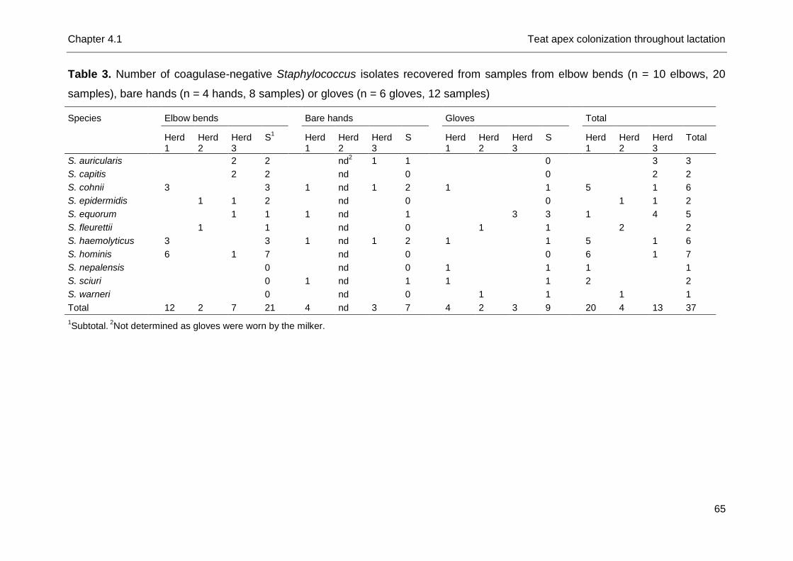
Chapter 4.1 Teat apex colonization throughout lactation
65
Table 3. Number of coagulase-negative Staphylococcus isolates recovered from samples from elbow bends (n = 10 elbows, 20
samples), bare hands (n = 4 hands, 8 samples) or gloves (n = 6 gloves, 12 samples)
Species Elbow bends Bare hands Gloves Total
Herd 1
Herd 2
Herd 3
S1 Herd
1 Herd 2
Herd 3
S Herd 1
Herd 2
Herd 3
S Herd 1
Herd 2
Herd 3
Total
S. auricularis
2 2 nd2 1 1
0 3 3
S. capitis
2 2 nd 0 0 2 2
S. cohnii
3 3
1 nd 1 2
1 1
5
1 6
S. epidermidis
1 1 2
nd 0
0 1 1 2
S. equorum
1 1
1 nd 1
3 3
1
4 5
S. fleurettii
1 1
nd 0 1
1
2
2
S. haemolyticus
3 3
1 nd 1 2
1 1
5 1 6
S. hominis
6 1 7
nd 0
0
6 1 7
S. nepalensis
0
nd 0
1 1
1 1
S. sciuri
0
1 nd 1
1 1
2 2
S. warneri
0
nd 0 1 1
1 1
Total
12 2 7 21
4 nd 3 7
4 2 3 9
20 4 13 37
1Subtotal.
2Not determined as gloves were worn by the milker.
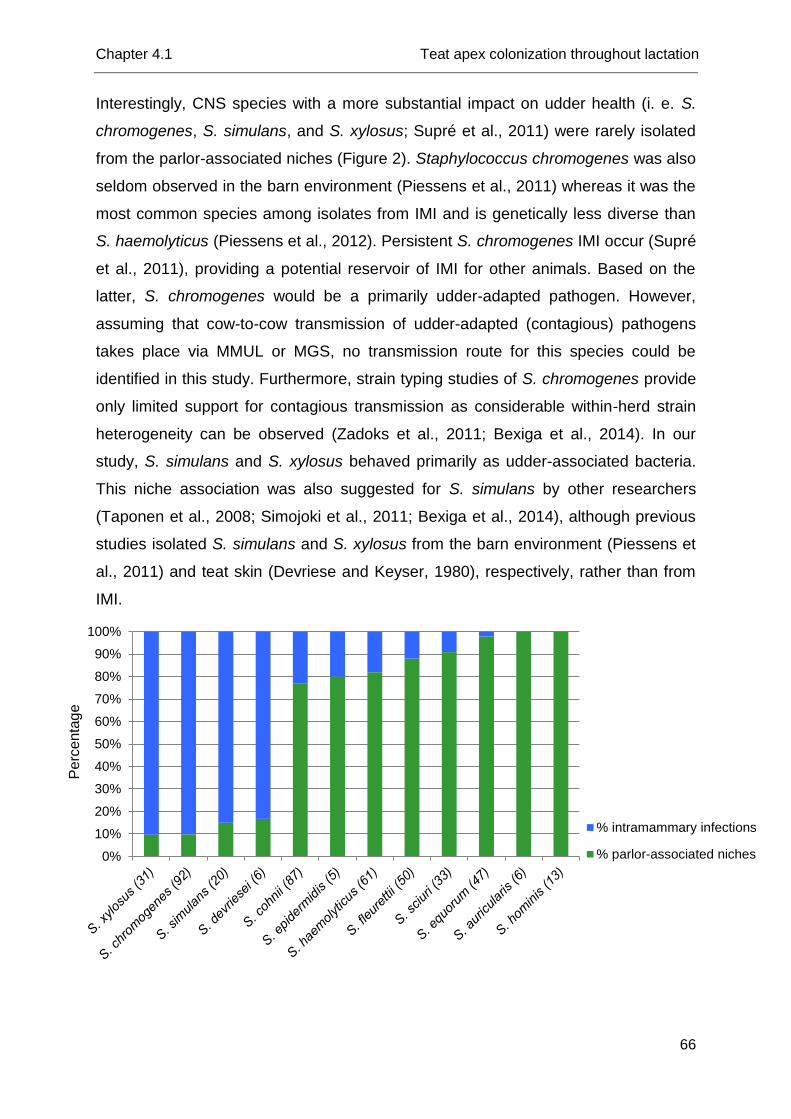
Chapter 4.1 Teat apex colonization throughout lactation
66
Interestingly, CNS species with a more substantial impact on udder health (i. e. S.
chromogenes, S. simulans, and S. xylosus; Supré et al., 2011) were rarely isolated
from the parlor-associated niches (Figure 2). Staphylococcus chromogenes was also
seldom observed in the barn environment (Piessens et al., 2011) whereas it was the
most common species among isolates from IMI and is genetically less diverse than
S. haemolyticus (Piessens et al., 2012). Persistent S. chromogenes IMI occur (Supré
et al., 2011), providing a potential reservoir of IMI for other animals. Based on the
latter, S. chromogenes would be a primarily udder-adapted pathogen. However,
assuming that cow-to-cow transmission of udder-adapted (contagious) pathogens
takes place via MMUL or MGS, no transmission route for this species could be
identified in this study. Furthermore, strain typing studies of S. chromogenes provide
only limited support for contagious transmission as considerable within-herd strain
heterogeneity can be observed (Zadoks et al., 2011; Bexiga et al., 2014). In our
study, S. simulans and S. xylosus behaved primarily as udder-associated bacteria.
This niche association was also suggested for S. simulans by other researchers
(Taponen et al., 2008; Simojoki et al., 2011; Bexiga et al., 2014), although previous
studies isolated S. simulans and S. xylosus from the barn environment (Piessens et
al., 2011) and teat skin (Devriese and Keyser, 1980), respectively, rather than from
IMI.
0%
10%
20%
30%
40%
50%
60%
70%
80%
90%
100%
% intramammary infections
% parlor-associated niches
Pe
rce
nta
ge
Chapter 4.1 Teat apex colonization throughout lactation
67
Figure 2. Distribution of the most common coagulase-negative Staphylococcus
species isolated from intramammary infections (blue) or found in parlour-associated
extramammary niches (green) per species.
One could argue only a limited number of cows (n = 18) and herds (n = 3) were
included in the current study. Still, a vast number of CNS isolates were collected and
identified using tDNA-PCR, a validated and accurate molecular identification
technique, giving a precise picture of their distribution in parlor-related
extramammary niches. We acknowledge the limited number of herds and animals
narrows the conclusions that can be drawn from the study. However, research based
on such precise molecular data as in our study, on possible infection sources of IMI-
causing CNS, will help to further increase our understanding of the epidemiology and
ecology of CNS species and will generate new hypotheses, useful for larger future
studies. Eventually, it can be helpful to determine prevention and control measures
for IMI with the species that have a more substantial impact on udder health, such as
S. chromogenes, S. simulans, and S. xylosus (Supré et al., 2011).
Conclusion
Exploring distributions of CNS reveals that each herd has its own CNS microbiota
in the extramammary niches. The most prevalent species in the extramammary
samples were S. cohnii, S. fleurettii, and S. equorum in herd 1 - 3, respectively, while
S. haemolyticus and S. sciuri were present in all herds. TA and MMUL shared similar
CNS distributions within each herd. The MSG seems to harbor a specific microbiota,
although some species might be shared between human skin and the other niches
sampled in this study. Staphylococcus chromogenes, S. simulans, and S. xylosus
favor the mammary gland and were barely found on TA, MMUL, and MSG, while S.
equorum and S. fleurettii preferred the extramammary niches. Staphylococcus
cohnii, S. haemolyticus, and S. sciuri were considered as less adapted to one of the
compartments in particular.
Chapter 4.1 Teat apex colonization throughout lactation
68
References
Bexiga, R., M. G. Rato, A. Lemsaddek, T. Semedo-Lemsaddek, C. Carneiro, H.
Pereira, D. J. Mellor, K. A. Ellis, and C. L. Vilela. 2014. Dynamics of bovine
intramammary infections due to coagulase-negative staphylococci on four farms.
J. Dairy Res. 81:208-2014.
Braem, G., S. De Vliegher, B. Verbist, V. Piessens, E. Van Coillie, L. De Vuyst, and
F. Leroy. 2013. Unraveling the microbiota of teat apices of clinically healthy
lactating dairy cows, with special emphasis on coagulase-negative staphylococci.
J. Dairy Sci. 96:1499-1510.
De Vliegher, S., H. Laevens, L. A. Devriese, G. Opsomer, J. L. M. Leroy, H. W.
Barkema, and A. de Kruif. 2003. Prepartum teat apex colonization with
Staphylococcus chromogenes in dairy heifers is associated with low somatic cell
count in early lactation. Vet. Microbiol. 92:245-252.
Dekio, I., H. Hayashi, M. Sakamoto, M. Kitahara, T. Nishikawa, M. Suematsu, and Y.
Benno. 2005. Detection of potentially novel bacterial components of the human
skin microbiota using culture-independent molecular profiling. J. Med. Microbiol.
54:1231-1238.
Devriese, L. A., and H. De Keyser. 1980. Prevalence of different species of
coagulase-negative staphylococci on teats and in milk samples from dairy cows.
J. Dairy Res. 47:155-158.
Gillespie, B. E., S. I. Headrick, S. Boonyayatra, and S. P. Oliver. 2009. Prevalence
and persistence of coagulase-negative Staphylococcus species in three dairy
research herds. Vet. Microbiol. 134:65-72.
Hogan, J. S., R. N. Gonzáles, R. J. Harmon, S. C. Nickerson, S. P. Oliver, J. W.
Pankey, and K. L. Smith. 1999. Laboratory Handbook on Bovine Mastitis. Rev.
Ed. National Mastitis Council, Madison, WI.
Piepers, S., G. Opsomer, H. W. Barkema, A. de Kruif, and S. De Vliegher. 2010.
Heifers infected with coagulase-negative staphylococci in early lactation have
fewer cases of clinical mastitis and a higher milk production in their first lactation
than non-infected heifers. J. Dairy Sci. 93:2014-2024.
Piepers, S., K. Peeters, G. Opsomer, H. W. Barkema, K. Frankena, and S. De
Vliegher. 2011. Pathogen group specific risk factors at herd, heifer and quarter

Chapter 4.1 Teat apex colonization throughout lactation
69
levels for intramammary infections in early lactating dairy heifers. Prev. Vet. Med.
99:91-101.
Piessens, V., E. Van Coillie, B. Verbist, K. Supré, G. Braem, A. Van Nuffel, L. De
Vuyst, M. Heyndrickx, and S. De Vliegher. 2011. Distribution of coagulase-
negative Staphylococcus species from dairy cows’ milk and environment differs
between herds. J. Dairy Sci. 94:2933-2944.
Piessens, V., S. De Vliegher, B. Verbist, G. Braem, A. Van Nuffel, L. De Vuyst, M.
Heyndrickx, and E. Van Coillie. 2012. Intra-species diversity and epidemiology
varies among coagulase-negative Staphylococcus species causing bovine
intramammary infections. Vet. Microbiol. 155:62-71.
Rasmussen, T. T., L. P. Kirkeby, K. Poulsen, J. Reinholdt, and M. Kilian. 2000.
Resident aerobic microbiota of the adult human nasal cavity. Acta Pathol. Mic. Sc.
108:663-675.
Roberson, J. R., L. K. Fox, D. D. Hancock, J. M. Gay, and T. E. Besser. 1994.
Ecology of Staphylococcus aureus isolated from various sites on dairy farms. J.
Dairy Sci. 77:3354-3364.
Sampimon, O. C, H. W. Barkema, I. M. G. A. Berends, J. Sol, and T. J. G. M. Lam.
2009. Prevalence and herd-level risk factors for intramammary infection with
coagulase-negative staphylococci in Dutch dairy herds. Vet. Microbiol. 134:37-44.
Simojoki, H., T. Salomäki, S. Taponen, A. Iivanainen, and S. Pyörälä. 2011. Innate
immune response in experimentally induced bovine intramammary infection with
Staphylococcus simulans and Staphylococcus epidermidis. Vet. Res. 42:49-59.
Supré, K., S. De Vliegher, O. C. Sampimon, R. N. Zadoks, M. Vaneechoutte, M.
Baele, E. De Graef, S. Piepers, and F. Haesebrouck. 2009. Technical note: use of
tRNA-intergenic spacer PCR combined with capillary electrophoresis to identify
coagulase-negative Staphylococcus species originating from bovine milk and teat
apices. J. Dairy Sci. 92:3204-3210.
Supré, K., F. Haesebrouck, R. N. Zadoks, M. Vaneechoutte, S. Piepers, and S. De
Vliegher. 2011. Some coagulase-negative Staphylococcus species are affecting
udder health more than others. J. Dairy Sci. 94:2329-2340.
Taponen, S., J. Koort, J. Björkroth, H. Saloniemi, and S. Pyörälä. 2007. Bovine
intramammary infections caused by coagulase-negative staphylococci may persist
throughout lactation according to amplified fragment length polymorphism-based
analysis. J. Dairy Sci., 90:3301-3307.
Chapter 4.1 Teat apex colonization throughout lactation
70
Taponen, S., J. Björkroth, and S. Pyörälä. 2008. Coagulase-negative staphylococci
isolated from bovine extramammary sites and intramammary infections in a single
dairy herd. J. Dairy Res. 75:422-429.
Thorberg, B. M., I. Kühn, F. M. Aarestrup, B. Brändströmd, P. Jonsson, and M. L.
Danielsson-Tham. 2006. Pheno- and genotyping of Staphylococcus epidermidis
isolated from bovine milk and human skin. Vet. Microbiol. 115:163-172.
Unal, S., J. Hoskins, J. E. Flokowitsch, C. Y. E. Wu, D. A. Preston, and P. L.
Skatrudi. 1992. Detection of methicillin-resistant staphylococci by using the
polymerase chain reaction. J. Clin. Microbiol. 30:1685-1691.
Zadoks, R. N., W. B. van Leeuwen, D. Kreft, L. K. Fox, H. W. Barkema, Y. H.
Schukken, and A. van Belkum. 2002. Comparison of Staphylococcus aureus
isolates from bovine and human skin, milking equipment, and bovine milk by
phage typing, pulsed-field gel electrophoresis, and binary typing. J. Clin. Microbiol.
40:3894-3902.
Zadoks, R. N., J. R. Middleton, S. McDougall, J. Katholm, and Y. H. Schukken. 2011.
Molecular epidemiology of mastitis pathogens of dairy cattle and comparative
relevance to humans. J. Mammary Gland Biol. 16:357-72.


Chapter 4.2
Teat Apex Colonization with Coagulase-
Negative Staphylococcus Species before
Parturition: Distribution and Species-Specific
Risk Factors
A. De Visscher1, S. Piepers1, F. Haesebrouck2, and S. De Vliegher1
1Department of Reproduction, Obstetrics, and Herd Health, Faculty of Veterinary
Medicine, Ghent University, Merelbeke, Belgium
2Department of Pathology, Bacteriology, and Avian Diseases, Faculty of Veterinary
Medicine, Ghent University, Merelbeke, Belgium
Journal of Dairy Science, 2016, 99:1427-1439

Chapter 4.2 Teat apex colonization before parturition
75
Abstract
Coagulase-negative staphylococci (CNS) are the main cause of bovine
intramammary infections and are also abundantly present in extramammary habitats
such as teat apices. Teat apex colonization (TAC) with CNS has already been
explored in lactating dairy cows at the species level, whereas this is not true for dry
cows and end-term heifers. Therefore, the aim of this observational study was to
describe CNS TAC in non-lactating dairy cows and end-term heifers in Flemish dairy
herds and to identify associated risk factors at the herd, cow and quarter level. All
CNS were molecularly identified to the species level using transfer RNA intergenic
spacer PCR (tDNA-PCR) and sequencing of the 16S rRNA gene, allowing for
species-specific statistical analyses using multivariable, multilevel logistic regression.
Staphylococcus devriesei, S. chromogenes, S. haemolyticus and S. equorum were
the most frequently isolated species. Staphylococcus chromogenes was the sole
species colonizing teat apices of cows and heifers in all herds whereas large
between-herd differences were observed for the other species. Teat apices of red
and white Holstein Friesians, of quarters dried-off without an internal teat sealer, and
swabbed in months with lower precipitation and higher ambient temperature were
significantly more likely to be colonized by S. devriesei. Slightly dirty teat apices and
teat apices swabbed in months with lower precipitation had higher odds of being
colonized by S. chromogenes whereas teat apices sampled in months with lower
precipitation and higher ambient temperature were more likely to be colonized by S.
haemolyticus. Dirty teat apices and teat apices swabbed in months with lower
ambient temperature in combination with low precipitation had higher odds of being
colonized by S. equorum. Diverse factors explaining CNS TAC, yet mostly related to
humidity, ambient temperature and hygiene, substantiate differences in
epidemiological behavior and ecology between species
Key words
Teat apex, Coagulase-negative Staphylococcus species
Chapter 4.2 Teat apex colonization before parturition
76
Introduction
Besides being the most common cause of intramammary infections in dairy cows
in many regions and countries (Vanderhaeghen et al., 2015), coagulase-negative
staphylococci (CNS) are abundantly present in extramammary habitats such as
sawdust and air (Piessens et al., 2011) and also colonize dairy cows’ teat apices
(Taponen et al., 2008; Braem et al., 2012; De Visscher et al., 2014). In fact, the
majority of teat apices colonizing microbiota belong to the group of the CNS (Braem
et al., 2013). Differences in ecological and epidemiological characteristics among
bovine-associated CNS species have been revealed (Vanderhaeghen et al., 2015),
yet many aspects remain undetermined. The presence of CNS on teat apices of
lactating dairy cows and heifers has already been explored at the species level
(Taponen et al., 2008; Braem et al., 2013; De Visscher et al., 2014; Vanderhaeghen
et al., 2015) but little is known about CNS teat apex colonization (TAC) in dry cows
and end-term heifers. A high prevalence of CNS colonized teat apices in pregnant
dairy heifers before calving has been described (De Vliegher et al., 2003; Piepers et
al., 2011) but those studies either solely concerned heifers, studied CNS as a group,
or focused specifically on Staphylococcus chromogenes only using non-molecular
identification. Large observational studies describing the species distribution on teat
apices in non-lactating dairy cows and end-term heifers and identifying factors
associated with their presence are needed to add to our understanding of the role of
CNS species in bovine udder health (Taponen et al., 2008; De Visscher et al., 2014;
Vanderhaeghen et al., 2015). Although we have started to learn about the relation
between CNS TAC and IMI, and associations seem to be present (Leroy et al.,
2015), potential protective aspects of TAC remain to be studied (Vanderhaeghen et
al., 2014).
Therefore, this study aimed to determine and to describe: (1) the species-specific
prevalence of CNS colonization of teat apices of dry cows and end-term heifers
before calving, (2) the CNS distribution, (3) associated herd-, cow-, and quarter-level
risk factors, and (4) the variance components of CNS TAC.
Chapter 4.2 Teat apex colonization before parturition
77
Materials and methods
Herds and cows
Thirteen commercial Flemish dairy herds were included, all of them participating in
the DHI program (CRV, Arnhem, The Netherlands), being a first selection criterion.
Other inclusion criteria were no pre-partum antibiotic treatment of heifers and the use
of artificial insemination as accurate expected calving dates were demanded. The
majority of farmers (n = 8) only housed dairy cattle, whereas the others also farmed
pigs (n = 1) or beef cattle (n = 4). The DHI-records and bulk milk quality data of the
Milk Control Centre Flanders (MCC Flanders, Lier, Belgium) allowed for calculating
the herd size and the bulk milk SCC, respectively, at the start of the sampling period
in July 2012. The entire sampling period lasted until February 2013. Before the start
of the study an average of 57 cows and heifers were in lactation (range = 30 - 95)
per herd (arithmetic mean of the 6 last test-day samples). The geometric mean bulk
milk SCC was 201,000 cells/mL (range = 79,000 cells/mL - 310,000 cells/mL). On 7
of the farms, dry cows were housed on concrete slatted floor with cubicles with mats
(n = 4), with mattresses (n = 2) or without bedding (n = 1). The other 6 farms kept the
dry cows on straw, either in a deep litter barn (n = 5) or in deep litter boxes with a
bottom layer of sand and a full concrete floor (n = 1). Pregnant heifers were typically
housed on a concrete slatted floor in cubicles (n = 11) with mats (n = 5), with
mattresses (n = 5) or without bedding (n = 1). The other 2 farms housed the
pregnant heifers either in a deep litter barn with straw (n = 1) or in deep litter boxes
with a bottom layer of sand and straw (n = 1). On the majority of farms, dry cows and
pregnant heifers (n = 12 and n = 11, respectively) were kept on pasture between
May and September. Seven of the herds housed the dry cows separated from the
lactating cows before calving, whereas in the majority of herds (n = 10) the pregnant
heifers were housed together with the lactating cows.
On each farm, 12 pregnant heifers and dry cows (total n = 156) were selected
according to the proportion of lactating cows and heifers present in the herd at the
start of the study, reflecting the parity distribution in the herd, resulting in a total of 53
pregnant heifers and 103 dry cows (range = start of 2nd lactation - start of 10th
lactation).
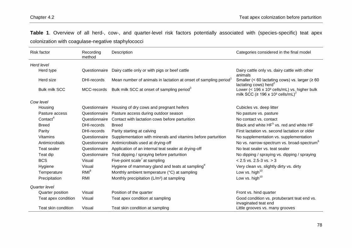
Chapter 4.2 Teat apex colonization before parturition
78
Table 1. Overview of all herd-, cow-, and quarter-level risk factors potentially associated with (species-specific) teat apex
colonization with coagulase-negative staphylococci
Risk factor Recording method
Description Categories considered in the final model
Herd level
Herd type Questionnaire Dairy cattle only or with pigs or beef cattle Dairy cattle only vs. dairy cattle with other animals
Herd size DHI-records Mean number of animals in lactation at onset of sampling period1 Smaller (< 60 lactating cows) vs. larger (≥ 60
lactating cows) herd2
Bulk milk SCC MCC-records Bulk milk SCC at onset of sampling period3 Lower (< 196 x 10³ cells/mL) vs. higher bulk
milk SCC (≥ 196 x 10³ cells/mL)2
Cow level
Housing Questionnaire Housing of dry cows and pregnant heifers Cubicles vs. deep litter
Pasture access Questionnaire Pasture access during outdoor season No pasture vs. pasture
Contact4 Questionnaire Contact with lactation cows before parturition No contact vs. contact
Breed DHI-records Breed Black and white HF5 vs. red and white HF
Parity DHI-records Parity starting at calving First lactation vs. second lactation or older
Vitamins Questionnaire Supplementation with minerals and vitamins before parturition No supplementation vs. supplementation
Antimicrobials Questionnaire Antimicrobials used at drying-off No vs. narrow-spectrum vs. broad-spectrum6
Teat sealer Questionnaire Application of an internal teat sealer at drying-off No teat sealer vs. teat sealer
Teat dip Questionnaire Teat dipping / spraying before parturition No dipping / spraying vs. dipping / spraying
BCS Visual Five-point scale7 at sampling < 2.5 vs. 2.5-3 vs. > 3
Hygiene Visual Hygiene of mammary gland and teats at sampling8 Very clean vs. slightly dirty vs. dirty
Temperature RMI9 Monthly ambient temperature (°C) at sampling Low vs. high
10
Precipitation RMI Monthly precipitation (L/m²) at sampling Low vs. high10
Quarter level
Quarter position Visual Position of the quarter Front vs. hind quarter
Teat apex condition Visual Teat apex condition at sampling Good condition vs. protuberant teat end vs. invaginated teat end
Teat skin condition Visual Teat skin condition at sampling Little grooves vs. many grooves
Chapter 4.2 Teat apex colonization before parturition
79
1Arithmetic mean based on 6 last DHI-records before the onset of the study (July 2012).
2Categorization based on median value of all mean values of all
herds. 3Geometric mean based on 6 last records of the Milk Control Centre Flanders before the onset of the study.
4Housing of dry cows and end-term heifers
together with lactating animals. 5
Holstein Friesian. 6Antimicrobials with a narrow spectrum include cloxacillin benzathine, cefalexine, and cefalonium whereas
cefquinome is a broad-spectrum antibiotic. 7
Edmonson et al. (1989). 8
Categorization based on a 5-point scale (Reneau et al., 2005). 9
Royal Meteorological Institute of Belgium.
10Categorization based on median value of all monthly records during 1 year (March 2012 until February 2013).
Chapter 4.2 Teat apex colonization before parturition
80
The majority of the selected animals were black and white Holstein Friesian (HF)
(85%, n = 132) and 15% were red and white HF (n = 24). Eighty-five percent (n =
132) of the animals were supplemented with minerals and vitamins before
parturition. On all farms, blanket dry cow treatment was applied using either
cloxacillin benzathine (47% of the 103 dry cows, n = 48 cows) or cephalosporins with
a broad or Gram-positive spectrum (40%, n = 41 cows and 10%, n = 10 cows,
respectively). Dry cow treatment information was lacking for a few cows only (3%, n
= 3) as these cows were purchased when dry. One cow did not receive
antimicrobials at drying-off due to a sudden drop in milk production. Only a minority
of the dry cows received an internal teat sealer (39% of the 103 dry cows, n = 40) at
drying-off. Iodine teat dip was applied to 38 dry cows and end-term heifers (24%)
before parturition.
Samples and data collection
To determine TAC before parturition, swabs of all teat apices of the 153 cows and
heifers (total n = 624) were collected 14 days before expected calving date. Visible
soil and manure were first removed. Further, a dry cotton swab (Copan, Novolab,
Belgium) was rotated gently on the teat apex as described by De Vliegher et al.
(2003). Swabs were transported under cooled condition (4°C) to the Mastitis and
Milk Quality Research Lab (Faculty of Veterinary Medicine, Ghent University,
Merelbeke, Belgium).
Several herd- and cow-level factors, potentially associated with CNS (species-
specific) TAC, were either calculated based on DHI-records or on data of the Milk
Control Centre Flanders (MCC Flanders, Lier, Belgium) or collected before the onset
of the study via a questionnaire (Table 1). Other potential cow- and quarter-level
factors were recorded at sampling (teat swabbing) (Table 1).
Laboratory analyses
All swabs were plated on mannitol salt agar (MSA) (Oxoid, Erembodegem, Aalst,
Belgium) (one plate per swab) (De Visscher et al., 2013). Plates were aerobically
incubated at 37°C and examined after 24h and 48h. One colony of all phenotypically
different colony types was picked up and subcultured on esculin blood agar (Oxoid)
(one quadrant per isolate) to obtain pure cultures for subsequent analysis. All
potential CNS isolates were stored at -80°C or immediately identified to the species
Chapter 4.2 Teat apex colonization before parturition
81
level using transfer RNA intergenic spacer PCR (tDNA-PCR). If no identification
could be obtained, sequencing of the 16S rRNA gene was performed (Supré et al.,
2009).
Descriptive and statistical analyses
Observations were checked for unlikely values before analyses were performed.
Complete data were available from 596 teat apices from 96 dry cows and 53 end-
term heifers.
Logistic multilevel regression models were fit using MLwiN 2.16 (Centre for
Multilevel Modeling, University of Bristol, Bristol, UK) applying reweighted iterative
generalized least squares and 1st order penalized quasi-likelihood. As quarters were
spatially clustered within cows and cows within herds, analyses were run with cow
and herd as random effects.
Five different binary outcome variables at the quarter level were used: (1) teat
apex colonized with CNS (whatever species) versus teat apex not colonized with
CNS and (2 to 5) teat apex colonized with 1 of the 4 most frequently isolated CNS
species: (2) S. devriesei, (3) S. chromogenes, (4) S. haemolyticus, and (5) S.
equorum, versus teat apex not colonized with CNS or colonized with (an)other CNS
species.
First, univariable associations were examined between the outcome variables and
the independent factors (Table 1) with statistical significance assessed at P < 0.15.
Second, Spearman rank correlation coefficients were calculated among the
significant independent variables to avoid multicollinearity in the next steps. A
correlation coefficient ≥ |0.6| among 2 factors led to the selection of 1 of the 2
variables for further analysis. In a third step, multivariable models were fit for all
outcome variables using backward stepwise elimination. Statistical significance was
assessed at P < 0.05. Biological relevant interaction terms were tested between all
remaining statistically significant factors and kept in the final multivariable model
when significant (P < 0.05). A Bonferroni-correction was applied to adjust for multiple
comparisons. The observational-level standardized residuals were plotted against
the observational-level predicted values to test the adequacy of all final models.
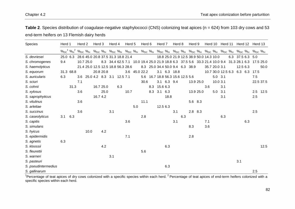
Chapter 4.2 Teat apex colonization before parturition
82
Table 2. Species distribution of coagulase-negative staphylococci (CNS) colonizing teat apices (n = 624) from 103 dry cows and 53
end-term heifers on 13 Flemish dairy herds
Species Herd 1 Herd 2 Herd 3 Herd 4 Herd 5 Herd 6 Herd 7 Herd 8 Herd 9 Herd 10 Herd 11 Herd 12 Herd 13
%DC1 %H
2 %DC %H %DC %H %DC %H %DC %H %DC %H %DC %H %DC %H %DC %H %DC %H %DC %H %DC %H %DC %H
S. devriesei 25.0 6.3 28.6 45.0 20.8 37.5 31.3 18.8 21.4 18.8 25.0 21.9 12.5 38.9 50.0 14.3 10.0 6.3 37.5 6.3 5.0
S. chromogenes 9.4 10.7 25.0 8.3 34.4 62.5 7.1 10.0 19.4 25.0 21.9 18.8 6.3 37.5 5.6 33.3 21.4 10.0 9.4 31.3 28.1 6.3 17.5 25.0
S. haemolyticus 21.4 25.0 12.5 12.5 18.8 56.3 28.6 8.3 25.0 34.4 50.0 9.4 6.3 38.9 35.7 20.0 3.1 12.5 6.3 50.0
S. equorum 31.3 68.8 20.8 20.8 3.6 45.0 22.2 3.1 6.3 18.8 10.7 30.0 12.5 6.3 6.3 6.3 17.5
S. auricularis 6.3 3.6 25.0 4.2 8.3 3.1 12.5 7.1 5.6 16.7 18.8 56.3 15.6 12.5 5.6 5.0 3.1 7.5
S. sciuri 7.1 30.6 3.1 6.3 9.4 13.9 25.0 10.0 3.1 22.5 37.5
S. cohnii 31.3 16.7 25.0 6.3 8.3 15.6 6.3 3.6 3.1
S. xylosus 3.6 25.0 10.7 8.3 3.1 6.3 13.9 25.0 5.0 3.1 2.5 12.5
S. saprophyticus 16.7 4.2 18.8 3.1 2.5
S. vitulinus 3.6 11.1 5.6 8.3
S. arlettae 5.0 12.5 6.3
S. succinus 3.6 3.1 3.1 2.8 8.3 2.5
S. caseolyticus 3.1 6.3 2.8 6.3 6.3
S. capitis 3.6 3.1 7.1 6.3
S. simulans 8.3 3.6
S. hyicus 10.0 4.2
S. epidermidis 7.1 2.8
S. agnetis 6.3
S. kloossii 4.2 6.3 12.5
S. fleurettii 5.6
S. warneri 3.1
S. pasteuri 3.1
S. pseudintermedius 6.3
S. gallinarum 2.5 1Percentage of teat apices of dry cows colonized with a specific species within each herd.
2 Percentage of teat apices of end-term heifers colonized with a
specific species within each herd.
Chapter 4.2 Teat apex colonization before parturition
83
In addition, the Hosmer-Lemeshow goodness-of-fit test and 2 residual log pseudo-
likelihood statistics were assessed on the fixed effect models only (SAS 9.3, SAS
Institute Inc., Cary, NC, USA) (Dohoo et al., 2009). The test was for none of the final
models statistically significant indicating good fit of our models. Data are presented
as odds ratio’s (OR) and 95% confidence intervals. The proportion of variation for the
outcome variables at the herd, cow and quarter level was estimated based on the
null and final models by assuming that the variance at the quarter level was π²/3
(Dohoo et al., 2001) as described by Piepers et al. (2011).
Results
Descriptive analyses
The majority of teat apices (87%, n = 540) yielded growth on MSA and per plate 0
to 4 phenotypically different colony types were present. Different colony types
sometimes appeared to belong to the same CNS species after tDNA-PCR or 16S
rRNA gene sequencing, eventually resulting in 619 CNS isolates available for further
analysis. Seventy-two percent (n = 448) of all teat apices were colonized with CNS
and harbored 1 up to 3 different CNS species. Twenty-four different CNS species
were identified.
The overall most frequently isolated species was S. devriesei (19% of all teat
apices colonized, n = 120 teat apices), followed by S. chromogenes (17%, n = 107),
S. haemolyticus (17%, n = 107), S. equorum (13%, n = 81), S. auricularis (8%, n =
49), S. sciuri (7%, n = 41), S. cohnii (4%, n = 25) and S. xylosus (4%, n = 25) (Table
2). Isolates (n = 390) other than CNS were phenotypically or genotypically (i.e. using
tDNA-PCR or 16S rRNA gene sequencing) identified as Staphylococcus aureus (n =
4), Arthrobacter sp. (n = 1), Bacillus spp. (n = 307), Jeotgallicoccus spp. (n = 28),
other Gram-positive cocci such as Aerococcus spp. (n = 12), Enterococcus sp. (n =
1) or Streptococcus spp. (n = 5), Gram-negative bacteria such as Pseudomonas spp.
(n = 16), Psychrobacter spp. (n = 4) or other (n = 7), and fungi (n = 5).
On each herd, between 6 (herd 12) and 12 (herd 7) different CNS species were
isolated from the teat apices. Staphylococcus chromogenes was the sole species
colonizing teat apices in all 13 herds whereas S. devriesei, S. haemolyticus, and S.
auricularis were colonizing teat apices on all farms except one.
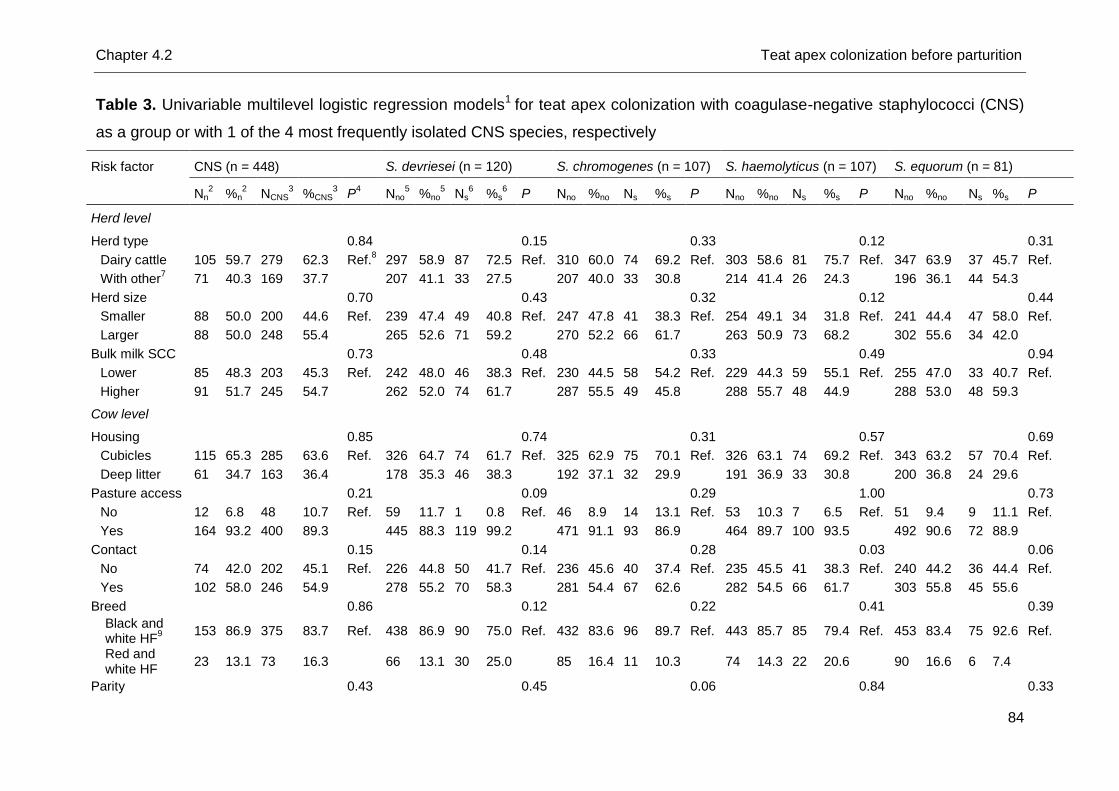
Chapter 4.2 Teat apex colonization before parturition
84
Table 3. Univariable multilevel logistic regression models1 for teat apex colonization with coagulase-negative staphylococci (CNS)
as a group or with 1 of the 4 most frequently isolated CNS species, respectively
Risk factor CNS (n = 448) S. devriesei (n = 120) S. chromogenes (n = 107) S. haemolyticus (n = 107) S. equorum (n = 81)
Nn2 %n
2 NCNS
3 %CNS
3 P
4 Nno
5 %no
5 Ns
6 %s
6 P Nno %no Ns %s P Nno %no Ns %s P Nno %no Ns %s P
Herd level
Herd type
0.84
0.15
0.33
0.12
0.31
Dairy cattle 105 59.7 279 62.3 Ref.8 297 58.9 87 72.5 Ref. 310 60.0 74 69.2 Ref. 303 58.6 81 75.7 Ref. 347 63.9 37 45.7 Ref.
With other7 71 40.3 169 37.7
207 41.1 33 27.5
207 40.0 33 30.8
214 41.4 26 24.3
196 36.1 44 54.3
Herd size
0.70
0.43
0.32
0.12
0.44
Smaller 88 50.0 200 44.6 Ref. 239 47.4 49 40.8 Ref. 247 47.8 41 38.3 Ref. 254 49.1 34 31.8 Ref. 241 44.4 47 58.0 Ref.
Larger 88 50.0 248 55.4
265 52.6 71 59.2
270 52.2 66 61.7
263 50.9 73 68.2
302 55.6 34 42.0
Bulk milk SCC
0.73
0.48
0.33
0.49
0.94
Lower 85 48.3 203 45.3 Ref. 242 48.0 46 38.3 Ref. 230 44.5 58 54.2 Ref. 229 44.3 59 55.1 Ref. 255 47.0 33 40.7 Ref.
Higher 91 51.7 245 54.7
262 52.0 74 61.7
287 55.5 49 45.8
288 55.7 48 44.9
288 53.0 48 59.3
Cow level
Housing
0.85
0.74
0.31
0.57
0.69
Cubicles 115 65.3 285 63.6 Ref. 326 64.7 74 61.7 Ref. 325 62.9 75 70.1 Ref. 326 63.1 74 69.2 Ref. 343 63.2 57 70.4 Ref.
Deep litter 61 34.7 163 36.4
178 35.3 46 38.3
192 37.1 32 29.9
191 36.9 33 30.8
200 36.8 24 29.6
Pasture access
0.21
0.09
0.29
1.00
0.73
No 12 6.8 48 10.7 Ref. 59 11.7 1 0.8 Ref. 46 8.9 14 13.1 Ref. 53 10.3 7 6.5 Ref. 51 9.4 9 11.1 Ref.
Yes 164 93.2 400 89.3
445 88.3 119 99.2
471 91.1 93 86.9
464 89.7 100 93.5
492 90.6 72 88.9
Contact
0.15
0.14
0.28
0.03
0.06
No 74 42.0 202 45.1 Ref. 226 44.8 50 41.7 Ref. 236 45.6 40 37.4 Ref. 235 45.5 41 38.3 Ref. 240 44.2 36 44.4 Ref.
Yes 102 58.0 246 54.9
278 55.2 70 58.3
281 54.4 67 62.6
282 54.5 66 61.7
303 55.8 45 55.6
Breed
0.86
0.12
0.22
0.41
0.39
Black and white HF
9
153 86.9 375 83.7 Ref. 438 86.9 90 75.0 Ref. 432 83.6 96 89.7 Ref. 443 85.7 85 79.4 Ref. 453 83.4 75 92.6 Ref.
Red and white HF
23 13.1 73 16.3
66 13.1 30 25.0
85 16.4 11 10.3
74 14.3 22 20.6
90 16.6 6 7.4
Parity
0.43
0.45
0.06
0.84
0.33
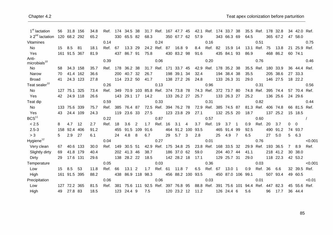
Chapter 4.2 Teat apex colonization before parturition
85
1st lactation 56 31.8 156 34.8 Ref. 174 34.5 38 31.7 Ref. 167 47.7 45 42.1 Ref. 174 33.7 38 35.5 Ref. 178 32.8 34 42.0 Ref.
≥ 2nd
lactation 120 68.2 292 65.2
330 65.5 82 68.3
350 67.7 62 57.9
343 66.3 69 64.5
365 67.2 47 58.0
Vitamines
0.14
0.24
0.16
0.51
0.75
No 15 8.5 81 18.1 Ref. 67 13.3 29 24.2 Ref. 87 16.8 9 8.4 Ref. 82 15.9 14 13.1 Ref. 75 13.8 21 25.9 Ref.
Yes 161 91.5 367 81.9
437 86.7 91 75.8
430 83.2 98 91.6
435 84.1 93 86.9
468 86.2 60 74.1
Anti-microbials
10
0.39
0.06
0.20
0.76
0.46
No 58 34.3 158 35.7 Ref. 178 36.2 38 31.7 Ref. 171 33.7 45 42.9 Ref. 178 35.2 38 35.5 Ref. 180 33.9 36 44.4 Ref.
Narow 70 41.4 162 36.6
200 40.7 32 26.7
198 39.1 34 32.4
194 38.4 38 35.5
205 38.6 27 33.3
Broad 41 24.3 123 27.8
114 23.2 50 41.7
138 27.2 26 24.8
133 26.3 31 29.0
146 27.5 18 22.2
Teat sealer10
0.26
0.13
0.96
0.31
0.56
No 127 75.1 325 73.4 Ref. 349 70.9 103 85.8 Ref. 374 73.8 78 74.3 Ref. 372 73.7 80 74.8 Ref. 395 74.4 57 70.4 Ref.
Yes 42 24.9 118 26.6
143 29.1 17 14.2
133 26.2 27 25.7
133 26.3 27 25.2
136 25.6 24 29.6
Teat dip
0.59
0.33
0.31
0.82
0.44
No 133 75.6 339 75.7 Ref. 385 76.4 87 72.5 Ref. 394 76.2 78 72.9 Ref. 385 74.5 87 81.3 Ref. 406 74.8 66 81.5 Ref.
Yes 43 24.4 109 24.3
119 23.6 33 27.5
123 23.8 29 27.1
132 25.5 20 18.7
137 25.2 15 18.5
BCS11
0.22
0.87
0.57
0.60
/
< 2.5 8 4.7 12 2.7 Ref. 18 3.6 2 1.7 Ref. 16 3.1 4 3.7 Ref. 19 3.7 1 0.9 Ref. 20 3.7 0 0
2.5-3 158 92.4 406 91.2
455 91.5 109 91.6
464 91.2 100 93.5
465 91.4 99 92.5
490 91.2 74 93.7
> 3 5 2.9 27 6.1
24 4.8 8 6.7
29 5.7 3 2.8
25 4.9 7 6.5
27 5.0 5 6.3
Hygiene12
0.04
0.27
0.01
0.76
<0.001
Very clean 67 40.6 133 30.0 Ref. 149 30.5 51 42.9 Ref. 175 34.8 25 23.8 Ref. 168 33.5 32 29.9 Ref. 193 36.5 7 8.9 Ref.
Slightly dirty 69 41.8 179 40.4
202 41.3 46 38.7
186 37.0 62 59.0
204 40.7 44 41.1
218 41.2 30 38.0
Dirty 29 17.6 131 29.6
138 28.2 22 18.5
142 28.2 18 17.1
129 25.7 31 29.0
118 22.3 42 53.2
Temperature 0.05 0.03 0.36 0.03 <0.001
Low 15 8.5 53 11.8 Ref. 66 13.1 2 1.7 Ref. 61 11.8 7 6.5 Ref. 67 13.0 1 0.9 Ref. 36 6.6 32 39.5 Ref.
High 161 91.5 395 88.2 438 86.9 118 98.3 456 88.2 100 93.5 450 87.0 106 99.1 507 93.4 49 60.5
Precipitation 0.06 0.06 0.03 0.01 <0.01
Low 127 72.2 365 81.5 Ref. 381 75.6 111 92.5 Ref. 397 76.8 95 88.8 Ref. 391 75.6 101 94.4 Ref. 447 82.3 45 55.6 Ref.
High 49 27.8 83 18.5 123 24.4 9 7.5 120 23.2 12 11.2 126 24.4 6 5.6 96 17.7 36 44.4
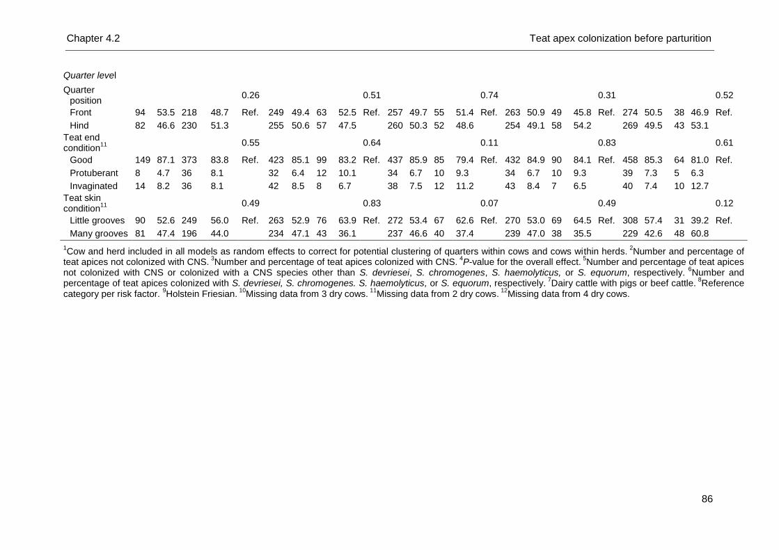
Chapter 4.2 Teat apex colonization before parturition
86
Quarter level
Quarter position
0.26
0.51
0.74
0.31
0.52
Front 94 53.5 218 48.7 Ref. 249 49.4 63 52.5 Ref. 257 49.7 55 51.4 Ref. 263 50.9 49 45.8 Ref. 274 50.5 38 46.9 Ref.
Hind 82 46.6 230 51.3
255 50.6 57 47.5
260 50.3 52 48.6
254 49.1 58 54.2
269 49.5 43 53.1
Teat end condition
11
0.55
0.64
0.11
0.83
0.61
Good 149 87.1 373 83.8 Ref. 423 85.1 99 83.2 Ref. 437 85.9 85 79.4 Ref. 432 84.9 90 84.1 Ref. 458 85.3 64 81.0 Ref.
Protuberant 8 4.7 36 8.1
32 6.4 12 10.1
34 6.7 10 9.3
34 6.7 10 9.3
39 7.3 5 6.3
Invaginated 14 8.2 36 8.1
42 8.5 8 6.7
38 7.5 12 11.2
43 8.4 7 6.5
40 7.4 10 12.7
Teat skin condition
11
0.49
0.83
0.07
0.49
0.12
Little grooves 90 52.6 249 56.0 Ref. 263 52.9 76 63.9 Ref. 272 53.4 67 62.6 Ref. 270 53.0 69 64.5 Ref. 308 57.4 31 39.2 Ref.
Many grooves 81 47.4 196 44.0
234 47.1 43 36.1
237 46.6 40 37.4
239 47.0 38 35.5
229 42.6 48 60.8
1Cow and herd included in all models as random effects to correct for potential clustering of quarters within cows and cows within herds.
2Number and percentage of
teat apices not colonized with CNS. 3Number and percentage of teat apices colonized with CNS.
4P-value for the overall effect.
5Number and percentage of teat apices
not colonized with CNS or colonized with a CNS species other than S. devriesei, S. chromogenes, S. haemolyticus, or S. equorum, respectively. 6
Number and percentage of teat apices colonized with S. devriesei, S. chromogenes. S. haemolyticus, or S. equorum, respectively.
7Dairy cattle with pigs or beef cattle.
8Reference
category per risk factor. 9Holstein Friesian.
10Missing data from 3 dry cows.
11Missing data from 2 dry cows.
12Missing data from 4 dry cows.
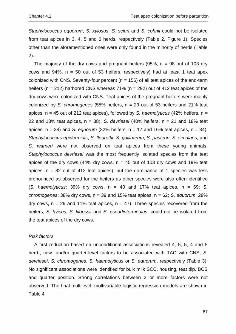
Chapter 4.2 Teat apex colonization before parturition
87
Staphylococcus equorum, S. xylosus, S. sciuri and S. cohnii could not be isolated
from teat apices in 3, 4, 5 and 6 herds, respectively (Table 2, Figure 1). Species
other than the aforementioned ones were only found in the minority of herds (Table
2).
The majority of the dry cows and pregnant heifers (95%, n = 98 out of 103 dry
cows and 94%, n = 50 out of 53 heifers, respectively) had at least 1 teat apex
colonized with CNS. Seventy-four percent (n = 156) of all teat apices of the end-term
heifers (n = 212) harbored CNS whereas 71% (n = 292) out of 412 teat apices of the
dry cows were colonized with CNS. Teat apices of the pregnant heifers were mainly
colonized by S. chromogenes (55% heifers, n = 29 out of 53 heifers and 21% teat
apices, n = 45 out of 212 teat apices), followed by S. haemolyticus (42% heifers, n =
22 and 18% teat apices, n = 38), S. devriesei (40% heifers, n = 21 and 18% teat
apices, n = 38) and S. equorum (32% heifers, n = 17 and 16% teat apices, n = 34).
Staphylococcus epidermidis, S. fleurettii, S. gallinarum, S. pasteuri, S. simulans, and
S. warneri were not observed on teat apices from these young animals.
Staphylococcus devriesei was the most frequently isolated species from the teat
apices of the dry cows (44% dry cows, n = 45 out of 103 dry cows and 19% teat
apices, n = 82 out of 412 teat apices), but the dominance of 1 species was less
pronounced as observed for the heifers as other species were also often identified
(S. haemolyticus: 39% dry cows, n = 40 and 17% teat apices, n = 69; S.
chromogenes: 38% dry cows, n = 39 and 15% teat apices, n = 62; S. equorum: 28%
dry cows, n = 29 and 11% teat apices, n = 47). Three species recovered from the
heifers, S. hyicus, S. kloossii and S. pseudintermedius, could not be isolated from
the teat apices of the dry cows.
Risk factors
A first reduction based on unconditional associations revealed 4, 5, 5, 4 and 5
herd-, cow- and/or quarter-level factors to be associated with TAC with CNS, S.
devriesei, S. chromogenes, S. haemolyticus or S. equorum, respectively (Table 3).
No significant associations were identified for bulk milk SCC, housing, teat dip, BCS
and quarter position. Strong correlations between 2 or more factors were not
observed. The final multilevel, multivariable logistic regression models are shown in
Table 4.
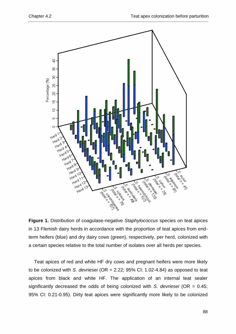
Chapter 4.2 Teat apex colonization before parturition
88
Figure 1. Distribution of coagulase-negative Staphylococcus species on teat apices
in 13 Flemish dairy herds in accordance with the proportion of teat apices from end-
term heifers (blue) and dry dairy cows (green), respectively, per herd, colonized with
a certain species relative to the total number of isolates over all herds per species.
Teat apices of red and white HF dry cows and pregnant heifers were more likely
to be colonized with S. devriesei (OR = 2.22; 95% CI: 1.02-4.84) as opposed to teat
apices from black and white HF. The application of an internal teat sealer
significantly decreased the odds of being colonized with S. devriesei (OR = 0.45;
95% CI: 0.21-0.95). Dirty teat apices were significantly more likely to be colonized
Chapter 4.2 Teat apex colonization before parturition
89
with CNS in general (OR = 2.91; 95% CI: 1.47-5.77) and S. equorum in particular
(OR = 5.24; 95% CI: 1.87-14.66) than very clean teat apices. Slightly dirty teat
apices had higher odds of being colonized with S. chromogenes (OR = 2.39; 95% CI:
1.30-4.40) as opposed to very clean teat apices. A higher ambient temperature
significantly increased the odds of being colonized with S. devriesei (OR = 6.63; 95%
CI: 1.37-32.13) or S. haemolyticus (OR = 10.89; 95% CI: 1.27-93.52) whereas the
opposite was true for S. equorum (OR = 0.30; 95% CI: 0.12-0.76) with the
association even being more pronounced in case of low precipitation than in case of
high precipitation (significant interaction term, see Figure 2). Teat apex colonization
with S. chromogenes was not associated with ambient temperature. Teat apices
swabbed in months with high precipitation were less likely to be colonized with CNS
in general (OR = 0.45; 95% CI: 0.24-0.84), and with S. devriesei (OR = 0.35; 95%
CI: 0.14-0.86), S. chromogenes (OR = 0.44; 95% CI: 0.21, 0.94) and S. haemolyticus
(OR = 0.23; 95% CI: 0.08-0.67) in particular than teat apices swabbed in months with
low precipitation. For S. equorum, the latter was only true in case of low ambient
temperature and when no Bonferroni-correction was applied (OR = 0.21; 95% CI:
0.05-0.93) (Table 4, Figure 2). In case of high ambient temperatures, teat apices
swabbed in months with high precipitation were even more likely to be colonized with
S. equorum than teat apices swabbed in months with low precipitation (Figure 2).
The highest variability was observed at the quarter level for all dependent
variables. For the outcome variable S. chromogenes TAC, the variation at the cow
level in the null model was much more pronounced as opposed to the herd level
whereas this discrepancy was less pronounced for S. haemolyticus or S. devriesei
and even absent for S. equorum (Table 5). For the latter species, the variation
occurred in an almost equal amount at the cow and herd level.
Discussion
This is the first large species-specific observational study that describes the
prevalence and distribution of CNS colonizing teat apices from dry cows and end-
term heifers and simultaneously identifies risk factors for TAC prior to calving at
different levels of the data hierarchy.
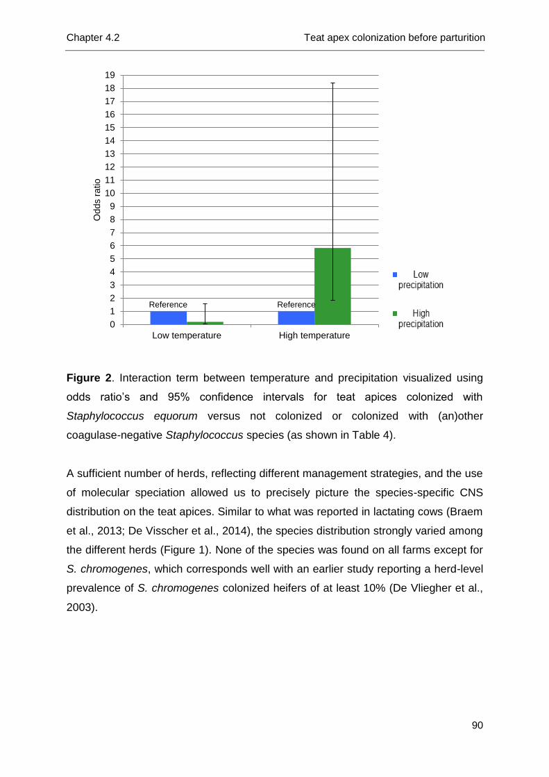
Chapter 4.2 Teat apex colonization before parturition
90
Figure 2. Interaction term between temperature and precipitation visualized using
odds ratio’s and 95% confidence intervals for teat apices colonized with
Staphylococcus equorum versus not colonized or colonized with (an)other
coagulase-negative Staphylococcus species (as shown in Table 4).
A sufficient number of herds, reflecting different management strategies, and the use
of molecular speciation allowed us to precisely picture the species-specific CNS
distribution on the teat apices. Similar to what was reported in lactating cows (Braem
et al., 2013; De Visscher et al., 2014), the species distribution strongly varied among
the different herds (Figure 1). None of the species was found on all farms except for
S. chromogenes, which corresponds well with an earlier study reporting a herd-level
prevalence of S. chromogenes colonized heifers of at least 10% (De Vliegher et al.,
2003).
0
1
2
3
4
5
6
7
8
9
10
11
12
13
14
15
16
17
18
19
Low temperature High temperature
Odds r
atio
Reference Reference
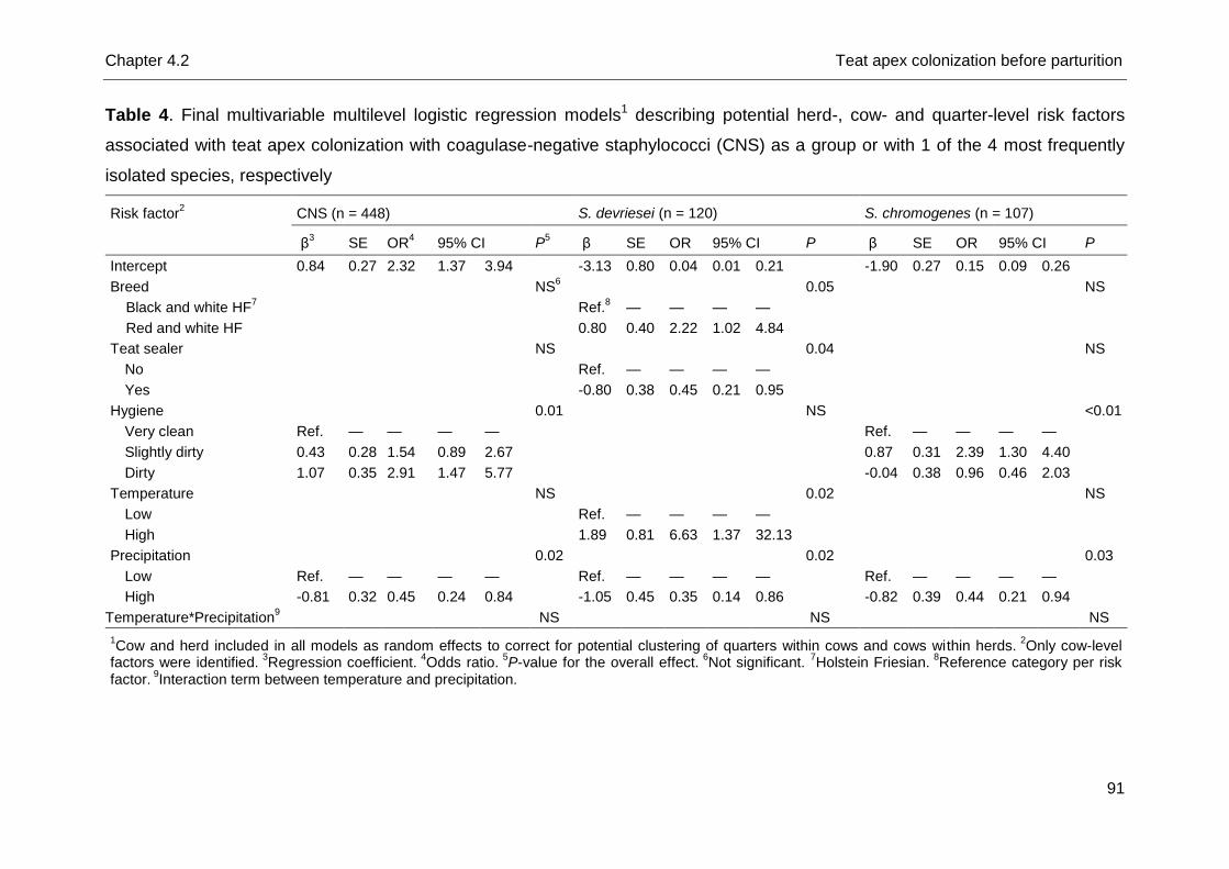
Chapter 4.2 Teat apex colonization before parturition
91
Table 4. Final multivariable multilevel logistic regression models1 describing potential herd-, cow- and quarter-level risk factors
associated with teat apex colonization with coagulase-negative staphylococci (CNS) as a group or with 1 of the 4 most frequently
isolated species, respectively
Risk factor2 CNS (n = 448) S. devriesei (n = 120) S. chromogenes (n = 107)
β3 SE OR
4 95% CI P
5 β SE OR 95% CI P β SE OR 95% CI P
Intercept 0.84 0.27 2.32 1.37 3.94
-3.13 0.80 0.04 0.01 0.21
-1.90 0.27 0.15 0.09 0.26
Breed
NS6
0.05
NS
Black and white HF7
Ref.
8 — — — —
Red and white HF
0.80 0.40 2.22 1.02 4.84
Teat sealer
NS
0.04
NS
No
Ref. — — — —
Yes
-0.80 0.38 0.45 0.21 0.95
Hygiene
0.01
NS
<0.01
Very clean Ref. — — — —
Ref. — — — —
Slightly dirty 0.43 0.28 1.54 0.89 2.67
0.87 0.31 2.39 1.30 4.40
Dirty 1.07 0.35 2.91 1.47 5.77
-0.04 0.38 0.96 0.46 2.03
Temperature
NS
0.02
NS
Low
Ref. — — — —
High
1.89 0.81 6.63 1.37 32.13
Precipitation
0.02
0.02
0.03
Low Ref. — — — —
Ref. — — — —
Ref. — — — —
High -0.81 0.32 0.45 0.24 0.84
-1.05 0.45 0.35 0.14 0.86
-0.82 0.39 0.44 0.21 0.94
Temperature*Precipitation9 NS NS NS
1Cow and herd included in all models as random effects to correct for potential clustering of quarters within cows and cows within herds.
2Only cow-level
factors were identified. 3
Regression coefficient. 4
Odds ratio. 5
P-value for the overall effect. 6
Not significant. 7Holstein Friesian.
8Reference category per risk
factor. 9Interaction term between temperature and precipitation.
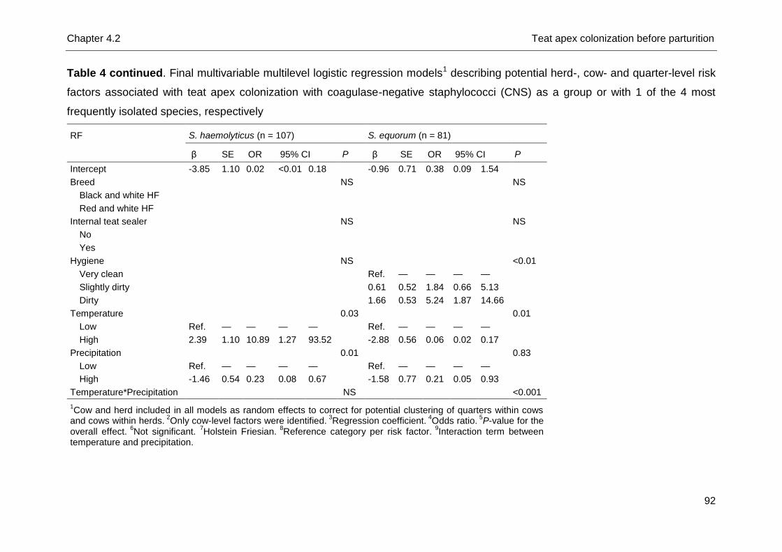
Chapter 4.2 Teat apex colonization before parturition
92
Table 4 continued. Final multivariable multilevel logistic regression models1 describing potential herd-, cow- and quarter-level risk
factors associated with teat apex colonization with coagulase-negative staphylococci (CNS) as a group or with 1 of the 4 most
frequently isolated species, respectively
RF S. haemolyticus (n = 107) S. equorum (n = 81)
β SE OR 95% CI P β SE OR 95% CI P
Intercept -3.85 1.10 0.02 <0.01 0.18
-0.96 0.71 0.38 0.09 1.54
Breed
NS
NS
Black and white HF
Red and white HF
Internal teat sealer
NS
NS
No
Yes
Hygiene
NS
<0.01
Very clean
Ref. — — — —
Slightly dirty
0.61 0.52 1.84 0.66 5.13
Dirty
1.66 0.53 5.24 1.87 14.66
Temperature
0.03
0.01
Low Ref. — — — —
Ref. — — — —
High 2.39 1.10 10.89 1.27 93.52
-2.88 0.56 0.06 0.02 0.17
Precipitation
0.01
0.83
Low Ref. — — — —
Ref. — — — —
High -1.46 0.54 0.23 0.08 0.67
-1.58 0.77 0.21 0.05 0.93
Temperature*Precipitation NS <0.001
1Cow and herd included in all models as random effects to correct for potential clustering of quarters within cows
and cows within herds. 2
Only cow-level factors were identified. 3
Regression coefficient. 4
Odds ratio. 5
P-value for the overall effect.
6Not significant.
7Holstein Friesian.
8Reference category per risk factor.
9Interaction term between
temperature and precipitation.
Chapter 4.2 Teat apex colonization before parturition
93
The prevalence of teat apices colonized with CNS as a group and with S.
chromogenes in particular was higher than those observed in previous studies
focusing on pre-partum TAC with CNS as a group (Piepers et al., 2011) and S.
chromogenes (De Vliegher et al., 2003), respectively. These differences might
partially be explained by using a more suitable and appropriate medium in our study
(De Visscher et al., 2013).
A lower percentage of teat apices was colonized with CNS in our previous work
(De Visscher et al, 2014) than in the current study in which only teat apices prior to
calving were swabbed. Routinely cleaning of the teats before cluster attachment, a
flushing effect of the milking process (De Visscher et al., 2014), and the application
of post-milking teat disinfection (Piessens et al., 2012; Quirk et al., 2012) may at
least partly explain this discrepancy. Further, a higher number of species and some
remarkable diversity in species distribution were found in our pre-partum study
compared with the studies concerning lactating cows (Braem et al., 2013; De
Visscher et al., 2014). Staphylococcus devriesei, the predominant species in the
current study, was only rarely isolated in one (Braem et al., 2013) and even absent in
another study (De Visscher et al., 2014), both focusing on TAC in lactating dairy
cows. Although less pronounced as for S. devriesei, the same was true for S.
auricularis (Braem et al., 2013; De Visscher et al., 2014). In turn, S. cohnii (Braem et
al., 2013; De Visscher et al., 2014), S. saprophyticus, S. simulans (Braem et al.,
2013), S. xylosus (Taponen et al., 2008) and S. fleurettii (De Visscher et al., 2014)
were less commonly isolated from teat apices from non-lactating cows and heifers as
opposed to teat apices from lactating dairy cows. The differences between lactating
and non-lactating animals are less consistent for S. chromogenes, the second most
frequently isolated species. Staphylococcus chromogenes could not or hardly be
isolated from teat apices from lactating dairy cows in some studies (Braem et al.,
2013; De Visscher et al., 2014), but was often isolated from bovine teat skin in some
other single-dairy-herd studies (White et al., 1989; Taponen et al., 2008).
Staphylococcus haemolyticus, S. equorum (Braem et al., 2013; De Visscher et al.,
2014; Vanderhaeghen et al., 2015), and to a lesser extent also S. sciuri (Braem et
al., 2013; De Visscher et al., 2014) were frequently isolated species from teat apices
from both lactating dairy cows and non-lactating cows and heifers. Again, our
findings suggest that herds have various CNS microbiota colonizing different habitats
implying the potential role of management strategies. Although we have included
Chapter 4.2 Teat apex colonization before parturition
94
herd-level factors in the current study, the number of herds was too small to identify
important ones. Other studies specifically designed for analyzing herd-level
variables, are needed.
Teat apices of red and white HF as opposed to teat apices of black and white HF
dry cows and pregnant heifers were more likely to be colonized with S. devriesei.
Also, the application of an internal teat sealer, a management tool useful in
preventing environmental pathogens invading quarters during the dry period (Huxley
et al., 2002), significantly decreased the odds of being colonized with S. devriesei in
this study. No well-defined interpretation can explain these observed associations,
yet they merit further study. Staphylococcus devriesei was also significantly more
present on teat apices in months with low precipitation and high ambient
temperatures comparable to what was observed for S. haemolyticus. Because
hygiene did not influence TAC with S. devriesei nor with S. haemolyticus in the
current study, an environmental nature of S. devriesei and S. haemolyticus could be
questioned as previously reported for S. haemolyticus by Leroy et al. (2015). Strain-
typing is necessary to come to better conclusions. As staphylococci prefer a higher
temperature to grow (Prescott et al., 2002) and dryness might cause imperceptible
teat end lesions, forming a good matrix for expanding the present CNS microbiota,
warmth and low precipitation could provide S. devriesei and S. haemolyticus with
good growth conditions.
On the other hand, TAC with S. equorum was significantly associated with
hygiene, with dirty teat apices being more colonized as opposed to very clean teat
apices. In addition, teat apices were more likely to be colonized with S. equorum
when swabbed under humid conditions in combination with a higher ambient
temperature. The latter climatic conditions could support the bacterial load from the
environment and thus could increase the exposure of teat apices to S. equorum
(Smith et al., 1985). Earlier studies also observed a more pronounced presence of S.
equorum in extramammary habitats (Piessens et al., 2011; De Visscher et al., 2014)
as opposed to milk (Supré et al., 2011; Piessens et al., 2011; Fry et al., 2014) and
our current findings seem to confirm the environmental nature of S. equorum.
Staphylococcus chromogenes was rarely isolated from the bovine environment
(Piessens et al., 2011), but was predominantly present in bovine IMI (Piessens et al.,
2011; Supré et al., 2011; Fry et al., 2014).
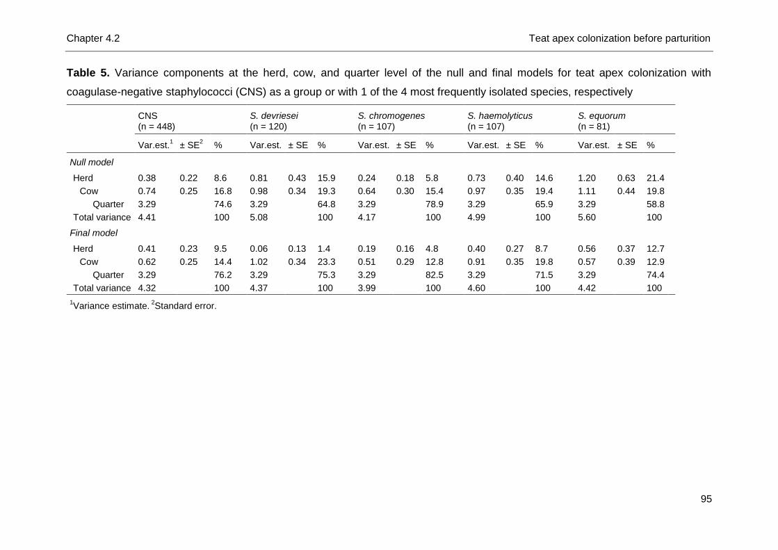
Chapter 4.2 Teat apex colonization before parturition
95
Table 5. Variance components at the herd, cow, and quarter level of the null and final models for teat apex colonization with
coagulase-negative staphylococci (CNS) as a group or with 1 of the 4 most frequently isolated species, respectively
CNS (n = 448)
S. devriesei (n = 120)
S. chromogenes (n = 107)
S. haemolyticus (n = 107)
S. equorum (n = 81)
Var.est.
1 ± SE
2 % Var.est. ± SE % Var.est. ± SE % Var.est. ± SE % Var.est. ± SE %
Null model
Herd 0.38 0.22 8.6 0.81 0.43 15.9 0.24 0.18 5.8 0.73 0.40 14.6 1.20 0.63 21.4
Cow 0.74 0.25 16.8 0.98 0.34 19.3 0.64 0.30 15.4 0.97 0.35 19.4 1.11 0.44 19.8
Quarter 3.29
74.6 3.29 64.8 3.29 78.9 3.29 65.9 3.29 58.8
Total variance 4.41
100 5.08 100 4.17 100 4.99 100 5.60 100
Final model
Herd 0.41 0.23 9.5 0.06 0.13 1.4 0.19 0.16 4.8 0.40 0.27 8.7 0.56 0.37 12.7
Cow 0.62 0.25 14.4 1.02 0.34 23.3 0.51 0.29 12.8 0.91 0.35 19.8 0.57 0.39 12.9
Quarter 3.29 76.2 3.29 75.3 3.29 82.5 3.29 71.5 3.29 74.4
Total variance 4.32 100 4.37 100 3.99 100 4.60 100 4.42 100
1Variance estimate.
2Standard error.
Chapter 4.2 Teat apex colonization before parturition
96
An earlier study (De Vliegher et al., 2003) and the current pre-partum one observed
S. chromogenes TAC in all included herds. The latter observations all indicate a
host-adapted nature of S. chromogenes. On the other hand, teat apices from
lactating dairy cows and heifers were hardly colonized with S. chromogenes
(Taponen et al., 2008; Braem et al., 2013; De Visscher et al., 2014). This can partly
be explained by a smaller number of included herds, different genotypic methods, or
both, yet needs further study.
Most of variation of the outcome variables in the null models resided at the quarter
level as reported before for CNS IMI (Piepers et al., 2011; Passchyn et al., 2014). As
no quarter-level factors were identified and the variation explained in the final models
was low, as seen before (Piepers et al., 2011; Verbeke et al., 2012; Passchyn et al.,
2014), factors others than the studied ones play a key role in explaining the
likelihood of colonization (or infection). Focusing on teat dimensions for instance
might be useful in improving udder health (Zwertvaegher et al., 2013) and a potential
pathway is diversity among cows and heifers due to their distinct genetic background
(Detilleux, 2009; Verbeke et al., 2012). Differences in susceptibility for CNS IMI
among cows and quarters could also rely on differences in immune status (Piepers
et al., 2009), which is also partially genetically determined. Still, calculation of the
variance components for binary outcome variables is not as straightforward as it is
for continuous outcomes (Dohoo et al., 2009) making comparisons with that level not
appropriate. Still, we hypothesize that factors at the quarter level rather than at the
cow and herd level play a more important role for S. chromogenes TAC than for TAC
with the other 3 species. For TAC with S. chromogenes, more variation was present
between cows than between herds when compared with the other species, which is
most likely related to the fact that S. chromogenes was the sole species colonizing
teat apices in all included herds. Regarding the proportion of variation being present
at the herd or cow level, analogous patterns could be observed for S. devriesei and
S. haemolyticus. Remarkably, for TAC with S. equorum an almost equal amount of
the total variation occurred at the herd and cow level despite being an environmental
species.
Chapter 4.2 Teat apex colonization before parturition
97
Conclusions
Staphylococcus devriesei, S. chromogenes, S. haemolyticus, and S. equorum
were the predominant species colonizing teat apices from dry dairy cows and end-
term heifers before calving. Staphylococcus chromogenes was present on teat
apices in all herds whereas large herd differences were observed for other species.
Diverse factors explaining CNS species-specific TAC, yet mostly related to humidity,
ambient temperature, and hygiene, substantiate differences in epidemiological
behavior and ecology between species. An environmental nature for S. equorum is
suggested whereas S. haemolyticus and S. devriesei seem to act as cow-adapted
bacteria, as S. chromogenes does.
Chapter 4.2 Teat apex colonization before parturition
98
References
Braem, G., S. De Vliegher, B. Verbist, M. Heyndrickx, F. Leroy, and L. De Vuyst.
2012. Culture-independent exploration of the teat apex microbiota of dairy cows
reveals a wide bacterial species diversity. Vet Microbiol. 157:383-390.
Braem, G., S. De Vliegher, B. Verbist, V. Piessens, E. Van Coillie, L. De Vuyst, and
F. Leroy. 2013. Unraveling the microbiota of teat apices of clinically healthy
lactating dairy cows, with special emphasis on coagulase-negative staphylococci.
J. Dairy Sci. 96:1499-1510.
Detilleux, J. C. 2009. Genetic factors affecting susceptibility to udder pathogens. Vet.
Microbiol. 134:157-164.
De Visscher, A., F. Haesebrouck, S. Piepers, W. Vanderhaeghen, K. Supré, F.
Leroy, E. Van Coillie, and S. De Vliegher. 2013. Assessment of the suitability of
mannitol salt agar for growing bovine-associated coagulase-negative
staphylococci and its use under field conditions. Res. Vet. Sci. 95:347-351.
De Visscher, A., K. Supré, F. Haesebrouck, R. N. Zadoks, V. Piessens, E. Van
Coillie, S. Piepers, and S. De Vliegher. 2014. Further evidence for the existence of
environmental and host-associated species of coagulase-negative staphylococci
in dairy cattle. Vet. Microbiol. 172:466-474.
De Vliegher, S., H. Laevens, L. A. Devriese, G. Opsomer, J. L. M. Leroy, H. W.
Barkema, and A. de Kruif. 2003. Prepartum teat apex colonization with
Staphylococcus chromogenes in dairy heifers is associated with low somatic cell
count in early lactation. Vet. Microbiol. 92:245-252.
Dohoo, I. R., E. Tillard, H. Stryhn, and B. Faye. 2001. The use of multilevel models
to evaluate sources of variation in reproductive performance in dairy cattle in
Reunion Island. Prev. Vet. Med. 50:127-144.
Dohoo, I., W. Martin, and H. Stryhn. 2009. Mixed models for discrete data. In:
Dohoo, I., W. Martin, and H. Stryhn, Veterinary Epidemiologic Research. 2nd
edition, AVC inc., Charlottetown, Canada, p. 579-606.
Edmonson, A. J., I. J. Lean, L. D. Weaver, T. Farver, and G. Webster. 1989. A body
condition scoring chart for Holstein dairy cows. J. Dairy Sci. 72:68-78.
Fry, P. R., J. R. Middleton, S. Dufour, J. Perry, D. Scholl, and I. Dohoo. 2014.
Association of coagulase-negative staphylococcal species, mammary quarter milk
Chapter 4.2 Teat apex colonization before parturition
99
somatic cell count, and persistence of intramammary infection in dairy cattle. J.
Dairy Sci. 97:4876-4885.
Huxley, J. N., M. J. Green, L. E. Green, and A. J. Bradley. 2002. Evaluation of the
efficacy of an internal teat sealer during the dry period. J. Dairy Sci. 85:551-561.
Leroy, F., E. Van Coillie, G. Braem, V. Piessens, B. Verbist, L. De Vuyst, and S. De
Vliegher. 2015. Short communication: subtyping of Staphylococcus haemolyticus
isolates from milk and corresponding teat apices to verify the potential teat-skin
origin of intramammary infections in dairy cows. J. Dairy Sci. 98:7893-7898.
Passchyn, P., S. Piepers, and S. De Vliegher. 2014. Pathogen group-specific risk
factors for intramammary infection in treated and untreated dairy heifers
participating in a prepartum antimicrobial treatment trial. J. Dairy Sci. 97:6260-
6270.
Piepers, S., G. Opsomer, E. Meyer, K. Demeyere, H. W. Barkema, A. de Kruif, and
S. De Vliegher. 2009. Heifer and quarter characteristics associated with
periparturient blood and milk neutrophil apoptosis in healthy heifers and in heifers
with subclinical mastitis. J. Dairy Sci. 92:4330-4339.
Piepers, S., K. Peeters, G. Opsomer, H. W. Barkema, K. Frankena, and S. De
Vliegher. 2011. Pathogen group specific risk factors at herd, heifer and quarter
levels for intramammary infections in early lactating dairy heifers. Prev. Vet. Med.
99:91-101.
Piessens, V., E. Van Coillie, B. Verbist, K. Supré, G. Braem, A. Van Nuffel, L. De
Vuyst M. Heyndrickx, and S. De Vliegher. 2011. Distribution of coagulase-
negative Staphylococcus species from dairy cows’ milk and environment differs
between herds. J. Dairy Sci. 94:2933-2944.
Piessens, V., S. De Vliegher, B. Verbist, G. Braem, A. Van Nuffel, L. De Vuyst, M.
Heyndrickx, and E. Van Coillie. 2012. Characterization of coagulase-negative
Staphylococcus species from cows’ milk and environment based on bap, icaA,
and mecA genes and phenotypic susceptibility to antimicrobials and teat dips. J.
Dairy Sci. 95:7027-7038.
Prescott, L. M., J. P. Harley, and D. A. Klein. 2002. Microbial growth. The influence
of environmental factors on growth. In: Prescott, L. M., J. P. Harley, and D. A.
Klein, Microbiology. 5th edition, The McGraw-Hill Inc., New York, USA, p. 118-125.
Quirk, T., L. K. Fox, D. D. Hancock, J. Capper, J. Wenz, and J. Park. 2012.
Intramammary infections and teat canal colonization with coagulase-negative
Chapter 4.2 Teat apex colonization before parturition
100
staphylococci after postmilking teat disinfection: species-specific responses. J.
Dairy Sci. 1906-1912.
Reneau, J. K., A. J. Seykora, B. J. Heins, M. I. Endres, R. J. Farnsworth, and R. F.
Bey. 2005. Associations between hygiene scores and somatic cell scores in dairy
cattle. J. Am. Vet. Med. Assoc. 227:1297-1301.
Smith, K. L., D. A. Todhunter, and P. S. Schoenberger. 1985. Environmental
mastitis: cause, prevalence and prevention. J. Dairy Sci. 68:1531-1553.
Supré, K., S. De Vliegher, O. C. Sampimon, R. N. Zadoks, M. Vaneechoutte, M.
Baele, E. De Graef, S. Piepers, and F. Haesebrouck. 2009. Technical note: Use of
transfer RNA-intergenic spacer PCR combined with capillary electrophoresis to
identify coagulase-negative Staphylococcus species originating from bovine milk
and teat apices. J. Dairy Sci. 92:3204-3210.
Supré, K., F. Haesebrouck, R. N. Zadoks, M. Vaneechoutte, S. Piepers, and S. De
Vliegher. 2011. Some coagulase-negative Staphylococcus species affect udder
health more than others. J. Dairy Sci. 94:2329-2340.
Taponen, S., J. Björkroth, and S. Pyörälä. 2008. Coagulase-negative staphylococci
isolated from bovine extramammary sites and intramammary infections in a single
dairy herd. J. Dairy Res. 75:422-429.
Vanderhaeghen, W., S. Piepers, F. Leroy, E. Van Coillie, F. Haesebrouck, and S. De
Vliegher. 2014. Invited review. Effect, persistence, and virulence of coagulase-
negative Staphylococcus species associated with ruminant udder health. J. Dairy
Sci. 97:5275-5293.
Vanderhaeghen, W., S. Piepers, F. Leroy, E. Van Coillie, F. Haesebrouck, and S. De
Vliegher. 2015. Identification, typing, ecology and epidemiology of coagulase-
negative staphylococci associated with ruminants. Vet. J. 203:44-51.
Verbeke, J., S. Piepers, L. Peelman, M. Van Poucke, and S. De Vliegher. 2012.
Pathogen-group specific association between CXCR1 polymorphisms and
subclinical mastitis in dairy heifers. J. Dairy Res. 79:341-351.
White, D. G., R. J. Harmon, J. E. S. Matos, and B. E. Langlois. 1989. Isolation and
identification of coagulase-negative Staphylococcus species from bovine body
sites and streak canals of nulliparous heifers. J. Dairy Sci. 72:1886-1892.
Zwertvaegher, I., S. De Vliegher, B. Verbist, A. Van Nuffel, J. Baert, and S. Van
Weyenberg. 2013. Short communication: Associations between teat dimensions
Chapter 4.2 Teat apex colonization before parturition
101
and milking-induced changes in teat dimensions and quarter milk somatic cell
counts in dairy cows. J. Dairy Sci. 96:1075-1080.

Chapter 5
Intramammary Infection with Coagulase-
Negative Staphylococcus Species in Early
Lactation and throughout Lactation

Chapter 5.1
Intramammary Infection with Coagulase-
Negative Staphylococci at Parturition: Species-
Specific Prevalence, Risk Factors, and Impact
on Udder Health
A. De Visscher1, S. Piepers1, F. Haesebrouck2, and S. De Vliegher1
1Department of Reproduction, Obstetrics, and Herd Health, Faculty of Veterinary
Medicine, Ghent University, Merelbeke, Belgium
2Department of Pathology, Bacteriology, and Avian Diseases, Faculty of Veterinary
Medicine, Ghent University, Merelbeke, Belgium
Journal of Dairy Science, 2016, In Review

Chapter 5.1 Intramammary infection in early lactation
107
Abstract
Coagulase-negative staphylococci (CNS) are the main cause of bovine
intramammary infections (IMI) in many countries. Despite a high prevalence of CNS
IMI at parturition, species-specific risk factor studies, relying on accurate
identification methods, are lacking. Therefore, this observational study aimed at
determining the prevalence and distribution of the different CNS species causing IMI
in fresh heifers and dairy cows in Flemish dairy herds and identifying associated
species- and subgroup-specific risk factors at the herd, cow, and quarter level. The
impact on udder health was investigated as well. Staphylococcus chromogenes, S.
sciuri, and S. cohnii were the most frequently isolated species. The only CNS
species causing IMI in fresh heifers and dairy cows in all herds was S. chromogenes
whereas large between-herd differences in distribution were observed for the other
species. Quarters from heifers (versus multiparous cows) and quarters with an
inverted teat end (versus good condition) had higher odds of being infected with S.
chromogenes, S. simulans, and S. xylosus and with S. chromogenes solely. Pre-
partum teat apex colonization with S. chromogenes increased the likelihood of S.
chromogenes IMI in the corresponding quarters at parturition. Quarters with dirty teat
apices were more likely to be infected with an environmental CNS species
supporting the environmental nature of S. cohnii, S. equorum, S. saprophyticus, and
S. sciuri. Three species (S. chromogenes, S. simulans, and S. xylosus) were
associated with a higher quarter somatic cell count at parturition as opposed to
uninfected quarters.
Key words
Dairy cattle, Mastitis, Coagulase-negative Staphylococcus, Risk factor
Chapter 5.1 Intramammary infection in early lactation
108
Introduction
Coagulase-negative staphylococci (CNS) are the most prevalent cause of bovine
intramammary infections in many countries and are most typically found in milk
samples of fresh heifers (De Vliegher et al., 2012). Recent work has shown CNS are,
in many aspects, not a homogeneous group (Vanderhaeghen et al., 2014, 2015). As
Staphylococcus chromogenes is the most prevalent species causing bovine IMI and
is rarely isolated from the bovine environment (Piessens et al., 2011, 2012), a host-
adapted nature is assumed. In turn, S. cohnii, S. equorum, S. saprophyticus, and S.
sciuri are more commonly present in environmental habitats (Piessens et al., 2011)
than in milk (Piessens et al., 2011; Supré et al., 2011; Fry et al., 2014), indicating an
environmental ecology. Furthermore, S. chromogenes, S. simulans, and S. xylosus
have a more substantial impact on udder health than other species. They can cause
a considerable increase in quarter SCC (Supré et al., 2011; Fry et al., 2014). A
species-specific distribution of CNS in fresh cows and heifers from different Flemish
dairy herds has not yet been described.
Despite the high prevalence of CNS IMI, only a few studies focused on the
identification of associated risk factors. One recent study identified CNS group-
specific predictors using molecular identification. Yet, the study solely concerned
CNS IMI throughout lactation (De Visscher et al., 2015). Other studies only included
fresh heifers (Piepers et al., 2011; Verbeke et al., 2012; Passchyn et al., 2014) or
were conducted at the CNS group level (Sampimon et al., 2009; Piepers et al., 2011;
Verbeke et al., 2012; Passchyn et al., 2014). A need exists to identify species-
specific risk factors for CNS IMI. However, large studies are required to reach a
sufficient number of isolates per species for this purpose. In the absence of
adequate numbers it is defendable to create subgroups of species, e.g. based on a
common impact on udder health, a common ecological nature, or a common
epidemiological behavior. This approach at least avoids studying CNS as a group, as
was commonly done before and respects recent findings indicating that CNS are not
a homogenous group.
This observational study aimed at (1) determining the species-specific prevalence
and distribution of CNS IMI in fresh heifers and cows in Flemish dairy herds, (2)
assessing the variance components of subgroup- and species-specific IMI, and (3)
identifying associated subgroup- and species-specific herd-, cow- and quarter-level
Chapter 5.1 Intramammary infection in early lactation
109
risk factors. In addition, (4) the impact on the quarter milk SCC in early lactation was
studied for a number of species.
Material and methods
Herd and cows
Thirteen commercial Flemish dairy herds were included. Herd inclusion criteria
were (1) participation in the DHI program in Flanders on an annual basis with an
interval of 4 to 6 weeks between 2 test-days (CRV, Arnhem, The Netherlands), (2)
no pre-partum antibiotic treatment of heifers, and (3) the use of artificial insemination
in order to predict the expected calving date as accurate as possible. On each farm,
12 end-term heifers and dry cows per herd (total n = 156) were randomly selected in
accordance with the proportion of lactating heifers and cows at the start of the study
period (July 2012), resulting in a total of 53 end-term heifers and 103 dry cows. The
total study period lasted till February 2013. Detailed herd and cow information can be
found elsewhere (De Visscher et al., 2016).
Samples and data collection
Within four days after parturition quarter milk samples (total n = 624) were
aseptically collected following the guidelines of the National Mastitis Council (Hogan
et al., 1999) for bacteriological culturing and determination of the quarter milk SCC
(qSCC). Milk samples were transported under cooled conditions (4°C) to the Mastitis
and Milk Quality Research Lab (Faculty of Veterinary Medicine, Ghent University,
Merelbeke, Belgium). Quarter milk SCC was immediately determined using a Direct
Cell Counter (DCC, Delaval, Belgium).
Various herd- and cow-level risk factors potentially associated with CNS
subgroup- or species-specific IMI at parturition were recorded or collected via a
questionnaire at the start of the study period (July 2012) (Table 1). The DHI-records
allowed for calculating the herd size. An average of 57 cows and heifers were in
lactation (range = 30 - 95) per herd (arithmetic mean of the 6 last test-day samples)
at the start of the sampling period.
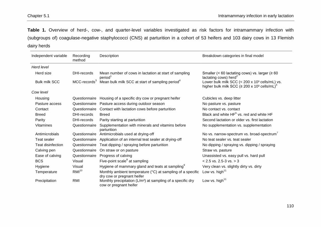
Chapter 5.1 Intramammary infection in early lactation
110
Table 1. Overview of herd-, cow-, and quarter-level variables investigated as risk factors for intramammary infection with
(subgroups of) coagulase-negative staphylococci (CNS) at parturition in a cohort of 53 heifers and 103 dairy cows in 13 Flemish
dairy herds
Independent variable Recording method
Description Breakdown categories in final model
Herd level
Herd size DHI-records Mean number of cows in lactation at start of sampling period
1
Smaller (< 60 lactating cows) vs. larger (≥ 60 lactating cows) herd
2
Bulk milk SCC MCC-records3 Mean bulk milk SCC at start of sampling period
4 Lower bulk milk SCC (< 200 x 10³ cells/mL) vs.
higher bulk milk SCC (≥ 200 x 10³ cells/mL)5
Cow level
Housing Questionnaire Housing of a specific dry cow or pregnant heifer Cubicles vs. deep litter
Pasture access Questionnaire Pasture access during outdoor season No pasture vs. pasture
Contact Questionnaire Contact with lactation cows before parturition No contact vs. contact
Breed DHI-records Breed Black and white HF6 vs. red and white HF
Parity DHI-records Parity starting at parturition Second lactation or older vs. first lactation
Vitamines Questionnaire Supplementation with minerals and vitamins before parturition
No supplementation vs. supplementation
Antimicrobials Questionnaire Antimicrobials used at drying-off No vs. narrow-spectrum vs. broad-spectrum7
Teat sealer Questionnaire Application of an internal teat sealer at drying-off No teat sealer vs. teat sealer
Teat disinfection Questionnaire Teat dipping / spraying before parturition No dipping / spraying vs. dipping / spraying
Calving pen Questionnaire On straw or on pasture Straw vs. pasture
Ease of calving Questionnaire Progress of calving Unassisted vs. easy pull vs. hard pull
BCS Visual Five-point scale8 at sampling < 2.5 vs. 2.5-3 vs. > 3
Hygiene Visual Hygiene of mammary gland and teats at sampling9 Very clean vs. slightly dirty vs. dirty
Temperature RMI10
Monthly ambient temperature (°C) at sampling of a specific dry cow or pregnant heifer
Low vs. high11
Precipitation RMI Monthly precipitation (L/m²) at sampling of a specific dry cow or pregnant heifer
Low vs. high11
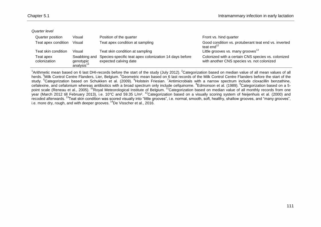
Chapter 5.1 Intramammary infection in early lactation
111
Quarter level
Quarter position Visual Position of the quarter Front vs. hind quarter
Teat apex condition Visual Teat apex condition at sampling Good condition vs. protuberant teat end vs. inverted teat end
12
Teat skin condition Visual Teat skin condition at sampling Little grooves vs. many grooves13
Teat apex colonization
Swabbing and genotypic analysis
14
Species-specific teat apex colonization 14 days before expected calving date
Colonized with a certain CNS species vs. colonized with another CNS species vs. not colonized
1Arithmetic mean based on 6 last DHI-records before the start of the study (July 2012).
2Categorization based on median value of all mean values of all
herds. 3
Milk Control Centre Flanders, Lier, Belgium. 4
Geometric mean based on 6 last records of the Milk Control Centre Flanders before the start of the study.
5Categorization based on Schukken et al. (2009).
6Holstein Friesian.
7Antimicrobials with a narrow spectrum include cloxacillin benzathine,
cefalexine, and cefalonium whereas antibiotics with a broad spectrum only include cefquinome. 8Edmonson et al. (1989).
9Categorization based on a 5-
point scale (Reneau et al., 2005). 10
Royal Meteorological Institute of Belgium. 11
Categorization based on median value of all monthly records from one year (March 2012 till February 2013), i.e. 10°C and 59.35 L/m².
12Categorization based on a visually scoring system of Neijenhuis et al. (2000) and
recoded afterwards. 13
Teat skin condition was scored visually into “little grooves”, i.e. normal, smooth, soft, healthy, shallow grooves, and “many grooves”, i.e. more dry, rough, and with deeper grooves.
14De Visscher et al., 2016.
Chapter 5.1 Intramammary infection in early lactation
112
Categorization of the herd size was based on the median value of all calculated
aforementioned arithmetic means: smaller herds, i.e. < 60 lactating animals, and
larger herds, i.e. ≥ 60 lactating animals. Bulk milk SCC was available through the
bulk milk quality data of the Milk Control Centre Flanders (MCC Flanders, Lier,
Belgium) and recoded into lower bulk milk SCC, i.e. < 200,000 cells/mL, and higher
bulk milk SCC, i.e. ≥ 200,000 cells/mL according to Schukken et al. (2009).
At each sampling of fresh cows and heifers (i.e. within 4 days after calving), other
potential cow- and quarter-level variables were recorded (Table 1). The Royal
Meteorological Institute of Belgium measures a monthly ambient temperature (°C)
and precipitation (L/m²). Classification of temperature and precipitation was based on
the median of all monthly values during the study period (from July 2012 till February
2013), i.e. 10°C and 59.35 L/m², respectively. Scoring of teat apex condition was
performed based on a visual scoring system (Neijenhuis et al., 2000) and recoded
afterwards into good condition, a protuberant teat end and an inverted teat end. Teat
skin condition was scored visually into “little grooves”, i.e. normal, smooth, soft,
healthy, shallow grooves, and “many grooves”, i.e. more dry, rough, and with deeper
grooves. Fourteen days before expected calving date, swabs of teat apices (n = 624)
have been collected. All swabs were plated on mannitol salt agar (MSA) as
described by De Visscher et al. (2016) and examined as described below in order to
identify the CNS species colonizing teat apices before parturition.
Laboratory analyses
All quarter milk samples (n = 624) were plated on MSA (Oxoid, Erembodegem,
Aalst, Belgium) (one quadrant per milk sample) and aerobically incubated at 37°C
(De Visscher et al., 2013) to recover CNS. Plates were examined after 24h and 48h.
All phenotypically different colony types were counted and 1 colony per colony type
was picked up and sub-cultured on esculin blood agar (Oxoid) (one quadrant per
colony) to obtain pure cultures. All potential CNS isolates were stored at -80°C for
subsequent analysis or immediately identified to the species level using transfer
RNA intergenic spacer PCR (tDNA-PCR) or sequencing of the 16S rRNA gene if no
identification was obtained (Supré et al., 2009).
All quarter milk samples were additionally plated on esculin blood agar (Oxoid)
and MacConkey agar (Oxoid) and examined according to the guidelines of the
National Mastitis Council (Hogan et al., 1999). This information was used only to
Chapter 5.1 Intramammary infection in early lactation
113
exclude quarters also infected with any major pathogen (Staphylococcus aureus,
Streptococcus agalactiae, Streptococcus dysgalactiae, Streptococcus uberis, other
esculin-positive streptococci, Escherichia coli, Klebsiella, Pseudomonas, yeast and
molds) from the subsequent statistical analyses.
Definition quarter IMI status
Quarters were considered to be infected with a specific CNS species when ≥ 1
colony forming unit per 0.01 mL milk of this species was observed on MSA,
according to the definition of Dohoo et al. (2011), in order to maximize the sensitivity.
Quarters infected with ≥ 3 genotypically different CNS species or with (a) major
pathogen(s), were excluded from the subsequent statistical analyses.
Descriptive and statistical analysis
Descriptive data analysis. The prevalence and herd-specific distribution of all CNS
species at parturition were described.
Risk factors. Before analyses were performed, observations were checked for
unlikely values. Complete data were available from 608 quarters from 99 fresh cows
and 53 fresh heifers. Dry cow treatment information was lacking for 3 few cows only
as these cows were purchased when dry. Some data were missing due to loss of
one questionnaire. However, data belonged to different quarters, cows, and herds.
The proportion of variation for the outcome variables at the herd, cow, and quarter
level was estimated for the null models. The variance at the quarter level was
assumed π²/3 (Goldstein et al., 2002) as described by Piepers et al. (2011).
Logistic multilevel regression models were fit (MLwiN 2.16, Centre for Multilevel
Modeling, University of Bristol, Bristol, UK) as follows:
Yijk ~ Binomial (πijk)
Logit (πijk) = β0ijk + β1 Xijk + v0k + uojk
Var (Yijk | πijk) = πijk (1 - πijk) with v0k ~ Normal (μk, σk) and uojk ~ Normal (μjk, σjk)
where Yijk is the infectious status of quarter i, from cow j, from herd k and πijk the
probability of this quarter (1) being infected at parturition with S. chromogenes, S.
simulans or S. xylosus, the so-called more relevant CNS species or uninfected or
infected with another CNS species, (2) being infected or not at parturition with a so-
called host-adapted species S. chromogenes, and (3) being infected or not at
parturition with a so-called environmental species, i.e. S. cohnii, S. equorum, S.
Chapter 5.1 Intramammary infection in early lactation
114
saprophyticus, or S. sciuri. Yijk is a function of the explanatory variable X through the
logit function, and approximately follows a binomial distribution; β0ijk is the intercept,
i.e. the baseline probability of infection when all predictors are equal to zero; β1 is the
regression coefficient for explanatory variable X; v0k and uojk are the herd and cow
random effects, respectively, and approximately follow normal distributions with
respective variances σk and σjk. Reweighted iterative generalized least squares and
1st order penalized quasi-likelihood estimation methods were applied.
Initially, univariable models examining associations between the outcome
variables and the independent variables (potential risk factors, see Table 1) were fit
and used as a first selection of variables. Statistical significance was assessed at P <
0.15. Afterwards, Spearman correlation coefficients among the statistically significant
variables were calculated to identify multi-collinearity in the multivariable models.
One out of 2 variables was selected, according to biological relevance, for further
analysis if a correlation coefficient ≥ |0.6| was calculated. Next, multivariable models
were fit for the significant variables from the univariable analysis using backward
stepwise elimination with statistical significance assessed at P < 0.05. Biological
relevant interaction terms were tested between all remaining statistically significant
risk factors and kept in the final multivariable model when significant (P < 0.05). Also,
confounding was investigated. A factor was considered a confounder if its removal
caused a relative change > 25% in the regression coefficients of the remaining
variables or with a regression coefficient between -0.4 and 0.4 if and absolute
change > 0.1 was observed (Noordhuizen et al., 2001).
In order to test the fitness of all final models, the observational-level standardized
residuals were plotted against the observational-level predicted values. Additionally,
Hosmer-Lemeshow goodness-of-fit tests were assessed on the fixed effect models
only (SAS 9.3, SAS Institute Inc., Cary, NC, USA) (Dohoo et al., 2009). The test was
statistically significant for none of the final models indicating good fit of our models.
Odds ratios (OR) and 95% confidence intervals were calculated to present the
magnitude of the associations.
Impact on quarter milk somatic cell count. A linear mixed regression model was fit
(MLwiN 2.16, Centre for Multilevel Modeling, University of Bristol, Bristol, UK), to
determine the association between subgroups of CNS species and the natural log
transformed qSCC, as follows:
Yijk = β0ijk + β1 X1ijk + β2 X2ijk + v0k + uojk + εijk
Chapter 5.1 Intramammary infection in early lactation
115
where Yijk is the natural log transformed qSCC of quarter i, from cow j, from herd k.
Yijk is a function of the explanatory variables X1 and X2 and approximately follows a
normal distribution; X1 is the fixed effect of the infectious status of the quarter (3
levels: uninfected, infected with the less relevant CNS, infected with the more
relevant CNS species, i.e. S. chromogenes, S. simulans, or S. xylosus); X2 is the
fixed effect to adjust for the day of sampling (3 levels: 1st day, 2nd day, 3rd day or
later); β0ijk is the intercept (overall mean); β1 and β2 are the regression coefficients for
explanatory variables X1 and X2, respectively; v0k and uojk are the herd and cow
random effects, respectively, and approximately follow normal distributions with
respective variances σk and σjk and εijk is the random error term, assumed to be
normally distributed with mean 0 and variance σ². Reweighted iterative generalized
least squares were applied. Quarters infected with a major pathogen were excluded
from this dataset.
Results
Distribution
Thirty-four percent (n = 211 out of 624 quarters) of all quarter milk samples
collected at parturition yielded growth on MSA. Per plate 0 to 5 phenotypically
different colony types were present. After tDNA-PCR and/or sequencing of the 16S
rRNA gene, a number of different colony types represented the same CNS species
resulting in 19 different species and 191 CNS isolates available for further analysis.
Twenty-six percent of all quarters (n = 163 out of 624 quarters) was infected at
parturition with 1 or 2 different CNS species. Staphylococcus chromogenes (13% of
all quarters and 41% of all isolates, n = 79) was the predominant species, followed
by S. sciuri (4% and 13%, respectively, n = 25), S. cohnii (3% and 11%, respectively,
n = 20), S. equorum (2% and 7%, respectively, n = 14), S. xylosus (2% and 7%,
respectively, n = 13), and S. haemolyticus (1% and 5%, respectively, n = 9).
Phenotypic or genotypic identification revealed 84 non-CNS-isolates on MSA
belonging to the phyla Firmicutes (Aerococcus spp., n = 3; Bacillus spp., n = 43;
Jeotgallicoccus sp., n = 1; S. aureus, n = 5; Streptococcus spp., n = 22) and
Proteobacteria (Pseudomonas, n = 8; Psychrobacter, n = 2).
In each herd, between 3 (herd 9) and 10 (herd 6) different CNS species were
isolated. The only CNS species causing IMI at parturition in all herds was S.
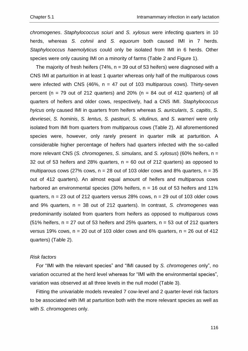
Chapter 5.1 Intramammary infection in early lactation
116
chromogenes. Staphylococcus sciuri and S. xylosus were infecting quarters in 10
herds, whereas S. cohnii and S. equorum both caused IMI in 7 herds.
Staphylococcus haemolyticus could only be isolated from IMI in 6 herds. Other
species were only causing IMI on a minority of farms (Table 2 and Figure 1).
The majority of fresh heifers (74%, n = 39 out of 53 heifers) were diagnosed with a
CNS IMI at parturition in at least 1 quarter whereas only half of the multiparous cows
were infected with CNS (46%, n = 47 out of 103 multiparous cows). Thirty-seven
percent (n = 79 out of 212 quarters) and 20% (n = 84 out of 412 quarters) of all
quarters of heifers and older cows, respectively, had a CNS IMI. Staphylococcus
hyicus only caused IMI in quarters from heifers whereas S. auricularis, S. capitis, S.
devriesei, S. hominis, S. lentus, S. pasteuri, S. vitulinus, and S. warneri were only
isolated from IMI from quarters from multiparous cows (Table 2). All aforementioned
species were, however, only rarely present in quarter milk at parturition. A
considerable higher percentage of heifers had quarters infected with the so-called
more relevant CNS (S. chromogenes, S. simulans, and S. xylosus) (60% heifers, n =
32 out of 53 heifers and 28% quarters, n = 60 out of 212 quarters) as opposed to
multiparous cows (27% cows, n = 28 out of 103 older cows and 8% quarters, n = 35
out of 412 quarters). An almost equal amount of heifers and multiparous cows
harbored an environmental species (30% heifers, n = 16 out of 53 heifers and 11%
quarters, n = 23 out of 212 quarters versus 28% cows, n = 29 out of 103 older cows
and 9% quarters, n = 38 out of 212 quarters). In contrast, S. chromogenes was
predominantly isolated from quarters from heifers as opposed to multiparous cows
(51% heifers, n = 27 out of 53 heifers and 25% quarters, n = 53 out of 212 quarters
versus 19% cows, n = 20 out of 103 older cows and 6% quarters, n = 26 out of 412
quarters) (Table 2).
Risk factors
For “IMI with the relevant species” and “IMI caused by S. chromogenes only”, no
variation occurred at the herd level whereas for “IMI with the environmental species”,
variation was observed at all three levels in the null model (Table 3).
Fitting the univariable models revealed 7 cow-level and 2 quarter-level risk factors
to be associated with IMI at parturition both with the more relevant species as well as
with S. chromogenes only.
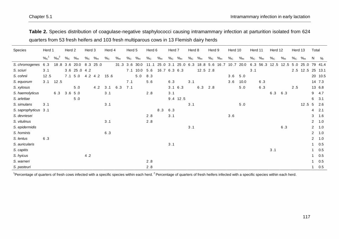
Chapter 5.1 Intramammary infection in early lactation
117
Table 2. Species distribution of coagulase-negative staphylococci causing intramammary infection at parturition isolated from 624
quarters from 53 fresh heifers and 103 fresh multiparous cows in 13 Flemish dairy herds
Species Herd 1 Herd 2 Herd 3 Herd 4 Herd 5 Herd 6 Herd 7 Herd 8 Herd 9 Herd 10 Herd 11 Herd 12 Herd 13 Total
%C
1 %H
2 %C %H %C %H %C %H %C %H %C %H %C %H %C %H %C %H %C %H %C %H %C %H %C %H N %
S. chromogenes 6 .3 18 .8 3 .6 20.0 8 .3 25 .0 31 .3 3 .6 30.0 11 .1 25 .0 3 .1 25 .0 6 .3 18 .8 5 .6 16 .7 10 .7 20.0 6 .3 56 .3 12 .5 12 .5 5 .0 25 .0 79 41.4
S. sciuri 3 .1 3 .6 25 .0 4 .2 7 .1 10.0 5 .6 16 .7 6 .3 6 .3 12 .5 2 .8 3 .1 2 .5 12 .5 25 13.1
S. cohnii 12 .5 7 .1 5 .0 4 .2 4 .2 15 .6 5 .0 8 .3 3 .6 5 .0 20 10.5
S. equorum 3 .1 12 .5 7 .1 5 .6 6 .3 3 .1 3 .6 10.0 6 .3 14 7.3
S. xylosus 5 .0 4 .2 3 .1 6 .3 7 .1 3 .1 6 .3 6 .3 2 .8 5 .0 6 .3 2 .5 13 6.8
S. haemolyticus 6 .3 3 .6 5 .0 3 .1 2 .8 3 .1 6 .3 6 .3 9 4.7
S. arlettae 5 .0 9 .4 12 .5 6 3.1
S. simulans 3 .1 3 .1 3 .1 5 .0 12 .5 5 2.6
S. saprophyticus 3 .1 8 .3 6 .3 4 2.1
S. devriesei 2 .8 3 .1 3 .6 3 1.6
S. vitulinus 3 .1 2 .8 2 1.0
S. epidermidis 3 .1 6 .3 2 1.0
S. hominis 6 .3 2 1.0
S. lentus 6 .3 2 1.0
S. auricularis 3 .1 1 0.5
S. capitis 3 .1 1 0.5
S. hyicus 4 .2 1 0.5
S. warneri 2 .8 1 0.5
S. pasteuri 2 .8 1 0.5
1Percentage of quarters of fresh cows infected with a specific species within each herd.
2 Percentage of quarters of fresh heifers infected with a specific species within each herd.
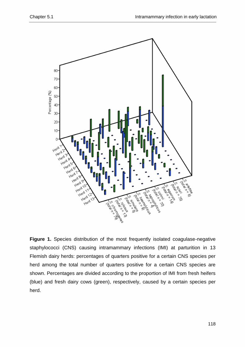
Chapter 5.1 Intramammary infection in early lactation
118
Figure 1. Species distribution of the most frequently isolated coagulase-negative
staphylococci (CNS) causing intramammary infections (IMI) at parturition in 13
Flemish dairy herds: percentages of quarters positive for a certain CNS species per
herd among the total number of quarters positive for a certain CNS species are
shown. Percentages are divided according to the proportion of IMI from fresh heifers
(blue) and fresh dairy cows (green), respectively, caused by a certain species per
herd.
Chapter 5.1 Intramammary infection in early lactation
119
Only 3 cow-level factors were unconditionally associated with IMI with the so-called
environmental species (Table 4). The risk factors “antimicrobials” (reflecting whether
or not narrow- or broad-spectrum antibiotics are used at drying-off) and “parity” were
correlated (Spearman rho = 0.85). The latter was expected as no antibiotics were
administered to end-term heifers which is in contrast with the multiparous cows, all
except one receiving antibiotics at drying-off. Only “parity” was used in the
subsequent models.
Table 3. Variance components at the herd, cow, and quarter level of the null models
for intramammary infections with (subgroups of) coagulase-negative Staphylococcus
(CNS) species1
Staphylococcus chromogenes, S. simulans, S. xylosus (n = 95)
1
Staphylococcus chromogenes (n = 79)
1
Staphylococcus cohnii, S. equorum, S. saprophyticus, S. sciuri (n = 61)
1
Var.est.2 ± SE
3 % Var.est. ± SE % Var.est. ± SE %
Herd 0.00 0.00 0.0 0.00 0.00 0.0 0.30 0.25 6.9
Cow 1.23 0.38 27.2 1.73 0.51 34.5 0.78 0.44 17.8
Quarter 3.29 - 72.8 3.29 - 65.5 3.29 - 75.3
Total Variance 4.52 100 5.02 100 4.37 100
1Staphylococcus chromogenes, S. simulans, and S. xylosus, represent the CNS species
being more relevant for udder health; S. chromogenes is representative for the host-adapted species; and S. cohnii, S. equorum, S. saprophyticus, and S. sciuri represent the environmental species.
2Variance components.
3Standard Error.
Table 5 presents results from the final multilevel, multivariable logistic regression
models. Quarters from heifers were more likely to be infected with the more relevant
species and with S. chromogenes, representing the host-adapted species, as
opposed to multiparous cows (OR = 3.9; 95% CI: 2.21-7.04 and OR = 4.2; 95% CI:
2.08-8.33, respectively). Quarters with an inverted teat end had increased odds of
being infected with the more relevant species and solely S. chromogenes as
opposed to quarters with a good teat end condition (OR = 2.8; 95% CI: 1.36-5.91
and OR = 3.7; 95% CI: 1.52-8.79, respectively). Quarters with teat apices colonized
with S. chromogenes prior to calving had higher odds of being infected at parturition
with this same species (OR = 3.3; 95% CI: 1.41-7.50). Quarters with dirty teat apices
were more likely to be infected with an environmental CNS species (OR = 6.4; 95%
CI: 1.31-30.90) as opposed to quarters with clean teats. In contrast, IMI with S.
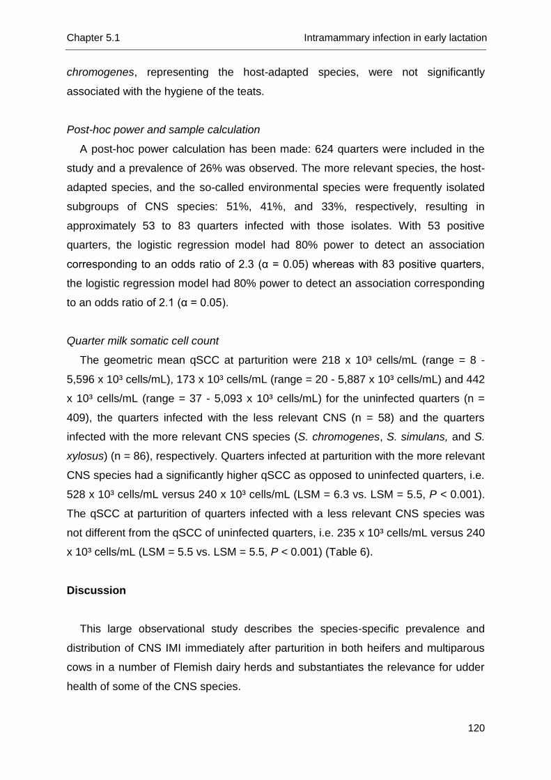
Chapter 5.1 Intramammary infection in early lactation
120
chromogenes, representing the host-adapted species, were not significantly
associated with the hygiene of the teats.
Post-hoc power and sample calculation
A post-hoc power calculation has been made: 624 quarters were included in the
study and a prevalence of 26% was observed. The more relevant species, the host-
adapted species, and the so-called environmental species were frequently isolated
subgroups of CNS species: 51%, 41%, and 33%, respectively, resulting in
approximately 53 to 83 quarters infected with those isolates. With 53 positive
quarters, the logistic regression model had 80% power to detect an association
corresponding to an odds ratio of 2.3 (α = 0.05) whereas with 83 positive quarters,
the logistic regression model had 80% power to detect an association corresponding
to an odds ratio of 2.1 (α = 0.05).
Quarter milk somatic cell count
The geometric mean qSCC at parturition were 218 x 10³ cells/mL (range = 8 -
5,596 x 10³ cells/mL), 173 x 10³ cells/mL (range = 20 - 5,887 x 10³ cells/mL) and 442
x 10³ cells/mL (range = 37 - 5,093 x 10³ cells/mL) for the uninfected quarters (n =
409), the quarters infected with the less relevant CNS (n = 58) and the quarters
infected with the more relevant CNS species (S. chromogenes, S. simulans, and S.
xylosus) (n = 86), respectively. Quarters infected at parturition with the more relevant
CNS species had a significantly higher qSCC as opposed to uninfected quarters, i.e.
528 x 10³ cells/mL versus 240 x 10³ cells/mL (LSM = 6.3 vs. LSM = 5.5, P < 0.001).
The qSCC at parturition of quarters infected with a less relevant CNS species was
not different from the qSCC of uninfected quarters, i.e. 235 x 10³ cells/mL versus 240
x 10³ cells/mL (LSM = 5.5 vs. LSM = 5.5, P < 0.001) (Table 6).
Discussion
This large observational study describes the species-specific prevalence and
distribution of CNS IMI immediately after parturition in both heifers and multiparous
cows in a number of Flemish dairy herds and substantiates the relevance for udder
health of some of the CNS species.
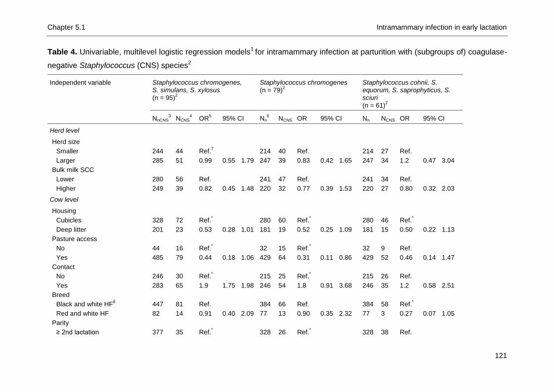
Chapter 5.1 Intramammary infection in early lactation
121
Table 4. Univariable, multilevel logistic regression models1 for intramammary infection at parturition with (subgroups of) coagulase-
negative Staphylococcus (CNS) species2
Independent variable Staphylococcus chromogenes, S. simulans, S. xylosus (n = 95)
2
Staphylococcus chromogenes (n = 79)
2
Staphylococcus cohnii, S. equorum, S. saprophyticus, S. sciuri (n = 61)
2
NnCNS
3 NCNS
4 OR
5 95% CI Nn
6 NCNS OR 95% CI Nn NCNS OR 95% CI
Herd level
Herd size
Smaller 244 44 Ref.7
214 40 Ref. 214 27 Ref.
Larger 285 51 0.99 0.55 1.79 247 39 0.83 0.42 1.65 247 34 1.2 0.47 3.04
Bulk milk SCC
Lower 280 56 Ref.
241 47 Ref. 241 34 Ref.
Higher 249 39 0.82 0.45 1.48 220 32 0.77 0.39 1.53 220 27 0.80 0.32 2.03
Cow level
Housing
Cubicles 328 72 Ref.*
280 60 Ref.
* 280 46 Ref.
*
Deep litter 201 23 0.53 0.28 1.01 181 19 0.52 0.25 1.09 181 15 0.50 0.22 1.13
Pasture access
No 44 16 Ref.*
32 15 Ref.
* 32 9 Ref.
Yes 485 79 0.44 0.18 1.06 429 64 0.31 0.11 0.86 429 52 0.46 0.14 1.47
Contact
No 246 30 Ref.*
215 25 Ref.
* 215 26 Ref.
Yes 283 65 1.9 1.75 1.98 246 54 1.8 0.91 3.68 246 35 1.2 0.58 2.51
Breed
Black and white HF8 447 81 Ref.
384 66 Ref. 384 58 Ref.
*
Red and white HF 82 14 0.91 0.40 2.09 77 13 0.90 0.35 2.32 77 3 0.27 0.07 1.05
Parity
≥ 2nd lactation 377 35 Ref.*
328 26 Ref.
* 328 38 Ref.
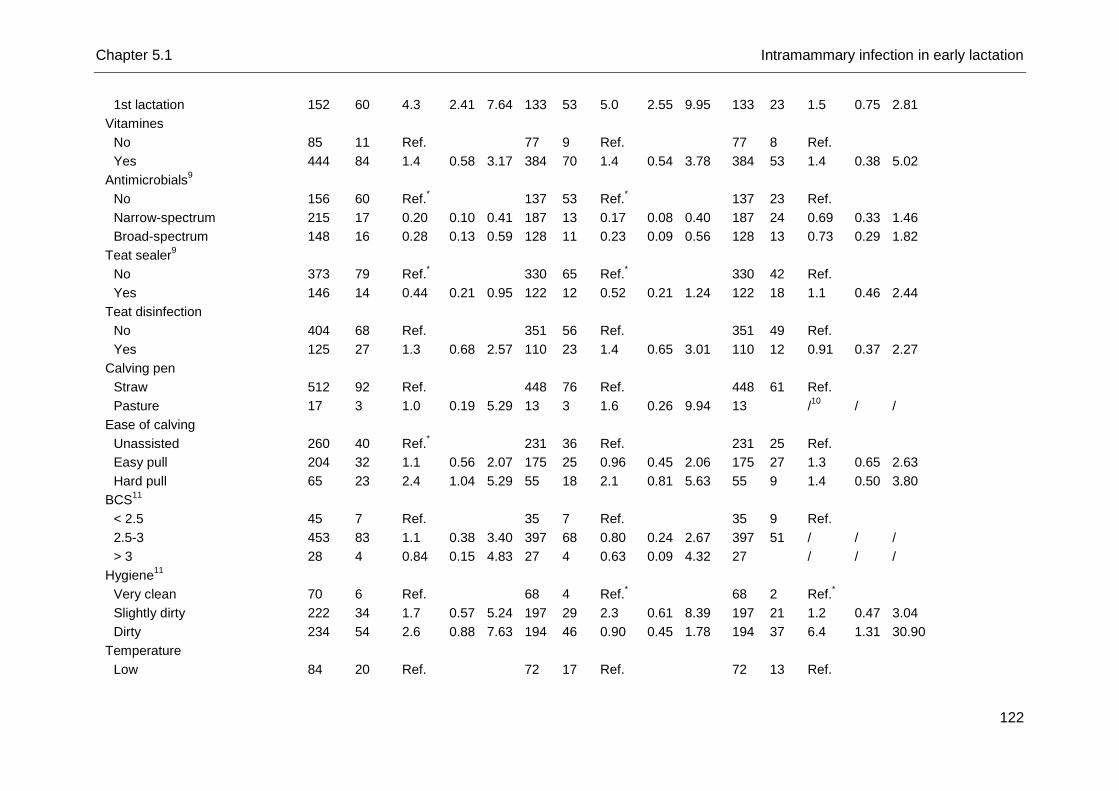
Chapter 5.1 Intramammary infection in early lactation
122
1st lactation 152 60 4.3 2.41 7.64 133 53 5.0 2.55 9.95 133 23 1.5 0.75 2.81
Vitamines
No 85 11 Ref.
77 9 Ref. 77 8 Ref.
Yes 444 84 1.4 0.58 3.17 384 70 1.4 0.54 3.78 384 53 1.4 0.38 5.02
Antimicrobials9
No 156 60 Ref.*
137 53 Ref.
* 137 23 Ref.
Narrow-spectrum 215 17 0.20 0.10 0.41 187 13 0.17 0.08 0.40 187 24 0.69 0.33 1.46
Broad-spectrum 148 16 0.28 0.13 0.59 128 11 0.23 0.09 0.56 128 13 0.73 0.29 1.82
Teat sealer9
No 373 79 Ref.*
330 65 Ref.
* 330 42 Ref.
Yes 146 14 0.44 0.21 0.95 122 12 0.52 0.21 1.24 122 18 1.1 0.46 2.44
Teat disinfection
No 404 68 Ref.
351 56 Ref. 351 49 Ref.
Yes 125 27 1.3 0.68 2.57 110 23 1.4 0.65 3.01 110 12 0.91 0.37 2.27
Calving pen
Straw 512 92 Ref.
448 76 Ref. 448 61 Ref.
Pasture 17 3 1.0 0.19 5.29 13 3 1.6 0.26 9.94 13
/10
/ /
Ease of calving
Unassisted 260 40 Ref.*
231 36 Ref. 231 25 Ref.
Easy pull 204 32 1.1 0.56 2.07 175 25 0.96 0.45 2.06 175 27 1.3 0.65 2.63
Hard pull 65 23 2.4 1.04 5.29 55 18 2.1 0.81 5.63 55 9 1.4 0.50 3.80
BCS11
< 2.5 45 7 Ref.
35 7 Ref. 35 9 Ref.
2.5-3 453 83 1.1 0.38 3.40 397 68 0.80 0.24 2.67 397 51 / / /
> 3 28 4 0.84 0.15 4.83 27 4 0.63 0.09 4.32 27
/ / /
Hygiene11
Very clean 70 6 Ref.
68 4 Ref.* 68 2 Ref.
*
Slightly dirty 222 34 1.7 0.57 5.24 197 29 2.3 0.61 8.39 197 21 1.2 0.47 3.04
Dirty 234 54 2.6 0.88 7.63 194 46 0.90 0.45 1.78 194 37 6.4 1.31 30.90
Temperature
Low 84 20 Ref.
72 17 Ref. 72 13 Ref.
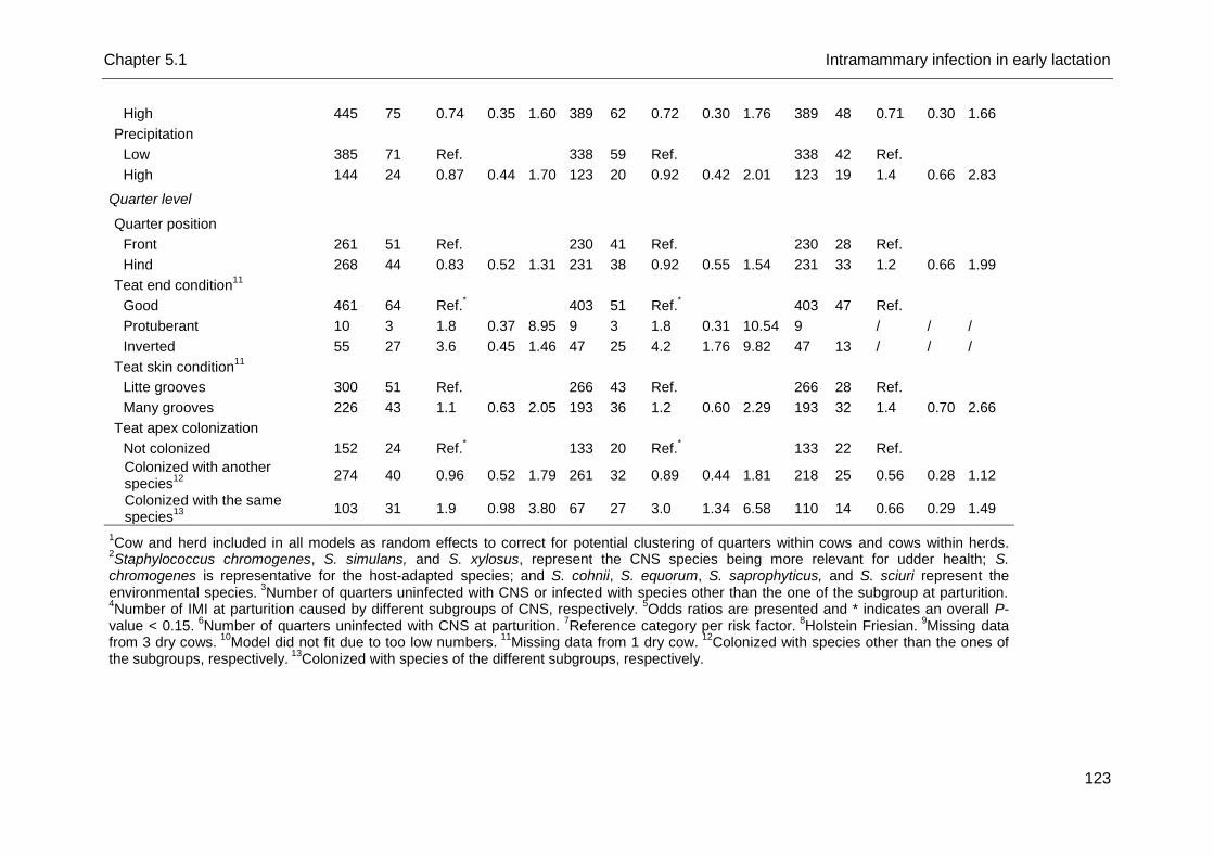
Chapter 5.1 Intramammary infection in early lactation
123
High 445 75 0.74 0.35 1.60 389 62 0.72 0.30 1.76 389 48 0.71 0.30 1.66
Precipitation
Low 385 71 Ref.
338 59 Ref. 338 42 Ref.
High 144 24 0.87 0.44 1.70 123 20 0.92 0.42 2.01 123 19 1.4 0.66 2.83
Quarter level
Quarter position
Front 261 51 Ref.
230 41 Ref. 230 28 Ref.
Hind 268 44 0.83 0.52 1.31 231 38 0.92 0.55 1.54 231 33 1.2 0.66 1.99
Teat end condition11
Good 461 64 Ref.*
403 51 Ref.
* 403 47 Ref.
Protuberant 10 3 1.8 0.37 8.95 9 3 1.8 0.31 10.54 9
/ / /
Inverted 55 27 3.6 0.45 1.46 47 25 4.2 1.76 9.82 47 13 / / /
Teat skin condition11
Litte grooves 300 51 Ref.
266 43 Ref. 266 28 Ref.
Many grooves 226 43 1.1 0.63 2.05 193 36 1.2 0.60 2.29 193 32 1.4 0.70 2.66
Teat apex colonization
Not colonized 152 24 Ref.*
133 20 Ref.
* 133 22 Ref.
Colonized with another species
12
274 40 0.96 0.52 1.79 261 32 0.89 0.44 1.81 218 25 0.56 0.28 1.12
Colonized with the same species
13
103 31 1.9 0.98 3.80 67 27 3.0 1.34 6.58 110 14 0.66 0.29 1.49
1Cow and herd included in all models as random effects to correct for potential clustering of quarters within cows and cows within herds.
2Staphylococcus chromogenes, S. simulans, and S. xylosus, represent the CNS species being more relevant for udder health; S.
chromogenes is representative for the host-adapted species; and S. cohnii, S. equorum, S. saprophyticus, and S. sciuri represent the environmental species.
3Number of quarters uninfected with CNS or infected with species other than the one of the subgroup at parturition.
4Number of IMI at parturition caused by different subgroups of CNS, respectively.
5Odds ratios are presented and * indicates an overall P-
value < 0.15. 6
Number of quarters uninfected with CNS at parturition. 7
Reference category per risk factor. 8
Holstein Friesian. 9
Missing data from 3 dry cows.
10Model did not fit due to too low numbers.
11Missing data from 1 dry cow.
12Colonized with species other than the ones of
the subgroups, respectively. 13
Colonized with species of the different subgroups, respectively.
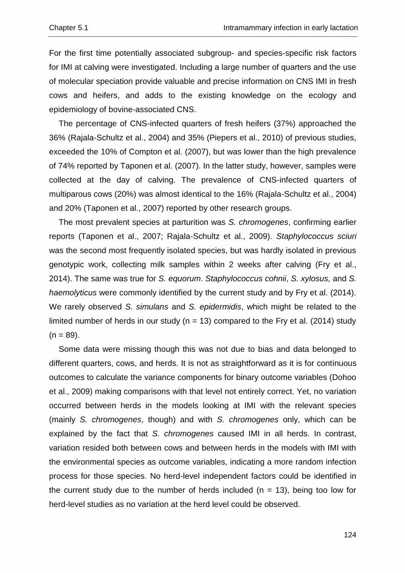
Chapter 5.1 Intramammary infection in early lactation
124
For the first time potentially associated subgroup- and species-specific risk factors
for IMI at calving were investigated. Including a large number of quarters and the use
of molecular speciation provide valuable and precise information on CNS IMI in fresh
cows and heifers, and adds to the existing knowledge on the ecology and
epidemiology of bovine-associated CNS.
The percentage of CNS-infected quarters of fresh heifers (37%) approached the
36% (Rajala-Schultz et al., 2004) and 35% (Piepers et al., 2010) of previous studies,
exceeded the 10% of Compton et al. (2007), but was lower than the high prevalence
of 74% reported by Taponen et al. (2007). In the latter study, however, samples were
collected at the day of calving. The prevalence of CNS-infected quarters of
multiparous cows (20%) was almost identical to the 16% (Rajala-Schultz et al., 2004)
and 20% (Taponen et al., 2007) reported by other research groups.
The most prevalent species at parturition was S. chromogenes, confirming earlier
reports (Taponen et al., 2007; Rajala-Schultz et al., 2009). Staphylococcus sciuri
was the second most frequently isolated species, but was hardly isolated in previous
genotypic work, collecting milk samples within 2 weeks after calving (Fry et al.,
2014). The same was true for S. equorum. Staphylococcus cohnii, S. xylosus, and S.
haemolyticus were commonly identified by the current study and by Fry et al. (2014).
We rarely observed S. simulans and S. epidermidis, which might be related to the
limited number of herds in our study (n = 13) compared to the Fry et al. (2014) study
(n = 89).
Some data were missing though this was not due to bias and data belonged to
different quarters, cows, and herds. It is not as straightforward as it is for continuous
outcomes to calculate the variance components for binary outcome variables (Dohoo
et al., 2009) making comparisons with that level not entirely correct. Yet, no variation
occurred between herds in the models looking at IMI with the relevant species
(mainly S. chromogenes, though) and with S. chromogenes only, which can be
explained by the fact that S. chromogenes caused IMI in all herds. In contrast,
variation resided both between cows and between herds in the models with IMI with
the environmental species as outcome variables, indicating a more random infection
process for those species. No herd-level independent factors could be identified in
the current study due to the number of herds included (n = 13), being too low for
herd-level studies as no variation at the herd level could be observed.
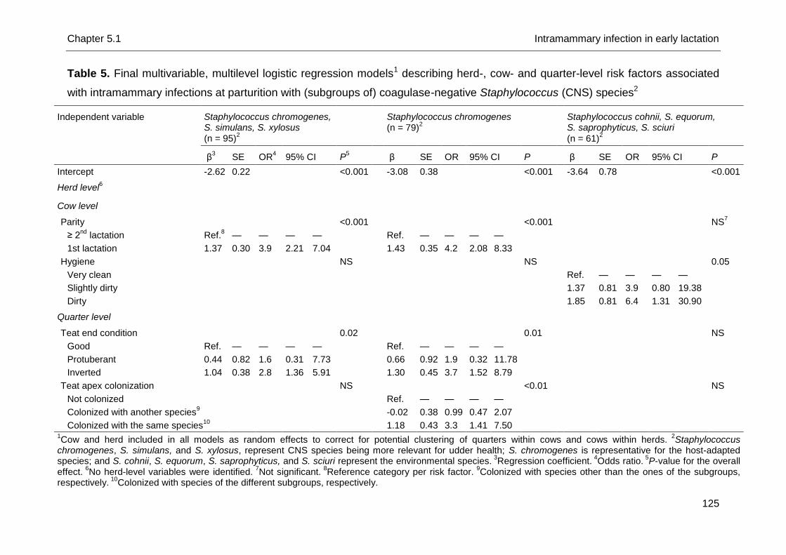
Chapter 5.1 Intramammary infection in early lactation
125
Table 5. Final multivariable, multilevel logistic regression models1 describing herd-, cow- and quarter-level risk factors associated
with intramammary infections at parturition with (subgroups of) coagulase-negative Staphylococcus (CNS) species2
Independent variable Staphylococcus chromogenes, S. simulans, S. xylosus (n = 95)
2
Staphylococcus chromogenes (n = 79)
2
Staphylococcus cohnii, S. equorum, S. saprophyticus, S. sciuri
(n = 61)2
β
3 SE OR
4 95% CI P
5
β SE OR 95% CI P
β SE OR 95% CI P
Intercept -2.62 0.22 <0.001
-3.08 0.38 <0.001
-3.64 0.78 <0.001
Herd level6
Cow level
Parity
<0.001
<0.001
NS7
≥ 2nd
lactation Ref.8 — — — —
Ref. — — — —
1st lactation 1.37 0.30 3.9 2.21 7.04
1.43 0.35 4.2 2.08 8.33
Hygiene
NS
NS
0.05
Very clean
Ref. — — — —
Slightly dirty
1.37 0.81 3.9 0.80 19.38
Dirty
1.85 0.81 6.4 1.31 30.90
Quarter level
Teat end condition
0.02
0.01
NS
Good Ref. — — — —
Ref. — — — —
Protuberant 0.44 0.82 1.6 0.31 7.73
0.66 0.92 1.9 0.32 11.78
Inverted 1.04 0.38 2.8 1.36 5.91
1.30 0.45 3.7 1.52 8.79
Teat apex colonization
NS
<0.01
NS
Not colonized
Ref. — — — —
Colonized with another species9
-0.02 0.38 0.99 0.47 2.07
Colonized with the same species
10
1.18 0.43 3.3 1.41 7.50
1Cow and herd included in all models as random effects to correct for potential clustering of quarters within cows and cows within herds.
2Staphylococcus
chromogenes, S. simulans, and S. xylosus, represent CNS species being more relevant for udder health; S. chromogenes is representative for the host-adapted species; and S. cohnii, S. equorum, S. saprophyticus, and S. sciuri represent the environmental species.
3Regression coefficient.
4Odds ratio.
5P-value for the overall
effect. 6
No herd-level variables were identified. 7
Not significant. 8
Reference category per risk factor. 9
Colonized with species other than the ones of the subgroups, respectively.
10Colonized with species of the different subgroups, respectively.
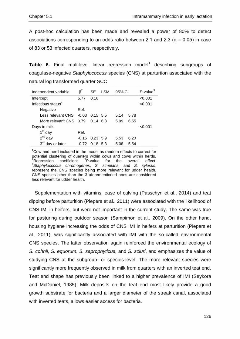
Chapter 5.1 Intramammary infection in early lactation
126
A post-hoc calculation has been made and revealed a power of 80% to detect
associations corresponding to an odds ratio between 2.1 and 2.3 (α = 0.05) in case
of 83 or 53 infected quarters, respectively.
Table 6. Final multilevel linear regression model1 describing subgroups of
coagulase-negative Staphylococcus species (CNS) at parturtion associated with the
natural log transformed quarter SCC
Supplementation with vitamins, ease of calving (Passchyn et al., 2014) and teat
dipping before parturition (Piepers et al., 2011) were associated with the likelihood of
CNS IMI in heifers, but were not important in the current study. The same was true
for pasturing during outdoor season (Sampimon et al., 2009). On the other hand,
housing hygiene increasing the odds of CNS IMI in heifers at parturition (Piepers et
al., 2011), was significantly associated with IMI with the so-called environmental
CNS species. The latter observation again reinforced the environmental ecology of
S. cohnii, S. equorum, S. saprophyticus, and S. sciuri, and emphasizes the value of
studying CNS at the subgroup- or species-level. The more relevant species were
significantly more frequently observed in milk from quarters with an inverted teat end.
Teat end shape has previously been linked to a higher prevalence of IMI (Seykora
and McDaniel, 1985). Milk deposits on the teat end most likely provide a good
growth substrate for bacteria and a larger diameter of the streak canal, associated
with inverted teats, allows easier access for bacteria.
Independent variable β2 SE LSM 95% CI P-value
3
Intercept 5.77 0.16 <0.001
Infectious status4 <0.001
Negative Ref.
Less relevant CNS -0.03 0.15 5.5 5.14 5.78
More relevant CNS 0.79 0.14 6.3 5.99 6.55
Days in milk <0.001
1st day Ref.
2nd
day -0.15 0.23 5.9 5.53 6.23
3rd
day or later -0.72 0.18 5.3 5.08 5.54
1Cow and herd included in the model as random effects to correct for
potential clustering of quarters within cows and cows within herds.
2Regression coefficient.
3P-value for the overall effect.
4Staphylococcus chromogenes, S. simulans, and S. xylosus,
represent the CNS species being more relevant for udder health. CNS species other than the 3 aforementioned ones are considered less relevant for udder health.
Chapter 5.1 Intramammary infection in early lactation
127
Teat apex colonization with S. chromogenes significantly increased the odds of S.
chromogenes IMI at parturition, a phenomenon that was not observed for the other
species (data not shown). The latter illustrates the host-adapted nature of S.
chromogenes as teat apices might act as a habitat for host-adapted species. This
has been suggested before for S. aureus IMI at parturition (Roberson et al., 1994)
and for S. chromogenes IMI throughout lactation (Taponen et al., 2008). An earlier
study from our group focusing on S. chromogenes, not using molecular speciation,
did not demonstrate this link (De Vliegher et al., 2003). To come to better
conclusions, strain-typing of all isolates should be performed, and is currently
ongoing.
It has previously been shown that CNS-infected quarters have a higher SCC as
opposed to negative control quarters (Taponen et al., 2007; Gillespie et al., 2009;
Schukken et al., 2009). However, recent research reported an impact on the qSCC
depending on the CNS species involved (Sampimon et al., 2009; Thorberg et al.,
2009; Simojoki et al., 2011). Our findings confirm this variation between species.
Actually, the more relevant species, S. chromogenes, S. simulans, and S. xylosus
induced a higher qSCC at parturition, as was reported before (Supré et al., 2011; Fry
et al., 2014; De Visscher et al., 2015). We also confirmed that quarters infected with
species other than the so-called relevant ones have a qSCC that is not different from
the qSCC of uninfected quarters (Supré et al., 2011; Fry et al., 2014). A limitation of
CNS research is the fact that the sensitivity can only reach a maximum of 86.7%
(Dohoo et al., 2011), indicating some quarters classified as negative ones can be
CNS-infected. The latter can explain the rather high qSCC of the negative quarters
observed in this study. However, in order to avoid an impact of a major pathogen on
the qSCC, all quarters infected with a major pathogen were removed from the
dataset.
Conclusions
Staphylococcus chromogenes, S. sciuri, and S. cohnii were the predominant species
causing IMI in fresh heifers and dairy cows. The only CNS species isolated from milk
in all herds was S. chromogenes; the presences of other species differed by herd.
The environmental nature was supported for S. cohnii, S. equorum, S.
saprophyticus, and S. sciuri whereas the host-adapted nature of S. chromogenes
Chapter 5.1 Intramammary infection in early lactation
128
was substantiated. Pre-partum teat apex colonization with S. chromogenes
increased the likelihood of S. chromogenes IMI in the corresponding quarters at
parturition. The more relevant species increased the quarter SCC at parturition as
opposed to uninfected quarters.
Chapter 5.1 Intramammary infection in early lactation
129
References
Compton, C. W. R., C. Heuer, K. Parker, and S. McDougall. 2007. Epidemiology of
mastitis in pasture-grazed peripartum dairy heifers and its effects on productivity.
J. Dairy Sci. 90:4157-4170.
De Visscher, A., F. Haesebrouck, S. Piepers, W. Vanderhaeghen, K. Supré, F.
Leroy, E. Van Coillie, and S. De Vliegher. 2013. Assessment of the suitability of
mannitol salt agar for growing bovine-associated coagulase-negative
staphylococci and its use under field conditions. Res. in Vet. Sci. 95:347-351.
De Visscher, A., S. Piepers, K. Supré, F. Haesebrouck, and S. De Vliegher. 2015.
Short communication. Species group-specific predictors at the cow and quarter
level for intramammary infection with coagulase-negative staphylococci in dairy
cattle throughout lactation. J. Dairy Sci. 98:5448-5453.
De Visscher, A., S. Piepers, F. Haesebrouck, and S. De Vliegher. 2016. Teat apex
colonization with coagulase-negative Staphylococcus species before parturition:
distribution and species-specific risk factors. J. Dairy Sci. 99:1427-1439.
De Vliegher, S., H. Laevens, L. A. Devriese, G. Opsomer, J. L. M. Leroy, H. W.
Barkema, and A. de Kruif. 2003. Prepartum teat apex colonization with
Staphylococcus chromogenes in dairy heifers is associated with low somatic cell
count in early lactation. Vet. Microbiol. 92:245-252.
De Vliegher, S., L. K. Fox, S. Piepers, S. McDougall, and H. W. Barkema. 2012.
Invited review: Mastitis in dairy heifers: Nature of the disease, potential impact,
prevention, and control. J. Dairy Sci. 95:1025-1040.
Dohoo, I., W. Martin, and H. Stryhn. 2009. Mixed models for discrete data. In: Doho,
I., W. Martin, and H. Stryhn, Veterinary Epidemiologic Research. 2nd edition, AVC
inc., Charlottetown, Canada, p. 579-606.
Dohoo, I. R., J. Smith, S. Andersen, D. F. Kelton, S. Godden and Mastitis Research
Workers’ Conference. 2011. Diagnosing intramammary infections: Evaluation of
definitions based on a single milk sample. J. Dairy Sci. 94:250-261.
Edmonson, A. J., I. J. Lean, L. D. Weaver, T. Farver, and G. Webster. 1989. A body
condition scoring chart for Holstein dairy cows. J. Dairy Sci. 72:68-78.
Fry, P. R., J. R. Middleton, S. Dufour, J. Perry, D. Scholl, and I. Dohoo. 2014.
Association of coagulase-negative staphylococcal species, mammary quarter milk
Chapter 5.1 Intramammary infection in early lactation
130
somatic cell count, and persistence of intramammary infection in dairy cattle. J.
Dairy Sci. 97:4876-4885.
Gillespie, B. E., S. I. Headrick, S. Boonyayatra, and S. P. Oliver. 2009. Prevalence
and persistence of coagulase-negative Staphylococcus species in three dairy
research herds. Vet. Microbiol. 134:65-72.
Goldstein, H., R. W. Browne, and J. Rasbash. 2002. Partitioning variation in
multilevel models. Understanding Stat. I 4:223-231.
Hogan, J. S., R. N. Gonzáles, R. J. Harmon, S. C. Nickerson, S. P. Oliver, J. W.
Pankey, and K. L. Smith. 1999. Laboratory Handbook on Bovine Mastitis. Rev.
Ed. National Mastitis Council, Madison, WI.
Neijenhuis, F., H. W. Barkema, H. Hogeveen, and J. P. T. M. Noordhuizen. 2000.
Classification and longitudinal examination of callused teat ends in dairy cows. J.
Dairy Sci. 83:2795-2804.
Noordhuizen, J. P. T. M., K. Frankena, M. V. Thursfield, and E. A. M. Graat. 2001.
Application of quantitative methods in veterinary epidemiology. Wageningen Pers,
Wageningen, The Netherlands.
Passchyn, P., S. Piepers, and S. De Vliegher. 2014. Pathogen group-specific risk
factors for intramammary infection in treated and untreated dairy heifers
participating in a prepartum antimicrobial treatment trial. J. Dairy Sci. 97:6260-
6270.
Piepers, S., G. Opsomer, H. W. Barkema, A. de Kruif, and S. De Vliegher. 2010.
Heifers infected with coagulase-negative staphylococci in early lactation have
fewer cases of clinical mastitis and a higher milk production in their first lactation
than non-infected heifers. J. Dairy Sci. 93:2014-2024.
Piepers, S., K. Peeters, G. Opsomer, H. W. Barkema, K. Frankena, and S. De
Vliegher. 2011. Pathogen group specific risk factors at herd, heifer and quarter
levels for intramammary infections in early lactating dairy heifers. Prev. Vet. Med.
99:91-101.
Piessens, V., E. Van Coillie, B. Verbist, K. Supré, G. Braem, A. Van Nuffel, L. De
Vuyst M. Heyndrickx, and S. De Vliegher. 2011. Distribution of coagulase-
negative Staphylococcus species from dairy cows’ milk and environment differs
between herds. J. Dairy Sci. 94:2933-2944.
Piessens, V., S. De Vliegher, B. Verbist, G. Braem, A. Van Nuffel, L. De Vuyst, M.
Heyndrickx, and E. Van Coillie. 2012. Characterization of coagulase-negative
Chapter 5.1 Intramammary infection in early lactation
131
Staphylococcus species from cows’ milk and environment based on bap, icaA,
and mecA genes and phenotypic susceptibility to antimicrobials and teat dips. J.
Dairy Sci. 95:7027-7038.
Rajala-Schultz P. J., K. L. Smith, J. S. Hogan, and B. C. Love. 2004. Antimicrobial
susceptibility of mastitis pathogens from first lactation and older cows. Vet.
Microbiol. 102:33-42.
Rajala-Schultz P. J., A. H. Torres, F. J. DeGraves, W. A. Gebreyes, and P.
Patchanee. 2009. Antimicrobial resistance and genotypic characterization of
coagulase-negative staphylococci over the dry period. Vet. Microbiol. 134:55-64.
Reneau, J. K., A. J. Seykora, B. J. Heins, M. I. Endres, R. J. Farnsworth, and R. F.
Bey. 2005. Associations between hygiene scores and somatic cell scores in dairy
cattle. J. Am. Vet. Med. Assoc. 227:1297-1301.
Roberson, J. R., L. K. Fox, D. D. Hancock, and J. M. Gay. 1994. Ecology of
Staphylococcus aureus isolated from various sites on dairy farms. J. Dairy Sci.
77:3354-3364.
Sampimon, O. C., H. W. Barkema, I. M. G. A. Berends, J. Sol, and T. J. G. M. Lam.
2009. Prevalence and herd-level risk factors for intramammary infection with
coagulase-negative staphylococci in Dutch dairy herds. Vet. Microbiol. 134:37-44.
Schukken, Y. H., R. N. González, L. L. Tikofsky, H. F. Schulte, C. G. Santisteban, F.
L. Welcome, G. J. Bennett, M. J. Zurakowski, and R. N. Zadoks. 2009. CNS
mastitis: Nothing to worry about? Vet. Microbiol. 134:9-14.
Seykora, A. J., and B. T. McDaniel. 1985. Udder and teat morphology related to
mastitis resistance. A review. J. Dairy Sci. 68:2087-2093.
Simojoki, H., T. Salomäki, S. Taponen, A. Iivanainen, and S. Pyörälä. 2011. Innate
immune response in experimentally induced bovine intramammary infection with
Staphylococcus simulans and S. epidermidis. Vet. Res. 42:49.
Supré, K., S. De Vliegher, O. C. Sampimon, R. N. Zadoks, M. Vaneechoutte, M.
Baele, E. De Graef, S. Piepers, and F. Haesebrouck. 2009. Technical note: Use of
transfer RNA-intergenic spacer PCR combined with capillary electrophoresis to
identify coagulase-negative Staphylococcus species originating from bovine milk
and teat apices. J. Dairy Sci. 92:3204-3210.
Supré, K., F. Haesebrouck, R. N. Zadoks, M. Vaneechoutte, S. Piepers, and S. De
Vliegher. 2011. Some coagulase-negative Staphylococcus species affect udder
health more than others. J. Dairy Sci. 94:2329-2340.
Chapter 5.1 Intramammary infection in early lactation
132
Taponen, S., J. Koort, J. Björkroth, H. Saloniemi, and S. Pyörälä. 2007. Bovine
intramammary infections caused by coagulase-negative staphylococci may persist
throughout lactation according to amplified fragment length polymorphism-based
analysis. J. Dairy Sci. 90:3301-3307.
Taponen, S., J. Björkroth, and S. Pyörälä. 2008. Coagulase-negative staphylococci
isolated from bovine extramammary sites and intramamamry infections in a single
dairy herd. J. Dairy Res. 75:422-429.
Thorberg, B.-M., M.-L. Danielsson-Tham, U. Emanuelson, and K. Persson Waller.
2009. Bovine subclinical mastitis caused by different types of coagulase-negative
staphylococci. J. Dairy Sci. 92:4962-4970.
Vanderhaeghen, W., S. Piepers, F. Leroy, E. Van Coillie, F. Haesebrouck, and S. De
Vliegher. 2014. Invited review. Effect, persistence, and virulence of coagulase-
negative Staphylococcus species associated with ruminant udder health. J. Dairy
Sci. 97:5275-5293.
Vanderhaeghen, W., S. Piepers, F. Leroy, E. Van Coillie, F. Haesebrouck, and S. De
Vliegher. 2015. Identification, typing, ecology and epidemiology of coagulase-
negative staphylococci associated with ruminants. Vet. J. 203:44-51.
Verbeke, J., S. Piepers, L. Peelman, M. Van Poucke, and S. De Vliegher. 2012.
Pathogen-group specific association between CXCR1 polymorphisms and
subclinical mastitis in dairy heifers. J. Dairy Res. 79:341-351.


Chapter 5.2
Species Group-Specific Predictors at the Cow
and Quarter Level for Intramammary Infection
with Coagulase-Negative Staphylococci in
Dairy Cattle Throughout Lactation
A. De Visscher1, S. Piepers1, K. Supré2, F. Haesebrouck3, and S. De
Vliegher1
1Department of Reproduction, Obstetrics, and Herd Health, Faculty of Veterinary
Medicine, Ghent University, Merelbeke, Belgium
2Milk Control Centre Flanders, Lier, Belgium
3Department of Pathology, Bacteriology, and Avian Diseases, Faculty of Veterinary
Medicine, Ghent University, Merelbeke, Belgium
Journal of Dairy Science, 2015, 98:5448-5453

Chapter 5.2 Intramammary infection throughout lactation
137
Abstract
Coagulase-negative staphylococci (CNS) are frequently isolated from quarters
with subclinical mastitis, teat apices and the cows’ environment. Virulence, ecology,
epidemiological behavior, and effect on udder health vary between the different
species. Staphylococcus chromogenes, S. simulans and S. xylosus are frequently
present in milk and have a more substantial effect on quarter milk somatic cell count
than other species. Therefore, these species are considered as the “more relevant”
CNS. As species-specific factors associated with CNS intramammary infection (IMI)
have not yet been identified and susceptibility for IMI differs between cows and
quarters, this study aimed to identify predictors for CNS IMI at the cow and quarter
level (some of them changing over time) with a specific focus on the aforementioned
more relevant CNS. Precise data were available from a longitudinal study (3,052
observations from 344 quarters from 86 dairy cows belonging to three commercial
dairy herds). All CNS were molecularly identified to the species level, and
multivariable, multilevel logistic regression models taking into account the
longitudinal nature of the data, were fit to study the likelihood of infection.
Staphylococcus chromogenes, S. xylosus and S. cohnii were the most frequently
isolated species from CNS IMI in older cows whereas S. chromogenes, S. xylosus
and S. simulans were the main species found in IMI in heifers. Quarters from heifers
(as opposed to multiparous cows), from heifers and multiparous cows in third or
fourth month in lactation (as opposed to early lactation, < 60 days in milk), and with
an increasing quarter milk SCC were more likely to be infected with the more
relevant CNS species. Quarter milk SCC was identified as the sole statistically
significant predictor for IMI with other CNS species, although the size of the effect
was lower [odds ratio of 1.6 (1.4 - 1.9) as compared with 2.1 (1.8 - 2.5)] than the
effect for IMI with the more relevant CNS. As a strong herd effect was present,
studying herd-level predictors is warranted.
Key words
Predictors, Coagulase-negative Staphylococcus species, Intramammary infection
Chapter 5.2 Intramammary infection throughout lactation
138
Introduction
Coagulase-negative staphylococci (CNS) are the most frequently isolated bacteria
from subclinical mastitis cases in dairy cows (Makovec and Ruegg, 2003; Piepers et
al., 2007; Sampimon et al., 2009a). In addition, they are abundantly present in the
cows’ environment (Piessens et al., 2011) and on their teat apices (Taponen et al.,
2008; Braem et al., 2012). Several factors associated with CNS-group IMI have been
identified (Sampimon et al., 2009a; Piepers et al., 2011), but in none of the studies
were CNS accurately identified to the species level and one study only included
heifers. As species-specific differences in protective effects, (presumed) virulence,
antimicrobial resistance, persistence, and effect on udder health have been revealed
(Sampimon et al., 2011; Vanderhaeghen et al., 2014, 2015), studying CNS as a
group is no longer recommended. Rather, accurate species-level studies or studies
at the subgroup level, when analyses at the species level are hindered by small
numbers, are needed.
Intramammary infection with Staphylococcus chromogenes, S. simulans and S.
xylosus is associated with a more pronounced increase in SCC compared with other
species (Supré et al., 2011; Fry et al., 2014), suggesting these species have a more
substantial effect on udder health and are for that reason considered as more
relevant than others. Differences in susceptibility for CNS IMI between cows and
quarters exist as some cows in a specific herd have no CNS infected quarters
whereas others have multiple quarters infected with CNS (Piessens et al., 2011;
Supré et al., 2011), indicating the likelihood of infections is explained not only by
CNS traits but also by host factors.
The aim of this study was to identify cow- and quarter-level predictors for IMI
throughout lactation caused by the more relevant CNS species for udder health, S.
chromogenes, S. simulans and S. xylosus, as well as other species.
Material and methods
Data were obtained from a longitudinal study conducted between September 2007
and January 2009 in 3 commercial dairy herds in Flanders (Belgium). On all farms,
cows were housed in freestall barns with cubicles with sawdust bedding and a
concrete slatted floor. Postmilking teat disinfection and blanket dry cow therapy
Chapter 5.2 Intramammary infection throughout lactation
139
combined with application of an internal teat sealant were applied. Detailed herd
information can be found elsewhere (Supré et al., 2011; De Visscher et al., 2014).
Single quarter milk samples were aseptically collected every month for determining
the quarter milk SCC (qSCC) and for bacteriological culturing. Several potential cow-
and quarter-level predictors were recorded at each sampling occasion, some of them
changing over time (observation level, lowest level of the dataset; Table 1).
Table 1. Overview of all potential cow-, quarter- and observation-level predictor
variables (independent variables)
Independent variable
Recording method Description Breakdown of categories in final model
Cow level
Breed DHI-records Two breeds Black and white HF1 vs.
red and white HF
Quarter level
Quarter position Visual Position of the quarter
Front vs. hind quarter
Observation level
Parity2 DHI-records Parity at test-day 1
st lactation vs. 2
nd
lactation or older Milk yield
3 DHI-records Milk yield at test-
day Low
4 (< 33.35 kg) vs. high
(≥ 33.35 kg) Stage of lactation
3 DHI-records Stage in lactation at
test-day 1 - 60 DIM vs. 61 - 120 DIM vs. 121 - 180 DIM vs. > 180 DIM
BSC3 Visual and palpation Five-point scale
5 < 2.5 vs. 2.5 - 3.5 vs. >
3.5 Quarter milk SCC
6 Fossomatic 5000/FC
7 Quarter SCC at
test-day Continuous
Teat skin condition6 Visual and palpation Nine-point scale
8 Without cracks vs. with
cracks Teat apex condition
6 Visual and palpation Nine-point scale
8 Without cracks vs. with
cracks
1Holstein Friesian.
2Cow-level variable potentially changing during the study.
3Cow-level variable
changing during the study. 4
Categorization based on median value of all test-day records of all cows. 5Edmonson et al. (1989).
6Quarter-level variable changing during the study.
7Foss, Hillerørd,
Denkmark. 8De Vliegher et al. (2003).
Animals with a test-day milk yield ≥ 33.35 kg/day (i.e. the median value of all test-
day records of all animals) were defined as high producing, whereas animals with a
test-day milk yield < 33.35 kg/day were defined as low producing. Stage in lactation
was categorized as described by Zadoks et al. (2001): 1 = 1 - 60 DIM; 2 = 61 - 120
DIM; 3 = 121 - 180 DIM; 4 ≥ 180 DIM. Body condition score of each animal was
scored on a 5-point scale divided into quarter-point increments as described by
Chapter 5.2 Intramammary infection throughout lactation
140
Edmonson et al. (1989). Recoding was applied for further analyses (1 = ≤ 2.5; 2 =
2.5 - 3.5; 3 = ≥ 3.5). The teat skin and teat apex condition were determined based on
a 9-point scale visual scoring system (De Vliegher et al., 2003) and recoded (0 =
without cracks; 1 = with cracks). Quarter milk SCC was determined at the Flanders
Milk Control Centre (Lier, Belgium) using a Fossomatic 5000/FC (Foss Electric,
Hillerød, Denmark). Bacteriological culturing of all milk samples was performed
following National Mastitis Council guidelines (Hogan et al., 1999). All CNS were
identified to the species level using transfer RNA intergenic spacer PCR (tDNA-PCR)
(Supré et al., 2009) and IMI status were assigned based on subsequent samplings
as described by Supré et al. (2011). Staphylococcus chromogenes, S. simulans, and
S. xylosus were considered as CNS species with a more substantial effect on udder
health (Supré et al., 2011; Fry et al., 2014) and referred to as “more relevant”
species throughout the paper; all other CNS species originating from milk in this
study are referred to as “less relevant” species. Before statistical analysis was
conducted, observations were revised and checked for unlikely values. Complete
data were available from 344 quarters from 86 cows, making 3,052 observations in
total. Logistic mixed regression models were fit in SAS 9.3 (SAS Institute Inc., Carry,
NC, USA). Clustering of observations occurred at multiple levels. Repeated
observations were clustered in time within quarters, whereas quarters within cows
and cows within herds were spatially clustered. For those reasons, analyses were
run with herd and sampling forced into all models as fixed effects and quarter as
random effect (REPEATED statement). Cow as random effect was omitted because
of non-convergence of the models when combined with a quarter random effect. The
contribution of each of the random effects was estimated (VARCOMP procedure)
and revealed that quarter accounted for more variability than cow (Zadoks et al.,
2001). Compound symmetry was used as the covariance structure for within-quarter
correlation (Barkema et al., 1997; Zadoks et al., 2001). Two different binary outcome
variables at the quarter level were used: (1) IMI with the more relevant CNS, and (2)
IMI with the less relevant CNS, both versus being non-infected. Initially, univariable
associations were tested between the 2 binary outcome variables and all predictor
variables at the cow, quarter and observation level. Statistical significance in this
step was assessed at P < 0.15. Second, Spearman correlation coefficients were
calculated among all statistically significant predictor variables to check for
multicollinearity. If 2 predictor variables had a correlation coefficient ≥ |0.60|, only
Chapter 5.2 Intramammary infection throughout lactation
141
one was selected for further analyses. In the third step, multivariable models were fit
for the 2 outcome variables using backward stepwise elimination. Statistical
significance in this step was assessed at P < 0.05. Interaction terms were tested
between all remaining statistically significant predictors. The goodness-of-fit of the
final models was evaluated by plotting the observational-level standardized residuals
against the observational-level predicted values.
Results
On average, 53 lactating cows were present in the herds (range = 39 - 70) with an
average of 29 animals per herd being included in the study, resulting in a total of
3,052 analyzed milk samples. One hundred twenty-eight IMI with the more relevant
CNS and 48 IMI with the less relevant CNS were available for analysis. Overall, S.
chromogenes caused most IMI (n = 83, 47% of all IMI) followed by S. xylosus (n =
28, 16%), S. cohnii (n = 19, 11%), S. simulans (n = 17, 10%), S. haemolyticus (n =
11, 6%), and S. fleurettii (n = 6, 3%). Sixty-nine percent (n = 121) of all CNS IMI (n =
176) originated from quarters of cows in second or higher lactation. However, only
0.07% of all quarters of older cows were infected with CNS, whereas 16.4% of all
quarters of heifers were infected. The majority of CNS IMI from quarters of heifers
(93%, n = 50) and multiparous cows (64%, n = 77) were caused by the more
relevant species. Staphylococcus simulans was mainly isolated from IMI in quarters
of heifers (82%, n = 14). Coagulase-negative staphylococci IMI in quarters of
multiparous cows were most frequently caused by S. chromogenes (43%, n = 52), S.
xylosus (18%, n = 22), and S. cohnii (15%, n = 18). Staphylococcus chromogenes
(56%, n = 31), S. simulans (26%, n = 14), and S. xylosus (11%, n = 6) were the
majority of CNS IMI in quarters of heifers. Overall, CNS species were equally
collected in any stage of lactation. Staphylococcus chromogenes (71%, n = 12), S.
haemolyticus (12%, n = 2), S. xylosus (12%, n = 2), and S. sciuri (6%, n = 1) were
the only species causing IMI in the first two months after parturition (1 to 60 DIM).
The more relevant CNS species were mostly isolated from IMI in quarters of heifers
in a later stage of lactation (> 180 DIM), whereas IMI in quarters of older cows
caused by those relevant species were equally present in any stage in lactation.
Most of the less relevant CNS (77%, n = 36) were isolated from IMI in quarters with a
qSCC < 200,000 cells/mL, whereas for the more relevant CNS this was only 45%.
Chapter 5.2 Intramammary infection throughout lactation
142
A first reduction based on the univariable models revealed 5 and 4 predictor
variables associated with IMI (P < 0.15) with the more relevant CNS and the less
relevant CNS, respectively (Table 2). As no correlation coefficient ≥ |0.6| between
the statistically significant predictors was calculated, all independent variables were
added in the multivariable models.
The final multivariable, multilevel models are presented in Table 3. Quarters from
heifers (as opposed to multiparous cows), from heifers and multiparous cows in third
or fourth month in lactation (as opposed to early lactation, < 60 DIM), and with an
increasing qSCC were more likely to be infected with the more relevant CNS.
Quarter SCC was identified as the sole statistically significant predictor for IMI with
other CNS species, although the size of the effect was lower [odds ratio of 1.6 (1.4 -
1.9) as compared with 2.1 (1.8 - 2.5)] than for IMI with the relevant species. None of
the tested interaction terms was significant. Statistically significant herd effects (herd
forced in all models to correct for clustering of cows within herds) for both IMI with
the more and less relevant CNS species, respectively, were present.
Discussion
This is to our opinion, the first study identifying CNS group-specific predictor
variables in a longitudinal setting using precise molecular techniques for CNS
speciation. Before the current study, past investigations examined factors associated
with CNS IMI, but researchers either did not differentiate CNS species or used
inaccurate phenotypic identification methods (Sampimon et al., 2009b). Several
studies demonstrated a high prevalence of CNS around calving. Still, to our
knowledge, in none of the other studies was the prevalence of CNS IMI in early
lactation compared with the prevalence of CNS IMI later in lactation.
We confirmed that heifers are more likely to be infected with (relevant) CNS in
comparison with multiparous cows (Sampimon et al., 2009a). Quarters from heifers
and cows in peak lactation (61 - 120 DIM) were more likely to be infected with the
more relevant species despite applying post-milking teat disinfection and using dry
cow antibiotics in all 3 herds. This might support the assumption of CNS being able
to develop resistance to antibiotics and teat disinfectants (Sampimon et al., 2011;
Piessens et al., 2012) .
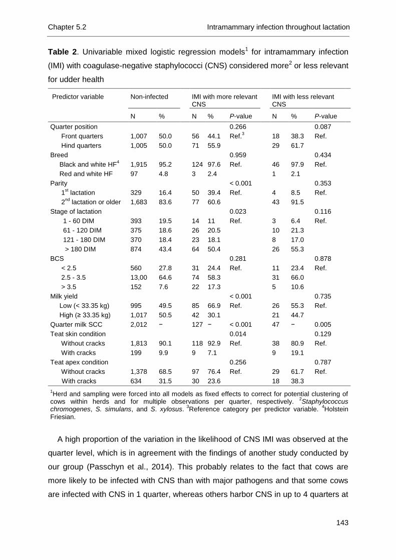
Chapter 5.2 Intramammary infection throughout lactation
143
Table 2. Univariable mixed logistic regression models1 for intramammary infection
(IMI) with coagulase-negative staphylococci (CNS) considered more2 or less relevant
for udder health
Predictor variable Non-infected IMI with more relevant CNS
IMI with less relevant CNS
N % N % P-value N % P-value
Quarter position
0.266
0.087
Front quarters 1,007 50.0
56 44.1 Ref.3
18 38.3 Ref.
Hind quarters 1,005 50.0
71 55.9
29 61.7
Breed
0.959
0.434
Black and white HF4 1,915 95.2
124 97.6 Ref.
46 97.9 Ref.
Red and white HF 97 4.8
3 2.4
1 2.1
Parity
< 0.001
0.353
1st lactation 329 16.4
50 39.4 Ref.
4 8.5 Ref.
2nd
lactation or older 1,683 83.6
77 60.6
43 91.5
Stage of lactation
0.023
0.116
1 - 60 DIM 393 19.5
14 11 Ref.
3 6.4 Ref.
61 - 120 DIM 375 18.6
26 20.5
10 21.3
121 - 180 DIM 370 18.4
23 18.1
8 17.0
> 180 DIM 874 43.4
64 50.4
26 55.3
BCS
0.281
0.878
< 2.5 560 27.8
31 24.4 Ref.
11 23.4 Ref.
2.5 - 3.5 13,00 64.6
74 58.3
31 66.0
> 3.5 152 7.6
22 17.3
5 10.6
Milk yield
< 0.001
0.735
Low (< 33.35 kg) 995 49.5
85 66.9 Ref.
26 55.3 Ref.
High (≥ 33.35 kg) 1,017 50.5
42 30.1
21 44.7
Quarter milk SCC 2,012 −
127 − < 0.001
47 − 0.005
Teat skin condition
0.014
0.129
Without cracks 1,813 90.1
118 92.9 Ref.
38 80.9 Ref.
With cracks 199 9.9
9 7.1
9 19.1
Teat apex condition
0.256
0.787
Without cracks 1,378 68.5
97 76.4 Ref.
29 61.7 Ref.
With cracks 634 31.5 30 23.6 18 38.3
1Herd and sampling were forced into all models as fixed effects to correct for potential clustering of
cows within herds and for multiple observations per quarter, respectively. 2Staphylococcus
chromogenes, S. simulans, and S. xylosus. 3
Reference category per predictor variable. 4Holstein
Friesian.
A high proportion of the variation in the likelihood of CNS IMI was observed at the
quarter level, which is in agreement with the findings of another study conducted by
our group (Passchyn et al., 2014). This probably relates to the fact that cows are
more likely to be infected with CNS than with major pathogens and that some cows
are infected with CNS in 1 quarter, whereas others harbor CNS in up to 4 quarters at
Chapter 5.2 Intramammary infection throughout lactation
144
the same time (Makovec and Ruegg, 2003; Piepers et al., 2007; Sampimon et al.,
2009a). In contrast, other studies (also) dealing with major pathogens, such as
Staphylococcus aureus and Streptococcus uberis, observed that a higher proportion
of the variation in the outcome resided at the cow level (Zadoks et al., 2001;
Passchyn et al., 2014).
More statistically significant predictors for IMI caused by the more relevant CNS
were identified than for IMI with the less relevant CNS. The greater number of
isolates of relevant CNS species in the dataset likely explains this, although we
hypothesize it also reflects differences in virulence and host adaptation.
On farms with excellent udder health, reflected by a bulk milk SCC < 200,000
cells/mL, CNS infections may be an important contributor to the total number of
somatic cells in the bulk milk (Schukken et al., 2009). In addition, a high proportion of
cows infected with the more relevant CNS might result in an unwarranted increase in
the bulk milk SCC in such a herd, also because IMI with the more relevant CNS last
longer, on average, than IMI with the less relevant CNS (Supré et al., 2011). We
hypothesize that our data are useful for selecting quarters and cows infected with the
more relevant CNS in herds in which major pathogens are not an issue. The results
suggest that the focus should be on quarters with an elevated qSCC from heifers
and on heifers and multiparous cows in peak lactation (61 - 120 DIM). In addition,
molecular techniques able to identify CNS at the species level or at least able to
distinguish the more relevant from the less relevant CNS would be very valuable
when making individual cow-level decisions in those herds. As statistically significant
differences were observed between herds in likelihood of CNS infection, species-
specific analysis at the herd level is required. This implies setting up studies that
include large numbers of herds based on quarter milk samples from an even larger
number of cows. Using bulk milk samples to identify herd-level predictor variables for
IMI with the more relevant CNS could be helpful to circumvent this inconvenience
and should be investigated
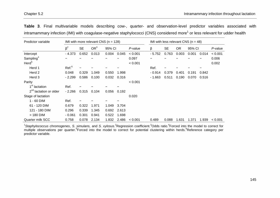
Chapter 5.2 Intramammary infection throughout lactation
145
Table 3. Final multivariable models describing cow-, quarter- and observation-level predictor variables associated with
intramammary infection (IMI) with coagulase-negative staphylococci (CNS) considered more1 or less relevant for udder health
Predictor variable IMI with more relevant CNS (n = 128) IMI with less relevant CNS (n = 48)
β2 SE OR
3 95% CI P-value β SE OR 95% CI P-value
Intercept - 4.373 0.652 0.013 0.004 0.045 < 0.001 - 5.752 0.763 0.003 0.001 0.014 < 0.001
Sampling4 − − − − − 0.097 − − − − − 0.006
Herd5
< 0.001 0.002
Herd 1 Ref.6 − − − −
Ref. − − − −
Herd 2 0.048 0.329 1.049 0.550 1.998 - 0.914 0.379 0.401 0.191 0.842
Herd 3 - 2.299 0.586 0.100 0.032 0.316 - 1.663 0.511 0.190 0.070 0.516
Parity
< 0.001
1st lactation Ref. − − − −
2nd
lactation or older - 2.266 0.315 0.104 0.056 0.192
Stage of lactation 0.020
1 - 60 DIM Ref. − − − −
61 - 120 DIM 0.679 0.322 1.971 1.049 3.704
121 - 180 DIM 0.296 0.339 1.345 0.692 2.613
> 180 DIM - 0.061 0.301 0.941 0.522 1.698
Quarter milk SCC 0.758 0.078 2.134 1.832 2.486 < 0.001 0.489 0.088 1.631 1.371 1.939 < 0.001
1Staphylococcus chromogenes, S. simulans, and S. xylosus.
2Regression coefficient.
3Odds ratio.
4Forced into the model to correct for
multiple observations per quarter.5Forced into the model to correct for potential clustering within herds.
6Reference category per
predictor variable.
Chapter 5.2 Intramammary infection throughout lactation
146
References
Barkema, H. W., Y. H. Schukken, T. J. G. M. Lam, D. T. Galligan, M. L. Beiboer, and
A. Brand. 1997. Estimation of interdependence among quarters of the bovine
udder with subclinical mastitis and implications for analysis. J. Dairy Sci. 80:1592-
1599.
Braem, G., S. De Vliegher, B. Verbist, M. Heyndrickx, F. Leroy, and L. De Vuyst.
2012. Culture-independent exploration of the teat apex microbiota of dairy cows
reveals a wide bacterial species diversity. Vet. Microbiol. 157:383-390.
De Visscher, A., K. Supré, F. Haesebrouck, R. N. Zadoks, V. Piessens, E. Van
Coillie, S. Piepers, and S. De Vliegher. 2014. Further evidence for the existence of
environmental and host-associated species of coagulase-negative staphylococci
in dairy cattle. Vet. Microbiol. 172:466:474.
De Vliegher, S., L. Laevens, H. W. Barkema, G. Opsomer, T. Hemling, and A. de
Kruif. 2003. Short-term effect of transition from conventional to automated milking
on teat skin and teat end condition. J. Dairy Sci. 86:1646-1652.
Edmonson, A. J., I. J. Lean, L. D. Weaver, T. Farver, and G. Webster. 1989. A body
condition scoring chart for Holstein dairy cows. J. Dairy Sci. 72:68-78.
Fry, P. R., J. R. Middleton, S. Dufour, J. Perry, D. Scholl, and I. Dohoo. 2014.
Association of coagulase-negative staphylococcal species, mammary quarter milk
somatic cell count, and persistence of intramammary infection in dairy cattle. J.
Dairy Sci. 97:1-10.
Hogan, J. S., R. N. González, R. J. Harmon, S. C. Nickerson, S. P. Oliver, J. W.
Pankey, and K. L. Smith. 1999. Laboratory Handbook on Bovine Mastitis. Rev.
Ed. National Mastitis Council, Madison, WI.
Makovec, J. A., and P. L. Ruegg. 2003. Results of milk samples submitted for
microbiological examination in Wisconsin from 1994 to 2001. J. Dairy Sci.
86:3466-3472.
Passchyn, P., S. Piepers, and S. De Vliegher. 2014. Pathogen group-specific risk
factors for intramammary infection in treated and untreated dairy heifers
participating in a prepartum antimicrobial treatment trial. J. Dairy Sci. 97:6926-
6934.
Chapter 5.2 Intramammary infection throughout lactation
147
Piepers, S., L. De Meulemeester, A. de Kruif, G. Opsomer, H. W. Barkema, and S.
De Vliegher. 2007. Prevalence and distribution of mastitis pathogens in
subclinically infected dairy cows in Flanders, Belgium. J. Dairy Res. 74:478-483.
Piepers, S., K. Peeters, G. Opsomer, H. W. Barkema, K. Frankena, and S. De
Vliegher. 2011. Pathogen group specific risk factors at herd, heifer and quarter
levels for intramammary infections in early lactating dairy heifers. Prev. Vet. Med.
99:91-101.
Piessens, V., E. Van Coillie, B. Verbist, K. Supré, G. Braem, A. Van Nuffel, L. De
Vuyst, M. Heyndrickx, and S. De Vliegher. 2011. Distribution of coagulase-
negative Staphylococcus species from milk and environment of dairy cows differs
between herd. J. Dairy Sci. 94:2933-2944.
Piessens, V., S. De Vliegher, B. Verbist, G. Braem, A. Van Nuffel, L. De Vuyst, M.
Heyndrickx, and E. Van Coillie. 2012. Characterization of coagulase-negative
Staphylococcus species from cows’ milk and environment based on bap, icaA,
and mecA genes and phenotypic susceptibility to antimicrobials and teat dips. J.
Dairy Sci. 95:7027-7038.
Sampimon, O. C., H. W. Barkema, I. M. G. A. Berends, J. Sol, and T. J. G. M. Lam.
2009a. Prevalence and herd-level risk factors for intramammary infection with
coagulase-negative staphylococci in Dutch dairy herds. Vet. Microbiol. 134:37-44.
Sampimon, O. C., R. N. Zadoks, S. De Vliegher, K. Supré, F. Haesebrouck, H. W.
Barkema, J. Sol, and T. J. G. M. Lam. 2009b. Performance of API Staph ID 32
and Staph-Zym for identification of coagulase-negative staphylococci isolated
from bovine milk samples. Vet. Microbiol. 136:300-305.
Sampimon, O. C., T. J. G. M. Lam, D. J. Mevius, Y. H. Schuken, and R. N Zadoks.
2011. Antimicrobial susceptibility of coagulase-negative staphylococci isolated
from bovine milk samples. Vet. Microbiol. 150:173-179.
Schukken, Y. H., R. N. González, L. L. Tikofsky, H. F. Schulte, C. G. Santisteban, F.
L. Welcome, G. J. Bennet, M. J. Zurakowski, and R. N. Zadoks. 2009. CNS
mastitis. Nothing to worry about? Vet. Microbiol. 134:9-14.
Supré, K., S. De Vliegher, O. C. Sampimon, R. N. Zadoks, M. Vaneechoutte, M.
Baele, E. De Graef, S. Piepers, and F. Haesebrouck. 2009. Technical note: Use of
transfer RNA-intergenic spacer PCR combined with capillary electrophoresis to
identify coagulase-negative Staphylococcus species originating from bovine milk
and teat apices. J. Dairy Sci. 92:3204-3210.
Chapter 5.2 Intramammary infection throughout lactation
148
Supré, K., F. Haesebrouck, R. N. Zadoks, M. Vaneechoutte, S. Piepers, and S. De
Vliegher. 2011. Some coagulase-negative Staphylococcus species affect udder
health more than others. J. Dairy Sci. 94:2329-2340.
Taponen, S., J. Björkroth, and S. Pyörälä. 2008. Coagulase-negative staphylococci
isolated from bovine extramammary sites and intramammary infections in a single
dairy herd. J. Dairy Res. 75:422-429.
Vanderhaeghen, W., S. Piepers, F. Leroy, E. Van Coillie, F. Haesebrouck, and S. De
Vliegher. 2014. Invited review: Effect, persistence, and virulence of coagulase-
negative Staphylococcus species associated with ruminant udder health. J. Dairy
Sci. 97:5275-5293.
Vanderhaeghen, W., S. Piepers, F. Leroy, E. Van Coillie, F. Haesebrouck, and S. De
Vliegher. 2015. Identification, typing, ecology and epidemiology of coagulase-
negative staphylococci associated with ruminants. Vet. J. 203:44-51.
Zadoks, R. N., H. G. Allore, H. W. Barkema, O. C. Sampimon, G. J. Wellenberg, Y.
T. Gröhn, and Y. H. Schukken. 2001. Cow- and quarter-level risk factors for
Streptococcus uberis and Staphylococcus aureus mastitis. J. Dairy Sci. 84:2649-
2663.


Chapter 6
Coagulase-Negative Staphylococcus Species
in Bulk Milk: Prevalence, Distribution, and
Associated Subgroup- and Species-Specific
Risk Factors
A. De Visscher1, S. Piepers1, F. Haesebrouck2, K. Supré3, and S. De
Vliegher1
1Department of Reproduction, Obstetrics, and Herd Health, Faculty of Veterinary
Medicine, Ghent University, Merelbeke, Belgium
2Department of Pathology, Bacteriology, and Avian Diseases, Faculty of Veterinary
Medicine, Ghent University, Merelbeke, Belgium
3Milk Control Centre Flanders (MCC), Lier, Belgium
In Preparation

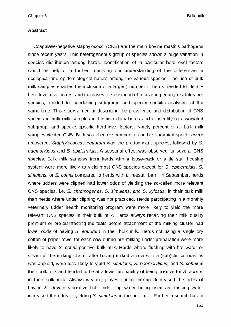
Chapter 6 Bulk milk
153
Abstract
Coagulase-negative staphylococci (CNS) are the main bovine mastitis pathogens
since recent years. This heterogeneous group of species shows a huge variation in
species distribution among herds. Identification of in particular herd-level factors
would be helpful in further improving our understanding of the differences in
ecological and epidemiological nature among the various species. The use of bulk
milk samples enables the inclusion of a large(r) number of herds needed to identify
herd-level risk factors, and increases the likelihood of recovering enough isolates per
species, needed for conducting subgroup- and species-specific analyses, at the
same time. This study aimed at describing the prevalence and distribution of CNS
species in bulk milk samples in Flemish dairy herds and at identifying associated
subgroup- and species-specific herd-level factors. Ninety percent of all bulk milk
samples yielded CNS. Both so-called environmental and host-adapted species were
recovered. Staphylococcus equorum was the predominant species, followed by S.
haemolyticus and S. epidermidis. A seasonal effect was observed for several CNS
species. Bulk milk samples from herds with a loose-pack or a tie stall housing
system were more likely to yield most CNS species except for S. epidermidis, S.
simulans, or S. cohnii compared to herds with a freestall barn. In September, herds
where udders were clipped had lower odds of yielding the so-called more relevant
CNS species, i.e. S. chromogenes, S. simulans, and S. xylosus, in their bulk milk
than herds where udder clipping was not practiced. Herds participating in a monthly
veterinary udder health monitoring program were more likely to yield the more
relevant CNS species in their bulk milk. Herds always receiving their milk quality
premium or pre-disinfecting the teats before attachment of the milking cluster had
lower odds of having S. equorum in their bulk milk. Herds not using a single dry
cotton or paper towel for each cow during pre-milking udder preparation were more
likely to have S. cohnii-positive bulk milk. Herds where flushing with hot water or
steam of the milking cluster after having milked a cow with a (sub)clinical mastitis
was applied, were less likely to yield S. simulans, S. haemolyticus, and S. cohnii in
their bulk milk and tended to be at a lower probability of being positive for S. aureus
in their bulk milk. Always wearing gloves during milking decreased the odds of
having S. devriesei-positive bulk milk. Tap water being used as drinking water
increased the odds of yielding S. simulans in the bulk milk. Further research has to
Chapter 6 Bulk milk
154
be carried out to investigate to what extent the observed associations are causal.
Contamination of the bulk milk with CNS might occur from environmental
transmission for some species, i.e. S. equorum or S. cohnii, or from within the udder
for other species, i.e. S. simulans. However, studies collecting bulk milk and quarter
milk samples in the same time frame along with environmental samples should be
conducted to determine the exact origin of CNS species in bulk milk.
Key words
Bulk milk, Coagulase-negative Staphylococcus species
Chapter 6 Bulk milk
155
Introduction
Coagulase-negative staphylococci (CNS) have become the main bovine mastitis
pathogens in recent years in several regions and countries (Piepers et al., 2007;
Schukken et al., 2009; Reyher et al., 2011). Research relying on genotypic
identification demonstrated the abundant presence of diverse CNS species in
different bovine habitats such as the cows’ environment (Piessens et al., 2011), milk
samples (Santos et al., 2008; Park et al., 2011; Persson Waller et al., 2011), and
other udder-related habitats (Taponen et al., 2008; De Visscher et al., 2014, 2016a).
Picturing the CNS prevalence and distribution in those habitats and conducting risk
factor studies, as have been done for other mastitis pathogens, is the obvious next
step towards a better understanding of the variation in epidemiological and
ecological nature among species (Zadoks et al., 2001; Østerås et al., 2006; Fox et
al., 2009). Still, species-specific research requires extensive studies including a
considerable number of different herds, cows and quarters in order to obtain enough
isolates of each (subgroup of) species for further (herd-level) analyses
(Vanderhaeghen et al., 2015).
Bulk milk is a convenient matrix as it is readily available as part of the (regulatory)
milk quality screening programs in different countries and regions and includes milk
of all lactating animals in the herd whose milk is not discarded. Bulk milk has already
shown its value for the identification of herd-level management practices associated
with the prevalence of Staphylococcus aureus (Olde Riekerink et al., 2010). Using
bulk milk instead of cow or quarter milk conveniently enables the inclusion of a larger
number of herds compared to collecting composite or quarter-level milk samples.
Bulk milk has only been used in one recent study, in which CNS were only
phenotypically identified to detect genes encoding for several virulence factors
(Bertelloni et al., 2015) and in another study evaluating the suitability of mannitol salt
agar for CNS recovery in field applications (De Visscher et al., 2013). However, bulk
milk has not been applied before as a matrix for determining the herd-level
prevalence and distribution of the different CNS species and for identifying
associated herd-level factors.
This study aimed (1) to describe the herd-level prevalence and distribution of CNS
species in Flemish dairy herds using bulk milk samples, (2) to assess the variation in
Chapter 6 Bulk milk
156
the presence of (subgroups of) different CNS species among herds, and (3) to
identify herd-level risk factors for the isolation of (subgroups of) CNS species.
Materials and methods
Herds, samples, and data
One hundred commercial Flemish dairy herds were randomly selected from the
database of the Milk Control Center Flanders (MCC Flanders, Lier, Belgium)
comprising all Flemish dairy producers delivering milk to a dairy factory. The Excel
RAND function (Excel 2010, Microsoft Corp., Redmond, WA) was applied for random
selection procedures. In the provinces Antwerp, Flemish Brabant, Limburg, East
Flanders, and West Flanders, 19, 5, 11, 28, and 37 herds were contacted,
respectively, matching the distribution of dairy herds per province in Flanders.
Fifty herds only housed dairy cattle, whereas the other half also farmed pigs, beef
cattle, or poultry. Herds had an average milk quota of 449,000 kg/year, ranging
between 92,000 kg and 1,500,000 kg. A fishbone milking parlor was the most
commonly found milking parlor set-up (n = 52), followed by a tie stall (n = 20), a
tandem parlor (n = 18), a side-by-side parlor (n = 6), an automated milking system (n
= 2), and a rotary parlor (n = 1). In 1 herd, a fishbone milking parlor was replaced by
an automated milking system during the year of sampling (i.e. 2013).
Bulk milk quality data were retrieved from MCC Flanders (Lier, Belgium) and
recoded into lower bulk milk SCC, i.e. < 200,000 cells/mL, and higher bulk milk SCC,
i.e. ≥ 200,000 cells/mL according to Schukken et al. (2009). MCC Flanders executes
the mandatory milk quality screening program in Flanders. Bulk milk samples were
collected 3 times with a 3 month-interval in 2013 (March, June, September) as part
of this program and used for this study. The geometric mean bulk milk SCC in the
month of sampling (March, June, September) was calculated based on 4 weekly
records and revealed a minimum, a maximum, and an average of 74,000 cells/mL,
539,000 cells/mL and 235,000 cells/mL, respectively.
Several additional herd-level factors, potentially associated with the presence of
(subgroups of) CNS species, were collected through a questionnaire and enclosed
the entire study period (i.e. January 2013 till December 2013) (Table 1).
Chapter 6 Bulk milk
157
Laboratory analyses
Bulk milk samples were collected at the dairy farm and plated on mannitol salt
agar (MSA) (Oxoid, Erembodegem, Aalst, Belgium) (one bulk milk sample per plate)
in the lab of MCC Flanders (De Visscher et al., 2013). After 24h aerobic incubation,
plates were transported under cooled condition (4°C) to the Mastitis and Milk Quality
Research Lab (Faculty of Veterinary Medicine, Ghent University, Merelbeke,
Belgium). All phenotypically different colony types were counted, picked up (one
colony per colony type) and subcultured on esculin blood agar (Oxoid) to obtain pure
cultures. Afterwards, plates were aerobically incubated (37°C) for another 24h and
again examined. All recovered isolates suspected of belonging to the group of CNS
were stored at -80°C or immediately subjected to species identification using transfer
RNA intergenic spacer PCR (tDNA-PCR) or 16S rRNA gene sequencing if no
identification could be obtained (Supré et al., 2009).
Descriptive and statistical analyses
Before any analysis was performed, observations were checked for unlikely
values. Complete data were available from 95 herds. Some data were missing, but
data belonged to different herds.
The proportion of variation for the outcome variables at the herd, cow and quarter
level was estimated for the null models using variance components analysis (MLWiN
2.16, Centre for Multilevel Modeling, University of Bristol, Bristol, UK). The variance
at the quarter level was assumed π²/3 (Goldstein et al., 2002) as described by
Piepers et al. (2011).
Logistic regression models were fit applying reweighted iterative generalized least
squares and 1st order penalized quasi-likelihood in MLwiN 2.16 (Centre for Multilevel
Modeling, University of Bristol, Bristol, UK). Month of sampling (March, June and
September) was forced into all models as fixed effect and herd was added as
random effect to account for clustering of observations (3 samplings) within herds.
First, separate models were fit to identify risk factors for different subgroups of CNS.
Several binary outcome variables were created: the likelihood of the presence of (1)
the more relevant CNS for udder health, i.e. Staphylococcus chromogenes, S.
simulans, and S. xylosus (Supré et al., 2011; Fry et al., 2014; De Visscher et al.,
2015), in a bulk milk sample versus CNS-negative bulk milk or bulk milk with
(an)other CNS species, (2) the so-called host-adapted CNS species, i.e. S.
Chapter 6 Bulk milk
158
chromogenes and S. epidermidis (Vanderhaeghen et al., 2015), versus CNS-
negative bulk milk, and (3) the so-called environmental CNS species, i.e. S. cohnii,
S. equorum, S. saprophyticus, and S. sciuri (Piessens et al., 2011), versus CNS-
negative bulk milk. Second, species-specific risk factors were identified for all
specific CNS species of which more than 25 isolates were available (presence of
such species versus CNS-negative bulk milk) and for S. aureus (presence of S.
aureus versus negative bulk milk).
Univariable models between all outcome variables and the independent factors
(Table 1) were fit. Statistical significance in this first step was assessed at P < 0.15.
Subsequently, Spearman rank correlation coefficients were calculated among all
significant independent variables, with a margin of |0.6|, in order to identify
multicollinearity in the next steps. Finally, multivariable models were fit using
backward stepwise elimination and assessing statistical significance at P < 0.05.
Biological relevant interaction terms among the remaining statistically significant
independent factors were tested and kept in the final models if significant (P < 0.05).
A factor was considered a confounder if its removal caused a relative change > 25%
in the regression coefficients of the remaining variables or with a regression
coefficient between -0.4 and 0.4 if and absolute change > 0.1 was observed
(Noordhuizen et al., 2001). To correct for multiple comparisons, a Bonferroni-
correction was applied. In order to test the goodness-of-fit of all models, the
observational-level standardized residuals were plotted against the observational-
level predicted values, all revealing good models. Odds ratio’s (OR) and 95%
confidence intervals were reported.
The relation between the presence of the more relevant CNS species, i.e. S.
chromogenes, S. simulans, and S. xylosus (Supré et al., 2011; Fry et al., 2014; De
Visscher et al., 2015), and the presence of the major pathogen S. aureus in bulk milk
was analyzed. A Fisher’s exact test (SPSS Statistics 22, SPSS Inc., Chicago, IL,
USA) on a contingency table was performed.
Results
Descriptive analysis
All except one of the collected bulk milk samples (n = 299) yielded growth on MSA
with a range between 0 and 9 phenotypically different colony types.
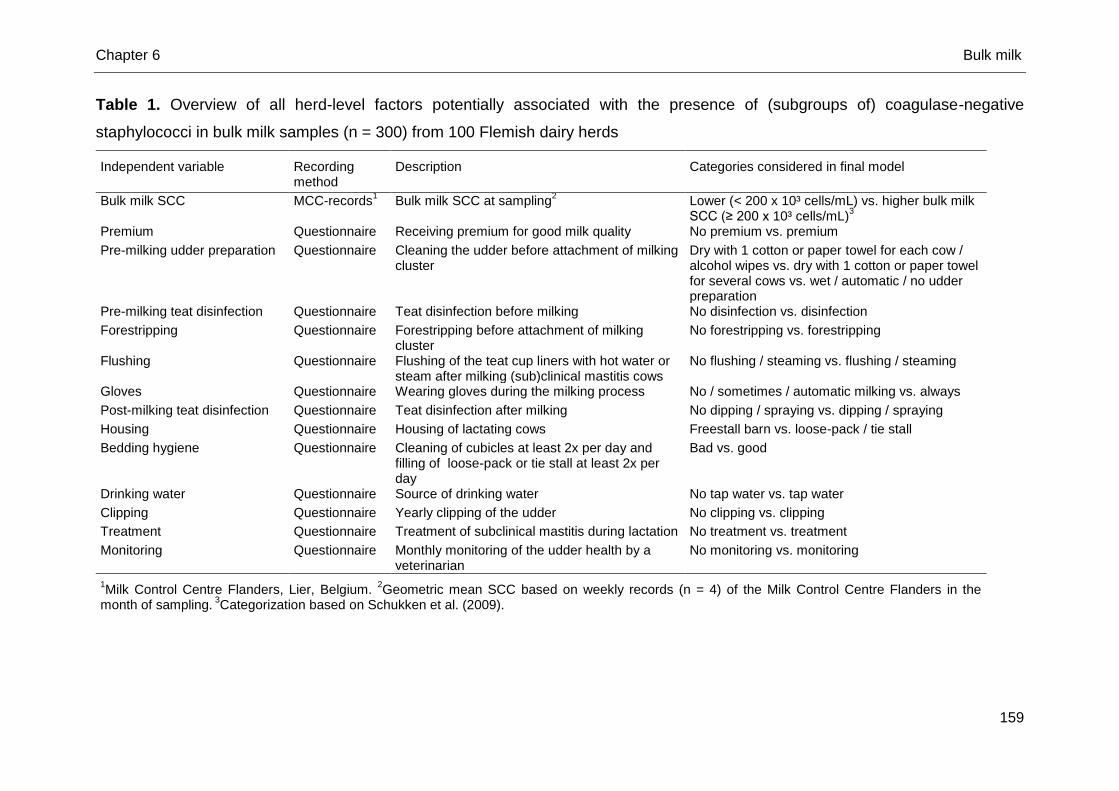
Chapter 6 Bulk milk
159
Table 1. Overview of all herd-level factors potentially associated with the presence of (subgroups of) coagulase-negative
staphylococci in bulk milk samples (n = 300) from 100 Flemish dairy herds
Independent variable Recording method
Description Categories considered in final model
Bulk milk SCC MCC-records1 Bulk milk SCC at sampling
2 Lower (< 200 x 10³ cells/mL) vs. higher bulk milk
SCC (≥ 200 x 10³ cells/mL)3
Premium Questionnaire Receiving premium for good milk quality No premium vs. premium
Pre-milking udder preparation Questionnaire Cleaning the udder before attachment of milking cluster
Dry with 1 cotton or paper towel for each cow / alcohol wipes vs. dry with 1 cotton or paper towel for several cows vs. wet / automatic / no udder preparation
Pre-milking teat disinfection Questionnaire Teat disinfection before milking No disinfection vs. disinfection
Forestripping Questionnaire Forestripping before attachment of milking cluster
No forestripping vs. forestripping
Flushing Questionnaire Flushing of the teat cup liners with hot water or steam after milking (sub)clinical mastitis cows
No flushing / steaming vs. flushing / steaming
Gloves Questionnaire Wearing gloves during the milking process No / sometimes / automatic milking vs. always
Post-milking teat disinfection Questionnaire Teat disinfection after milking No dipping / spraying vs. dipping / spraying
Housing Questionnaire Housing of lactating cows Freestall barn vs. loose-pack / tie stall
Bedding hygiene Questionnaire Cleaning of cubicles at least 2x per day and filling of loose-pack or tie stall at least 2x per day
Bad vs. good
Drinking water Questionnaire Source of drinking water No tap water vs. tap water
Clipping Questionnaire Yearly clipping of the udder No clipping vs. clipping
Treatment Questionnaire Treatment of subclinical mastitis during lactation No treatment vs. treatment
Monitoring Questionnaire Monthly monitoring of the udder health by a veterinarian
No monitoring vs. monitoring
1Milk Control Centre Flanders, Lier, Belgium.
2Geometric mean SCC based on weekly records (n = 4) of the Milk Control Centre Flanders in the
month of sampling. 3Categorization based on Schukken et al. (2009).
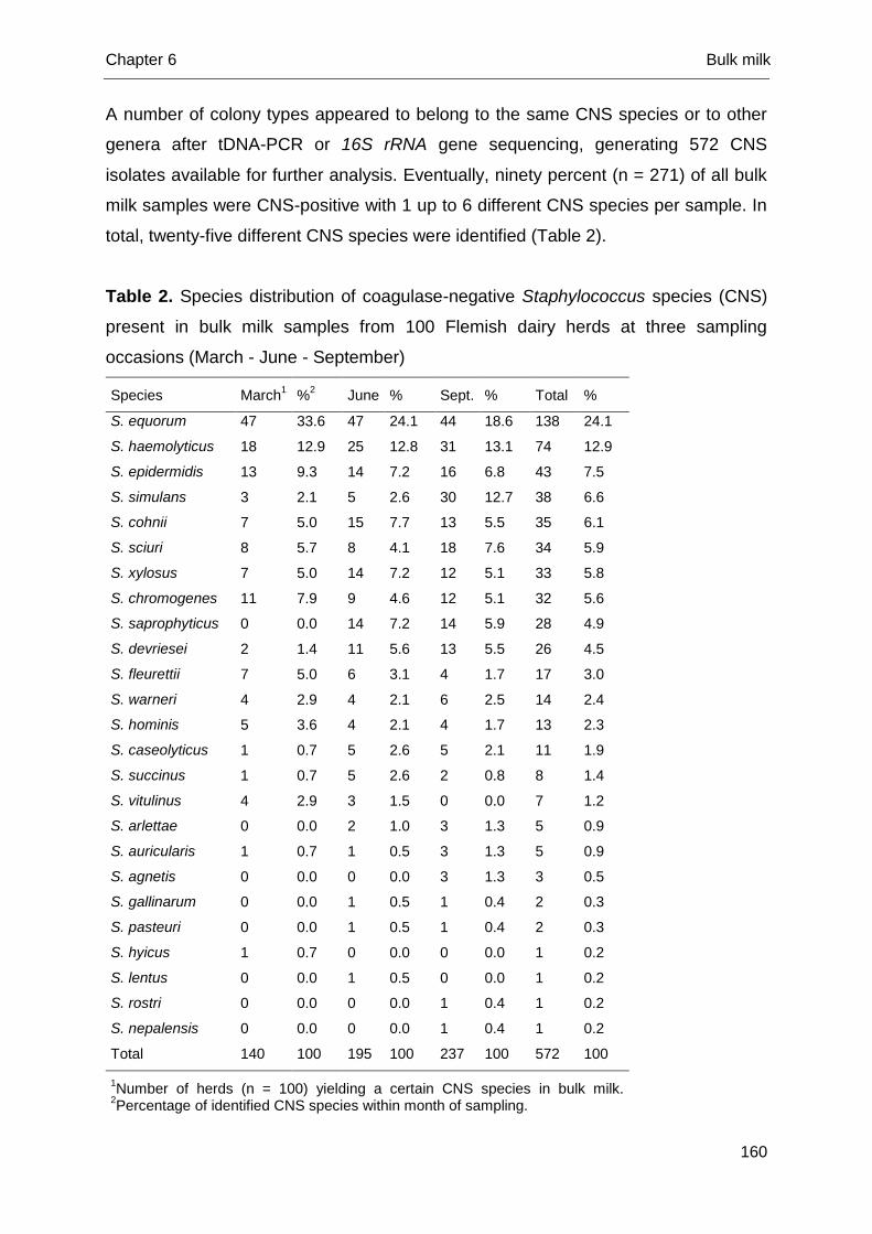
Chapter 6 Bulk milk
160
A number of colony types appeared to belong to the same CNS species or to other
genera after tDNA-PCR or 16S rRNA gene sequencing, generating 572 CNS
isolates available for further analysis. Eventually, ninety percent (n = 271) of all bulk
milk samples were CNS-positive with 1 up to 6 different CNS species per sample. In
total, twenty-five different CNS species were identified (Table 2).
Table 2. Species distribution of coagulase-negative Staphylococcus species (CNS)
present in bulk milk samples from 100 Flemish dairy herds at three sampling
occasions (March - June - September)
Species March1 %
2 June % Sept. % Total %
S. equorum 47 33.6 47 24.1 44 18.6 138 24.1
S. haemolyticus 18 12.9 25 12.8 31 13.1 74 12.9
S. epidermidis 13 9.3 14 7.2 16 6.8 43 7.5
S. simulans 3 2.1 5 2.6 30 12.7 38 6.6
S. cohnii 7 5.0 15 7.7 13 5.5 35 6.1
S. sciuri 8 5.7 8 4.1 18 7.6 34 5.9
S. xylosus 7 5.0 14 7.2 12 5.1 33 5.8
S. chromogenes 11 7.9 9 4.6 12 5.1 32 5.6
S. saprophyticus 0 0.0 14 7.2 14 5.9 28 4.9
S. devriesei 2 1.4 11 5.6 13 5.5 26 4.5
S. fleurettii 7 5.0 6 3.1 4 1.7 17 3.0
S. warneri 4 2.9 4 2.1 6 2.5 14 2.4
S. hominis 5 3.6 4 2.1 4 1.7 13 2.3
S. caseolyticus 1 0.7 5 2.6 5 2.1 11 1.9
S. succinus 1 0.7 5 2.6 2 0.8 8 1.4
S. vitulinus 4 2.9 3 1.5 0 0.0 7 1.2
S. arlettae 0 0.0 2 1.0 3 1.3 5 0.9
S. auricularis 1 0.7 1 0.5 3 1.3 5 0.9
S. agnetis 0 0.0 0 0.0 3 1.3 3 0.5
S. gallinarum 0 0.0 1 0.5 1 0.4 2 0.3
S. pasteuri 0 0.0 1 0.5 1 0.4 2 0.3
S. hyicus 1 0.7 0 0.0 0 0.0 1 0.2
S. lentus 0 0.0 1 0.5 0 0.0 1 0.2
S. rostri 0 0.0 0 0.0 1 0.4 1 0.2
S. nepalensis 0 0.0 0 0.0 1 0.4 1 0.2
Total 140 100 195 100 237 100 572 100
1Number of herds (n = 100) yielding a certain CNS species in bulk milk.
2Percentage of identified CNS species within month of sampling.
Chapter 6 Bulk milk
161
The most commonly isolated species were S. equorum, S. haemolyticus, S.
epidermidis, S. simulans, S. cohnii, S. sciuri, S. xylosus, S. chromogenes, S.
saprophyticus, and S. devriesei (Table 2). Eighty-nine bulk milk samples (n total =
300) (30%) collected in 61 out of 100 herds were S. aureus-positive (n March = 45, n
June = 21, n September = 23) and ninety-two bulk milk samples (31%) collected in
62 out of 100 herds harbored at least one of the more relevant CNS species for
udder health, i.e. S. chromogenes, S. simulans, and S. xylosus (n March = 19, n
June = 27, n September = 46). Pathogens other than staphylococci were either
phenotypically or genotypically (i.e. using tDNA-PCR or 16S rRNA gene sequencing)
identified as esculin-positive streptococci (n = 156), Bacillus spp. (n = 116),
Acinetobacter spp. (n = 33), Corynebacterium spp. (n = 28), Pseudomonas (n = 26),
Alcaligenaceae (n = 14), Psychrobacter spp. (n = 12), Jeotgallicoccus spp. (n = 11),
and other Gram-negative bacteria (n = 2). Also, fungi (n = 3) and yeasts (n = 3) were
identified.
The number of herds (n = 100) yielding a certain CNS species in March and/or
June and/or September are shown in Figure 1. Staphylococcus equorum, S.
haemolyticus, S. epidermidis, S. sciuri, S. cohnii, S. xylosus and S. devriesei, as well
as S. aureus were on at least one herd isolated on all three sampling occasions in
the same herd(s).
Statistical analyses
Variance components. In all null-models, most of the variation in the outcome
variables resided at the observation level. The herd level variation was highest for
the subgroup of host-adapted CNS (34%) and S. epidermidis (36%), S. xylosus
(32%), and S. haemolyticus (32%) solely whereas only 24% of the variation was
observed at the herd level for the subgroup of environmental CNS and 13% for S.
aureus.
Univariable analyses. Univariable analyses revealed in each model 4 factors
significantly associated with the presence of different subgroups of CNS species
(Table 3). The species-specific univariable analyses showed 5 herd-level factors to
be significantly associated with the presence of S. haemolyticus, S. devriesei, or S.
equorum, in bulk milk, 4 factors for the presence of S. epidermidis, S. xylosus, or S.
sciuri and 3 factors for the presence of S. chromogenes, S. simulans, or S. cohnii
(Table 4). Univariable analyses with S. saprophyticus (n = 28) did not fit due to low
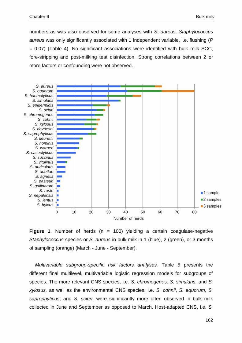
Chapter 6 Bulk milk
162
numbers as was also observed for some analyses with S. aureus. Staphylococcus
aureus was only significantly associated with 1 independent variable, i.e. flushing (P
= 0.07) (Table 4). No significant associations were identified with bulk milk SCC,
fore-stripping and post-milking teat disinfection. Strong correlations between 2 or
more factors or confounding were not observed.
Figure 1. Number of herds (n = 100) yielding a certain coagulase-negative
Staphylococcus species or S. aureus in bulk milk in 1 (blue), 2 (green), or 3 months
of sampling (orange) (March - June - September).
Multivariable subgroup-specific risk factors analyses. Table 5 presents the
different final multilevel, multivariable logistic regression models for subgroups of
species. The more relevant CNS species, i.e. S. chromogenes, S. simulans, and S.
xylosus, as well as the environmental CNS species, i.e. S. cohnii, S. equorum, S.
saprophyticus, and S. sciuri, were significantly more often observed in bulk milk
collected in June and September as opposed to March. Host-adapted CNS, i.e. S.
0 10 20 30 40 50 60 70 80 90
S. hyicusS. lentus
S. nepalensisS. rostri
S. gallinarumS. pasteuriS. agnetisS. arlettae
S. auricularisS. vitulinusS. succinus
S. caseolyticusS. warneriS. hominisS. fleurettii
S. saprophyticusS. devriesei
S. xylosusS. cohnii
S. chromogenesS. sciuri
S. epidermidisS. simulans
S. haemolyticusS. equorum
S. aureus
Number of herds
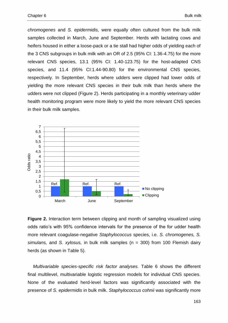
Chapter 6 Bulk milk
163
chromogenes and S. epidermidis, were equally often cultured from the bulk milk
samples collected in March, June and September. Herds with lactating cows and
heifers housed in either a loose-pack or a tie stall had higher odds of yielding each of
the 3 CNS subgroups in bulk milk with an OR of 2.5 (95% CI: 1.36-4.75) for the more
relevant CNS species, 13.1 (95% CI: 1.40-123.75) for the host-adapted CNS
species, and 11.4 (95% CI:1.44-90.80) for the environmental CNS species,
respectively. In September, herds where udders were clipped had lower odds of
yielding the more relevant CNS species in their bulk milk than herds where the
udders were not clipped (Figure 2). Herds participating in a monthly veterinary udder
health monitoring program were more likely to yield the more relevant CNS species
in their bulk milk samples.
Figure 2. Interaction term between clipping and month of sampling visualized using
odds ratio’s with 95% confidence intervals for the presence of the for udder health
more relevant coagulase-negative Staphylococcus species, i.e. S. chromogenes, S.
simulans, and S. xylosus, in bulk milk samples (n = 300) from 100 Flemish dairy
herds (as shown in Table 5).
Multivariable species-specific risk factor analyses. Table 6 shows the different
final multilevel, multivariable logistic regression models for individual CNS species.
None of the evaluated herd-level factors was significantly associated with the
presence of S. epidermidis in bulk milk. Staphylococcus cohnii was significantly more
0
0,5
1
1,5
2
2,5
3
3,5
4
4,5
5
5,5
6
6,5
7
March June September
No clipping
Clipping
Odds r
atio
Ref. Ref. Ref.
Chapter 6 Bulk milk
164
often observed in bulk milk collected in June and September as opposed to March. A
seasonal effect was also seen for S. simulans, S. devriesei as well as for S. sciuri, as
they were all more recovered in bulk milk in September as opposed to March. For
the three latter species, no difference was observed among bulk milk samples
collected in June and March. Staphylococcus epidermidis, S. chromogenes, and S.
equorum were equally present in the samples collected in March, June and
September. Housing cows in in loose-pack stall and a tie stall increased the odds of
yielding S. chromogenes, S. xylosus, S. haemolyticus, S. devriesei, S. equorum, and
S sciuri. Herds where the milking cluster was not flushed with hot water or steam
after having milked a cow with (sub)clinical mastitis were more likely to have S.
simulans-, S. haemolyticus-, and S. cohnii-positive bulk milk. Herds with drinking
water being tap water had higher odds of yielding S. simulans in their bulk milk. Not
or only sometimes wearing gloves during milking increased the odds of having S.
devriesei in the bulk milk. Herds always receiving their milk quality premium were
less likely to yield S. equorum in their bulk milk. Teat disinfection before attachment
of the milking cluster decreased the odds of yielding S. equorum in the bulk milk.
Herds using a dry cotton or paper towel for several cows during the pre-milking
udder preparation were more likely to have S. cohnii-positive bulk milk opposed to
those that used a dry cotton or paper towel for each cow.
Fisher’s exact test. The presence of the more relevant CNS, i.e. S. chromogenes,
S. simulans, and S. xylosus, was not associated with the presence of the major
pathogen S. aureus in bulk milk (P = 0.347) (SPSS Statistics 22, SPSS Inc.,
Chicago, IL, USA).
Discussion
This is the first extensive study describing the prevalence and distribution of CNS
species in bulk milk of a large number of samples and dairy herds. The use of MSA
as a (quasi-) selective medium along with the molecular speciation of all
phenotypically different CNS isolates allowed us to precisely picture the species-
specific prevalence and distribution of CNS in bulk milk within 1 year and to identify
subgroup- and even species-specific herd-level risk factors whereas previous studies
identified herd-level risk factors for CNS as a group (Sampimon et al., 2009; Piepers
et al., 2011).
Chapter 6 Bulk milk
165
Coagulase-negative staphylococci are prevalent in bulk milk in Flanders (De
Visscher et al., 2013), though the species-specific prevalence and distribution greatly
varied among herds. Staphylococcus aureus and the more relevant CNS species for
udder health were recovered in almost equal numbers, however their presence was
not associated and no seasonal effect was observed for S. aureus. The prevalence
of S. aureus corresponds well with earlier reported results on the bulk milk
prevalence of S. aureus (Olde Riekerink et al., 2006, 2010). Bacteria in bulk milk are,
however, not solely originating from infected quarters. Environmental contamination
or insufficient hygiene and poor preservation conditions can also cause positive bulk
milk samples, potentially hampering the interpretation of the data (Elmoslemany et
al., 2009a, 2009b), which also applies for the current study. Although our study
revealed the presence of both so-called host-adapted and environmental CNS
species in bulk milk, studying the presence of CNS in bulk milk samples and the
presence in quarter milk and environmental samples at the same time, relying on
strain-typing, is warranted to determine the exact origin of the CNS in bulk milk.
A seasonal effect was observed for several species which is in contrast with
earlier CNS group-studies (Gillespie et al., 2012). Bad housing conditions and poor
hygiene are important factors increasing the total bacterial count in bulk milk (Olde
Riekerink et al., 2010). Housing of lactating animals most likely also plays an
important role in our study as the presence of only 3 species was not associated with
this risk factor, i.e. S. cohnii, S. epidermidis, and S. simulans. The latter might either
indicate an ubiquitous nature of those 3 species both in free-stall and loose-pack or
tie stall barns (perhaps for S. cohnii) or suggest a more cow-dependent presence in
bulk milk (perhaps for the cow-adapted CNS S. epidermidis and S. simulans).
Cleaning of the cubicles or loose-pack was not associated with the presence of any
CNS species in our study, however, bedding hygiene was not visually checked.
Caution is thus needed in drawing conclusions.
Clipping the udder reduced the odds of having bulk milk contaminated with a more
relevant CNS species, i.e. S. chromogenes, S. simulans, and S. xylosus (Supré et
al., 2011; Fry et al., 2014; De Visscher et al., 2015). A previous study reported a
negative association between clipping of the udder prior to calving and CNS IMI at
parturition (Piepers et al., 2011). The latter might indicate that the presence of the
relevant CNS in bulk milk results from a transmission via the teat skin and teat hairs
as well as via quarters infected with those species.
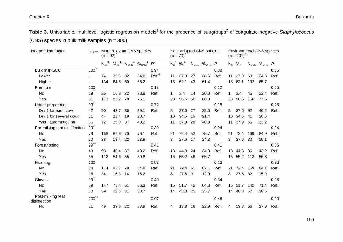
Chapter 6 Bulk milk
166
Table 3. Univariable, multilevel logistic regression models1 for the presence of subgroups2 of coagulase-negative Staphylococcus
(CNS) species in bulk milk samples (n = 300)
Independent factor NHerds More relevant CNS species (n = 92)
2
Host-adapted CNS species (n = 70)
2
Environmental CNS species (n = 201)
2
Nno3 %no
3 NCNS
4 %CNS
4 P
5 Nn
6 %n
6 NCNS %CNS P Nn %n NCNS %CNS P
Bulk milk SCC 1007
0.94
0.88
0.85
Lower - 74 35.6 32 34.8 Ref.8
11 37.9 27 38.6 Ref.
11 37.9 69 34.3 Ref.
Higher - 134 64.6 60 65.2
18 62.1 43 61.4
18 62.1 132 65.7
Premium 100
0.18
0.12
0.05
No 19 35 16.8 22 23.9 Ref.
1 3.4 14 20.0 Ref.
1 3.4 45 22.4 Ref.
Yes 81 173 83.2 70 76.1
28 96.6 56 80.0
28 96.6 156 77.6
Udder preparation 999
0.72
0.18
0.26
Dry 1 for each cow 42 90 43.7 36 39.1 Ref.
8 27.6 27 38.6 Ref.
8 27.6 92 46.2 Ref.
Dry 1 for several cows 21 44 21.4 19 20.7
10 34.5 15 21.4
10 34.5 41 20.6
Wet / automatic / no 36 72 35.0 37 40.2
11 37.9 28 40.0
11 37.9 66 33.2
Pre-milking teat disinfection 999
0.30
0.94
0.24
No 79 168 81.6 70 76.1 Ref.
21 72.4 53 75.7 Ref.
21 72.4 169 84.9 Ref.
Yes 20 38 18.4 22 23.9
8 27.6 17 24.3
8 27.6 30 15.1
Forestripping 9910
0.41
0.41
0.86
No 43 93 45.4 37 40.2 Ref.
13 44.8 24 34.3 Ref.
13 44.8 86 43.2 Ref.
Yes 55 112 54.6 55 59.8
16 55.2 46 65.7
16 55.2 113 56.8
Flushing 100
0.82
0.13
0.23
No 84 174 83.7 78 84.8 Ref.
21 72.4 61 87.1 Ref.
21 72.4 169 84.1 Ref.
Yes 16 34 16.3 14 15.2
8 27.6 9 12.9
8 27.6 32 15.9
Gloves 999
0.40
0.34
0.08
No 69 147 71.4 61 66.3 Ref.
15 51.7 45 64.3 Ref.
15 51.7 142 71.4 Ref.
Yes 30 59 28.6 31 33.7
14 48.3 25 35.7
14 48.3 57 28.6
Post-milking teat disinfection
10011
0.97
0.48
0.20
No 21 49 23.6 22 23.9 Ref.
4 13.8 16 22.9 Ref.
4 13.8 56 27.9 Ref.
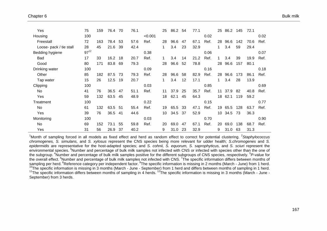
Chapter 6 Bulk milk
167
Yes 75 159 76.4 70 76.1
25 86.2 54 77.1
25 86.2 145 72.1
Housing 100
<0.001
0.02
0.02
Freestall 72 163 78.4 53 57.6 Ref.
28 96.6 47 67.1 Ref.
28 96.6 142 70.6 Ref.
Loose- pack / tie stall 28 45 21.6 39 42.4
1 3.4 23 32.9
1 3.4 59 29.4
Bedding hygiene 9712
0.38
0.06
0.07
Bad 17 33 16.2 18 20.7 Ref.
1 3.4 14 21.2 Ref.
1 3.4 39 19.9 Ref.
Good 80 171 83.8 69 79.3
28 96.6 52 78.8
28 96.6 157 80.1
Drinking water 100
0.09
0.16
0.18
Other 85 182 87.5 73 79.3 Ref.
28 96.6 58 82.9 Ref.
28 96.6 173 86.1 Ref.
Tap water 15 26 12.5 19 20.7
1 3.4 12 17.1
1 3.4 28 13.9
Clipping 100
0.03
0.85
0.69
No 41 76 36.5 47 51.1 Ref.
11 37.9 25 35.7 Ref.
11 37.9 82 40.8 Ref.
Yes 59 132 63.5 45 48.9
18 62.1 45 64.3
18 62.1 119 59.2
Treatment 100
0.22
0.15
0.77
No 61 132 63.5 51 55.4 Ref.
19 65.5 33 47.1 Ref.
19 65.5 128 63.7 Ref.
Yes 39 76 36.5 41 44.6
10 34.5 37 52.9
10 34.5 73 36.3
Monitoring 100
0.03
0.70
0.90
No 69 152 73.1 55 59.8 Ref.
20 69.0 47 67.1 Ref.
20 69.0 138 68.7 Ref.
Yes 31 56 26.9 37 40.2
9 31.0 23 32.9
9 31.0 63 31.3
1Month of sampling forced in all models as fixed effect and herd as random effect to correct for potential clustering.
2Staphylococcus
chromogenes, S. simulans, and S. xylosus represent the CNS species being more relevant for udder health; S.chromogenes and S. epidermidis are representative for the host-adapted species; and S. cohnii, S. equorum, S. saprophyticus, and S. sciuri represent the environmental species.
3Number and percentage of bulk milk samples not infected with CNS or infected with species other than the one of
the subgroup. 4
Number and percentage of bulk milk samples positive for the different subgroups of CNS species, respectively. 5
P-value for the overall effect.
6Number and percentage of bulk milk samples not infected with CNS.
7The specific information differs between months of
sampling per herd. 8Reference category per independent factor.
9The specific information is missing in 2 months (March - June) from 1 herd.
10The specific information is missing in 3 months (March - June - September) from 1 herd and differs between months of sampling in 1 herd.
11The specific information differs between months of sampling in 4 herds.
12The specific information is missing in 3 months (March - June -
September) from 3 herds.
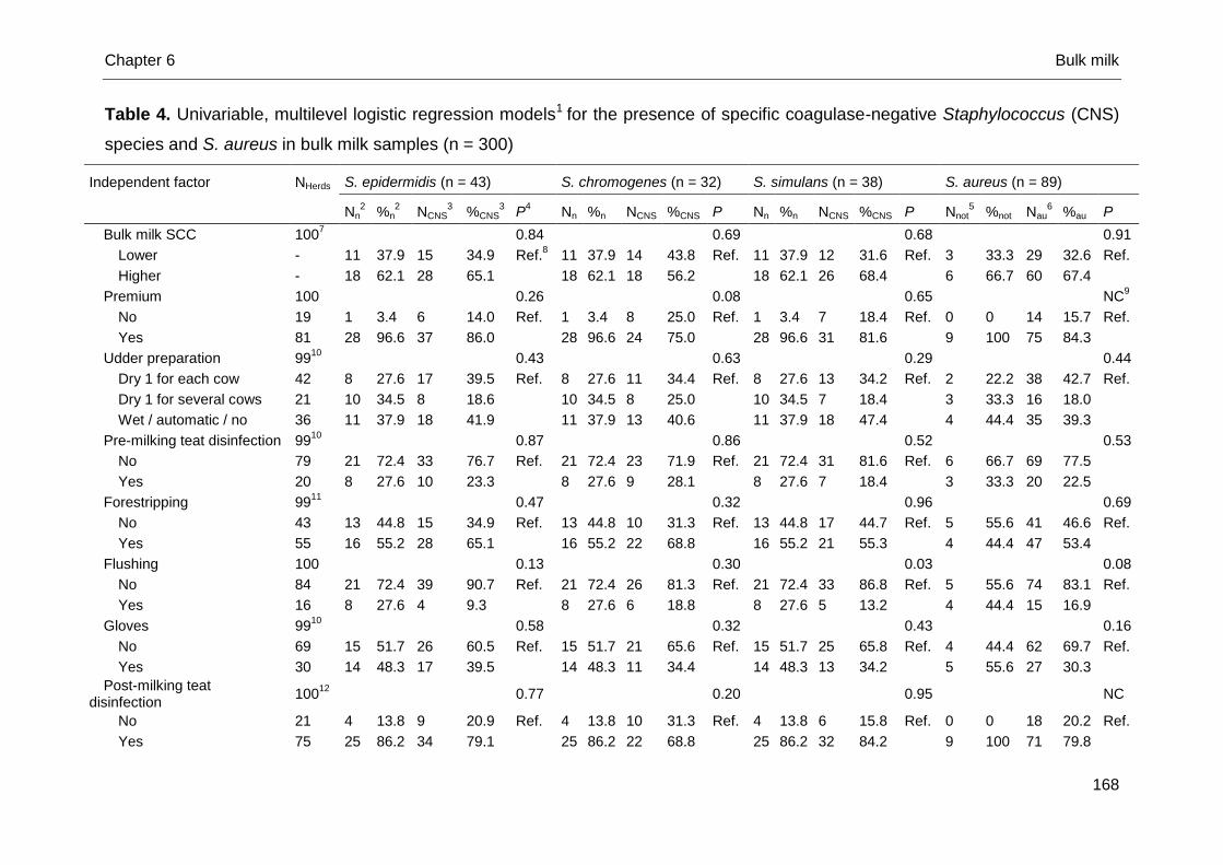
Chapter 6 Bulk milk
168
Table 4. Univariable, multilevel logistic regression models1 for the presence of specific coagulase-negative Staphylococcus (CNS)
species and S. aureus in bulk milk samples (n = 300)
Independent factor NHerds S. epidermidis (n = 43) S. chromogenes (n = 32) S. simulans (n = 38) S. aureus (n = 89)
Nn
2 %n
2 NCNS
3 %CNS
3 P
4 Nn %n NCNS %CNS P Nn %n NCNS %CNS P Nnot
5 %not Nau
6 %au P
Bulk milk SCC 1007
0.84
0.69
0.68 0.91
Lower - 11 37.9 15 34.9 Ref.8 11 37.9 14 43.8 Ref. 11 37.9 12 31.6 Ref. 3 33.3 29 32.6 Ref.
Higher - 18 62.1 28 65.1
18 62.1 18 56.2
18 62.1 26 68.4
6 66.7 60 67.4
Premium 100
0.26
0.08
0.65 NC9
No 19 1 3.4 6 14.0 Ref. 1 3.4 8 25.0 Ref. 1 3.4 7 18.4 Ref. 0 0 14 15.7 Ref.
Yes 81 28 96.6 37 86.0
28 96.6 24 75.0
28 96.6 31 81.6
9 100 75 84.3
Udder preparation
9910
0.43
0.63
0.29 0.44
Dry 1 for each cow 42 8 27.6 17 39.5 Ref. 8 27.6 11 34.4 Ref. 8 27.6 13 34.2 Ref. 2 22.2 38 42.7 Ref.
Dry 1 for several cows 21 10 34.5 8 18.6
10 34.5 8 25.0
10 34.5 7 18.4
3 33.3 16 18.0
Wet / automatic / no 36 11 37.9 18 41.9
11 37.9 13 40.6
11 37.9 18 47.4
4 44.4 35 39.3
Pre-milking teat disinfection
9910
0.87
0.86
0.52 0.53
No 79 21 72.4 33 76.7 Ref. 21 72.4 23 71.9 Ref. 21 72.4 31 81.6 Ref. 6 66.7 69 77.5
Yes 20 8 27.6 10 23.3
8 27.6 9 28.1
8 27.6 7 18.4
3 33.3 20 22.5
Forestripping
9911
0.47
0.32
0.96 0.69
No 43 13 44.8 15 34.9 Ref. 13 44.8 10 31.3 Ref. 13 44.8 17 44.7 Ref. 5 55.6 41 46.6 Ref.
Yes 55 16 55.2 28 65.1
16 55.2 22 68.8
16 55.2 21 55.3
4 44.4 47 53.4
Flushing 100
0.13
0.30
0.03 0.08
No 84 21 72.4 39 90.7 Ref. 21 72.4 26 81.3 Ref. 21 72.4 33 86.8 Ref. 5 55.6 74 83.1 Ref.
Yes 16 8 27.6 4 9.3
8 27.6 6 18.8
8 27.6 5 13.2
4 44.4 15 16.9
Gloves 9910
0.58
0.32
0.43 0.16
No 69 15 51.7 26 60.5 Ref. 15 51.7 21 65.6 Ref. 15 51.7 25 65.8 Ref. 4 44.4 62 69.7 Ref.
Yes 30 14 48.3 17 39.5
14 48.3 11 34.4
14 48.3 13 34.2
5 55.6 27 30.3
Post-milking teat disinfection
10012
0.77
0.20
0.95 NC
No 21 4 13.8 9 20.9 Ref. 4 13.8 10 31.3 Ref. 4 13.8 6 15.8 Ref. 0 0 18 20.2 Ref.
Yes 75 25 86.2 34 79.1
25 86.2 22 68.8
25 86.2 32 84.2
9 100 71 79.8
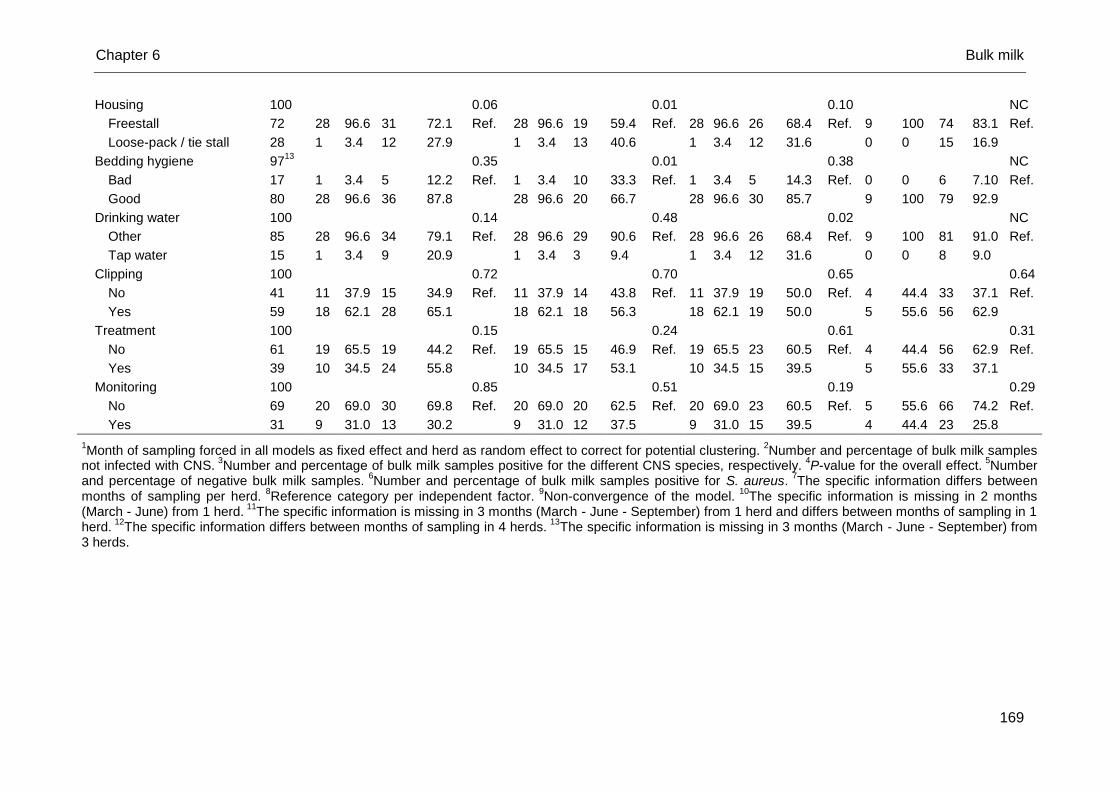
Chapter 6 Bulk milk
169
Housing 100
0.06
0.01
0.10 NC
Freestall 72 28 96.6 31 72.1 Ref. 28 96.6 19 59.4 Ref. 28 96.6 26 68.4 Ref. 9 100 74 83.1 Ref.
Loose-pack / tie stall 28 1 3.4 12 27.9
1 3.4 13 40.6
1 3.4 12 31.6
0 0 15 16.9
Bedding hygiene
9713
0.35
0.01
0.38 NC
Bad 17 1 3.4 5 12.2 Ref. 1 3.4 10 33.3 Ref. 1 3.4 5 14.3 Ref. 0 0 6 7.10 Ref.
Good 80 28 96.6 36 87.8
28 96.6 20 66.7
28 96.6 30 85.7
9 100 79 92.9
Drinking water 100
0.14
0.48
0.02 NC
Other 85 28 96.6 34 79.1 Ref. 28 96.6 29 90.6 Ref. 28 96.6 26 68.4 Ref. 9 100 81 91.0 Ref.
Tap water 15 1 3.4 9 20.9
1 3.4 3 9.4
1 3.4 12 31.6
0 0 8 9.0
Clipping 100
0.72
0.70
0.65 0.64
No 41 11 37.9 15 34.9 Ref. 11 37.9 14 43.8 Ref. 11 37.9 19 50.0 Ref. 4 44.4 33 37.1 Ref.
Yes 59 18 62.1 28 65.1
18 62.1 18 56.3
18 62.1 19 50.0
5 55.6 56 62.9
Treatment 100
0.15
0.24
0.61 0.31
No 61 19 65.5 19 44.2 Ref. 19 65.5 15 46.9 Ref. 19 65.5 23 60.5 Ref. 4 44.4 56 62.9 Ref.
Yes 39 10 34.5 24 55.8
10 34.5 17 53.1
10 34.5 15 39.5
5 55.6 33 37.1
Monitoring 100
0.85
0.51
0.19 0.29
No 69 20 69.0 30 69.8 Ref. 20 69.0 20 62.5 Ref. 20 69.0 23 60.5 Ref. 5 55.6 66 74.2 Ref.
Yes 31 9 31.0 13 30.2
9 31.0 12 37.5
9 31.0 15 39.5
4 44.4 23 25.8
1Month of sampling forced in all models as fixed effect and herd as random effect to correct for potential clustering.
2Number and percentage of bulk milk samples
not infected with CNS. 3
Number and percentage of bulk milk samples positive for the different CNS species, respectively. 4
P-value for the overall effect. 5
Number and percentage of negative bulk milk samples.
6Number and percentage of bulk milk samples positive for S. aureus.
7The specific information differs between
months of sampling per herd. 8
Reference category per independent factor. 9
Non-convergence of the model. 10
The specific information is missing in 2 months (March - June) from 1 herd.
11The specific information is missing in 3 months (March - June - September) from 1 herd and differs between months of sampling in 1
herd. 12
The specific information differs between months of sampling in 4 herds. 13
The specific information is missing in 3 months (March - June - September) from 3 herds.
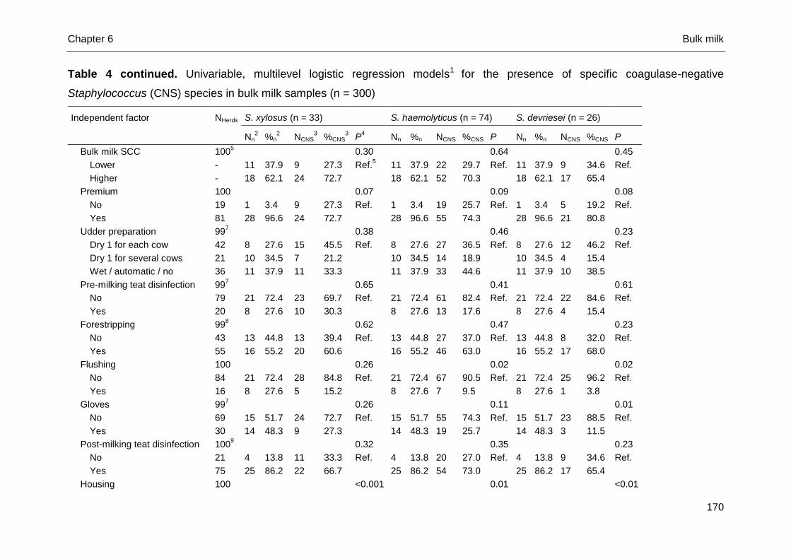
Chapter 6 Bulk milk
170
Table 4 continued. Univariable, multilevel logistic regression models1 for the presence of specific coagulase-negative
Staphylococcus (CNS) species in bulk milk samples (n = 300)
Independent factor NHerds S. xylosus (n = 33) S. haemolyticus (n = 74) S. devriesei (n = 26)
Nn2
%n2
NCNS3
%CNS3
P4
Nn %n NCNS %CNS P Nn %n NCNS %CNS P
Bulk milk SCC 1005
0.30
0.64
0.45
Lower - 11 37.9 9 27.3 Ref.5
11 37.9 22 29.7 Ref. 11 37.9 9 34.6 Ref.
Higher - 18 62.1 24 72.7
18 62.1 52 70.3
18 62.1 17 65.4
Premium 100
0.07
0.09
0.08
No 19 1 3.4 9 27.3 Ref. 1 3.4 19 25.7 Ref. 1 3.4 5 19.2 Ref.
Yes 81 28 96.6 24 72.7
28 96.6 55 74.3
28 96.6 21 80.8
Udder preparation
997
0.38
0.46
0.23
Dry 1 for each cow 42 8 27.6 15 45.5 Ref. 8 27.6 27 36.5 Ref. 8 27.6 12 46.2 Ref.
Dry 1 for several cows 21 10 34.5 7 21.2
10 34.5 14 18.9
10 34.5 4 15.4
Wet / automatic / no 36 11 37.9 11 33.3
11 37.9 33 44.6
11 37.9 10 38.5
Pre-milking teat disinfection
997
0.65
0.41
0.61
No 79 21 72.4 23 69.7 Ref. 21 72.4 61 82.4 Ref. 21 72.4 22 84.6 Ref.
Yes 20 8 27.6 10 30.3
8 27.6 13 17.6
8 27.6 4 15.4
Forestripping
998
0.62
0.47
0.23
No 43 13 44.8 13 39.4 Ref. 13 44.8 27 37.0 Ref. 13 44.8 8 32.0 Ref.
Yes 55 16 55.2 20 60.6
16 55.2 46 63.0
16 55.2 17 68.0
Flushing 100
0.26
0.02
0.02
No 84 21 72.4 28 84.8 Ref. 21 72.4 67 90.5 Ref. 21 72.4 25 96.2 Ref.
Yes 16 8 27.6 5 15.2
8 27.6 7 9.5
8 27.6 1 3.8
Gloves
997
0.26
0.11
0.01
No 69 15 51.7 24 72.7 Ref. 15 51.7 55 74.3 Ref. 15 51.7 23 88.5 Ref.
Yes 30 14 48.3 9 27.3
14 48.3 19 25.7
14 48.3 3 11.5
Post-milking teat disinfection 1009
0.32
0.35
0.23
No 21 4 13.8 11 33.3 Ref. 4 13.8 20 27.0 Ref. 4 13.8 9 34.6 Ref.
Yes 75 25 86.2 22 66.7
25 86.2 54 73.0
25 86.2 17 65.4
Housing 100
<0.001
0.01
<0.01
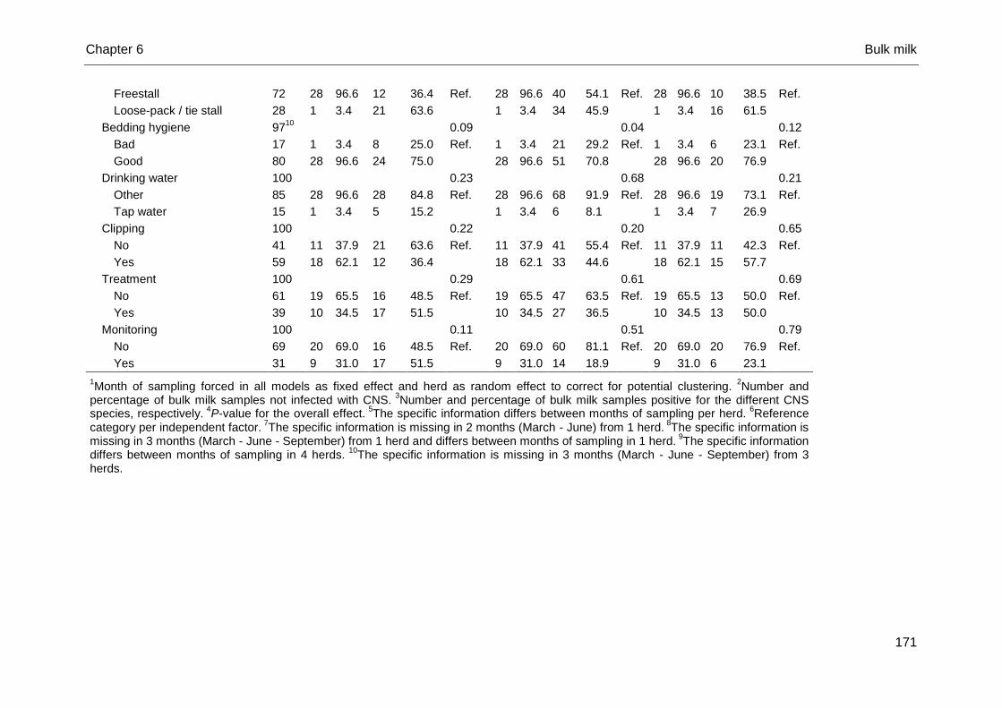
Chapter 6 Bulk milk
171
Freestall 72 28 96.6 12 36.4 Ref. 28 96.6 40 54.1 Ref. 28 96.6 10 38.5 Ref.
Loose-pack / tie stall 28 1 3.4 21 63.6
1 3.4 34 45.9
1 3.4 16 61.5
Bedding hygiene
9710
0.09
0.04
0.12
Bad 17 1 3.4 8 25.0 Ref. 1 3.4 21 29.2 Ref. 1 3.4 6 23.1 Ref.
Good 80 28 96.6 24 75.0
28 96.6 51 70.8
28 96.6 20 76.9
Drinking water 100
0.23
0.68
0.21
Other 85 28 96.6 28 84.8 Ref. 28 96.6 68 91.9 Ref. 28 96.6 19 73.1 Ref.
Tap water 15 1 3.4 5 15.2
1 3.4 6 8.1
1 3.4 7 26.9
Clipping 100
0.22
0.20
0.65
No 41 11 37.9 21 63.6 Ref. 11 37.9 41 55.4 Ref. 11 37.9 11 42.3 Ref.
Yes 59 18 62.1 12 36.4
18 62.1 33 44.6
18 62.1 15 57.7
Treatment 100
0.29
0.61
0.69
No 61 19 65.5 16 48.5 Ref. 19 65.5 47 63.5 Ref. 19 65.5 13 50.0 Ref.
Yes 39 10 34.5 17 51.5
10 34.5 27 36.5
10 34.5 13 50.0
Monitoring 100
0.11
0.51
0.79
No 69 20 69.0 16 48.5 Ref. 20 69.0 60 81.1 Ref. 20 69.0 20 76.9 Ref.
Yes 31 9 31.0 17 51.5
9 31.0 14 18.9
9 31.0 6 23.1
1Month of sampling forced in all models as fixed effect and herd as random effect to correct for potential clustering.
2Number and
percentage of bulk milk samples not infected with CNS. 3
Number and percentage of bulk milk samples positive for the different CNS species, respectively.
4P-value for the overall effect.
5The specific information differs between months of sampling per herd.
6Reference
category per independent factor. 7The specific information is missing in 2 months (March - June) from 1 herd.
8The specific information is
missing in 3 months (March - June - September) from 1 herd and differs between months of sampling in 1 herd. 9The specific information
differs between months of sampling in 4 herds. 10
The specific information is missing in 3 months (March - June - September) from 3 herds.
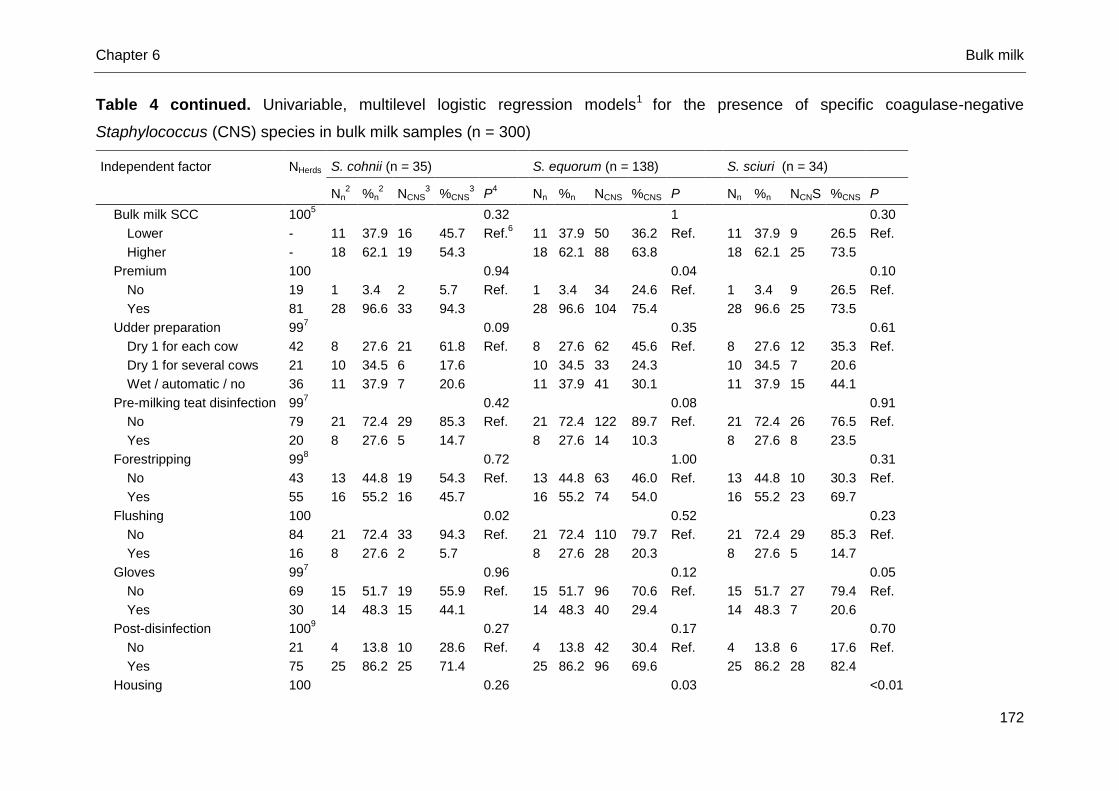
Chapter 6 Bulk milk
172
Table 4 continued. Univariable, multilevel logistic regression models1 for the presence of specific coagulase-negative
Staphylococcus (CNS) species in bulk milk samples (n = 300)
Independent factor NHerds S. cohnii (n = 35) S. equorum (n = 138) S. sciuri (n = 34)
Nn2 %n
2 NCNS
3 %CNS
3 P
4 Nn %n NCNS %CNS P Nn %n NCNS %CNS P
Bulk milk SCC 1005
0.32
1
0.30
Lower - 11 37.9 16 45.7 Ref.6
11 37.9 50 36.2 Ref.
11 37.9 9 26.5 Ref.
Higher - 18 62.1 19 54.3
18 62.1 88 63.8
18 62.1 25 73.5
Premium 100
0.94
0.04
0.10
No 19 1 3.4 2 5.7 Ref.
1 3.4 34 24.6 Ref.
1 3.4 9 26.5 Ref.
Yes 81 28 96.6 33 94.3
28 96.6 104 75.4
28 96.6 25 73.5
Udder preparation
997
0.09
0.35
0.61
Dry 1 for each cow 42 8 27.6 21 61.8 Ref.
8 27.6 62 45.6 Ref.
8 27.6 12 35.3 Ref.
Dry 1 for several cows 21 10 34.5 6 17.6
10 34.5 33 24.3
10 34.5 7 20.6
Wet / automatic / no 36 11 37.9 7 20.6
11 37.9 41 30.1
11 37.9 15 44.1
Pre-milking teat disinfection
997
0.42
0.08
0.91
No 79 21 72.4 29 85.3 Ref.
21 72.4 122 89.7 Ref.
21 72.4 26 76.5 Ref.
Yes 20 8 27.6 5 14.7
8 27.6 14 10.3
8 27.6 8 23.5
Forestripping
998
0.72
1.00
0.31
No 43 13 44.8 19 54.3 Ref.
13 44.8 63 46.0 Ref.
13 44.8 10 30.3 Ref.
Yes 55 16 55.2 16 45.7
16 55.2 74 54.0
16 55.2 23 69.7
Flushing 100
0.02
0.52
0.23
No 84 21 72.4 33 94.3 Ref.
21 72.4 110 79.7 Ref.
21 72.4 29 85.3 Ref.
Yes 16 8 27.6 2 5.7
8 27.6 28 20.3
8 27.6 5 14.7
Gloves
997
0.96
0.12
0.05
No 69 15 51.7 19 55.9 Ref.
15 51.7 96 70.6 Ref.
15 51.7 27 79.4 Ref.
Yes 30 14 48.3 15 44.1
14 48.3 40 29.4
14 48.3 7 20.6
Post-disinfection 1009
0.27
0.17
0.70
No 21 4 13.8 10 28.6 Ref.
4 13.8 42 30.4 Ref.
4 13.8 6 17.6 Ref.
Yes 75 25 86.2 25 71.4
25 86.2 96 69.6
25 86.2 28 82.4
Housing 100
0.26
0.03
<0.01
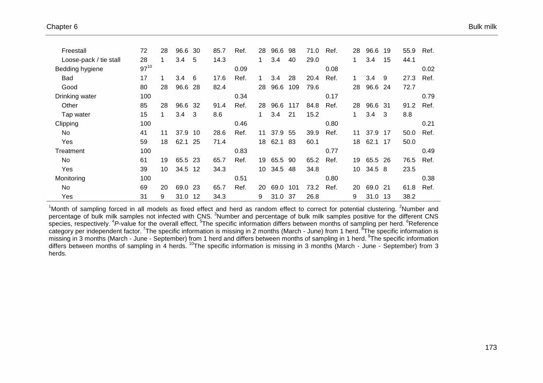
Chapter 6 Bulk milk
173
Freestall 72 28 96.6 30 85.7 Ref.
28 96.6 98 71.0 Ref.
28 96.6 19 55.9 Ref.
Loose-pack / tie stall 28 1 3.4 5 14.3
1 3.4 40 29.0
1 3.4 15 44.1
Bedding hygiene
9710
0.09
0.08
0.02
Bad 17 1 3.4 6 17.6 Ref.
1 3.4 28 20.4 Ref.
1 3.4 9 27.3 Ref.
Good 80 28 96.6 28 82.4
28 96.6 109 79.6
28 96.6 24 72.7
Drinking water 100
0.34
0.17
0.79
Other 85 28 96.6 32 91.4 Ref.
28 96.6 117 84.8 Ref.
28 96.6 31 91.2 Ref.
Tap water 15 1 3.4 3 8.6
1 3.4 21 15.2
1 3.4 3 8.8
Clipping 100
0.46
0.80
0.21
No 41 11 37.9 10 28.6 Ref.
11 37.9 55 39.9 Ref.
11 37.9 17 50.0 Ref.
Yes 59 18 62.1 25 71.4
18 62.1 83 60.1
18 62.1 17 50.0
Treatment 100
0.83
0.77
0.49
No 61 19 65.5 23 65.7 Ref.
19 65.5 90 65.2 Ref.
19 65.5 26 76.5 Ref.
Yes 39 10 34.5 12 34.3
10 34.5 48 34.8
10 34.5 8 23.5
Monitoring 100
0.51
0.80
0.38
No 69 20 69.0 23 65.7 Ref.
20 69.0 101 73.2 Ref.
20 69.0 21 61.8 Ref.
Yes 31 9 31.0 12 34.3
9 31.0 37 26.8
9 31.0 13 38.2
1Month of sampling forced in all models as fixed effect and herd as random effect to correct for potential clustering.
2Number and
percentage of bulk milk samples not infected with CNS. 3
Number and percentage of bulk milk samples positive for the different CNS species, respectively.
4P-value for the overall effect.
5The specific information differs between months of sampling per herd.
6Reference
category per independent factor. 7The specific information is missing in 2 months (March - June) from 1 herd.
8The specific information is
missing in 3 months (March - June - September) from 1 herd and differs between months of sampling in 1 herd. 9The specific information
differs between months of sampling in 4 herds. 10
The specific information is missing in 3 months (March - June - September) from 3 herds.
Chapter 6 Bulk milk
174
Nineteen herds did not receive their milk quality premium at least one time during
the study period. The main reason for not receiving the milk quality premium in
Flemish dairy herds is an elevated coli count (Passchyn et al., submitted). Therefore,
it is not that surprising that especially the presence of S. equorum in the bulk milk
samples was associated with not achieving the milk quality premium.
Staphylococcus equorum has mainly been isolated from extramammary habitats
such as teat apices (Braem et al., 2013; De Visscher et al., 2014, 2016a), milking
machine unit liners and milkers’ skin and gloves (De Visscher et al., 2014), and the
cow’s environment (Piessens et al., 2011). In that respect, the S. equorum bulk milk
contamination in the current study might occur from an environmental transfer via the
teat surface. The latter hypothesis is supported by the fact that the presence of S.
equorum in the bulk milk was also negatively associated with pre-disinfection of the
teats. Pre-milking teat disinfection lowered the likelihood of finding CNS in bulk milk
in another study as well, which can most probably be attributed to a more careful
removal of environmental teat skin organisms before milking (Jayarao et al., 2004).
The use of a single towel for multiple cows enlarged the probability of isolating S.
cohnii from the bulk milk. The latter practice increases both the risk of contamination
of quarters and cows with environmental organisms and the risk of transmission of
the more udder-adapted pathogens to uninfected quarters and cows. Also, flushing
of the milking cluster with hot water or steam after having milked a cow with
(sub)clinical mastitis was negatively associated with the presence of S. cohnii in bulk
milk. This association suggests the presence of S. cohnni in the teat liners which is
in accordance with previous work (De Visscher et al., 2014). The latter finding was
explained by direct contact between teat skin and liners (De Visscher et al., 2014).
Staphylococcus cohnii rather seems to be an ecological equivalent of S. equorum
although in at least some studies S. cohnii has been more commonly isolated from
bovine milk (Supré et al., 2011; Fry et al., 2014; De Visscher et al., 2016b). Bulk milk
contamination with this species presumably occurred from an environmental
transmission via the teat surface and via the teat liners. However, the presence of S.
cohnii in the bulk milk via teat liners contaminated with milk residues of S. cohnii-
infected animals cannot be excluded.
Flushing with hot water or steam after having milked a cow with (sub)clinical
mastitis was also negatively associated with the isolation of S. simulans and S.
haemolyticus in the bulk milk. Both species have recently been assumed to be
Chapter 6 Bulk milk
175
opportunistic in nature as they have frequently been isolated from bovine IMI as well
as from several extramammary habitats including the cow’s environment (Piessens
et al., 2011; Supré et al., 2011; Vanderhaeghen et al., 2015). The latter findings
along with the results from the current study let us assume that the presence of S.
simulans and S. haemolyticus in the bulk milk is rather the result of contamination
from infected milk than from an environmental transfer.
Wearing gloves decreased the odds of having S. devriesei-positive bulk milk. This
is interesting as in recent work S. devriesei could be hardly isolated from the milker’s
hands or gloves (De Visscher et al., 2014). Staphylococcus devriesei has been
mainly isolated from extramammary habitats including teat apices from dry cows and
end-term heifers and the cow’s environment (Piessens et al., 2011; De Visscher et
al., 2016a) and less from bovine IMI (Piessens et al., 2011; Supré et al., 2011; De
Visscher et al., 2016b). Teat apices of lactating animals appear to be not (Taponen
et al., 2008; De Visscher et al., 2014) or hardly colonized (Braem et al., 2013) with S.
devriesei. The presence of this species in the bulk milk might be a result from an
environmental transfer via the teat surface along with contamination of the teats via
the milker’s hands. However, more in-depth research concerning the ecology and
epidemiological nature of S. devriesei is needed before final conclusions can be
drawn.
Quarters infected with CNS are important contributors to the total number of
somatic cells in herds with low bulk milk SCC (Piepers et al., 2009; Schukken et al.,
2009; Sampimon et al., 2010). Also, mean bulk milk SCC was significantly
associated with the mean CNS bulk milk count and isolation of S. aureus in bulk milk
(Jayarao et al., 2004). In our study, however, no association between the bulk milk
SCC and the presence of any of the CNS species or S. aureus was revealed.
Although post-milking teat disinfection was found to decrease the total bacterial
count in bulk milk (Jayarao et al., 2004) and is considered an effective control
measure against both the host-adapted and environmental bacteria (Hogeveen et
al., 2011), no species-specific associations were observed in this study.
Interestingly, participation in a monthly veterinary udder health program was
positively associated with the presence of the for udder health more relevant CNS. A
cause-effect reversal is suspected, yet the true nature of this association remains
unknown.
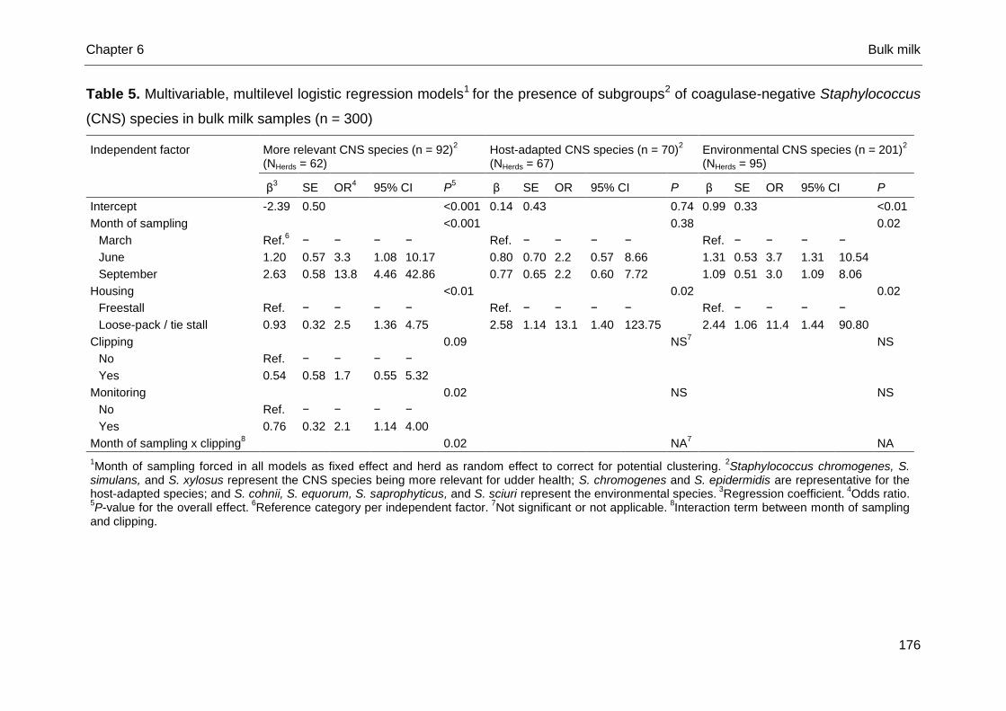
Chapter 6 Bulk milk
176
Table 5. Multivariable, multilevel logistic regression models1 for the presence of subgroups2 of coagulase-negative Staphylococcus
(CNS) species in bulk milk samples (n = 300)
Independent factor More relevant CNS species (n = 92)2
(NHerds = 62) Host-adapted CNS species (n = 70)
2
(NHerds = 67) Environmental CNS species (n = 201)
2
(NHerds = 95)
β3 SE OR
4 95% CI P
5 β SE OR 95% CI P β SE OR 95% CI P
Intercept -2.39 0.50 <0.001 0.14 0.43 0.74 0.99 0.33 <0.01
Month of sampling
<0.001
0.38
0.02
March Ref.6 − − − −
Ref. − − − −
Ref. − − − −
June 1.20 0.57 3.3 1.08 10.17
0.80 0.70 2.2 0.57 8.66
1.31 0.53 3.7 1.31 10.54
September 2.63 0.58 13.8 4.46 42.86
0.77 0.65 2.2 0.60 7.72
1.09 0.51 3.0 1.09 8.06
Housing
<0.01
0.02
0.02
Freestall Ref. − − − −
Ref. − − − −
Ref. − − − −
Loose-pack / tie stall 0.93 0.32 2.5 1.36 4.75
2.58 1.14 13.1 1.40 123.75
2.44 1.06 11.4 1.44 90.80
Clipping
0.09
NS7
NS
No Ref. − − − −
Yes 0.54 0.58 1.7 0.55 5.32
Monitoring
0.02
NS
NS
No Ref. − − − −
Yes 0.76 0.32 2.1 1.14 4.00
Month of sampling x clipping8
0.02
NA
7
NA
1Month of sampling forced in all models as fixed effect and herd as random effect to correct for potential clustering.
2Staphylococcus chromogenes, S.
simulans, and S. xylosus represent the CNS species being more relevant for udder health; S. chromogenes and S. epidermidis are representative for the host-adapted species; and S. cohnii, S. equorum, S. saprophyticus, and S. sciuri represent the environmental species.
3Regression coefficient.
4Odds ratio.
5P-value for the overall effect.
6Reference category per independent factor.
7Not significant or not applicable.
8Interaction term between month of sampling
and clipping.
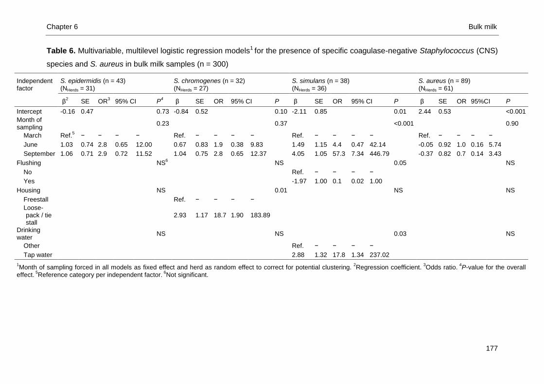
Chapter 6 Bulk milk
177
Table 6. Multivariable, multilevel logistic regression models1 for the presence of specific coagulase-negative Staphylococcus (CNS)
species and S. aureus in bulk milk samples (n = 300)
Independent factor
S. epidermidis (n = 43) (NHerds = 31)
S. chromogenes (n = 32) (NHerds = 27)
S. simulans (n = 38) (NHerds = 36)
S. aureus (n = 89) (NHerds = 61)
β
2 SE OR
3 95% CI P
4 β SE OR 95% CI P β SE OR 95% CI P β SE OR 95%CI P
Intercept -0.16 0.47 0.73 -0.84 0.52 0.10 -2.11 0.85 0.01 2.44 0.53 <0.001
Month of sampling
0.23
0.37
<0.001
0.90
March Ref.5 − − − −
Ref. − − − −
Ref. − − − −
Ref. − − − −
June 1.03 0.74 2.8 0.65 12.00
0.67 0.83 1.9 0.38 9.83
1.49 1.15 4.4 0.47 42.14
-0.05 0.92 1.0 0.16 5.74
September 1.06 0.71 2.9 0.72 11.52
1.04 0.75 2.8 0.65 12.37
4.05 1.05 57.3 7.34 446.79
-0.37 0.82 0.7 0.14 3.43
Flushing
NS6
NS
0.05
NS
No
Ref. − − − −
Yes
-1.97 1.00 0.1 0.02 1.00
Housing
NS 0.01
NS
NS
Freestall
Ref. − − − −
Loose-pack / tie stall
2.93 1.17 18.7 1.90 183.89
Drinking water
NS
NS
0.03
NS
Other
Ref. − − − −
Tap water
2.88 1.32 17.8 1.34 237.02
1Month of sampling forced in all models as fixed effect and herd as random effect to correct for potential clustering.
2Regression coefficient.
3Odds ratio.
4P-value for the overall
effect. 5Reference category per independent factor.
6Not significant.
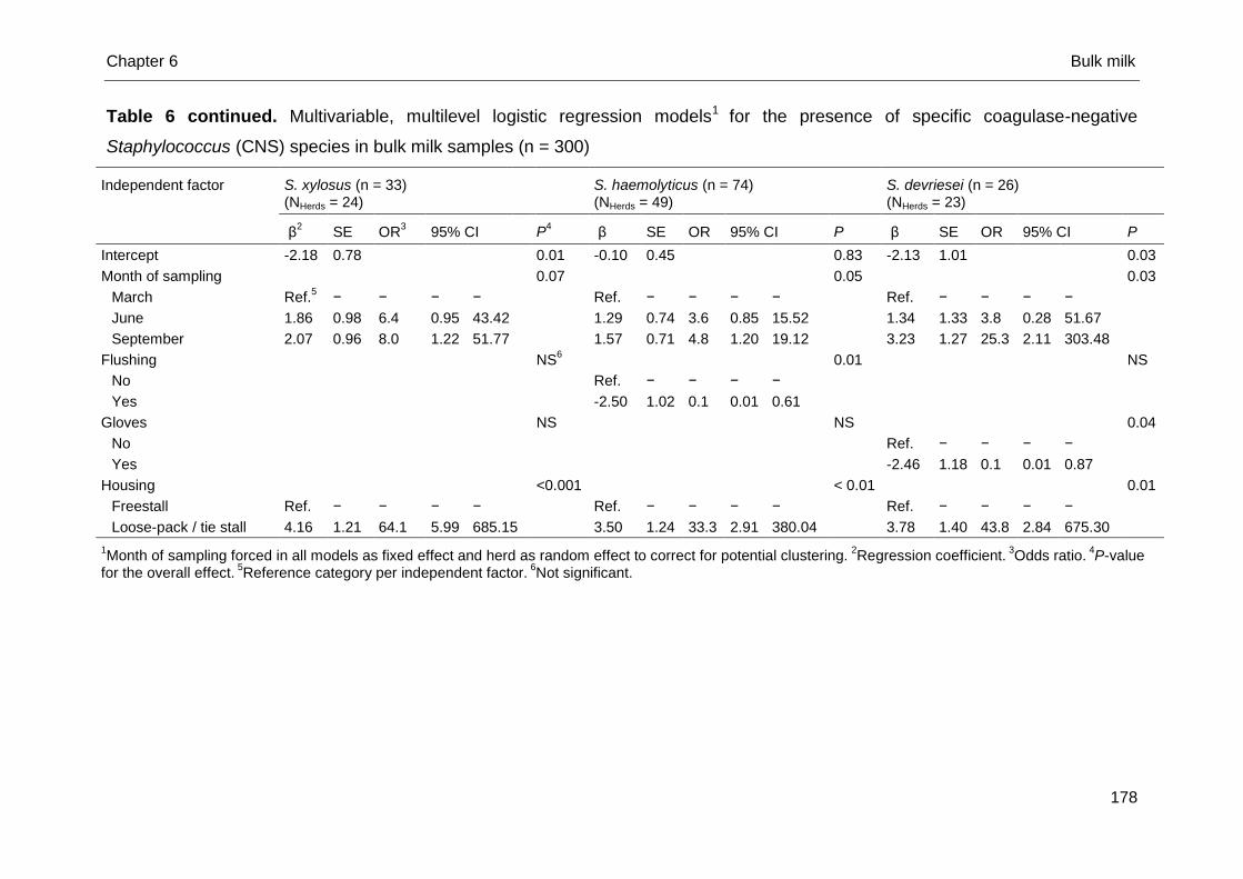
Chapter 6 Bulk milk
178
Table 6 continued. Multivariable, multilevel logistic regression models1 for the presence of specific coagulase-negative
Staphylococcus (CNS) species in bulk milk samples (n = 300)
Independent factor S. xylosus (n = 33) (NHerds = 24)
S. haemolyticus (n = 74) (NHerds = 49)
S. devriesei (n = 26) (NHerds = 23)
β2 SE OR
3 95% CI P
4 β SE OR 95% CI P β SE OR 95% CI P
Intercept -2.18 0.78 0.01 -0.10 0.45 0.83 -2.13 1.01 0.03
Month of sampling
0.07
0.05
0.03
March Ref.5 − − − −
Ref. − − − −
Ref. − − − −
June 1.86 0.98 6.4 0.95 43.42
1.29 0.74 3.6 0.85 15.52
1.34 1.33 3.8 0.28 51.67
September 2.07 0.96 8.0 1.22 51.77
1.57 0.71 4.8 1.20 19.12
3.23 1.27 25.3 2.11 303.48
Flushing
NS
6
0.01
NS
No
Ref. − − − −
Yes
-2.50 1.02 0.1 0.01 0.61
Gloves
NS
NS
0.04
No
Ref. − − − −
Yes
-2.46 1.18 0.1 0.01 0.87
Housing <0.001 < 0.01 0.01
Freestall Ref. − − − − Ref. − − − − Ref. − − − −
Loose-pack / tie stall 4.16 1.21 64.1 5.99 685.15 3.50 1.24 33.3 2.91 380.04 3.78 1.40 43.8 2.84 675.30
1Month of sampling forced in all models as fixed effect and herd as random effect to correct for potential clustering.
2Regression coefficient.
3Odds ratio.
4P-value
for the overall effect. 5Reference category per independent factor.
6Not significant.
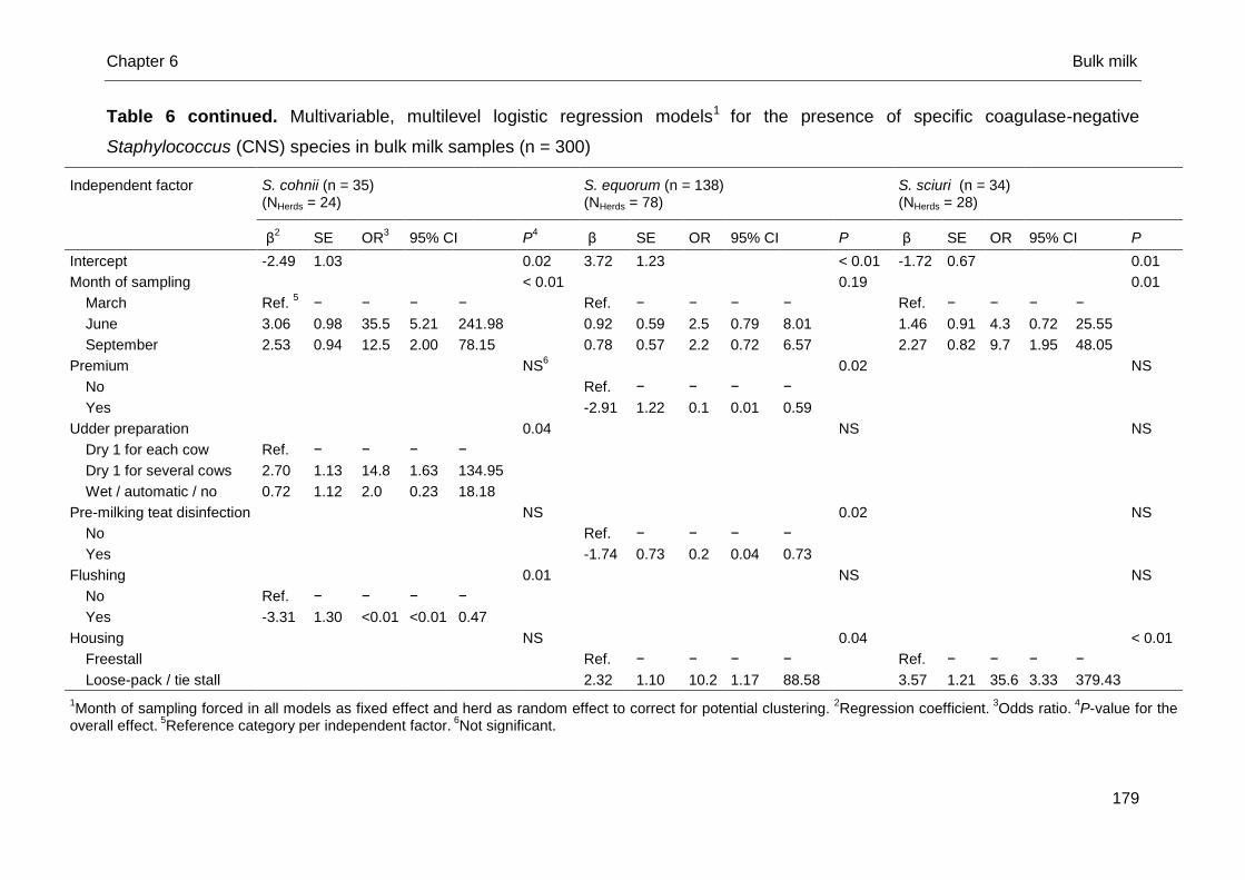
Chapter 6 Bulk milk
179
Table 6 continued. Multivariable, multilevel logistic regression models1 for the presence of specific coagulase-negative
Staphylococcus (CNS) species in bulk milk samples (n = 300)
Independent factor
S. cohnii (n = 35) (NHerds = 24)
S. equorum (n = 138) (NHerds = 78)
S. sciuri (n = 34) (NHerds = 28)
β2 SE OR
3 95% CI P
4 β SE OR 95% CI P β SE OR 95% CI P
Intercept -2.49 1.03
0.02 3.72 1.23 < 0.01 -1.72 0.67 0.01
Month of sampling
< 0.01
0.19
0.01
March Ref. 5 − − − −
Ref. − − − −
Ref. − − − −
June 3.06 0.98 35.5 5.21 241.98
0.92 0.59 2.5 0.79 8.01
1.46 0.91 4.3 0.72 25.55
September 2.53 0.94 12.5 2.00 78.15
0.78 0.57 2.2 0.72 6.57
2.27 0.82 9.7 1.95 48.05
Premium
NS
6
0.02
NS
No
Ref. − − − −
Yes
-2.91 1.22 0.1 0.01 0.59
Udder preparation
0.04
NS
NS
Dry 1 for each cow Ref. − − − −
Dry 1 for several cows 2.70 1.13 14.8 1.63 134.95
Wet / automatic / no 0.72 1.12 2.0 0.23 18.18
Pre-milking teat disinfection
NS
0.02
NS
No
Ref. − − − −
Yes
-1.74 0.73 0.2 0.04 0.73
Flushing
0.01
NS
NS
No Ref. − − − −
Yes -3.31 1.30 <0.01 <0.01 0.47
Housing
NS
0.04
< 0.01
Freestall
Ref. − − − −
Ref. − − − −
Loose-pack / tie stall
2.32 1.10 10.2 1.17 88.58
3.57 1.21 35.6 3.33 379.43
1Month of sampling forced in all models as fixed effect and herd as random effect to correct for potential clustering.
2Regression coefficient.
3Odds ratio.
4P-value for the
overall effect. 5Reference category per independent factor.
6Not significant.
Chapter 6 Bulk milk
180
As the more relevant CNS, i.e. S. chromogenes, S. simulans, and S. xylosus, have
already been associated with an increased quarter SCC and have been shown to
cause persistent IMI (Supré et al., 2011; Fry et al., 2014; De Visscher et al., 2015),
we hypothesize that herds suffering from a high proportion of cows with an elevated
SCC might have decided to monitor their udder health more closely with their
veterinarian. An explanation for the association of drinking water with the isolation of
S. simulans from bulk milk is also difficult. Sampimon et al. (2009) observed a
negative association between drinking water and the presence of CNS in quarter
milk samples. The positive association in the current study might also be attributed to
a cause-effect reversal.
Conclusions
Bulk milk yields a wide range of different CNS species. Host-adapted as well as
environmental CNS species are present. Several herd-level risk factors were
associated with the presence of subgroups of as well as specific CNS species.
Contamination of the bulk milk with CNS might occur from environmental
transmission for some species, i.e. S. equorum or S. cohnii, or from within the udder
for other species, i.e. S. simulans. However, studies collecting bulk milk and quarter
milk samples in the same time frame along with environmental samples should be
conducted to determine the exact origin of CNS species in bulk milk.
.
Chapter 6 Bulk milk
181
References
Bertelloni, F., F. Fratini, V. V. Ebani, A. Galiero, B. Turchi, and D. Cerri. 2015.
Detection of genes encoding for enterotoxines, TSST-1, and biofilm production in
coagulase-negative staphylococci from bovine bulk tank milk. Dairy Sci. Technol.
95:341-352.
Braem, G., S. De Vliegher, B. Verbist, V. Piessens, E. Van Coillie, L. De Vuyst, and
F. Leroy. 2013. Unraveling the microbiota of teat apices of clinically healthy
lactating dairy cows, with special emphasis on coagulase-negative staphylococci.
J. Dairy Sci. 96:1499-1510.
De Visscher, A., F. Haesebrouck, S. Piepers, W. Vanderhaeghen, K. Supré, F.
Leroy, E. Van Coillie, and S. De Vliegher. 2013. Assessment of the suitability of
mannitol salt agar for growing bovine-associated coagulase-negative
staphylococci and its use under field conditions. Res. Vet. Sci. 95:347-351.
De Visscher, A., K. Supré, F. Haesebrouck, R. N. Zadoks, V. Piessens, E. Van
Coillie, S. Piepers, and S. De Vliegher. 2014. Further evidence for the existence of
environmental and host-associated species of coagulase-negative staphylococci
in dairy cattle. Vet. Microbiol. 172:466-474.
De Visscher, A., S. Piepers, K. Supré, F. Haesebrouck, and S. De Vliegher. 2015.
Short communication: Species group-specific predictors at the cow and quarter
level for intramammary infection with coagulase-negative staphylococci in dairy
cattle throughout lactation. J. Dairy Sci. 98:5448-5453.
De Visscher, A., S. Piepers, F. Haesebrouck, and S. De Vliegher. 2016a. Teat apex
colonization with coagulase-negative Staphylococcus species before parturition:
distribution and species-specific risk factors. J. Dairy Sci. 99:1427-1439.
De Visscher, A., S. Piepers, F. Haesebrouck, and S. De Vliegher. 2016b.
Intramammary infection with coagulase-negative Staphylococcus species at
parturition: species-specific prevalence, risk factors, and impact on udder health.
J. Dairy Sci. In review.
Elmoslemany, A. M., G. P. Keefe, I. R. Dohoo, and T. R. Dingwell. 2009a.
Microbiological quality of bulk tank raw milk in Prince Edward Island dairy herds.
J. Dairy Sci. 92:4239-4248.
Chapter 6 Bulk milk
182
Elmoslemany, A. M., G. P. Keefe, I. R. Dohoo, and B. M. Jayarao. 2009b. Risk
factors for bacteriological quality of bulk tank milk in Prince Edward Island dairy
herds. Part 2: Bacteria count-specific risk factors. J. Dairy Sci. 92:2644-2652.
Fox, L. K. 2009. Prevalence, incidence and risk factors for heifer mastitis. Vet.
Microbiol. 134:82-88.
Fry, P. R., J. R. Middleton, S. Dufour, J. Perry, D. Scholl, and I. Dohoo. 2014.
Association of coagulase-negative staphylococcal species, mammary quarter milk
somatic cell count, and persistence of intramammary infection in dairy cattle. J.
Dairy Sci. 97:4876-4885.
Gillespie, B. E., M. J. Lewis, S. Boonyayatra, M. L. Maxwell, A. Saxton, S. P. Oliver,
and R. A. Almeida. 2012. Short communication: evaluation of bulk tank milk
microbiological quality of nine dairy farms in Tennessee. J. Dairy Sci. 95:4275-
4279.
Goldstein, H., R. W. Browne, and J. Rasbash. 2002. Partitioning variation in
multilevel models. Understanding Stat. I 4:223-231.
Hogeveen, H., K. Huijps, and T. J. G. M. Lam. 2011. Economic aspects of mastitis:
New developments. New Zeal. Vet. J. 59:16-23.
Jayarao, B. M., S. R. Pillai, A. A. Sawant, D. R. Wolfgang, and N. V. Hegde. 2004.
Guidelines for monitoring bulk milk somatic cell count and bacterial counts. J.
Dairy Sci. 87:3561-3573.
Noordhuizen, J. P. T. M., K. Frankena, M. V. Thursfield, and E. A. M. Graat. 2001.
Application of quantitative methods in veterinary epidemiology. Wageningen Pers,
Wageningen, The Netherlands.
Olde Riekerink, R. G. M., H. W. Barkema, S. Veenstra, D. E. Poole, R. T. Dingwell,
and G. P. Keefe. 2006. Prevalence of contagious mastitis pathogens in bulk tank
milk in Prince Edward Island. Can. Vet. J. 47:567-572.
Olde Riekerink, R. G. M., H. W. Barkema, D. T. Scholl, D. E. Poole, and D. F. Kelton.
2010. Management practices associated with the bulk-milk prevalence of
Staphylococcus aureus in Canadian dairy farms. Prev. Vet. Med. 97:20-28.
Østerås, O., L. Sølverød, and O. Reksen. 2006. Milk culture results in a large
Norwegian survey. Effects of season, parity, days in milk, resistance, and
clustering. J. Dairy Sci. 89:1010-1023.
Park, J. Y., L. K. Fox, K. S. Seo, M. A. McGuire, Y. H. Park, F. R. Rurangirwa, W. M.
Sischo, and G. A. Bohach. 2011. Comparison of phenotypic and genotypic
Chapter 6 Bulk milk
183
methods for the species identification of coagulase-negative staphylococcal
isolates from bovine intramammary infections. Vet. Microbiol. 2011:142-148.
Persson Waller, K., A. Aspán, A. Nyman, Y. Persson, and U. Grönlund Andersson.
2011. CNS species and antimicrobial resistance in clinical and subclinical bovine
mastitis. Vet. Microbiol. 152:112-116.
Piepers, S., L. De Meulemeester, A. de Kruif, G. Opsomer, H. W. Barkema, and S.
De Vliegher. 2007. Prevalence and distribution of mastitis pathogens in
subclinically infected dairy cows in Flanders, Belgium. J. Dairy Res. 74:478-483.
Piepers, S., S. De Vliegher, A. de Kruif, G. Opsomer, and H. W. Barkema. 2009.
Impact of intramammary infections in dairy heifers on future udder health, milk
production, and culling. Vet. Microbiol. 134:113-121.
Piepers, S., K. Peeters, G. Opsomer, H. W. Barkema, K. Frankena, and S. De
Vliegher. 2011. Pathogen group specific risk factors at herd, heifer and quarter
levels for intramammary infections in early lactating dairy heifers. Prev. Vet. Med.
99:91-101.
Piessens, V., E. Van Coillie, B. Verbist, K. Supré, G. Braem, A. Van Nuffel, L. De
Vuyst M. Heyndrickx, and S. De Vliegher. 2011. Distribution of coagulase-
negative Staphylococcus species from dairy cows’ milk and environment differs
between herds. J. Dairy Sci. 94:2933-2944.
Reyher, K. K., S. Dufour, H. W. Barkema, L. Des Côteaux, T. J. DeVries, I. R Dohoo,
G. P. Keefe, J.-P. Roy, and D. T. Scholl. 2011. The national cohort of dairy farms.
A data collection platform for mastitis research in Canada. J. Dairy Sci. 94:1616-
1626.
Sampimon, O. C., H. W. Barkema, I. M. G. A. Berends, J. Sol, and T. J. G. M. Lam.
2009. Prevalence and herd-level risk factors for intramammary infection with
coagulase-negative staphylococci in Dutch dairy herds. Vet. Microbiol. 134:37-44.
Sampimon, O., B. H. P. van den Borne, I. Santman-Berends, H. W. Barkema, and T.
Lam. 2010. Effect of coagulase-negative staphylococci on somatic cell count in
Dutch dairy herds. J. Dairy Res. 77:318-324.
Santos, O. C. S., E. M. Barros, M. A. V. P. Brito, M. C. F. Bastos, K. R. N. Santos,
and M. Giambiagi-deMarval. 2008. Identification of coagulase-negative
staphylococci from bovine mastitis using RFLP-PCR of the groEL gene. Vet.
Microbiol. 130:134-140.
Chapter 6 Bulk milk
184
Schukken, Y. H., R. N. González, L. L. Tikofsky, H. F. Schulte, C. G. Santisteban, F.
L. Welcome, G. J. Bennett, M. J. Zurakowski, and R. N. Zadoks. 2009. CNS
mastitis: Nothing to worry about? Vet. Microbiol. 134:9-14.
Supré, K., S. De Vliegher, O. C. Sampimon, R. N. Zadoks, M. Vaneechoutte, M.
Baele, E. De Graef, S. Piepers, and F. Haesebrouck. 2009. Technical note: Use of
transfer RNA-intergenic spacer PCR combined with capillary electrophoresis to
identify coagulase-negative Staphylococcus species originating from bovine milk
and teat apices. J. Dairy Sci. 92:3204-3210.
Supré, K., F. Haesebrouck, R. N. Zadoks, M. Vaneechoutte, S. Piepers, and S. De
Vliegher. 2011. Some coagulase-negative Staphylococcus species affect udder
health more than others. J. Dairy Sci. 94:2329-2340.
Taponen, S., J. Björkroth, and S. Pyörälä. 2008. Coagulase-negative staphylococci
isolated from bovine extramammary sites and intramammary infections in a single
dairy herd. J. Dairy Res. 75:422-429.
Vanderhaeghen, W., S. Piepers, F. Leroy, E. Van Coillie, F. Haesebrouck, and S. De
Vliegher. 2015. Identification, typing, ecology and epidemiology of coagulase-
negative staphylococci associated with ruminants. Vet. J. 203:44-51.
Zadoks, R. N., H. G. Allore, H. W. Barkema, O. C. Sampimon, G. J. Wellenberg, Y.
T. Gröhn, and Y. H. Schukken. 2001. Cow- and quarter-level risk factors for
Streptococcus uberis and Staphylococcus aureus mastitis. J. Dairy Sci. 84:2649-
2663.


Chapter 7
General Discussion
A. De Visscher
Department of Reproduction, Obstetrics, and Herd Health
Faculty of Veterinary Medicine
Ghent University, Merelbeke, Belgium

Chapter 7 General discussion
189
Introduction
Effort is ongoing to broaden our knowledge of the ecological and epidemiological
nature and the relevance for udder health of the bovine-related coagulase-negative
Staphylococcus species (CNS). This thesis also aimed to do so. Mannitol salt agar
was considered as a suitable medium for CNS recovery (Chapter 3) as all bovine-
associated CNS were able to grow, and was further applied in Chapter 4.2, 5.1 and
6. A predominance of Staphylococcus chromogenes in quarter milk samples and the
recovery in all herds was observed in early lactation (Chapter 5.1), whereas bulk milk
harboured a wide variety of CNS species (Chapter 6). Herd-specific CNS microbiota
were detected (Chapter 4.2, 5.1, 5.2, 6). Quarters of heifers (Chapter 5.1, 5.2) and
quarters of animals in peak lactation (Chapter 5.2) were more likely to be infected
with the so-called more relevant CNS species, i.e. S. chromogenes, S. simulans, and
S. xylosus. The udder-adapted, and by extension host-adapted, nature of S.
chromogenes was substantiated (Chapter 4.2, 5.1). Also, the environmental ecology
of S. cohnii (Chapter 5.1), S. equorum (Chapter 4.2, 5.1), S. saprophyticus and S.
sciuri (Chapter 5.1) was confirmed whereas a more host-adapted nature was
evidenced for S. devriesei and S. haemolyticus (Chapter 4.2). Staphylococcus
chromogenes was also shown to be a predominant teat apex colonizer prior to
calving (Chapter 4.2) whereas teat apices of lactating dairy cows and heifers were
colonized to a lesser extent with this species (Chapter 4.1). Teat apices colonized
with S. chromogenes prior to parturition had higher odds of having S. chromogenes
intramammary infection (IMI) in early lactation (Chapter 5.1). The higher relevance for
udder health of S. chromogenes, S. simulans, and S. xylosus was confirmed as those
3 species were able to increase the quarter SCC both in early and in a later stage in
lactation (Chapter 5.1, 5.2) whereas other species were not.
All recovered CNS in this thesis were accurately identified to the species level
providing detailed and unique information. The first applied genotypic identification
method was transfer RNA intergenic spacer PCR (tDNA-PCR) as this technique was
validated for bovine-related CNS species by our research group (Supré et al., 2009),
implying all necessary equipment and a comprehensive library were available. If no
identification could be obtained, sequencing of the rpoB gene was performed.
However, the presences of several genera other than the Staphylococcus genus
(Chapter 3, 4.2, 5.1, 6), though often phenotypically indistinguishable from them,
Chapter 7 General discussion
190
made us prefer sequencing of the 16S rRNA gene, which relies on universal primers
in contrast with the rpoB gene. The lower accuracy of our preferred sequencing
method did not affect our results as sequencing of the 16S rRNA gene was mostly
performed to identify genera other than the Staphylococcus genus and was never
used alone, though in combination with tDNA-PCR, for CNS species identification.
Prevalence data and species distribution in Flanders were completed as several
unexplored bovine habitats were studied and for the first time, subgroup- and
species-specific risk factors and predictors were identified. The results in this work
considerably enhance our insights in bovine-associated CNS ecology and
epidemiology, and their relevance for udder health. Besides, large collections of CNS
species and associated data are available for further investigations. In this final
chapter our findings are summarized, compared to other research and discussed.
Future CNS research will benefit from the use of mannitol salt agar
Research intending to examine the prevalence and distribution of specific bacterial
species requires the use of a (quasi-)selective medium, allowing the genera / species
of interest to grow as well as circumventing suppression by other pathogens at the
same time. The latter prerequisite was unconfirmed for CNS and was therefore
investigated in Chapter 3. The ability of all bovine-associated CNS to grow on MSA
was demonstrated, though some aspects should be taken into account. First, a few
isolates of some CNS species, i.e. S. chromogenes, S. equorum, S. haemolyticus, S.
hominis, and S. lugdunensis, did not show growth within 24h aerobic incubation at
37°C (Chapter 3). A 48h incubation time should thus be respected, especially in
those studies that aim at identifying all present CNS species in a specific habitat. In
fact, studies using MSA with different incubation conditions (Piessens et al., 2011; El-
Jakee et al., 2013; Abbas et al., 2014; Li et al., 2015) might actually have missed the
presence of some species in their samples for that reason. Secondly, both growth
characteristics and mannitol fermentation seem to strongly vary among isolates
within species (Chapter 3), emphasizing caution is needed in the interpretation.
Therefore, mannitol fermentation cannot be used solely in differentiating bovine S.
aureus from non-S. aureus staphylococci (Cheng et al., 2010; Kateete et al., 2010;
Chavhan et al; 2012; Oliveira et al., 2015; Chapter 3), as was wrongly assumed
before (Chapman, 1945; Addis et al., 2011; Singh et al., 2013). The need for
Chapter 7 General discussion
191
supplementary methods for phenotypic identification of S. aureus does, however, not
outweigh the advantages of this conscientiously tested agar for overcrowded bovine-
associated habitats. The suitability of MSA for CNS recovery under field conditions
was tested as well (Chapter 3) and the practicability of this agar in comparison with
non-selective media was demonstrated. The results made us decide to use MSA for
CNS recovery from all other habitats and matrices in our studies, namely teat apices
(Chapter 4.2), quarter milk samples (Chapter 5.1) and bulk milk samples (Chapter 6).
In the aforementioned studies, pathogens other than staphylococci were isolated
though they never hindered the recovery of all phenotypically different CNS colony
types (Chapman et al., 1945; Graber et al., 2013). Actually, the number of
phenotypically different colony types ranged from 0 (no growth) to 4 (teat apices,
Chapter 4.2), 5 (quarter milk samples, Chapter 5.1), and 9 (bulk milk samples,
Chapter 6). Other pathogens recovered from the studied habitats belonged to the
phyla Firmicutes (Chapman, 1945; Han et al., 2007; Braem et al., 2013; Chapter 3,
4.2, 5.1, 6), Proteobacteria (Chapter 4.2, 5.1, 6), and Actinobacteria (Braem et al.,
2013; Chapter 3, 4.2, 6) and a minority of isolates were fungi (Chapter 4.2, 6).
In conclusion, as MSA is the only thoroughly examined (quasi-)selective agar for
the recovery of bovine-associated CNS from multiple bovine-associated habitats and
non-selective media are inappropriate for CNS recovery from overcrowded habitats,
such as teat apices, bulk milk and manure, future studies should take advantage of
this medium. However, if one wants to recover other (mastitis-causing) microbiota
besides staphylococci, non-selective media can be used for quarter milk samples
while for overpopulated habitats several other (tested) (quasi-)selective media along
with MSA should be applied.
Bovine-associated habitats have been further explored
Our work contributed to mapping the species-specific CNS prevalence and
distribution of IMI both in early lactation and throughout lactation (Table 1, Chapter
5.1). Yet, several other studies have genotypically identified different CNS species.
Still, not all of those genotypic studies were set up for determining the CNS species-
specific quarter IMI prevalence (Santos et al., 2008; Sampimon et al., 2009b; Park et
al., 2011; Persson Waller et al., 2011; Quirk et al., 2012; Fry et al., 2014b; Lange et
al., 2015). Additionally, in some other studies not all isolated CNS were retained for
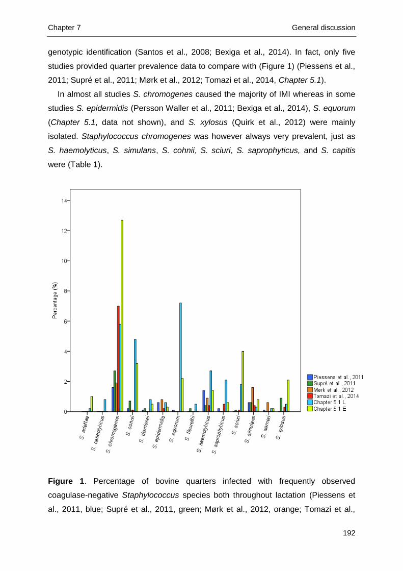
Chapter 7 General discussion
192
genotypic identification (Santos et al., 2008; Bexiga et al., 2014). In fact, only five
studies provided quarter prevalence data to compare with (Figure 1) (Piessens et al.,
2011; Supré et al., 2011; Mørk et al., 2012; Tomazi et al., 2014, Chapter 5.1).
In almost all studies S. chromogenes caused the majority of IMI whereas in some
studies S. epidermidis (Persson Waller et al., 2011; Bexiga et al., 2014), S. equorum
(Chapter 5.1, data not shown), and S. xylosus (Quirk et al., 2012) were mainly
isolated. Staphylococcus chromogenes was however always very prevalent, just as
S. haemolyticus, S. simulans, S. cohnii, S. sciuri, S. saprophyticus, and S. capitis
were (Table 1).
Figure 1. Percentage of bovine quarters infected with frequently observed
coagulase-negative Staphylococcus species both throughout lactation (Piessens et
al., 2011, blue; Supré et al., 2011, green; Mørk et al., 2012, orange; Tomazi et al.,
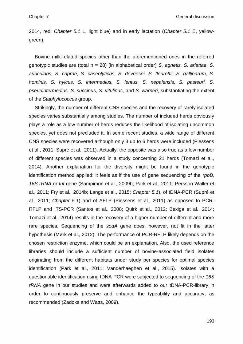
Chapter 7 General discussion
193
2014, red; Chapter 5.1 L, light blue) and in early lactation (Chapter 5.1 E, yellow-
green).
Bovine milk-related species other than the aforementioned ones in the referred
genotypic studies are (total n = 28) (in alphabetical order) S. agnetis, S. arlettae, S.
auricularis, S. caprae, S. caseolyticus, S. devriesei, S. fleurettii, S. gallinarum, S.
hominis, S. hyicus, S. intermedius, S. lentus, S. nepalensis, S. pasteuri, S.
pseudintermedius, S. succinus, S. vitulinus, and S. warneri, substantiating the extent
of the Staphylococcus group.
Strikingly, the number of different CNS species and the recovery of rarely isolated
species varies substantially among studies. The number of included herds obviously
plays a role as a low number of herds reduces the likelihood of isolating uncommon
species, yet does not precluded it. In some recent studies, a wide range of different
CNS species were recovered although only 3 up to 6 herds were included (Piessens
et al., 2011; Supré et al., 2011). Actually, the opposite was also true as a low number
of different species was observed in a study concerning 21 herds (Tomazi et al.,
2014). Another explanation for the diversity might be found in the genotypic
identification method applied: it feels as if the use of gene sequencing of the rpoB,
16S rRNA or tuf gene (Sampimon et al., 2009b; Park et al., 2011; Persson Waller et
al., 2011; Fry et al., 2014b; Lange et al., 2015; Chapter 5.1), of tDNA-PCR (Supré et
al., 2011; Chapter 5.1) and of AFLP (Piessens et al., 2011) as opposed to PCR-
RFLP and ITS-PCR (Santos et al., 2008; Quirk et al., 2012; Bexiga et al., 2014;
Tomazi et al., 2014) results in the recovery of a higher number of different and more
rare species. Sequencing of the sodA gene does, however, not fit in the latter
hypothesis (Mørk et al., 2012). The performance of PCR-RFLP likely depends on the
chosen restriction enzyme, which could be an explanation. Also, the used reference
libraries should include a sufficient number of bovine-associated field isolates
originating from the different habitats under study per species for optimal species
identification (Park et al., 2011; Vanderhaeghen et al., 2015). Isolates with a
questionable identification using tDNA-PCR were subjected to sequencing of the 16S
rRNA gene in our studies and were afterwards added to our tDNA-PCR-library in
order to continuously preserve and enhance the typeability and accuracy, as
recommended (Zadoks and Watts, 2009).
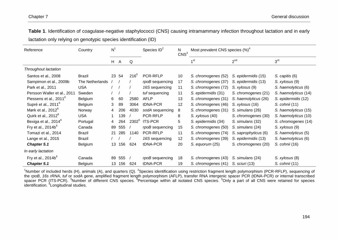
Chapter 7 General discussion
194
Table 1. Identification of coagulase-negative staphylococci (CNS) causing intramammary infection throughout lactation and in early
lactation only relying on genotypic species identification (ID)
Reference Country N1 Species ID
2 N
CNS3
Most prevalent CNS species (%)4
H A Q 1st 2
nd 3
rd
Throughout lactation
Santos et al., 2008 Brazil 23 54 2165 PCR-RFLP 10 S. chromogenes (52) S. epidermidis (15) S. capitis (6)
Sampimon et al., 2009b The Netherlands / / / rpoB sequencing 17 S. chromogenes (37) S. epidermidis (13) S. xylosus (9)
Park et al., 2011 USA / / / 16S sequencing 11 S. chromogenes (72) S. xylosus (9) S. haemolyticus (6)
Persson Waller et al., 2011 Sweden / / / tuf sequencing 11 S. epidermidis (31) S. chromogenes (21) S. haemolyticus (14)
Piessens et al., 20116 Belgium 6 60 2580 AFLP 13 S. chromogenes (31) S. haemolyticus (26) S. epidermidis (12)
Supré et al., 20116 Belgium 3 89 3064 tDNA-PCR 12 S. chromogenes (46) S. xylosus (16) S. cohnii (11)
Mørk et al., 20126 Norway 4 206 4030 sodA sequencing 8 S. chromogenes (31) S. simulans (26) S. haemolyticus (15)
Quirk et al., 20126 USA 1 139 / PCR-RFLP 8 S. xylosus (40) S. chromogenes (30) S. haemolyticus (10)
Bexiga et al., 20146 Portugal 4 264 2302
5 ITS-PCR 5 S. epidermidis (34) S. simulans (32) S. chromogenes (14)
Fry et al., 2014b6 Canada 89 555 / rpoB sequencing 15 S. chromogenes (50) S. simulans (24) S. xylosus (9)
Tomazi et al., 2014 Brazil 21 285 1140 PCR-RFLP 11 S. chromogenes (74) S. saprophyticus (6) S. haemolyticus (5)
Lange et al., 2015 Brazil / / / 16S sequencing 12 S. chromogenes (39) S. epidermidis (13) S. haemolyticus (6)
Chapter 5.1 Belgium 13 156 624 tDNA-PCR 20 S. equorum (25) S. chromogenes (20) S. cohnii (16)
In early lactation
Fry et al., 2014b6 Canada 89 555 / rpoB sequencing 18 S. chromogenes (43) S. simulans (24) S. xylosus (8)
Chapter 5.1 Belgium 13 156 624 tDNA-PCR 19 S. chromogenes (41) S. sciuri (13) S. cohnii (11)
1Number of included herds (H), animals (A), and quarters (Q).
2Species identification using restriction fragment length polymorphism (PCR-RFLP), sequencing of
the rpoB, 16s rRNA, tuf or sodA gene, amplified fragment length polymorphism (AFLP), transfer RNA intergenic spacer PCR (tDNA-PCR) or internal transcribed spacer PCR (ITS-PCR).
3Number of different CNS species.
4Percentage within all isolated CNS species.
5Only a part of all CNS were retained for species
identification. 6Longitudinal studies.
Chapter 7 General discussion
195
The use of MSA can also explain the observed difference as overgrowth of
pathogens other than CNS was circumvented and all phenotypically different colonies
were thus picked up and subjected to further analysis. Quirk et al. (2012), however,
also used MSA though in combination with another identification method.
Chapter 6 presents the first species-specific herd-level study using bulk milk as an
interesting matrix in CNS research and relying on genotypic identification. Both host-
adapted CNS species, e.g. S. chromogenes, and environmental species, e.g. S.
equorum, were often recovered from bulk milk. We hypothesize that the
predominance of either host-adapted or environmental CNS species in bulk milk is
associated with a domination of major pathogens with the same ecology in the herd.
In that respect, screening the CNS distribution in bulk milk could provide information
on the herd health status and certain management issues, as already seen for so-
called contagious major pathogens (Olde Riekerink et al., 2006, 2010; Boss et al.,
2011). If this assumption is true, screening of the species-specific CNS distribution in
bulk milk could eventually become an efficient instrument to improve udder health
and milk quality through an optimised management.
Chapter 4.1 and Chapter 4.2 add to our knowledge about teat apex colonization
(TAC) both prior to parturition and throughout lactation. The predominant species
colonizing teat apices of dairy cows and heifers are shown in Figure 2. Differences in
CNS prevalence and distribution among lactation studies might be explained by
variation in the number of included herds, ranging between 1 (Taponen et al., 2008)
and 3 (Chapter 4.1), the amount of swabbed teat apices being 65 (Taponen et al.,
2008), 72 (Braem et al., 2013) and 140 (Chapter 4.1), and the used identification
method, i.e. ribotyping (Taponen et al., 2008), (GTG)5-PCR (Braem et al., 2011;
Braem et al., 2013) and tDNA-PCR (Supré et al., 2009; Chapter 4.1). A larger
number of CNS species colonized teat apices prior to parturition (24 species, Chapter
4.2) than after calving (in lactation), with 10 (Taponen et al., 2008), 13 (Chapter 4.1)
and 14 (Braem et al., 2013) different species, respectively, colonizing teat apices.
Routinely cleaning of the teat end of lactating dairy cows and heifers as part of udder
preparation before milking, the flushing effect of lactation (Chapter 4.1), and the
application of post-milking teat disinfection (Piessens et al., 2012; Quirk et al., 2012)
potentially clarify this discrepancy. Recovery of CNS on MSA (Chapter 4.2) and a
considerably higher number of included herds likely enlarged the number of detected
species as well in our work (Chapter 4.2). On the other hand, only limited variation in
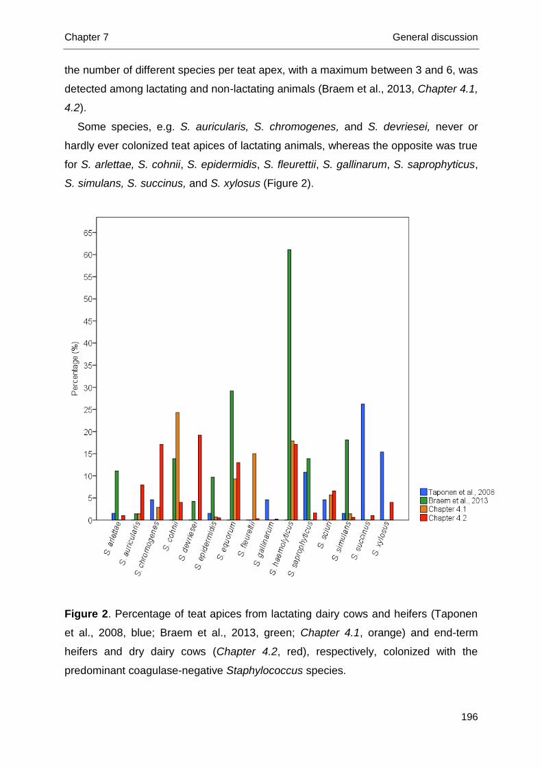
Chapter 7 General discussion
196
the number of different species per teat apex, with a maximum between 3 and 6, was
detected among lactating and non-lactating animals (Braem et al., 2013, Chapter 4.1,
4.2).
Some species, e.g. S. auricularis, S. chromogenes, and S. devriesei, never or
hardly ever colonized teat apices of lactating animals, whereas the opposite was true
for S. arlettae, S. cohnii, S. epidermidis, S. fleurettii, S. gallinarum, S. saprophyticus,
S. simulans, S. succinus, and S. xylosus (Figure 2).
Figure 2. Percentage of teat apices from lactating dairy cows and heifers (Taponen
et al., 2008, blue; Braem et al., 2013, green; Chapter 4.1, orange) and end-term
heifers and dry dairy cows (Chapter 4.2, red), respectively, colonized with the
predominant coagulase-negative Staphylococcus species.
Chapter 7 General discussion
197
Staphylococcus equorum, S. haemolyticus, and S. sciuri, though to a lesser extent,
frequently colonized teat apices from both lactating and non-lactating animals
(Taponen et al., 2008; Braem et al., 2013; Chapter 4.1, 4.2), suggesting an
omnipresent nature of these particular species. Especially S. chromogenes grabbed
our attention, being a dominant species (Chapter 4.2) and the sole species present
on teat apices in all herds pre-partum (De Vliegher et al., 2003; Chapter 4.2).
Additionally, this species was abundantly present on the udder skin of lactating
animals (Taponen et al., 2008). Despite the latter findings, S. chromogenes was
hardly recovered from teat apices of cows in lactation (Taponen et al., 2008; Braem
et al., 2013; Chapter 4.1), suggesting a different CNS microbiota on teat skin
compared to teat apices.
A herd-specific CNS microbiota exists and demands further study
Staphylococcus chromogenes is highly prevalent in bovine milk in all herds
whereas for other species large within-herd differences are observed (Piessens et al.,
2011; Supré et al., 2011; Mørk et al., 2012; Bexiga et al., 2014; Chapter 5.1, 5.2).
Research exploring TAC agrees on the finding that every herd seems to have its own
CNS microbiota (Braem et al., 2013; Chapter 4.1, 4.2). Also, S. equorum was
recovered from the bulk milk in almost all herds. Other species such as S.
haemolyticus, S. cohnii, and S. epidermidis were in none of the three bulk milk
samples detected in some herds whereas in other herds they were isolated from all
three bulk milk samples (Chapter 6). The observed herd-dependency in the different
habitats highlights the potential role of factors such as management and climatic
conditions, and requires herd-level analyses to identify variables associated with the
presence of CNS (species).
Chapter 6 identifies for the first time herd-level management factors associated
with individual CNS species, using bulk milk samples. In contrast with earlier studies
(Gillespie et al., 2012), Chapter 6 revealed a strong seasonal effect of CNS recovery
of environmental CNS species, i.e. S. cohnii, S. equorum, S. saprophyticus, and S.
sciuri, from bulk milk whereas the host-adapted species, i.e. S. chromogenes and S.
epidermidis, were equally prevalent in all seasons. Climatic conditions such as
temperature and precipitation are significantly affecting the TAC of several species
(Chapter 4.2). Contamination of bulk milk with teat apex colonizing species might
Chapter 7 General discussion
198
clarify the observed seasonal species distribution in bulk milk. Contrastingly,
(species-specific) CNS IMI did not depend on climatic conditions (Chapter 5.1). Still,
no quarter milk samples were available and only 3 bulk milk samples with a 3-month
interval were collected (Chapter 6) rather than e.g. weekly samples, potentially
affecting sensitivity (Godkin and Leslie, 1993; Olde Riekerink et al., 2010). The
conclusions from Chapter 6 remain thus preliminary, as working solely with bulk milk
has its limitations, and we look forward to the results from new studies combining the
CNS recovery from bulk milk with the CNS isolation from quarter milk samples in the
same herds.
Staphylococcus chromogenes should be easily differentiated from other CNS
species
Our work substantiates the higher relevance for udder health of S. chromogenes,
put forward by Supré et al. (2011) and Fry et al. (2014b), in early lactation (Chapter
5.1) and throughout lactation (Chapter 5.2). Still, Bexiga et al. (2014) questioned this
species-specific impact, and suggested a cow-dependent response on CNS IMI
rather than a species-dependent effect. In that study, however, only 5 different CNS
species were identified (Table 1) and the predominant species, S. chromogenes, S.
simulans, and S. epidermidis, were isolated in low numbers. The latter might explain
why the numerically higher SCC in S. chromogenes-infected quarters was not found
to be significantly different from the other CNS-infected quarters. In our opinion,
enough evidence exists that S. chromogenes is more relevant for udder health than
other CNS species, although we would not classify it as a major pathogen. The same
is likely true for S. simulans and S. xylosus, yet in the lack of sufficient numbers final
conclusions cannot be drawn. Besides the ability to elevate quarter SCC, S.
chromogenes is able to cause persistent IMI (Taponen et al., 2007; Thorberg et al.,
2009; Piessens et al., 2011; Chapter 5.1: data not shown). Others have confirmed
the persistency potential using pulsed-field gel electrophoresis (Gillespie et al., 2009;
Mørk et al., 2012; Bexiga et al., 2014; Fry et al., 2014b) and ongoing strain-typing,
using multi-locus sequence typing (MLST), may provide definite conclusions (Chapter
5.1). Interestingly, quarters non-cured from S. chromogenes in early lactation had a
significantly higher quarter SCC as opposed to uninfected quarters (Chapter 5.1: data
not shown), suggesting at least some S. chromogenes strains can survive and
Chapter 7 General discussion
199
maintain themselves in the mammary gland despite the established host immune
response.
Altogether, there are enough reasons to strive in the near future for an easy
differentiation of S. chromogenes from other CNS in both clinical and subclinical
mastitis in routine milk quality laboratories. Differentiation should rely on genotypic
methods as differences in phenotypic expression of several characteristics, e.g.
DNase reaction and mannitol fermentation, among isolates within species were
observed (Chapter 3). Eventually, it would allow to quantify the true relevance of S.
chromogenes. Nowadays, it looks as if prevention of S. chromogenes IMI in well-
managed dairy herds with an already good udder health should be encouraged to
keep bulk milk SCC low. As quarters with S. chromogenes colonized teat apices
were more likely to be infected with S. chromogenes in early lactation and
subsequently were more likely to be S. chromogenes-infected in that quarter one
month later (Chapter 4.2, 5.1), prevention of TAC might be the key. This hypothesis
should, however, be confirmed by strain-typing, which is ongoing. How to prevent
and control TAC with S. chromogenes is the next question that needs to be
answered. It looks as if teat dipping cannot bring merit as this was not identified as a
factor associated with TAC (Chapter 4.2). Yet, quarters with inverted teat ends had
higher odds of S. chromogenes infection (Chapter 5.1). Teat characteristics
potentially depend on the genetic background, implying genetic selection should be
investigated (Detilleux, 2009; Verbeke et al., 2012). Only if S. chromogenes TAC
during lactation also increases the likelihood of S. chromogenes IMI, the role of
milking machine settings and the choice of teat cup liners should be scrutinized as
well (Bhutto et al., 2010; Zwertvaegher et al., 2013).
Subgroup- and species-specific risk factors have been identified for the first
time
Along with the increased prevalence and relevance of CNS IMI, the need for risk
factor studies, identifying associations with the presence of CNS in different habitats,
arose. Several studies conducted multilevel, multivariable analysis and assessed
variance components before (Sampimon et al., 2009a; Piepers et al., 2011;
Passchyn et al., 2014), though CNS were always considered as one homogeneous
group.
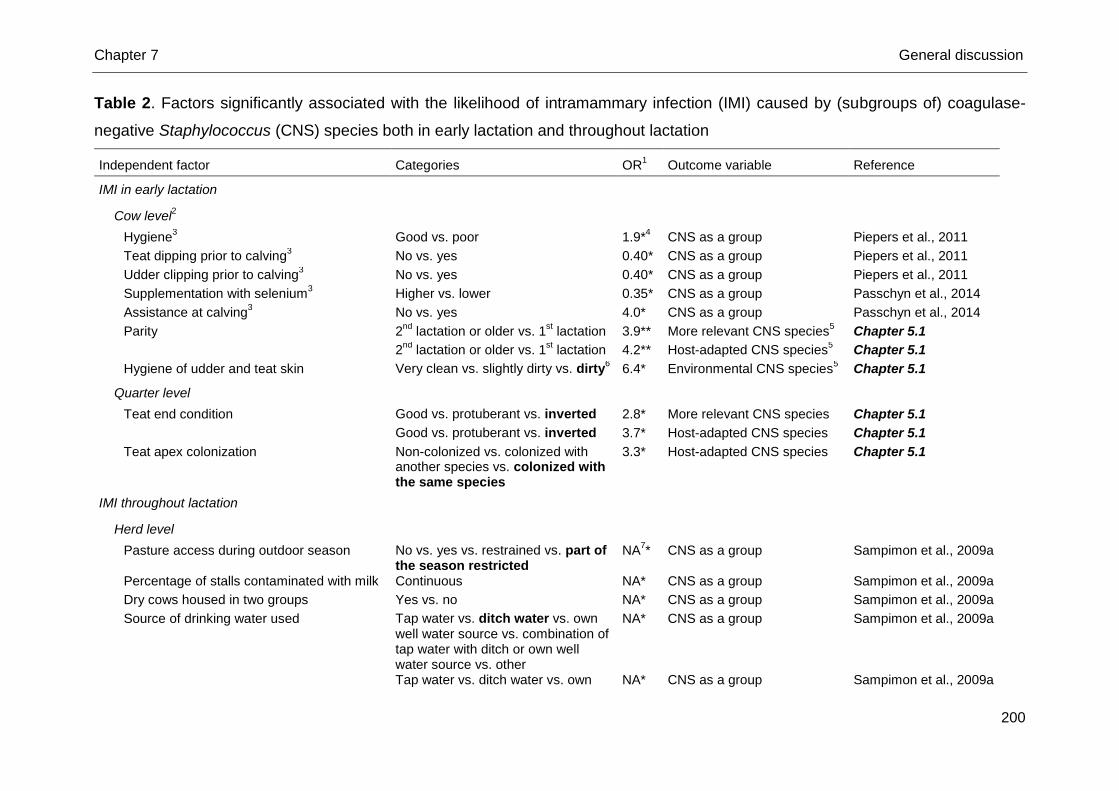
Chapter 7 General discussion
200
Table 2. Factors significantly associated with the likelihood of intramammary infection (IMI) caused by (subgroups of) coagulase-
negative Staphylococcus (CNS) species both in early lactation and throughout lactation
Independent factor Categories OR1 Outcome variable Reference
IMI in early lactation
Cow level2
Hygiene3 Good vs. poor 1.9*
4 CNS as a group Piepers et al., 2011
Teat dipping prior to calving3 No vs. yes 0.40* CNS as a group Piepers et al., 2011
Udder clipping prior to calving3 No vs. yes 0.40* CNS as a group Piepers et al., 2011
Supplementation with selenium3 Higher vs. lower 0.35* CNS as a group Passchyn et al., 2014
Assistance at calving3 No vs. yes 4.0* CNS as a group Passchyn et al., 2014
Parity 2nd
lactation or older vs. 1st lactation 3.9** More relevant CNS species
5 Chapter 5.1
2nd
lactation or older vs. 1st lactation 4.2** Host-adapted CNS species
5 Chapter 5.1
Hygiene of udder and teat skin Very clean vs. slightly dirty vs. dirty6 6.4* Environmental CNS species
5 Chapter 5.1
Quarter level
Teat end condition Good vs. protuberant vs. inverted 2.8* More relevant CNS species Chapter 5.1
Good vs. protuberant vs. inverted 3.7* Host-adapted CNS species Chapter 5.1
Teat apex colonization Non-colonized vs. colonized with another species vs. colonized with the same species
3.3* Host-adapted CNS species Chapter 5.1
IMI throughout lactation
Herd level
Pasture access during outdoor season No vs. yes vs. restrained vs. part of the season restricted
NA7* CNS as a group Sampimon et al., 2009a
Percentage of stalls contaminated with milk Continuous NA* CNS as a group Sampimon et al., 2009a
Dry cows housed in two groups Yes vs. no NA* CNS as a group Sampimon et al., 2009a
Source of drinking water used Tap water vs. ditch water vs. own well water source vs. combination of tap water with ditch or own well water source vs. other
NA* CNS as a group Sampimon et al., 2009a
Tap water vs. ditch water vs. own NA* CNS as a group Sampimon et al., 2009a
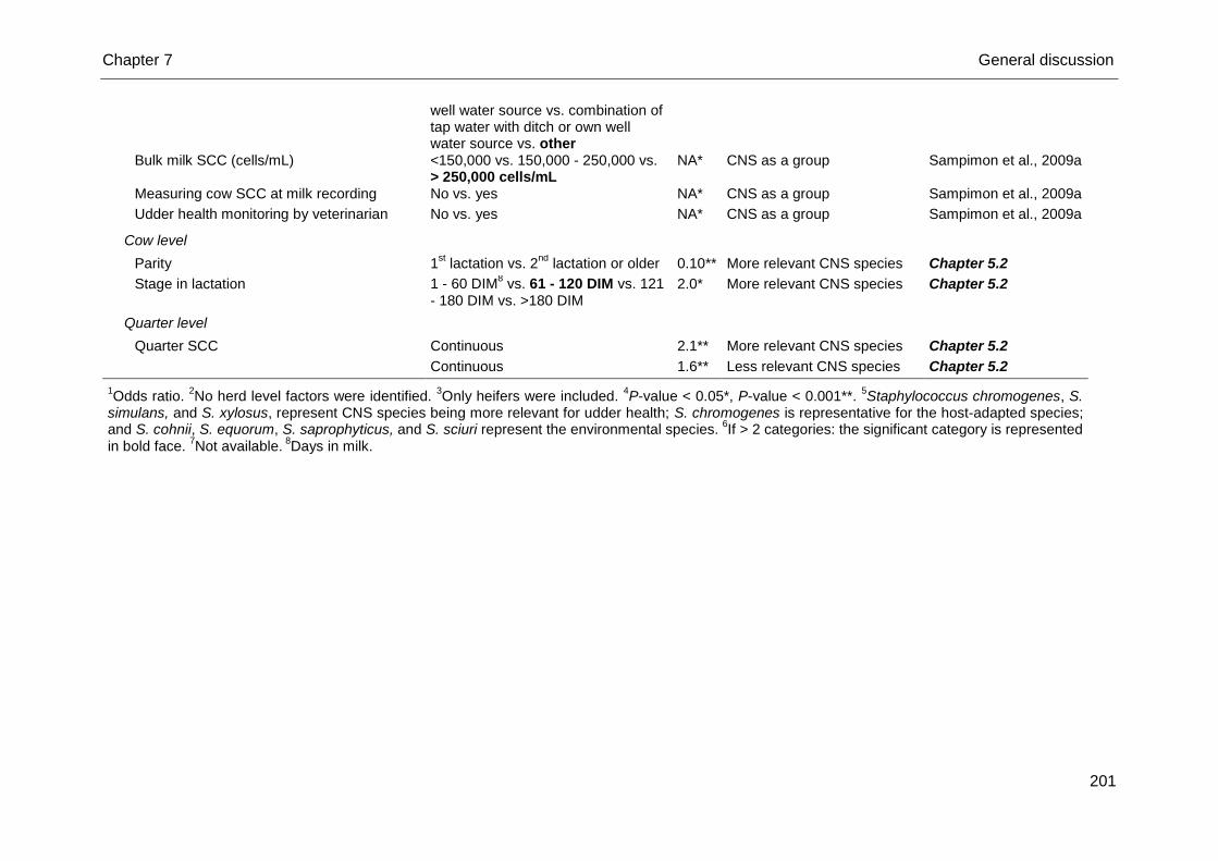
Chapter 7 General discussion
201
well water source vs. combination of tap water with ditch or own well water source vs. other
Bulk milk SCC (cells/mL) <150,000 vs. 150,000 - 250,000 vs. > 250,000 cells/mL
NA* CNS as a group Sampimon et al., 2009a
Measuring cow SCC at milk recording No vs. yes NA* CNS as a group Sampimon et al., 2009a
Udder health monitoring by veterinarian No vs. yes NA* CNS as a group Sampimon et al., 2009a
Cow level
Parity 1st lactation vs. 2
nd lactation or older 0.10** More relevant CNS species Chapter 5.2
Stage in lactation 1 - 60 DIM8 vs. 61 - 120 DIM vs. 121
- 180 DIM vs. >180 DIM 2.0* More relevant CNS species Chapter 5.2
Quarter level
Quarter SCC Continuous 2.1** More relevant CNS species Chapter 5.2
Continuous 1.6** Less relevant CNS species Chapter 5.2
1Odds ratio.
2No herd level factors were identified.
3Only heifers were included.
4P-value < 0.05*, P-value < 0.001**.
5Staphylococcus chromogenes, S.
simulans, and S. xylosus, represent CNS species being more relevant for udder health; S. chromogenes is representative for the host-adapted species; and S. cohnii, S. equorum, S. saprophyticus, and S. sciuri represent the environmental species.
6If > 2 categories: the significant category is represented
in bold face. 7Not available.
8Days in milk.
Chapter 7 General discussion
202
Our studies identified risk factors associated with IMI caused by subgroups of
species or individual CNS species at first (Chapter 5.1, 5.2) (Table 2). The apparently
higher susceptibility of heifers for CNS IMI compared to multiparous cows is intriguing
and requests further study. Fresh animals also seemed to be more susceptible for
CNS IMI (Chapter 5.1: 26%) as opposed to animals in a later stage in lactation
(Piessens et al., 2011: 6%; Supré et al., 2011: 6%; Tomazi et al., 2014: 7%; Mørk et
al., 2012: 13%). Interestingly, the latter seems to mainly concern S. arlettae, S.
chromogenes, S. sciuri, and S. xylosus (Figure 1). For other species, the effect of
stage in lactation on their occurrence seems to depend on the parity of the animals
and vice versa. Throughout lactation, S. epidermidis was solely isolated from heifers
(Chapter 5.2), while shortly after parturition S. epidermidis IMI were observed in both
heifers and older cows (Chapter 5.1). The opposite was true for S. equorum. In a
later stage in lactation only older cows seemed to be infected, while shortly after
parturition this species was recovered from IMI in both heifers and older cows.
Staphylococcus devriesei and S. pasteuri were only isolated from older cows both in
early lactation (Chapter 5.1) and a later stage in lactation (Chapter 5.2), whereas S.
hyicus was solely isolated from one fresh heifer (Chapter 5.1) and one older cow in a
later stage in lactation (Chapter 5.2).
The ecological and the epidemiological nature of the predominant coagulase-
negative staphylococci becomes clear
In recent years, different bovine-associated habitats of CNS species have been
studied. In general, this has helped to distinguish within the large group of CNS
between the so-called host-adapted species (e.g. S. chromogenes, S. epidermidis, S.
simulans) and environmental species (e.g. S. cohnii, S. equorum, S. fleurettii, S.
saprophyticus, S. sciuri). Besides, species can behave in an opportunistic (e.g. S.
equorum) or contagious (e.g. S. chromogenes) manner (Piessens et al., 2012;
Vanderhaeghen et al., 2014, 2015).
Our studies (Chapter 4.1, 4.2, 5.1) allowed verification of previous findings, as
summarized in Figure 3. Indeed, the so-called environmental species were clearly
more present in environmental samples, only S. haemolyticus interrupts the
ascendant trend-line, substantiating its potential opportunistic nature. The
Chapter 7 General discussion
203
predominance of some CNS species causing IMI is also clearly shown (Figure 3),
reinforcing the host-adapted nature of S. chromogenes and S. simulans. The latter
observations along with the obtained results from our risk factor studies and those
conducted by other research groups (Chapter 4.2, 5.1) (Table 2) further substantiate
the stratification in host-adapted and environmental species. The ecology of S.
xylosus, a skin-opportunist (Verdier-Metz et al., 2012), still remains to be elucidated
(Taponen et al., 2008, Figure 3).
Teat apices of non-lactating animals seem to mainly yield more skin-opportunistic
and host-adapted species, such as S. chromogenes and S. xylosus, and are less
subjected to environmental influences (Figure 3). In that respect, the high number of
S. haemolyticus and S. devriesei colonized teat apices of non-lactating cows and
heifers suggests a rather host-adapted nature of both species as was also proposed
in Chapter 4.2. Contrastingly, teat apices of lactating cows and heifers as well as teat
skin harbor both environmental and host-adapted species. Different housing
conditions and the milking process, influencing teat apex microbiota of lactating
animals, most likely explain this discrepancy. Similar strains of S. chromogenes were,
however, found on the teat skin and in bovine milk, indicating teats are a possible
infection source of IMI in the corresponding quarter (Taponen et al., 2008). Chapter
5.1 additionally revealed a significant association between S. chromogenes TAC and
IMI as was also recently suggested for S. haemolyticus (Leroy et al., 2015). It is very
plausible that at least part of the S. chromogenes IMI in early lactation originated
from the dry period and were established via penetration into the mammary gland of
S. chromogenes bacteria that first colonized the teat apex.
Up till now, a contagious epidemiology of the host-adapted species has not been
proven, in contrast with S. aureus (Zadoks et al., 2002; Smith et al., 2005a), as
milking machine unit liners seem to be mainly contaminated with the environmental
species (Figure 3) and no transmission during milking process was observed
(Chapter 4.1). However, a contagious behavior was suspected for S. simulans and S.
epidermidis as clonality was shown within herds, i.e. dendrograms demonstrated that
isolates were more grouped together per herd (Bexiga et al., 2014). Also, the
presence of both host-adapted and environmental species on milkers’ skin and
gloves suggest contamination rather than transmission through this vector. Strains
other than milk-related ones were detected on milkers’ skin, justifying our hypothesis
(Taponen et al., 2008). Also, human skin yields similar species, such as S.
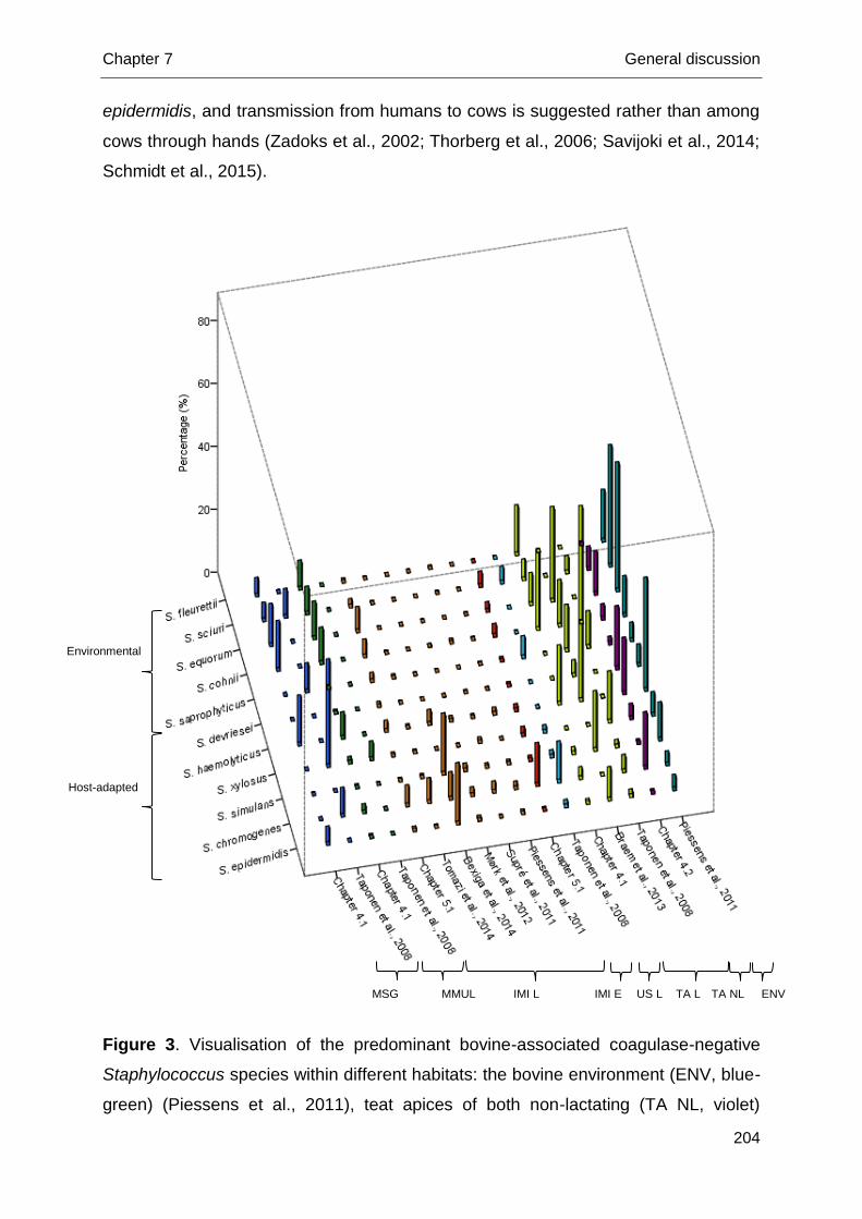
Chapter 7 General discussion
204
epidermidis, and transmission from humans to cows is suggested rather than among
cows through hands (Zadoks et al., 2002; Thorberg et al., 2006; Savijoki et al., 2014;
Schmidt et al., 2015).
Figure 3. Visualisation of the predominant bovine-associated coagulase-negative
Staphylococcus species within different habitats: the bovine environment (ENV, blue-
green) (Piessens et al., 2011), teat apices of both non-lactating (TA NL, violet)
Environmental
Host-adapted
US L IMI E IMI L MSG TA L MMUL ENV TA NL
Chapter 7 General discussion
205
(Chapter 4.2) and lactating animals (TA L, yellow-green) (Taponen et al., 2008;
Braem et al., 2013; Chapter 4.1), udder skin of lactating cows and heifers (US L, light
blue) (Taponen et al., 2008), intramammary infection (IMI) both in early lactation (IMI
E, red) (Chapter 5.1) and throughout lactation (IMI L, orange) (Piessens et al., 2011;
Supré et al., 2011; Mørk et al., 2012; Bexiga et al., 2014; Tomazi et al., 2014;
Chapter 5.1), milking machine unit liners (MMUL, green) and milkers’ skin and gloves
(MSG, blue) (Taponen et al., 2008; Chapter 4.1). Results are displayed as
percentages of habitats (e.g. all swabbed teat apices, all isolated milk samples)
harbouring a specific CNS species as reported by different studies. The species are
ordered based on their potential ecological nature with S. fleurettii and S. epidermidis
at the ends of the spectrum representing the environmental and host-adapted
species, respectively.
Preliminary screening of the species-specific protective effect of teat apex
colonization
Protective effects of CNS TAC (De Vliegher et al., 2003; Piepers et al., 2011) and
of pre-existing CNS IMI (Poutrel and Lerondelle, 1980; Rainard and Poutrel, 1988;
Nickerson and Boddie, 1994; Green et al., 2005) have been suggested. However, a
recently conducted meta-analysis showed that the outcome of analyses, i.e. finding
protective effects, depends on several factors such as differences in study design
(i.e. dose of inoculated major pathogens, size of the study, study duration), IMI
definition, and underlying undetected risk. Smaller challenge studies rather than large
observational studies, using a higher major pathogen challenge dose, covering a
short period, relying on more stringent IMI definitions and with a higher underlying
risk for IMI with major pathogens in the population showed more and stronger
protective effects (Reyher et al., 2012).
Although it was not a specific aim of this thesis, our data provide a unique
opportunity to preliminary analyze whether pre-partum species-specific CNS TAC
protects against IMI in early lactation as suggested before by De Vliegher et al.
(2003) for S. chromogenes and Piepers et al. (2011) for the CNS group (Chapter 4.2,
5.1). The number of included herds and sampled animals are comparable with the
aforementioned observational studies (De Vliegher et al., 2003: 8 herds - 123
animals; Piepers et al., 2011: 20 herds - 191 animals; Chapter 5.1: 13 herds - 156
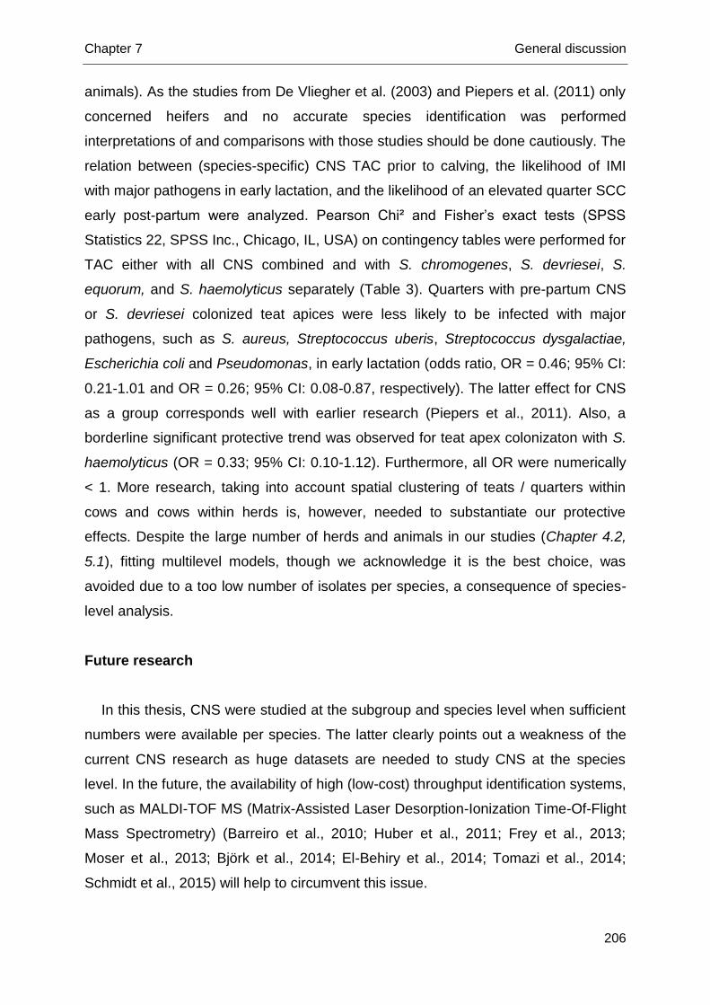
Chapter 7 General discussion
206
animals). As the studies from De Vliegher et al. (2003) and Piepers et al. (2011) only
concerned heifers and no accurate species identification was performed
interpretations of and comparisons with those studies should be done cautiously. The
relation between (species-specific) CNS TAC prior to calving, the likelihood of IMI
with major pathogens in early lactation, and the likelihood of an elevated quarter SCC
early post-partum were analyzed. Pearson Chi² and Fisher’s exact tests (SPSS
Statistics 22, SPSS Inc., Chicago, IL, USA) on contingency tables were performed for
TAC either with all CNS combined and with S. chromogenes, S. devriesei, S.
equorum, and S. haemolyticus separately (Table 3). Quarters with pre-partum CNS
or S. devriesei colonized teat apices were less likely to be infected with major
pathogens, such as S. aureus, Streptococcus uberis, Streptococcus dysgalactiae,
Escherichia coli and Pseudomonas, in early lactation (odds ratio, OR = 0.46; 95% CI:
0.21-1.01 and OR = 0.26; 95% CI: 0.08-0.87, respectively). The latter effect for CNS
as a group corresponds well with earlier research (Piepers et al., 2011). Also, a
borderline significant protective trend was observed for teat apex colonizaton with S.
haemolyticus (OR = 0.33; 95% CI: 0.10-1.12). Furthermore, all OR were numerically
< 1. More research, taking into account spatial clustering of teats / quarters within
cows and cows within herds is, however, needed to substantiate our protective
effects. Despite the large number of herds and animals in our studies (Chapter 4.2,
5.1), fitting multilevel models, though we acknowledge it is the best choice, was
avoided due to a too low number of isolates per species, a consequence of species-
level analysis.
Future research
In this thesis, CNS were studied at the subgroup and species level when sufficient
numbers were available per species. The latter clearly points out a weakness of the
current CNS research as huge datasets are needed to study CNS at the species
level. In the future, the availability of high (low-cost) throughput identification systems,
such as MALDI-TOF MS (Matrix-Assisted Laser Desorption-Ionization Time-Of-Flight
Mass Spectrometry) (Barreiro et al., 2010; Huber et al., 2011; Frey et al., 2013;
Moser et al., 2013; Björk et al., 2014; El-Behiry et al., 2014; Tomazi et al., 2014;
Schmidt et al., 2015) will help to circumvent this issue.
Chapter 7 General discussion
207
Table 3. Association between pre-partum teat apex colonization and the likelihood of intramammary infection (IMI) with major
pathogens in early lactation, and the likelihood of an elevated quarter milk SCC in early lactation, respectively
Teat apex colonization
IMI major pathogens IMI Gram-positive major pathogens
IMI Gram-negative major pathogens
Quarter milk SCC1
Nn2 Ni
3 P
4 OR
5 95% CI Nn Ni P OR 95% CI Nn Ni P OR 95% CI N
<200
N
≥ 200
P OR 95% CI
CNS 0.05 0.46 0.21 1.01 0.21 0.51 0.18 1.50 0.17 0.44 0.15 1.30 0.40 0.80 0.47 1.36
Nn 55 10 55 5 55 5 26 40
Nc3 278 23 278 13 278 11 165 202
S. chromogenes 0.46 0.68 0.24 1.90 0.74 0.77 0.20 3.03 0.74 0.77 0.20 3.03 0.21 0.67 0.35 1.27
Nn 55 10 55 5 55 5 26 40
Nc 57 7 57 4 57 4 44 45
S. devriesei 0.02 0.26 0.08 0.87 0.12 0.26 0.05 1.38 0.12 0.26 0.05 1.38 0.88 0.95 0.51 1.78
Nn 55 10 55 5 55 5 26 40
Nc 85 4
85 2 85 2 43 63
S. equorum 0.16 0.42 0.13 0.43 0.44 0.42 0.08 2.28 0.44 0.42 0.08 2.28 0.40 0.75 0.38 1.48
Nn 55 10 55 5 55 5 26 40
Nc 52 4
52 2 52 2 33 38
S. haemolyticus 0.07 0.33 0.10 1.12 0.10 0.17 0.02 1.47 0.47 0.50 0.11 2.19 0.28 0.70 0.36 1.34
Nn 55 10 55 5 55 5 26 40
Nc 66 4 66 1 66 3 41 44
1Categorization based on De Vliegher et al. (2003), i.e. < 200,000 cells/mL vs. ≥ 200,000 cells/mL.
2Number of uninfected quarters or non-colonized (Nn)
teat apices. 3Number of quarters infected (Ni) or teat apices colonized (Nc) with the bacteria belonging to the different subgroups.
4 P-value for the overall
effect. 5Odds ratio
Chapter 7 General discussion
208
It all, however, starts with conducting large scale studies including enormous
numbers of samples from a huge number of herds.
We believe that, combined with the studies of other research groups around the
world, good steps have been taken in further elucidating the differences among CNS
species in many respects. The need to include strain-typing in large studies clearly
exists as differences between strains within species have been revealed and can be
relevant (Vanderhaeghen et al., 2015). In our work, strain-typing was not performed,
although this will be done in the future to reinforce or counter several (of our) findings
and hypotheses such as the association between S. chromogenes colonized teat
apices and S. chromogenes IMI in the same quarters in early lactation (Chapter 4.2,
5.1) and the ability of CNS species to cause persistent IMI (Chapter 5.1: data not
shown). Pulsed-field gel electrophoresis (PFGE), a highly discriminatory strain-typing
method (Rabello et al., 2007; Holmes and Zadoks, 2011; Vanderhaeghen et al.,
2015), able to distinguish CNS strains, can be used for that purpose. Pulsed-field gel
electrophoresis is, however, labour intensive, expensive (Vanderhaeghen et al.,
2015), and interlaboratory comparisons are difficult (Rabello et al., 2007), though a
CNS-specific protocol exist as PFGE has already been used before (Shimizu et al.,
1997; Thorberg et al., 2006; Taponen et al., 2008; Gillespie et al., 2009; Rajala-
Schultz et al., 2009; Sawant et al., 2009; Feβler et al., 2010; Jaglic et al., 2010; Mørk
et al., 2012; Pate et al., 2012; Bexiga et al., 2014; Fry et al., 2014b). The latter
makes PFGE the most suitable and quickest method at the moment to distinguish
different CNS strains obtained in 1 study. In order to determine the global
epidemiology and population structure of CNS species, MLST (Smith et al., 2005a;
Holmes and Zadoks, 2011; Vanderhaeghen et al., 2015) or whole genome
sequencing (WGS) (Vanderhaeghen et al., 2015) are however more appropriate. In
contrast to PFGE, being already applicable for all CNS species, a suitable MLST
protocol only exists for bovine S. epidermidis (Frey et al., 2013) and differences
among CNS species are expected.Currently, a MLST-protocol for bovine S.
chromogenes isolates is being validated, partly based on some of our isolates
(Chapter 4.2, 5.1). The same inconvenience occurs for WGS as only the genome of
a bovine S. chromogenes (Fry et al., 2014a), a bovine S. epidermidis (Savijoki et al.,
2014), and a bovine S. xylosus isolate (Harrison et al., 2013) have been sequenced.
Bovine S. aureus-protocols can be valuable in developing WGS for other CNS
species (Herron-Olson et al., 2007; Garcia-Alvarez et al., 2011; Wolf et al., 2011).
Chapter 7 General discussion
209
The possibility to differentiate protective CNS species / strains from those that are
more relevant for udder health should be examined using the above mentioned
strain-typing techniques. Several hypotheses have been suggested to explain
protection of CNS species, all demanding further investigation. Competitive
exclusion or an activated immune response from pre-existing CNS IMI (Rainard and
Poutrel, 1988; Matthews et al., 1990; Piepers et al., 2009), inhibiting or quickly
eliminating infection and colonization of the mammary gland with major pathogens,
have been proposed. This could be examined in challenge trials, i.e. inoculation of
uninfected quarters with major pathogens after establishing TAC with protective CNS
strains using dairy cows, although other target species such as mice could be used
as well (Breyne et al., 2015). Protective CNS strains might be able to produce
bacteriocins, antimicrobial peptides / proteins able of inhibiting pathogens such as S.
aureus and Streptococcus agalactiae (Nascimento et al., 2005; Coelho et al., 2007).
Production of bacteriocins has been reported for several staphylococci (Allgaier et
al., 1985; Sashihara et al., 2000; Nascimento et al., 2005; Coelho et al., 2007;
Wilaipun et al., 2008; Ceotto et al., 2010; Fagundes et al., 2011; Braem et al., 2014).
The contagious or opportunistic epidemiology of a number of species such as S.
chromogenes, S. devriesei, and S. haemolyticus has not yet been substantiated and
needs examination. A large study, sampling more parlor-associated habitats and a
larger number of samples per habitat over time than in Chapter 4.1, is warranted to
identify the role of several potential vectors, such as milking machine unit liners, and
quarters having CNS IMI. In Chapter 4.1, swabs of unit liners and teat apices were
collected before and after milking though the link with infected quarters was not
studied. Analogous to S. aureus, studies should rely on adequate strain-typing
(Zadoks et al., 2000, 2002; Smith et al., 2005a, 2005b).
The association among CNS species recovered from bulk milk samples and CNS
species causing IMI in the same herd should be investigated. An extensive study,
including a large number of herds and a huge number of cows and quarters within
each herd is needed to do so. This would allow to compare the distribution of CNS
causing IMI and CNS present in the bulk milk. Also, bulk milk might be used to
screen the herd udder health status (Gillespie et al., 2012; Santos et al., 2014) and
to pinpoint shortcomings in management. Possibly, the predominance of either host-
adapted or environmental CNS in bulk milk is associated with the presence of certain
major pathogens possessing the same ecological nature. In that respect, the
Chapter 7 General discussion
210
presence of several CNS species in bulk milk could reveal the predominant major
pathogen in the herd, potentially not present in bulk milk due to intermittent excretion
or a lower prevalence (as opposed to CNS species). Screening CNS in bulk milk
could be helpful for the development of a herd-specific and effective prevention and
control program without the need of sampling all high SCC-cows in the herd.
The search for an accurate (quick, cheap) CNS identification method is urgent as
current routine diagnostics, i.e. bacteriological culturing followed by phenotypic
identification, are too limited to differentiate among species or strains. Strains able to
persist and elevate the SCC are carrying virulence factors (Vanderhaeghen et al.,
2014; Zuniga et al., 2015) that other strains do not. A polymerase chain reaction
(PCR), targeting genes encoding those factors should be set up allowing accurate
identification of S. chromogenes in the first place. Probably, the combination of
several genes, i.e. a genetic profile, will be associated with virulence and persistency
of S. chromogenes rather than a single gene effect (Bertelloni et al., 2015; Zuniga et
al., 2015). Multiplex PCR methods, able to detect several of these genes that are
solely present in S. chromogenes in a single reaction, such as the commercially
available real-time PCR “PathoProof Mastitis PCR Assay” (Finnzymes Oy, Espoo,
Finland) (Pitkälä et al., 2007; Koskinen et al., 2009), should be adapted and
validated for detection of S. chromogenes. Analogous to S. aureus, virulence gene
patterns highly linking different genotypes to ecological behavior and pathogenicity
should thus be searched for (Graber et al., 2009; Boss et al., 2011).
Chapter 7 General discussion
211
References
Abbas, B. A., M. H. Khudor, and B. M. Hanoon. 2014. Isolation and identification of
Staphylococcus aureus from bovine and the detection of its coagulase gene (coa)
using polymerase chain reaction (PCR). Sci. Res. Essays. 9:864-868.
Addis M., P. Mahindra, and N. K. Moses. 2011. Isolation and identification of
Staphylococcus species from raw bovine milk in Debre Zeit, Ethiopia. Vet. Res. 4,
45-49.
Allgaier, H., G. Jung, R. G. Werner, U. Schneider, and H. Zähner. 1985. Elucidation
of the structure of epidermin, a ribosomally synthesized, tetracyclic heterodetic
polypeptide antibiotic. Angew. Chem. Int. Edit. 24:1051-1053.
Barreiro, J. R., C. R. Ferreira, G. B. Sanvido, M. Kostrzewa, T. Maier, B. Wegemann,
V. Bötttcher, M. N. Eberlin, and M. V. Dos Santos. 2010. Short communication:
Identification of subclinial cow mastitis pathogens in milk by matrix-assisted laser
desorption/ionization time-of-flight mass spectrometry. J. Dairy Sci. 93:5661-5667.
Bertelloni, F., F. Fratini, V. V. Ebani, A. Galiero, B. Turchi, and D. Cerri. 2015.
Detecion of genes encoding for enterotoxines, TSST-1, and biofilm production in
coagulase-negative staphylococci from bovine bulk tank milk. Dairy Sci. Technol.
95:341-352.
Bexiga, R., M. G. Rato, A. Lemsaddek, T. Semedo-Lemsaddek, C. Carneiro, H.
Pereira, D. J. Mellor, K. A. Ellis, and C. L. Vilela. 2014. Dynamics of bovine
intramammary infections due to coagulase-negative staphylococci on four farms.
J. Dairy Res. 81:208-214.
Bhutto, A. L., R. D. Murray, and Z. Woldehiwet. 2010. Udder shape and teat-end
lesions as potential risk factors for high somatic cell counts and intra-mammary
infections in dairy cows. Vet. J. 183:63-67.
Björk, S., R. Båge, B. M. Kanyima, S. André, M. G. Nassuna-Musoke, D. O. Owiny,
and Y. Persson. 2014. Characterization of coagulase-negative staphylococci from
cases of subclinial mastitis in dairy cattle in Kampala, Uganda. Irish Vet. J. 67:12.
Boss, R., J. Naskova, A. Steiner, and H. U. Graber. 2011. Mastitis diagnostics:
quantitative PCR for Staphylococcus aureus genotype B in bulk tank milk. J. Dairy
Sci. 94:128-137.
Braem, G., S. De Vliegher, K. Supré, F. Haesebrouck, F. Leroy and L. De Vuyst.
2011. (GTG)5-PCR fingerprinting for the classification and identification of
Chapter 7 General discussion
212
coagulase-negative Staphylococcus species from bovine milk and teat apices: A
comparison of type strains and field isolates. Vet. Microbiol. 147:67-74.
Braem, G., S. De Vliegher, B. Verbist, V. Piessens, E. Van Coillie, L. De Vuyst, and
F. Leroy. 2013. Unraveling the microbiota of teat apices of clinically healthy
lactating dairy cows, with special emphasis on coagulase-negative staphylococci.
J. Dairy Sci. 96:1499-1510.
Braem, G., B. Stijlemans, W. Van Haken, S. De Vliegher, L. De Vuyst, and F. Leroy.
2014. Antibacterial activities of coagulase-negative staphylococci from bovine teat
apex skin and their inhibitory effect on mastitis-related pathogens. J. App.
Microbiol. 116:1084-1093.
Breyne, K., S. De Vliegher, A. De Visscher, S. Piepers, and E. Meyer. 2015.
Technical note: A pilot study using a mouse mastitis model to study differences
between bovine associated coagulase-negative staphylococci. J. Dairy Sci.
98:1090-1100.
Ceotto, H., H. Holo, K. F. S. Costa, J. S. Nascimento, A. Salehian, I. F. Nes, and M.
C. F. Bastos. 2010. Nukacin 3299, a lantibiotic produced by Staphylococcus
simulans 3299 identical to nukacin ISK-1. Vet. Microbiol. 146:124-131.
Chapman, G. H. 1945. The significance of sodium chloride in studies of
staphylococci. J. Bacteriol. 50:201-203.
Chavhan, S. K., D. R. Kalorey, A. A. Nagdive, H. J. Purohit, S. B. Barbuddhe, and N.
V. Kurkure. 2012. Molecular characterization of intercellular adhesion gene in
Staphylococcus aureus isolated from bovine mastitis milk. Trop. Anim. Health
Prod. 44:247-252.
Cheng, D. R., S. Y. Zhu, Z. H. Yin, W. W. Ding, Z. X. Mu, Z. R. Su, and H. C. Sun.
2010. Prevalence of bacterial infection responsible for bovine mastitis. Afr. J.
Microbiol. Res. 4:1110-1116.
Coelho, M. L. V., J. S. Nascimento, P. C. Fagundes, D. J. Madureira, S. S. Oliveira,
M. A. V. P. Brito, and M. C. F. Bastos. 2007. Activity of staphylococcal
bactericocins against Staphylococcus aureus and Streptococcus agalactiae
involved in bovine mastitis. Res. Microbiol. 158:625-630.
Detilleux, J. C. 2009. Genetic factors affecting susceptibility to udder pathogens. Vet.
Microbiol. 134:157-164.
De Vliegher, S., H. Laevens, L. A. Devriese, G. Opsomer, J. L. M. Leroy, H. W.
Barkema, and A. de Kruif. 2003. Prepartum teat apex colonization with
Chapter 7 General discussion
213
Staphylococcus chromogenes in dairy heifers is associated with low somatic cell
count in early lactation. Vet. Microbiol. 92:245-252.
El-Behiry, A., R. N. Zahran, R. Tarabees, E. Marzouk, and M. Al-Dubaib. 2014.
Phenotyical and genotypical assessment techniques for identification of some
contagious mastitis pathogens. Int. Scholarly Sci. Res. Inn. 8:231-237.
El-Jakee, J. K., N. E. Aref, A. Gomaa, M. D. El-Hariri, H. M. Galal, S. A. Omar, and
A. Samir. 2013. Emerging of coagulase-negative staphylococci as a cause of
mastitis in dairy animals: an environmental hazard. Int. J. Vet. Sci. Med. 1:74-78.
Fagundes, P. C., H. Ceotto, A. Potter, M. A. V. P. Brito, D. Brede, I. F. Nes, and M.
C. F. Bastos. 2011. Hyicin 3682, a bioactive peptide produced by Staphylococcus
hyicus 3682 with potential applications for food preservation. Res. Microbiol.
162:1052-1059.
Feβler, A. T., C. Billerbeck, K. Kadlec, and S. Schwarz. 2010. Identification and
characterization of methicillin-resistant coagulase-negative staphylococci from
bovine mastitis. J. Antimicrob. Chemother. DOI:10.1093/jac/dkq172.
Frey, Y., J. P. Rodriguez, A. Thomann, S. Schwendener, and V. Perreten. 2013.
Genetic characterization of antimicrobial resistance in coagulase-negative
staphylococci from bovine mastitis milk. J. Dairy Sci. 96:1-11.
Fry, P. R., M. J. Calcutt, M. F. Foecking, H.-Y. Hsieh, D. G. Suntrup, J. Perry, G. C.
Stewart, and J. R. Middleton. 2014a. Draft genome sequence of Staphylococcus
chromogenes strain MU 970, isolated from a case of chronic bovine mastitis.
Genome Announc. 2:1-2.
Fry, P. R., J. R. Middleton, S. Dufour, J. Perry, D. Scholl, and I. Dohoo. 2014b.
Association of coagulase-negative staphylococcal species, mammary quarter milk
somatic cell count, and persistence of intramammary infection in dairy cattle. J.
Dairy Sci. 97:4876-4885.
Garcia-Alvarez, L., M. T. G. Holden, H. Lindsay, C. R. Webb, D. F. J. Brown, M. D.
Curran, E. Walpole, K. Brooks, D. J. Pickard, C. Teale, J. Parkhill, S. D. Bentley,
G. F. Edwards, E. K. Girvan, A. M. Kearns, B. Pichon, R. L. R. Hill, A. R. Larsen,
R. L. Skov, S. J. Peacock, D. J. Maskell, and M. A. Holmes. 2011. Meticillin-
resistant Staphylococcus aureus with a novel mecA homologue in human and
bovine populations in the UK and Denmark: A descriptive study. Lancet. Infect.
Dis. 11:595-603.
Chapter 7 General discussion
214
Gillespie, B. E., S. I. Headrick, S. Boonyayatra, and S. P. Oliver. 2009. Prevalence
and persistence of coagulase-negative Staphylococcus species in three dairy
research herds. Vet. Microbiol. 2009: 65-72.
Gillespie, B. E., M. J. Lewis, S. Boonyayatra, M. L. Maxwell, A. Saxton, S. P. Oliver,
and R. A. Almeida. 2012. Short communication: evaluation of bulk tank milk
microbiological quality of nine dairy farms in Tennessee. J. Dairy Sci. 95:4275-
4279.
Godkin, M. A., and K. E. Leslie. 1993. Culture of bulk tank milk as a mastitis
screening test: a brief review. Can. Vet. J. 34:601-605.
Graber, H. U., J. Naskova, E. Studer, T. Kaufmann, M. Kirchhofer, M. Brechbühl, W.
Schaeren, A. Steiner, and C. Fournier. 2009. Mastitis-related subtypes of bovine
Staphylococcus aureus are characterized by different clinical properties. J. Dairy
Sci. 92:1442-1451.
Graber, H. U., S. Pfister, P. Burgener, R. Boss, M. Meylan, and J. Hummerjohann.
2013. Bovine Staphylococcus aureus: Diagnostic properties of specific media.
Res. Vet. Sci. 38-44.
Green, M. J., L. E. Green, A. J. Bradley, P. R. Burton, Y. H. Schukken, and G. F.
Medley. 2005. Prevalence and association between bacterial isolates from dry
mammary glands of dairy cows. Vet. Rec. 156:71-77.
Han, Z., E. Lautenbach, N. Fishman, and I. Nachamkin. 2007. Evaluation of mannitol
salt agar, CHROMagar Staph aureus and CHROMagar MRSA for detection of
methicillin-resistant Staphylococcus aureus from nasal swab specimens. J. Med.
Microbiol. 56:43-46.
Harrison, E. M., G. K. Paterson, M. T. G. Holden, F. J. E. Morgan, A. R. Larsen, A.
Petersen, S. Leroy, S. De Vliegher, V. Perreten, L. K. Fox, T. J. G. M. Lam, O. C.
Sampimon, R. N. Zadoks, S. J. Peacock, J. Parkhill, and M. A. Homes. 2013. A
Staphylococcus xylosus isolate with a new mecC allotype. Antimicrob. Agents Ch.
57:1524-1528.
Herron-Olson, L., J. R. Fitzgerald, J. M. Musser, and V. Kapur. 2007. Molecular
correlates of host specialization in Staphylococcus aureus. PLos ONE
2:e1120.doi.10.1371/journal.pone.0001120.
Holmes, M. A., and R. N. Zadoks. 2011. Methicillin resistant S. aureus in human and
bovine mastitis. J. Mammary Gland. Biol. Neoplasia 16:373-382.
Chapter 7 General discussion
215
Huber, H., D. Ziegler, V. Pflüger, G. Vogel, C. Zweifel, and R. Stephan. 2011.
Prevalence and characteristics of methicillin-resistant coagulase-negative
staphylococci from livestock, chicken carcasses, bulk tank milk, minced meat, and
contact persons. BMC Vet. Res. 7:6.
Jaglic, Z., E. Michu, M. Holasova, H. Vlkova, V. Babak, M. Kolar, J. Bardon, and J.
Schlegelova. 2010. Epidemiology and characterization of Staphylococcus
epidermidis isolates from humans, raw bovine milk and a dairy plant. Epidemiol.
Infect. 138:772-782.
Kateete, D. P., C. N. Kimani, F. A. Katabazi, A. Okeng, M. S. Okee, A. Nanteza, M.
L. Joloba, and F. C. Najjuka. 2010. Identificiation of Staphylococcus aureus:
DNase and mannitol salt agar improve the efficiency of the tube coagulase test.
Ann. Clin. Microbiol. Antimicrob. 9:23.
Koskinen, M. T., J. Holopainen, S. Pyörälä, P. Bredbacka, A. Pitkälä, H. W.
Barkema, R. Bexiga, J. Roberson, L. Sølverød, R. Piccinini, D. Kelton, H.
Lehmusto, S. Niskala, and L. Salmikivi. 2009. Analytical specificity and sensitivity
of a real-time polymerase chain reaction assay for indentification of bovine
mastitis pathogens. J. Dairy Sci. 92: 952-959.
Lange, C. C., M. A. V. P. Brito, D. R. L. Reis, M. A. Machado, A. S. Guimarães, A. L.
S. Azevedo, E. B. Salles, M. C. T. Alvim, F. S. Silva, and I. R. Meurer. 2015.
Species-level identification of staphylococci isolated from bovine mastitis in Brazil
using partial 16S rRNA sequencing. Vet. Microbiol. 176:382-388.
Leroy, F., E. Van Coillie, G. Braem, V. Piessens, B. Verbist, L. De Vuyst, and S. De
Vliegher. 2015. Short communication: subtyping of Staphylococcus haemolyticus
isolates from milk and corresponding teat apices to verify the potential teat-skin
origin of intramammary infections in dairy cows. J. Dairy Sci. 98:7893-7898.
Li, L., L. Zhou, L. Wang, H. Xue, and X. Zhao. 2015. Characterization of methicillin-
resistant and –susceptible staphylococcal isolates from bovine milk in
Northwestern China. Plos One, DOI:10.1371/journal.pone.0116699.
Matthews, K. R., R. J. Harmon, and B. A. Smith. 1990. Protective effect of
Staphylococcus chromogenes infection against Staphylococcus aureus infection
in the lactating bovine mammary gland. J. Dairy Sci. 73:3457-3462.
Mørk, T., H. J. Jørgensen, M. Sunde, B. Kvitle, S. Sviland, S. Waage, and T.
Tollersrud. 2012. Persistence of staphylococcal species and genotypes in the
bovine udder. Vet. Microbiol. 159:171-180.
Chapter 7 General discussion
216
Moser, A., R. Stephan, D. Ziegler, and S. Johler. 2013. Species distribution and
resistance profiles of coagulase-negative staphylococci isolated from bovine
mastitis in Switzerland. Schweiz. Arch. Tierh. 155:333-338.
Nascimento, J. S., P. C. Fagundes, M. A. V. P. Brito, K. R. N. Santos, and M. C. F.
Bastos. 2005. Production of bacteriocins by coagulase-negative staphylococci
involved in bovine mastitis. Vet. Microbiol. 106:61-71.
Nickerson, S. C., and R. L. Boddie. 1994. Effect of naturally occurring coagulase-
negative staphylococcal infections on experimental challenge with major mastitis
pathogens. J. Dairy Sci. 77:2526-2536.
Olde Riekerink, R. G. M., H. W. Barkema, S. Veenstra, D. E. Poole, R.T. Dingwell,
and G. P. Keefe. 2006. Prevalence of contagious mastitis pathogens in bulk tank
milk in Prince Edward Island. Can. Vet. J. 47:567-572.
Olde Riekerink, R. G. M., H. W. Barkema, D. T. Scholl, D. E. Poole, and D. F. Kelton.
2010. Management practices associated with the bulk-milk prevalence of
Staphylococcus aureus in Canadian dairy farms. Prev. Vet. Med. 97:20-28.
Oliveira, C. J. B., N. Tiao, F. G. C. de Sousa, J. F. P. de Moura, L. S. Filho, and W.
A. Gebreyes. 2015. Methicillin-resistant Staphylococcus aureus from Brazilian
dairy farms and identification of novel sequence types. Zoonoses Public Hlth. DOI:
10.1111/zph.12209.
Park, J. Y., L. K. Fox, K. S. Seo, M. A. McGuire, Y. H. Park, F. R. Rurangirwa, W. M.
Sischo, and G. A. Bohach. 2011. Comparison of phenotypic and genotypic
methods for the species identification of coagulase-negative staphylococcal
isolates from bovine intramammary infections. Vet. Microbiol. 2011:142-148.
Passchyn, P., S. Piepers, and S. De Vliegher. 2014. Pathogen group-specific risk
factors for intramammary infection in treated and untreated dairy heifers
participating in a prepartum antimicrobial treatment trial. J. Dairy Sci. 97:6260-
6270.
Pate, M., I. Zdovc, J. Avberšek, M. Ocepek, A. Pengov, and O. Podpečan. 2012.
Coagulase-negative staphylococci from non-mastitic bovine mammary gland:
Characterization of Staphylococcus chromogenes and Staphylococcus
haemolyticus by antibiotic susceptibility testing and pulse-field gel electrophoresis.
J. Dairy Res. 79:129-134.
Chapter 7 General discussion
217
Persson Waller, K., A. Aspán, A. Nyman, Y. Persson, and U. Grönlund Andersson.
2011. CNS species and antimicrobial resistance in clinical and subclinical bovine
mastitis. Vet. Microbiol. 152:112-116.
Piepers, S., G. Opsomer, E. Meyer, K. Demeyere, H. W. Barkema, A. de Kruif, and
S. De Vliegher. 2009. Heifer and quarter characteristics associated with
periparturient blood and milk neutrophil apoptosis in healthy heifers and in heifers
with subclinical mastitis. J. Dairy Sci. 92:4330-4339.
Piepers, S., K. Peeters, G. Opsomer, H. W. Barkema, K. Frankena, and S. De
Vliegher. 2011. Pathogen group specific risk factors at herd, heifer and quarter
levels for intramammary infections in early lactating dairy heifers. Prev. Vet. Med.
99:91-101.
Piessens, V., E. Van Coillie, B. Verbist, K. Supré, G. Braem, A. Van Nuffel, L. De
Vuyst, M. Heyndrickx, and S. De Vliegher. 2011. Distribution of coagulase-
negative Staphylococcus species from cows’ milk and environment differs
between herds. J. Dairy Sci. 94:2933-2944.
Piessens, V., S. De Vliegher, B. Verbist, G. Braem, A. Van Nuffel, L. De Vuyst, M.
Heyndrickx, and E. Van Coillie. 2012. Characterization of coagulase-negative
Staphylococcus species from cows’ milk and environment based on bap, icaA,
and mecA genes and phenotypic susceptibility to antimicrobials and teat dips. J.
Dairy Sci. 95:7027-7038.
Pitkälä, A., L. Salmikivi, P. Bredbacka, A.-L. Myllyniemi, and M. T. Koskinen. 2007.
Comparison of test of dection of β-lactamase-producing staphylococci. J. Clin.
Microbiol. 45:2031-2033.
Poutrel, B., and C. Lerondelle. 1980. Protective effect in the lactating bovine
mammary gland induced by coagulase-negative staphylococci against
experimental Staphylococcus aureus infections. Ann. Rech. Vet. 11:327-332.
Quirk, T., L. K. Fox, D. D. Hancock, J. Capper, J. Wenz, and J. Park. 2012.
Intramammary infections and teat canal colonization with coagulase-negative
staphylococci after postmilking teat disinfection: Species-specific responses. J.
Dairy Sci. 95:1906-1912.
Rabello, R. F., B. M. Moreira, R. M. M. Lopes, L. M. Teixeira, L. W. Riley, and A. C.
D. Castro. 2007. Multilocus sequence typing of Staphylococcus aureus isolates
recovered from cows with mastitis in Brazilian dairy herds. J. Med. Microbiol.
56:1505-1511.
Chapter 7 General discussion
218
Rainard, P., and B. Poutrel. 1988. Effect of naturally occurring intramammary
infections by minor pathogens on new infections by major pathogens in cattle. Am.
J. Vet. Res. 49:327-329.
Rajala-Schultz, P. J., A. H. Torres, F. J. DeGraves, W. A. Gebreyes, and P.
Patchanee. 2009. Antimicrobial resistance and genotypic characterization of
coagulase-negative staphylococci over the dry period. Vet. Microbiol. 134:55-64.
Reyher, K. K., D. Haine, I. R. Dohoo, and C. W. Revie. 2012. Examining the effect of
intramammary infections with mastitis pathogens on the acquisition of new
intramammary infections with major mastitis pathogens - A systematic review and
meta-analysis. J. Dairy Sci. 95:6483-6502.
Sampimon, O. C., H. W. Barkema, I. M. G. A. Berends, J. Sol, and T. J. G. M. Lam.
2009a. Prevalence and herd-level risk factors for intramammary infection with
coagulase-negative staphylococci in Dutch dairy herds. Vet. Microbiol. 134:37-44.
Sampimon, O. C., R. N. Zadoks, S. De Vliegher, K. Supré, F. Haesebrouck, H. W.
Barkema, J. Sol, and T. J. G. M. Lam. 2009b. Performance of API Staph ID 32
and Staph-Zym for identification of coagulase-negative staphylococci isolated
from bovine milk samples. Vet. Microbiol. 136:300-305.
Santos, O. C. S., E. M. Barros, M. A. V. P. Brito, M. C. F. Bastos, K. R. N. Santos,
and M. Giambiagi-deMarval. 2008. Identification of coagulase-negative
staphylococci from bovine mastitis using RFLP-PCR of the groEL gene. Vet.
Microbiol. 130:134-140.
Santos, V. M., H. B. Martinus, I. S. Rezende, M. S. Barbosa, E. F. Andrade, S. G.
Souza, G. B. Campos, P. S. Oliveira, D. S. Sousa, D. C. C. Da Silva, A. T.
Amorim, J. Timenetsky, M. P. Cruz, R. Yatsuda, and L. M. Marques. 2014.
Virulence factor profile of Staphylococcus aureus isolated from bovine milk from
Brazil. Food Nutr. Sci. 5:1496-1505.
Sashihara, T., H. Kimura, T. Higuchi, A. Adachi, H. Matsusaki, K. Sonomoto, and A.
Ishizaki. 2000. A novel lantibiotic, nukacin ISK-1, of Staphylococcus warneri ISK-
1: Cloning of the structural gene and identification of the structure. Biosci.
Biotechnol. Biochem. 64:2420-2428.
Savijoki, K., A. Iivanainen, P. Siljamäki, P. K. Laine, L. Paulin, T. Karonen, S.
Pyörälä, M. Kankainen, T. A. Nyman, T. Salomäki, P. Koskinen, L. Holm, H.
Simojoki, S. Taponen, A. Sukura, N. Kalkkinen, P. Auvinen, and P. Varmanen.
2014. Genomics and proteomics provide new insight into the commensal and
Chapter 7 General discussion
219
pathogenic lifestyles of bovine- and human-associated Staphylococcus
epidermidis strain. J. Proteome. Res. 13:3748-3762.
Sawant, A. A., B. E. Gillespie, and S. P. Oliver. 2009. Antimicrobial susceptibility of
coagulase-negative Staphylococcus species isolated from bovine milk. Vet.
Microbiol. 134:73-81.
Schmidt, T., M. M. Kock, and M. M. Ehlers. 2015. Diversity and antimicrobial
susceptibility profiling of staphylococci isolated from bovine mastitis cases and
close human contacts. J. Dairy Sci. 98:6256-6269.
Shimizu, A., W. E. Kloos, H. A. Berkhoff, C. G. George, and D. N. Ballard. 1997.
Pulsed-field gel electrophoresis of Staphylococcus hyicus and Staphylococcus
chromogenes genomic DNA and its taxonomic, epidemiologic and ecologic
applications in veterinary medicine. J. Vet. Med. Sci. 59:443-450.
Singh, M., A. Sharma, and A. Singh. 2013. Duplex polymerase chain reaction for
detection of coagulase and beta-haemolysin genes of Staphylococcus aureus
isolated from bovine clinical mastitis. Vet. Pract. 14:258-260.
Smith, E. M., L. E. Green, G. F. Medley, H. E. Bird, and C. G. Dowson. 2005a.
Multilocus sequence typing of Staphylococcus aureus isolated from high-somatic-
cell-count cows and the environment of an organic dairy farm in the United
Kingdom. J. Clin. Microbiol. 43:4731-4736.
Smith, E. M., L. E. Green, G. F. Medley, H. E. Bird, L. K. Fox, Y. H. Schukken, J. V.
Kruze, A. J. Bradley, R. N. Zadoks, and C. G. Dowson. 2005b. Multilocus
sequence typing of intercontinental bovine Staphylococcus aureus isolates. J.
Clin. Microbiol. 43:4737-4743.
Supré, K., S. De Vliegher, O. C. Sampimon, R. N. Zadoks, M. Vaneechoutte, M.
Baele, E. De Graef, S. Piepers, and F. Haesebrouck. 2009. Technical note: Use of
tRNA-intergenic spacer PCR combined with capillary electrophoresis to identify
coagulase-negative Staphylcococcus species originating from bovine milk and
teat apices. J. Dairy Sci. 92:3204-3210.
Supré, K., F. Haesebrouck, R. N. Zadoks, M. Vaneechoutte, S. Piepers, and S. De
Vliegher. 2011. Some coagulase-negative Staphylococcus species affect udder
health more than others. J. Dairy Sci. 94:2329-2340.
Taponen, S., J. Koort, J. Björkroth, H. Saloniemi, and S. Pyörälä. 2007. Bovine
intramammary infections caused by coagulase-negative staphylococci may persist
Chapter 7 General discussion
220
throughout lactation according to amplified fragment length polymorphism-based
analysis. J. Dairy Sci. 90:3301-3307.
Taponen, S., J. Björkroth, and S. Pyörälä. 2008. Coagulase-negative staphylococci
isolated from bovine extramammary sites and intramammary infections in a single
dairy herd. J. Dairy Res. 75:422-429.
Thorberg, B.-M., I. Kühn, F. M. Aarestrup, B. Brändström, P. Jonsson, and M.-L.
Danielsson-Tham. 2006. Pheno- and genotyping of Staphylococcus epidermidis
isolated from bovine milk and human skin. Vet. Microbiol. 115:163-172.
Thorberg, B.-M., M.-L. Danielsson-Tham, U. Emanuelson, and K. Persson Waller.
2009. Bovine subclinical mastitis caused by different types of coagulase-negative
staphylococci. J. Dairy Sci. 92:4962-4970.
Tomazi, T., J. L. Gonçalves, J. R. Barreiro, P. A. C. Braga, L. F. P. Silva, M. N.
Eberlin, and M. V. Santos. 2014. Identification of coagulase-negative
staphylococci from bovine intramammary infection by Matrix-Assisted Laser
Desorption Ionization - Time of Flight Mass Spectrometry. J. Clin. Microbiol.
52:1658-1663.
Vanderhaeghen, W., S. Piepers, F. Leroy, E. Van Coillie, F. Haesebrouck, and S. De
Vliegher. 2014. Invited review. Effect, persistence, and virulence of coagulase-
negative Staphylococcus species associated with ruminant udder health. J. Dairy
Sci. 97:5275-5293.
Vanderhaeghen, W., S. Piepers, F. Leroy, E. Van Coillie, F. Haesebrouck, and S. De
Vliegher. 2015. Identification, typing, ecology and epidemiology of coagulase-
negative staphylococci associated with ruminants. Vet. J. 203:44-51.
Verbeke, J., S. Piepers, L. Peelman, M. Van Poucke, and S. De Vliegher. 2012.
Pathogen-group specific association between CXCR1 polymorphisms and
subclinical mastitis in dairy heifers. J. Dairy Res. 79:341-351.
Verdier-Metz, I., G. Gagne, S. Bornes, F. Monsallier, P. Veisseire, C. Delbès-Paus
and M.-C. Montel. 2012. Cow teat skin, a potential source of diverse microbial
populations for cheese production. Appl. Environ. Microbiol. 78:326-333.
Wilaipun, P., T. Zendo, K. Okuda, J. Nakayama, and K. Sonomoto. 2008.
Identification of the nukacin KQU-131, a new type-A(II) lantibiotic produced by
Staphylococcus hominis KQU-131 isolated from Thai fermented fish product (Pla-
ra). Biosci. Biotechnol. Biochem. 72:2232-2235.
Chapter 7 General discussion
221
Wolf, C., H. Kusch, S. Monecke, D. Albrecht, S. Holtfreter, C. Eiff, W. Petzl, P.
Rainard, B. M. Bröker, and S. Engelmann. 2011. Genomic and proteomic
characterization of Staphylococcus aureus mastitis isolates of bovine origin.
Proteomics 11:2491-2502.
Zadoks, R. N., W. van Leeuwen, H. Barkema, O. Sampimon, H. Verbrugh, Y. H.
Schukken, and A. van Belkum. 2000. Application of pulsed-field gel
electrophoresis and binary typing as tools in veterinary clinical microbiology and
molecular epidemiologic analysis of bovine and human Staphylococcus aureus
isolates. J. Clin. Microbiol. 38:1931-1939.
Zadoks, R. N., W. B. van Leeuwen, D. Kreft, L. K. Fox, H. W. Barkema, Y. H.
Schukken, and A. van Belkum. 2002. Comparison of Staphylococcus aureus
isolates from bovine and human skin, milking equipment, and bovine milk by
phage typing, pulsed-field gel electrophoresis, and binary typing. J. Clin. Microbiol.
40:3894-3902.
Zadoks, R. N., and J. L. Watts. 2009. Species identification of coagulase-negative
staphylococci: Genotyping is superior to phenotyping. Vet. Microbiol. 134:20-28.
Zuniga, E., P. A. Melville, A. B. S. Saidenberg, M. A. Laes, F. F. Gonsales, S. R. S.
Salaberry, F. Gregori, P. E. Brandão, F. G. B. Santos, N. E. Lincopan, and N. R.
Benites. 2015. Occurrence of genes coding for MSCRAMM and biofilm-associated
protein Bap in Staphylococcus spp. isolated from bovine subclinical mastitis and
relationship with somatic cell counts. Microb. Pathogenesis 89:1-6.
Zwertvaegher, I., S. De Vliegher, B. Verbist, A. Van Nuffel, J. Baert, and S. Van
Weyenberg. 2013. Short communication: Associations between teat dimensions
and milking-induced changes in teat dimensions and quarer milk somatic cell
counts in dairy cows. J. Dairy Sci. 96:1075-1080.

Summary
A. De Visscher
Department of Reproduction, Obstetrics, and Herd Health
Faculty of Veterinary Medicine
Ghent University, Merelbeke, Belgium

Summary
225
Worldwide, mastitis is one of the most prevalent and costly diseases in the dairy
industry. Besides huge economic losses, milk quality and animal well-fare are
affected. Mastitis is mainly caused by bacteria, based on their ecological nature
classified into host-adapted and environmental pathogens. Currently, coagulase-
negative staphylococci (CNS) are the main causative agents of intramammary
infection (IMI) in dairy cows. Literature was reviewed and knowledge and
assumptions concerning the CNS group were reported (Chapter 1). Coagulase-
negative staphylococci are able to cause mild (sub)clinical mastitis and have as a
group been considered as minor pathogenic for udder health. Aside from the
negative impact, protective effects have been suggested as well. Differences among
species within this group have been reported, emphasizing the necessity of research
at the species level rather than at the group level.
The aim of this thesis was to get more insight in the ecological nature and
epidemiological behavior of the different bovine-associated CNS species (Chapter
2).
Several habitats other than the mammary gland, such as teat apices and bulk
milk, yield CNS species. Picturing the CNS distribution in those habitats is hampered
by the presence of several bacteria other than CNS. The suitability of a (quasi-)
selective medium, mannitol salt agar (MSA) was assessed for that reason (Chapter
3). Both reference strains and field isolates of the twenty-five predominant bovine-
related CNS species were included in the study (n = 50). Also, 10 isolates of the 6
and 4 most frequently isolated species from milk and teat apices (n = 100),
respectively, were tested. All isolates were plated on MSA and aerobically incubated
at 37°C. Growth was examined after 24h and 48h and after another 24h incubation
at room temperature. Aerobic incubation for 48h at 37°C was recommended.
Additionally, the applicability of MSA for routine recovery of CNS species was
assessed using bulk milk samples (n = 20) and swabs of teat apices (n = 20). All
isolated CNS were identified to the species level using tDNA-PCR or sequencing of
the rpoB or 16S rRNA gene. Twenty and 34 CNS isolates were recovered from bulk
milk and teat apices, respectively, the majority being Staphylococcus equorum, S.
chromogenes, S. fleurettii, and S. haemolyticus. Advantages of the use of MSA were
detected over the use of a non-selective medium as overgrowth of bacteria other
than CNS was avoided and could thus not hinder us from picking up different
colonies on MSA. Mannitol salt agar was considered as a highly suitable medium for
Summary
226
the growth and recovery of bovine-associated CNS, and was further applied in
Chapters 4.2, 5.1 and 6.
The species-specific CNS distribution on teat apices of lactating animals was
described through a cross-sectional sampling in 3 Flemish dairy herds (Chapter 4.1).
Swabs of teat apices were taken before and after milking (n = 140). Parlour-
associated habitats were explored as well and compared to CNS species causing
IMI in the herds to investigate potential transmission routes. Milking machine unit
liners (MMUL) and gloves or hands and elbows of milkers (MSG) were swabbed,
again before and after milking (n = 252 and n = 40, respectively). All CNS were
identified to the species level using tDNA-PCR or rpoB gene sequencing. The most
frequently isolated species were S. cohnii, S. haemolyticus, S. equorum, S. fleurettii,
and S. sciuri. A herd-specific CNS microbiota was observed and up to 4 different
species were found per teat apex. The likelihood of presence of CNS on teat apices
was significantly lower after milking than before and milk was thus excluded as
source of teat apex colonizing species. Contamination from the cows’ environment or
colonization with udder-adapted species were 2 potential hypotheses that could
explain the observed CNS distribution on teat apices. An efficient cleaning procedure
of the MMUL in between milking was observed. The MMUL and teat apices shared a
similar CNS distribution, suggesting CNS species were flushed off teat apices during
milking, contaminating the MMUL. Environmental contamination of the MSG was
observed, though for some species MSG appeared to act as a potential vector
during milking process. A shared microbiota with human skin was also suggested. A
contagious nature for the more relevant species for udder health, i.e. S.
chromogenes, S. simulans, and S. xylosus, all favouring the mammary gland, could
not be demonstrated. Staphylococcus fleurettii and S. equorum preferred the
extramammary niches rather than the mammary gland.
Besides teat apices of lactating dairy cows, teat apex colonization (TAC) of non-
lactating animals was examined (Chapter 4.2). Thirteen Flemish dairy herds were
enrolled in the study and on each farm, 12 pregnant end-term heifers and dry cows
(n = 156) were selected. Swabs of teat apices (n = 624) were collected 14 days prior
to the expected calving date. All potential CNS isolates were genotypically identified
at the species level using tDNA-PCR or 16S rRNA gene sequencing. Logistic
multilevel regression models were fit in order to identify risk factors associated with
TAC prior to parturition. Seventy-two percent of all teat apices (n = 624) yielded CNS
Summary
227
with a range from 1 to 3 different CNS species per teat apex. The predominant
species were S. devriesei, S. chromogenes, S. haemolyticus, and S. equorum.
Staphylococcus chromogenes was the sole species colonizing teat apices in all
herds. Large herd-dependent differences in CNS TAC distribution were observed for
the other species. Dirty teat apices or teat apices swabbed in months with low
precipitation were more likely to yield whatever CNS species. Teat apices of red and
white Holstein Friesians, of cows dried-off without an internal teat sealer and
swabbed in months with lower precipitation and higher ambient temperature had
higher odds of S. devriesei TAC. Slightly dirty teat apices were more likely to be
colonized with S. chromogenes. Also, low precipitation was significantly associated
with S. chromogenes TAC. Staphylococcus haemolyticus TAC only depended on the
ambient temperature and precipitation with teat apices swabbed in months with
higher temperature and lower precipitation being more likely to yield S. haemolyticus.
Dirty teat apices had higher odds of being colonized with S. equorum. High ambient
temperature in combination with high precipitation increased the likelihood of S.
equorum TAC. Our findings confirmed the environmental nature of S. equorum
whereas a host-adapted ecology was reinforced for S. chromogenes and suggested
for S. devriesei and S. haemolyticus.
The abovementioned herds (n = 13) and animals (n = 156) were also used for
determining the species-specific prevalence and distribution of CNS IMI in fresh
heifers and cows (Chapter 5.1). Quarter milk samples (n = 1248) were collected
within four days after parturition and approximately one month later. Again, CNS
were accurately identified to the species level using tDNA-PCR or 16S rRNA gene
sequencing. Several potential herd-, cow- and quarter-level risk factors were
identified using logistic multilevel regression models. Twenty-six percent of all
quarters were infected with 1 or 2 CNS species at parturition. The most frequently
isolated species were S. chromogenes, S. sciuri, and S. cohnii. Only S.
chromogenes was able to cause IMI in all herds, whereas other species showed a
herd-dependent distribution. Quarters from heifers and with an inverted teat end had
higher odds of having IMI with the for udder health more relevant CNS or with S.
chromogenes solely. Dirty quarters were more likely to be infected with S. cohnii, S.
equorum, S. saprophyticus, and S. sciuri, substantiating their environmental nature.
Staphylococcus chromogenes, on the other hand, was reinforced as a host-adapted
species. Teat apices colonized with S. chromogenes pre-partum had higher odds of
Summary
228
S. chromogenes IMI in the same quarters at parturition. Furthermore, the impact on
udder health was examined. The more relevant species, i.e. S. chromogenes, S.
simulans, and S. xylosus increased the quarter SCC at parturition.
Potential cow-, and quarter-level predictors associated with IMI throughout
lactation were also analysed (Chapter 5.2). Data were obtained from a previously
conducted longitudinal study in 3 Flemish dairy herds, as described in Chapter 4.1.
Quarter milk samples were collected and CNS were identified to the species level
relying on genotypic identification (i.e. tDNA-PCR or rpoB gene sequencing). Logistic
multilevel regression models were fit. Quarters from heifers, from animals in peak
lactation and with an increased quarter milk SCC were more likely to be infected with
the more relevant CNS species, i.e. S. chromogenes, S. simulans, and S. xylosus.
Quarters infected with species other than the aforementioned ones were also
associated with an increased quarter SCC, though to a lesser extent. Especially in
herds with a low bulk milk SCC, the predictors will be useful to select quarters and
cows infected with the more relevant CNS species.
Identification of herd-level factors was performed to gain more insight in the
variation in ecological and epidemiological nature among CNS species as well as to
explain the herd-dependency in CNS distribution. Bulk milk samples (n = 300) were
collected in 100 Flemish dairy herds 3 times with a 3-month interval and allowed the
recovery of a sufficient number of isolates per CNS species, required for species-
specific analyses (Chapter 6). Potential CNS species were genotypically identified to
the species level using tDNA-PCR and 16S rRNA gene sequencing. Logistic
multilevel regression models, analysing potential herd-level variables, were fit. Ninety
percent of all bulk milk samples yielded CNS. A wide variety of CNS species was
observed, belonging to both the environmental and host-adapted subgroup. The
most frequently isolated species were S. equorum, S. haemolyticus, and S.
epidermidis. A seasonal effect in CNS species recovery was observed. Bulk milk
from herds with lactating cows and heifers housed in a loose-pack or a tie-stall were
more likely to yield whatever CNS species and subgroups of species except for S.
epidermidis, S. simulans, or S. cohnii. Bulk milk from herds participating in a monthly
veterinary udder health monitoring program were more likely to yield the more
relevant CNS species. Herds constantly obtaining the milk quality premium had
decreased odds of yielding S. equorum in their bulk milk whereas herds using dry
cotton or paper towels for several cows as opposed to 1 cow during the pre-milking
Summary
229
udder preparation were more likely to have S. cohnii-positive bulk milk. Pre-
disinfection of the teats before attachment of the milking cluster decreased the odds
of having S. equorum in the bulk milk. Herds where flushing with hot water or steam
of the milking cluster after milking a cow with a (sub)clinical mastitis was applied,
were less likely to have S. simulans-, S. haemolyticus- and S. cohnii-positive bulk
milk. Always wearing gloves during milking process decreased the odds of having S.
devriesei in the bulk milk and tap water being used as drinking water increased the
odds of yielding S. simulans.
Finally, all obtained results were summarized, compared to other CNS research
and discussed in Chapter 7.

Samenvatting
A. De Visscher
Department of Reproduction, Obstetrics, and Herd Health
Faculty of Veterinary Medicine
Ghent University, Merelbeke, Belgium

Samenvatting
233
De meest voorkomende en duurste aandoening op melkveebedrijven over de hele
wereld is mastitis of een ontsteking van de melkklier. Uierontstekingen zorgen voor
grote economische verliezen en een verminderde melkkwaliteit. Ook het welzijn van
de dieren wordt er door aangetast. De voornaamste mastitisverwekkers zijn
bacteriën. Op basis van hun ecologie kunnen deze ingedeeld worden in gastheer-
(en koe-) gebonden of omgevingsgebonden kiemen. De belangrijkste bacteriële
mastitispathogenen zijn momenteel de coagulase-negatieve stafylokokken (CNS).
De literatuur werd bestudeerd en de huidige kennis en vooropgestelde hypotheses
over deze groep stafylokokken werden weergegeven in Hoofdstuk 1. Coagulase-
negatieve stafylokokken veroorzaken milde klinische en subklinische
uierontstekingen. Als groep worden ze aanzien als minder relevante
mastitisverwekkers voor de uiergezondheid in vergelijking met zeer schadelijke
pathogenen zoals Staphylococcus aureus en Streptococcus uberis. Behalve hun
geringe virulente karakter werden ook beschermende eigenschappen toegekend aan
de CNS-groep. Recent wetenschappelijk onderzoek bracht aan het licht dat er
duidelijke verschillen bestaan tussen de vele Staphylococcus species binnen deze
groep. Studies op groepsniveau zijn dus niet langer aangewezen.
De doelstelling van deze doctoraatsthesis is meer inzicht te verwerven in de
ecologie en epidemiologie van de diverse Staphylococcus species (Hoofdstuk 2).
Coagulase-negatieve stafylokokken worden ook teruggevonden in andere habitats
dan de melkklier, zoals op de speentoppen en in tankmelk. Het nagaan van de
prevalentie en de distributie van de verschillende CNS-species in deze habitats
wordt bemoeilijkt door de aanwezigheid van vele andere bacteriën. Het gebruik van
een (semi-) selectief medium, mannitol salt agar (MSA), voor het isoleren van
boviene CNS uit deze habitats werd daarom onderzocht (Hoofdstuk 3). De groei van
de vijventwintig meest voorkomende boviene CNS-species werd eerst nagegaan.
Zowel referentiestammen als veldisolaten (n = 50) werden uitgeënt op MSA, alsook
10 isolaten van de 6 belangrijkste CNS-species uit melk en de 4 voornaamste CNS-
species geïsoleerd van speentoppen (n = 100). De platen werden onderzocht na 24u
en 48u aerobe incubatie bij 37°C en vervolgens nogmaals na 24u aerobe incubatie
bij kamertemperatuur. Aerobe incubatie gedurende 48u bij 37°C bleek noodzakelijk.
De praktische bruikbaarheid van MSA werd vervolgens getest met behulp van
tankmelkstalen (n = 20) en swabs van speentoppen (n = 20). Alle gevonden
stafylokokken werden geïdentificeerd op speciesniveau met behulp van accurate
Samenvatting
234
moleculaire technieken (tDNA-PCR en rpoB en 16S rRNA gensequenering). De
belangrijkste CNS-species gevonden in tankmelk (20 isolaten) en van speentoppen
(34 isolaten) waren Staphylococcus equorum, S. chromogenes, S. fleurettii en S.
haemolyticus. Mannitol salt agar verhinderde overgroei door andere pathogenen,
wat een voordeel is t.o.v. niet-selectieve media. Aangezien MSA een uiterst geschikt
medium bleek te zijn voor het isoleren van boviene CNS-species werd het verder
aangewend in de Hoofdstukken 4.2, 5.1 en 6.
De CNS-distributie op speentoppen van lacterende koeien en vaarzen werd
beschreven op basis van de resultaten van een reeds uitgevoerde cross-sectionele
studie op 3 Vlaamse melkveebedrijven (Hoofdstuk 4.1). Er werden zowel voor als na
het melken swabs van de speentoppen genomen (n = 140). Habitats gerelateerd
aan het melkproces werden ook onderzocht, i.e. swabs van speenvoeringen (n =
252) en van handen en ellebogen van de melkers (n = 40) werden verzameld. De
geïsoleerde CNS-species werden vergeleken met species die intramammaire
infecties (IMI) veroorzaakten op de bedrijven om mogelijke vectoren voor CNS-
overdracht op te sporen. Na accurate speciesidentificatie (tDNA-PCR en rpoB
gensequenering) bleken S. cohnii, S. haemolyticus, S. equorum, S. fleurettii en S.
sciuri de voornaamste species in de onderzochte extramammaire habitats. Elk
bedrijf vertoonde een eigen CNS-microbiota. Per speentop werden tot 4
verschillende CNS-species geïsoleerd. Er werden significant minder CNS-species
gevonden op speentoppen na het melken dan ervoor, waardoor melk kon worden
uitgesloten als oorzaak van de CNS-speentopkolonisatie. Twee hypotheses voor de
CNS-species distributie op speentoppen werden naar voren gebracht: contaminatie
vanuit de omgeving of kolonisatie met meer uiergebonden species. Een efficiënte
reiniging van de speenbekers tussen de melkbeurten werd bevestigd op alle
onderzochte bedrijven. Er werd vermoed dat tijdens het melkproces CNS-species
van de speentoppen worden gespoeld en in de speenvoeringen achter blijven.
Handen en ellebogen van de melkers bleken besmet vanuit de omgeving. Een
mogelijke transmissie van CNS-species tijdens het melken door de handen van de
melkers werd voorgesteld alsook een gedeelde microbiota tussen mens en dier.
Staphylococcus chromogenes, S. simulans en S. xylosus werden meer
teruggevonden als oorzaak van IMI dan in de onderzochte extramammaire habitats.
De overdracht van deze koegebonden species tijdens het melkproces kon niet
Samenvatting
235
aangetoond worden. Staphylococcus fleurettii en S. equorum werden daarentegen
voornamelijk in de extramammaire habitats aangetroffen.
Ook de speentopkolonisatie met CNS-species bij niet-lacterende koeien en
vaarzen werd grondig onderzocht (Hoofdstuk 4.2). Dertien Vlaamse
melkveebedrijven werden geselecteerd en per bedrijf werden 12 drachtige vaarzen
en droge koeien uitgekozen (n = 156). Er werden swabs genomen van alle
speentoppen (n = 624) veertien dagen voor de verwachte kalfdatum. Alle
geïsoleerde CNS werden moleculair geïdentificeerd met behulp van tDNA-PCR en
16S rRNA gensequenering. Logistische regressiemodellen werden opgesteld om
potentiële risicofactoren, geassocieerd met speentopkolonistie voor afkalven, te
analyseren. Tweeënzeventig procent van alle speentoppen bleek gekoloniseerd met
1 tot 3 verschillende CNS-species. De voornaamste species waren S. devriesei, S.
chromogenes, S. haemolyticus en S. equorum. Staphylococcus chromogenes werd
als enig CNS-species terug gevonden op speentoppen in álle onderzochte bedrijven.
Een grote variatie in CNS-distributie werd aangetroffen voor de andere species.
Meer bevuilde speentoppen en speentoppen onderzocht in drogere maanden
vertoonden vaker CNS-kolonisatie. Speentoppen van roodbonte Holstein Friesian-
dieren, van koeien die drooggezet werden zonder toediening van een inwendige
speenafsluiter en die werden onderzocht in drogere en warmere maanden bleken
vaker gekoloniseerd te zijn met S. devriesei. Een lichte bevuiling van de speentop
was positief geassocieerd met het voorkomen van S. chromogenes op deze
speentop. Ook drogere weersomstandigheden bevorderden de S. chromogenes-
kolonisatie. Speentopkolonisatie met S. haemolyticus werd enkel door
klimatologische omstandigheden (hogere temperatuur en minder neerslag)
beïnvloed. Sterk bevuilde speentoppen waren vaker gekoloniseerd met S. equorum.
Bovendien zorgde een hogere omgevingstemperatuur en een grotere hoeveelheid
neerslag voor meer S. equorum-positieve speentoppen. De omgevingsgebonden
ecologie van S. equorum werd bevestigd. Een koegebonden aard werd daarentegen
bekrachtigd voor S. chromogenes en vooropgesteld voor S. devriesei en S.
haemolyticus.
De hierboven beschreven bedrijven (n = 13) en dieren (n = 156) werden eveneens
aangewend om de CNS-prevalentie en -distributie in de melk bij pas gekalfde
vaarzen en koeien te bepalen (Hoofdstuk 5.1). Kwartiermelkstalen (n = 1248)
werden genomen binnen de 4 dagen na afkalven en ongeveer 1 maand later.
Samenvatting
236
Geïsoleerde CNS werden opnieuw op een accurate manier geïdentificeerd op
speciesniveau (tDNA-PCR en 16S rRNA gensequenering). Verschillende
risicofactoren, zowel op bedrijfs-, koe- als kwartierniveau, werden geanalyseerd met
behulp van logistische, multilevel regressiemodellen. Zesentwintig procent van alle
kwartieren bleek geïnfecteerd met 1 tot 2 verschillende CNS-species bij afkalven.
Staphylococcus chromogenes, S. sciuri en S. cohnii werden het vaakst terug
gevonden. Opnieuw was S. chromogenes het enige species dat op álle bedrijven
werd geïsoleerd en werden grote bedrijfsverschillen gevonden in de distributie van
de andere CNS-species. Kwartieren van vaarzen en kwartieren met een ingestulpte
speentop waren vaker geïnfecteerd met één van de voor uiergezondheid relevantere
species, i.e. S. chromogenes, S. simulans en S. xylosus en met S. chromogenes
afzonderlijk. Sterk bevuilde kwartieren waren vaker geïnfecteerd met S. cohnii, S.
equorum, S. saprophyticus en S. sciuri, wat de omgevingsgebonden ecologie van
deze species bevestigde. Staphylococcus chromogenes werd benoemd als
koegebonden species. Speentoppen die voor afkalven gekoloniseerd waren met S.
chromogenes bleken positief geassocieerd te zijn met IMI veroorzaakt door S.
chromogenes bij afkalven in diezelfde kwartieren. Staphylococcus chromogenes, S.
simulans en S. xylosus waren in staat het kwartiercelgetal bij afkalven te verhogen
en zijn dus meer relevant dan andere species.
Voorspellende factoren op koe- en kwartierniveau, geassocieerd met IMI
doorheen de lactatie, werden ook geïdentificeerd (Hoofdstuk 5.2). Een bestaande
dataset van een longitudinale studie op drie Vlaamse melkveebedrijven werd
hiervoor gebruikt, zoals in Hoofdstuk 4.1. Kwartiermelkstalen waren verzameld en
CNS waren geïdentificeerd met behulp van de moleculaire technieken tDNA-PCR en
rpoB gensequenering. Logistische multilevel regressiemodellen werden opgesteld.
Kwartieren van vaarzen en van dieren in pieklactatie en met een verhoogd
kwartiercelgetal bleken vaker geïnfecteerd met de meer relevante CNS-species, i.e.
S. chromogenes, S. simulans en S. xylosus. Kwartieren geïnfecteerd met andere
CNS-species hadden ook een verhoogd celgetal, maar de verhoging van het
celgetal was minder uitgesproken. Voornamelijk op bedrijven met een laag
tankmelkcelgetal kunnen deze voorspellende factoren aangewend worden om de
koeien en vaarzen geïnfecteerd met de meer relevante CNS-species op te sporen.
Om meer inzicht te verwerven in de ecologische en epidemiologische variatie
tussen de verschillende CNS-species en om de bedrijfsafhankelijke CNS-distributie
Samenvatting
237
trachten te verklaren, werden tenslotte risicofactoren op bedrijfsniveau geanalyseerd
(Hoofdstuk 6). Hiervoor werden 3 maal tankmelkstalen (n = 300) verzameld op 100
Vlaamse melkveebedrijven met een interval van 3 maanden. Door het gebruik van
tankmelk op een groot aantal bedrijven werden genoeg isolaten per CNS-species
geïsoleerd, wat species-specifieke analyses mogelijk maakte. Er werd opnieuw
gebruik gemaakt van genotypische identificatie (tDNA-PCR en 16S rRNA
gensequenering) en logistische multilevel regressiemodellen. Negentig procent van
de tankmelkstalen bevatte CNS-species. Een groot aantal verschillende
koegebonden en omgevingsgebonden species werden gevonden. Staphylococcus
equorum, S. haemolyticus en S. epidermidis waren de meest voorkomende CNS-
species in tankmelk. Een seizoensgebonden effect kwam naar voren. In tankmelk
van bedrijven waar lacterende dieren in een potstal of bindstal gehuisvest zijn,
werden alle geteste CNS-species, met uitzondering van S. epidermidis, S. simulans
en S. cohnii, vaker teruggevonden. Het maandelijks monitoren van de
uiergezondheid in samenspraak met de (bedrijfs-)dierenarts was geassocieerd met
het voorkomen van de meer relevante CNS-species, i.e. S. chromogenes, S.
simulans en S. xylosus, in tankmelk. Het altijd ontvangen van de premie voor een
goede uiergezondheid deed daarentegen het voorkomen van S. equorum in
tankmelk dalen. Tankmelk van bedrijven waar bij de voorbehandeling tijdens het
melkproces per koe telkens minstens één droge katoenen of papieren doek werd
gebruikt t.o.v. het gebruik van één doek bij meerdere dieren was minder vaak S.
cohnii-positief. Het gebruik van een voorschuimproduct voor het melken was
negatief geassocieerd met S. equorum in de tankmelk. Het ontsmetten met heet
water of stoom van het melkstel na het melken van een dier met een klinische of
subklinische uierontsteking verlaagde het voorkomen van S. simulans, S.
haemolyticus en S. cohnii in de tankmelk. Tankmelk van bedrijven waar gedurende
het hele melkproces steeds handschoenen gedragen werden, bevatte minder S.
devriesei. Het gebruik van leidingwater als drinkwater voor de lacterende dieren
verhoogde het voorkomen van S. simulans in de tankmelk.
Alle verkregen resultaten werden uiteindelijk samengevat, vergeleken met ander
wetenschappelijk CNS-onderzoek en besproken in Hoofdstuk 7.


Curriculum Vitae and Publications
A. De Visscher
Department of Reproduction, Obstetrics, and Herd Health
Faculty of Veterinary Medicine
Ghent University, Merelbeke, Belgium

Curriculum vitae and publications
241
Curriculum vitae
Anneleen De Visscher werd op 19 maart 1986 geboren te Lokeren. Zij behaalde in
2004 haar diploma hoger secundair onderwijs in de richting Latijn-Wiskunde aan het
Sint-Lodewijkscollege te Lokeren. Vervolgens vatte ze de studie Diergeneeskunde
aan te Gent. In 2010 studeerde ze met grote onderscheiding af als dierenarts en
ontving tevens voor haar scriptie de prijs “Beste masterproef in de optie Herkauwers”.
In juli 2010 trad ze in dienst als doctoraatsbursaal bij de “Mastitis and Milk Quality
Research Unit” van de vakgroep Voortplanting, Verloskunde en
Bedrijfsdiergeneeskunde aan de Faculteit Diergeneeskunde van de Universiteit Gent.
Na het behalen van een specialisatiebeurs werd haar onderzoek vanaf januari 2012
gefinancierd door het Agentschap voor Innovatie door Wetenschap en Technologie
(IWT). Haar onderzoek focust op de ecologie en epidemiologie van boviene
coagulase-negatieve Staphylococcus species met als promotoren Prof. dr. Sarne De
Vliegher, Prof. dr. Freddy Haesebrouck en Dr. Sofie Piepers. Anneleen is auteur en
co-auteur van meerdere wetenschappelijke publicaties. Haar onderzoeksresultaten
werden ook gepresenteerd op verschillende internationale congressen waaronder de
Annual Meeting van de National Mastitis Council in 2014, waarvoor zij een
Scholarship ontving. Ze volgde eveneens verscheidene specialisatiecursussen en
mag daardoor het diploma van de “Doctoral Schools of Life Science and Medicine” in
ontvangst nemen.
Anneleen was bovendien werkzaam als dierenarts bij het M-teamUGent en begeleidde
meerdere melkveebedrijven in Vlaanderen. Ze gaf zowel aan studenten, veehouders
als dierenartsen voordrachten en opleidingen over uiergezondheid, melkkwaliteit en
microbiologie. Anneleen ondersteunde, als promotor, verschillende thesisstudenten.
Het belang van een goede uiergezondheid en het nut van bacteriologisch onderzoek
werden meermaals neergeschreven en gepubliceerd in nationale
landbouwtijdschriften. Anneleen hielp tevens in het routinelabo van het M-teamUGent en
draaide graag mee in de dag-, nacht-, en weekenddiensten van de kliniek
Verloskunde.
Curriculum vitae and publications
242
Publications in international journals
Koop, G., A. De Visscher, C. A. Collar, D. A. Bacon, E. A. Maga, J. D. Murray, K.
Supré, S. De Vliegher, F. Haesebrouck, J. D. Rowe, M. Nielen, and T. van
Werven. 2012. Short communication. Identification of coagulase-negative
Staphylococcus species from goat milk with the API Staph identification test and
with transfer RNA-intergenic spacer PCR combined with capillary electrophoresis.
J. Dairy Sci. 95:7200-7205.
Koop, G., S. De Vliegher, A. De Visscher, K. Supré, F. Haesebrouck, M. Nielen, and
T. van Werven. 2012. Differences between coagualse-negative Staphylococcus
species in persistence and in effect on somatic cell count and milk yield in dairy
goats. J. Dairy Sci. 95:5075-5084.
De Visscher, A., F. Haesebrouck, S. Piepers, W. Vanderhaeghen, K. Supré, F.
Leroy, E. Van Coillie, and S. De Vliegher. 2013. Assessment of the suitability of
mannitol salt agar for growing bovine-associated coagulase-negative
staphylococci and its use under field conditions. Res. Vet. Sci. 95:347-351.
De Visscher, A., K. Supré, F. Haesebrouck, R. N. Zadoks, V. Piessens, E. Van
Coillie, S. Piepers, and S. De Vliegher. 2014. Further evidence for the existence of
environmental and host-associated species of coagulase-negative staphylococci in
dairy cattle. Vet. Microbiol. 172:466-474.
Breyne, K., S. De Vliegher, A. De Visscher, S. Piepers, and E. Meyer. 2015.
Technical note: A pilot study using a mouse mastitis model to study differences
between bovine associated coagulase-negative staphylococci. J. Dairy Sci.
98:1090-1100.
De Visscher, A., S. Piepers, K. Supré, F. Haesebrouck, and S. De Vliegher. 2015.
Short communication. Species group-specific predictors at the cow and quarter
level for intramamary infection with coagulase-negative staphylococci in dairy
cattle throughout lactation. J. Dairy Sci. 98:5448-5453.
Verbeke, J., K. Piccart, S. Piepers, M. Van Poucke, L. Peelman, A. De Visscher, and
S. De Vliegher. 2015. Somatic cell count and milk neutrophil viability of dairy
heifers with specific CXCR1 genotypes following experimental intramammary
infection with Staphylococcus chromogenes originating from milk. Vet. J. 204:322-
326.
Curriculum vitae and publications
243
De Visscher, A., S. Piepers, F. Haesebrouck, and S. De Vliegher. 2016. Teat apex
colonization with coagulase-negative Staphylococcus species before parturition:
Distribution and species-specific risk factors. J. Dairy Sci. 99:1427-1439.
De Visscher, A., S. Piepers, F. Haesebrouck, and S. De Vliegher. 2016.
Intramammary infection with coagulase-negative staphylococci at parturition:
species-specific prevalence, risk factors, and impact on udder health. J. Dairy Sci.
In review.
Piccart, K., J. Verbeke, A. De Visscher, S. Piepers, F. Haesebrouck, and S. De
Vliegher. 2016. Host response of dairy heifers following an intramammary
challenge with different bovine-associated coagulase-negative staphylococci. Vet.
Res. Submitted.
De Visscher, A., S. Piepers, F. Haesebrouck, K. Supré, and S. De Vliegher. 2016.
Coagulase-negative Staphylococcus species in bulk milk: prevalence, distribution,
and subgroup- and species-specific risk factors. In preparation.
Oral and poster presentations and contributions
De Visscher, A., F. Haesebrouck, K. Supré, S. Piepers, and S. De Vliegher. Outline
of a new study: Coagulase-negative staphylococci: Some species do cause
trouble: Risk factors, persistence and sources of infection. Oral presentation, 4th
Annual Meeting Dutch Mastitis Research Workers, November 2010, Utrecht, The
Netherlands.
Deberdt, K., S. Piepers, R. March, R. Guix, A. De Visscher, J. Verbeke, and S. De
Vliegher. Immunological response to an experimental intramammary inocculation
with a killed Staphylococcus aureus strain in vaccinated and non-vaccinated
lactating dairy cows. Oral and poster presentation, 6th European Congress of
Bovine Health Management, September 2011, Luik, Belgium.
De Visscher, A., F. Haesebrouck, K. Supré, S. Piepers, and S. De Vliegher. Risk
factors associated with intramammary infections caused by the more pathogenic
CNS species. Oral and poster presentation, 6th European Congress of Bovine
Health Management, September 2011, Luik, Belgium.
Piepers, S., K. Deberdt, A. De Visscher, J. Verbeke, and S. De Vliegher. 2011.
Immunological response to an experimental intramammary inocculation with a
killed Staphylococcus aureus strain in vaccinated and non-vaccinated lactating
Curriculum vitae and publications
244
dairy cows. Oral presentation, 3rd International Symposium on Mastitis and Milk
Quality, September 2011, St. Louis, USA.
Deberdt, K., S. Piepers, A. Prenafeta, R. March, A. Foix, R. Guix, A. De Visscher, J.
Verbeke, and S. De Vliegher. Immunological response to an experimental
intramammary inoculation with a killed Staphylococcus aureus strain in vaccinated
and non-vaccinated lactating dairy cows. Poster presentation, International
Conference on Udder Health and Communication, October 2011, Utrecht, The
Netherlands.
De Visscher, A., F. Haesebrouck, K. Supré, S. Piepers, and S. De Vliegher. Risk
factors associated with intramammary infections caused by the more pathogenic
CNS species. Poster presentation, International Conference on Udder Health and
Communication, October 2011, Utrecht, The Netherlands.
De Visscher, A., F. Haesebrouck, K. Supré, S. Piepers, and S. De Vliegher. Risk
factors associated with intramammry infections caused by the more pathogenic
CNS species. Oral presentation, Annual Meeting Mastitis Research Workers,
November 2011, Chicago, USA.
De Visscher, A., F. Haesebrouck, S. Piepers, K. Supré, L. De Vuyst, E. Van Coillie,
and S. De Vliegher. Mannitol salt agar suits for the selective growth of the most
commonly reported coagulase-negative staphylococci originating from dairy cows
and dairy cows’ environment. Oral presentation, 5th Annual Meeting Dutch Mastitis
Research Workers, November 2011, Merelbeke, Belgium.
De Visscher, A., F. Haesebrouck, S. Piepers, K. Supré, and S. De Vliegher.
Assessment of the suitability of MSA for growing bovine-associated CNS and its
use under field conditions. Oral presentation, Annual Meeting Mastitis Research
Workers, November 2012, Chicago, USA.
De Visscher, A., F. Haesebrouck, S. Piepers, and S. De Vliegher. Cow and quarter
level risk factors for IMI with the more pathogenic CNS species: Outline of a new
study and preliminary results. Oral presentation, 6th Annual Meeting Dutch
Mastitis Research Workers, April 2013, Utrecht, The Netherlands.
De Visscher, A., F. Haesebrouck, S. Piepers, K. Supré, and S. De Vliegher. Herd
level risk factors associated with the presence of coagulase-negative
Staphylococcus species with substantial impact on udder health: Outline of a new
study and preliminary results. Oral and poster presentation, 53rd Annual Meeting
National Mastitis Council, January 2014, Texas, USA.
Curriculum vitae and publications
245
De Visscher A., F. Haesebrouck, S. Piepers, K. Supré, and S. De Vliegher. Herd
level risk factors explaining the presence of coagulase-negative staphylococci in
bulk milk. Poster presentation. Regional Meeting National Mastitis Council, August
2014, Ghent, Belgium.
De Visscher A., F. Haesebrouck, S. Piepers, K. Supré, and S. De Vliegher. Herd
level risk factors associated with the presence of coagulase-negative
Staphylococcus species in bulk milk. Oral presentation. Annual Meeting Mastitis
Research Workers, August 2014, Ghent, Belgium.
De Visscher A., F. Haesebrouck, S. Piepers, and S. De Vliegher. Teat apex
colonization with coagulase-negative staphylococci before parturition and
consequences at the start of lactation. Oral presentation, 54rd Annual Meeting
National Mastitis Council and Annual Meeting Mastitis Research Workers,
February 2015, Memphis, USA.

Acknowledgements - Dankwoord
A. De Visscher
Department of Reproduction, Obstetrics, and Herd Health
Faculty of Veterinary Medicine
Ghent University, Merelbeke, Belgium

Acknowledgements - Dankwoord
249
Nu het voltooien van dit proefschrift bijna een feit is, blik ik graag terug op al die
jaren waarin naarstig naar dit eindpunt toegewerkt werd. Alleen kon ik deze weg niet
afleggen. Ik wil dan ook oprecht iedereen danken die mij bijgestaan en gesteund
heeft, in welke vorm dan ook.
Mijn promotoren waren uiteraard het nauwst betrokken. Beste Sarne, bedankt
voor je bijdrage aan alle hoofdstukken en artikels van dit werk. Ondanks je drukke
agenda (zoals je zelf vaak aangeeft), nam je toch de tijd om mij bij te sturen en aan
te moedigen wanneer nodig. Je deed me volhouden door te beloven dat de
voldoening aan het einde heel groot zou zijn. Wel, nu ik bijna aan die eindmeet ben,
begin ik het al een beetje te voelen . Ik ben dus blij geluisterd te hebben. Dank je
wel ook voor het vertrouwen dat je in me stelde. Ik kijk uit naar een vlotte verdere
samenwerking. Beste Sofie, jij stond aan de wieg van het M-team en jouw
enthousiasme ervoor is nog steeds volop te voelen. Bedankt voor menig uitleg over
statistische analyses en je kritische doch steeds heldere blik op mijn artikels. Dank je
wel voor alle hulp en steun! Ik wens je heel veel succes met je nieuwe functie als
CEO. Als derde promotor, wil ik jou, professor Freddy Haesebrouck, oprecht
bedanken voor alle kansen die ik kreeg in het labo van de vakgroep bacteriologie.
Ook voor het nalezen van de artikels, je goede suggesties en je advies: heel veel
dank.
Beste voorzitter, professor de Kruif, en beste leden van de examencommissie,
professor Herman Barkema, professor Frédéric Leroy, professor Evelyne Meyer,
professor Filip Van Immerseel, en doctors Els en Karlien, bedankt voor de nuttige
suggesties, goede voorstellen en vragen tijdens de interne verdediging van mijn
proefschrift. Hopelijk kruisen onze wegen nogmaals, bv. op een congres of bij een
toekomstige samenwerking.
Zonder melkveehouders en hun melkkoeien en vaarzen had ik geen onderzoek
kunnen voeren. Daarom verdienen zij ook een bijzondere plaats in dit dankwoord. De
samenwerking verliep op alle bedrijven enorm vlot. Ik werd goed geholpen en
ontvangen (zelfs met koffie en heerlijke taart!). Het was een intensieve, fysiek heel
drukke periode, maar ik verkies ze stiekem toch tot de aangenaamste tijd van mijn
doctoraat. Dank jullie wel allemaal.
Ook een woord van dank voor de firma’s die het drukken van dit proefschrift mede
mogelijk maakten: MSD Animal Health en Boehringer Ingelheim. Het was steeds
aangenaam om samen met jullie een voordracht te geven op bv. een klantenavond.

Acknowledgements - Dankwoord
250
Hans H., Geert, Simke, Hans V., Benjamin (vooral met jullie werkte ik samen):
bedankt voor het vertrouwen dat jullie in mij stelden. Ook in samenwerking met het
Melkcontrolecentrum Vlaanderen werd een studie uitgevoerd. Luc, Koen, Karlien en
medewerkers, veel dank voor het vlotte verloop.
Diegenen met wie ik de laatste jaren (letterlijk) de meeste tijd doorbracht, zijn
zonder twijfel mijn bureaugenootjes. Joren, al van in het begin was jij er bij. Het
waren leuke tijden. Ik weet zeker dat we elkaar nog zullen blijven zien, o.a. mede
dankzij Bert (laten we die dan maar heel even de goede herder noemen ) aan wie
we onzer beider geweldige hobby te danken hebben: schapen scheren. Succes
samen met Ilse in het prachtige Brugge. Marina en Kristine, of beter, Rina en Pikkert,
het was even wennen toen jullie ons team vorig jaar verlieten. Ik was het zo gewoon:
wij met ons drietjes op een rij. Maar gelukkig is ondertussen al gebleken dat uit het
oog niet uit het hart is. Ik beloof plechtig hier verder over te waken en de (kleine)
afstand niet tussen ons te laten komen. Want ach, Vlaams Brabant en Limburg liggen
toch ook maar “de straat uit en den hoek om”. Bedankt voor de fijne jaren, enorm
veel succes (o.a. met jullie doctoraat en niet in het minst met jullie carrièrewending)
en geniet van het leven met Ruben en Hans (respectievelijk wel te verstaan )! Drie
nieuwe, jonge (ik laat de term moderne even ter zijde) dames vervoegden ons team.
Justine, jij was mijn CNS-poulain. Je bent een harde werker en was bovenal een
aangename collega. Ik duim voor je nieuwe carrière en wens je het allerbeste met
Bart! Ameline, ik kijk uit naar je komst (voor de veldproef), want weet ondertussen dat
je positivisme en energie onze bureau vrolijker maken. Geniet verder van het leven
met Gaëtan! Zyncke, het was me een aangename tijd tot dusver. Ik zal nooit zulke
reismicrobe te pakken krijgen als jij, maar je verhalen zorgen voor afwisseling .
Veel succes nog met je verdere werk en met Geert! Dimitri, ondertussen zijn wij de
overgebleven oude garde. Ik stel voor dat we daar bij gelegenheid nog eens een
Amerikaanse pint op drinken (bij voorkeur op een zonnige congresplaats) . Alle
geluk met Lynn en Alina. Dagmar, je verblijf bij ons was van korte duur. Bedankt voor
de hulp in het routinelabo. Ons team wordt gelukkig hard ondersteund door Steven.
Toen mijn computer ophield te bestaan vlak voor het indienen op de faculteitsraad en
ook Word besloot het lootje te leggen, wist je heel snel al mijn documenten te
recupereren. Bedankt daarvoor en uiteraard voor de (vele ) uitgevoerde
interventies. Thanks, Stevie Wonder, en een mooie toekomst gewenst samen met
Kim! Els, met jouw warme persoonlijkheid stond je steeds klaar om te luisteren en
Acknowledgements - Dankwoord
251
helpen. Bedankt voor je medeleven en zorgen. Geniet verder van je gezin. Pieter, het
was heel fijn met jou op congres te gaan. Je vrolijkheid en humor zijn vaak een
welkome afwisseling. Het allerbeste met Annelies en de hele kroost. Lars, het samen
sequeneren was vaak een race tegen de klok . Bedankt voor alle hulp. Veel succes
met je nieuwe loopbaan en uiteraard met Fré. Reshat, jij was steeds de rust zelve.
Het ga je goed in het verre Canada! Karlien, jij was het die mij wegwijs maakte in het
identificeren van CNSjes. Bedankt voor die mooie tijd dat we samen in de M-bureau
werkten. Veel geluk nog samen met Seppe, Jozefien en Jef. Bert, jij bracht
verstrooiing, nuchterheid (en schapengeur ) in onze M-bureau. Bedankt voor je
eerlijkheid en je medeleven. Ik wens je het allerbeste met Stéphanie, je kindjes en al
je schapen. Wannes, jij verbleef (tijdelijk) bij ons toen ik mijn eerste artikel moest
afwerken en submitten. Dankzij jou heb ik de revisions tot een goed einde kunnen
brengen. Bedankt voor al die handige tips en vele raad. Johan, ook jij was
kortstondig een bureaugenoot. Dank voor je nuchterheid en veel succes! Gorik,
Ingrid en Veerle, toen ik als nieuweling bij het M-team begon, zaten jullie (bijna) in de
afwerkingsfase. Toch leerde ik op die korte tijd ook van jullie bij. Bedankt! Gerrit en
Koen, dankzij onze samenwerking leerden we allen meer over CNS bij geiten en
muizen, dank jullie wel!
Naast teamgenoten telt onze vakgroep nog menig ander persoon. Bedankt Cyriel,
Davy, Emilie H., Hilde, Jan G., Jef, Kim R., Marcel, Sofie en Vanessa voor het
aanleren van de keizersnedes op verloskunde. Ik heb er een leuke tijd beleefd en
draaide graag mee met de wachtdiensten. Het team werd in de loop der jaren (al dan
niet tijdelijk) versterkt door Ameline, Anita, Annelies S., Bart, Dagmar, Dimitri, Eline
C., Elke, Eva, Geoffrey, Hannes, Ilse, Jenne, Judith, Justine, Karina, Kim M., Kristof,
Margot, Marina, Mieke, Philippe, Sander, Sarah, Steven, Valérie en Zyncke. De
weekenddiensten werden vaak aangenaam gemaakt door etentjes met deze
dienstdoende collega’s in onze vakgroepkeuken. Dirk, Marnik, Véronique, Wilfried en
Willy, bedankt voor de fijne babbels in de stallen. Ilse en Mieke, ik zal onze
bloedtransfusieperikelen bij de kameel nooit vergeten. Dank jullie wel allemaal voor
deze fijne kliniek-tijd! Ria, jij stond steeds klaar om een koffie of een tasje thee te
halen wanneer ik dat zo nodig had (bij veel labowerk). Je zorgde voor veel
afwisseling en aanmoediging. Bedankt daarvoor. Véronique, ook op jou kon ik steeds
rekenen en je leefde oprecht mee. Heel veel dank! Sandra en Leïla, ik telefoneerde
menig maal om raad te vragen bij administratieve beslommeringen. Dank jullie wel
Acknowledgements - Dankwoord
252
om mij bij te staan wanneer nodig! Isabel en Petra, onze nieuwe labosamenwerking
loopt heel vlot! Dank voor de tijd en moeite! Lotte, jouw verdriet en pijn zijn haast niet
te omvatten. Ik zal er proberen zijn wanneer nodig. Je hebt al een hele weg afgelegd.
Je kan trots zijn. Ann en Marjolijn, ik denk dat we blij mogen zijn met de
verwezenlijkingen van het Anders Werken-team . Catharina, bedankt voor de
vrolijke en eerlijke noot in de keuken. Nog veel geluk met de kroost (en Maarten ).
Ook nog niet nader genoemde (ex-) collega’s (na al die jaren op de DI08 leerde ik er
heel veel kennen): Alfonso, Anneleen M., Annelies M., Bart D.T., Bénédicte, Bert,
Branko, Cornell, Dominiek, Eline W., Ellen, Emily, Evelyne, Féline, Geert, Hans, Ilias,
Ioannis, Iris, Janne, Jan V., Jeroen, Josine, Kaatje, Katrien, Liesbet, Linda, Loes,
Luca, Lynn, Maaike, Maarten, Marilena, Marlijn, Maya, Merel, Miel, Mostofa, Nerea,
Osvaldo, Peter, Pierre-Alexander, Pouya, Ruben, Rubén, Ruth, Sonia, Stefaan, Tom,
Tommy (mea culpa indien ik iemand vergeet): dank jullie voor de afwisselende
praatjes en verstrooiing in gang en keuken, elk op jullie eigen manier.
Op bacteriologie kon ik steeds hulp vragen aan Annemieke, Arlette, Connie, Filip,
Marleen, Sofie, Venessa en menig doctoraatsstudent. Bedankt om mij wegwijs te
maken in jullie laboratoria! Ook de samenwerking met inwendige ziekten zorgde voor
veel plezier en animo. Kirsten, je sloeg een nieuwe weg in en kwam zo de liefde van
je leven tegen. Wat een mooi verhaal . Veel succes nog. Ook dank aan Barbara,
Bart, Bonnie, Caroline, Hans, Laurence en Peter voor de hulp en toffe tijd.
Met veel vreugde denk ik terug aan onze studietijd. Ondanks de drukte in ieders
(al dan niet veterinaire) werkleven proberen we elkaar niet uit het oog te verliezen.
Bedankt voor jullie aanmoedigingen en steun vooraf. Ook jullie aanwezigheid
vandaag doet veel deugd. Bedankt David en Kathleen, Diederik en Inge, Dirk en
Martine, Ilse en Lorenz, Leander en Eva, Lieven en Hedwige, Mathias en Joke,
Stephanie en Koen, Vicky en Dimitri en respectievelijke kindjes . Het allerbeste
toegewenst!
Ilse, maatje , samen hebben we al menig buitenlands avontuur achter de rug. Je
was een studiegenoot, werd een collega, maar bent bovenal een ongelofelijk
geweldige vriendin. Je bent er steeds wanneer ik een ventilatiekanaal(tje) nodig heb.
Dank je wel. Succes nog met je doctoraat. Je verzette al bergen werk. Het wordt
prachtig. Ik duim voor een schitterende toekomst samen met Lorenz en je meisjes,
Marie en Leonie. Evelyn, ik denk met veel vreugde en plezier terug aan onze etentjes
en boeiende gespreksonderwerpen tijdens onze avonden dienst doen. Je bent nog
Acknowledgements - Dankwoord
253
steeds een geoefend oor en daar ben ik je heel dankbaar voor. Evelyn en
Christophe, het allerbeste samen. Eva, samen reden we in de beginjaren van onze
studie diergeneeskunde naar de les. Nadat we beiden het mooie Lokeren verlieten,
wonen we toevallig weer op een boogscheut van elkaar . Succes nog met je
doctoraat en alle geluk toegewenst met Jonathan, Quinten en Lander. Sven, jouw
werk- en woongebied ligt iets verder, maar toch mogen we de afstand niet laten
winnen! Ik duim voor een mooie verdere toekomst met Mathieu en de kindjes! Stijn,
we beleefden een leuke tijd samen op de faculteit. Maar vooral onze opbeurende
gesprekken nadien zullen me altijd bijblijven. Bedankt voor je luisterend oor. Je
verdient het allerbeste! Jan en Annelies, ik kijk al uit naar ons volgende etentje. Jullie
zijn twee aangename, warme persoonlijkheden. Dank voor het medeleven.
Sabine, Binie, onze Waaslandse fietstochtjes op warme zomerdagen deden
steeds deugd. Naast het letterlijk uitwaaien kon ik steeds bij jou terecht als klankbord.
Ondertussen hebben we allebei onze gedroomde levensgezel ontmoet . Bedankt
om er steeds voor mij te zijn! Ik wens je een prachtige verdere levensweg! Lien, het
Sint-Teresiacollege bracht ons samen. Je bent altijd eerlijk en steeds bereid te
luisteren. Dank je wel voor al je steun en nog veel succes met je prachtige gezin!
Margot, onze herinneringen gaan nog veel verder terug: samen doopsuiker uitdelen
in de kleuterklas. We gingen elk onze eigen weg, maar keren regelmatig terug naar
elkaar voor wat aanmoedigende woorden of een steunbetuiging. Bedankt!. Nele, het
brieven schrijven in de studie was het begin van een mooie vriendschap. Ik heb een
enorme bewondering voor de weg die jij al aflegde. Steeds kunnen we op elkaar
rekenen. Vielen dank, dr. frau Nele! Evy, Marijke en Vanessa, onze kletsmadammen-
avonden zijn vaak hilarisch en heel ontspannend. Bedankt voor jullie medeleven en
aanmoedigingen. Reindert, Björn, Davy, Ward en Jo, ik denk dat ons gelach vaak
onbegrijpelijk en ondoorgrondelijk moet zijn , ook een dankjewel voor jullie!
Kletsvriendinnen, ik wens jullie het allerbeste toe! Roep gerust als geïnteresseerde
kindjes een boerderijgids nodig hebben (met alle plezier )! Brecht, het even rustig
bijpraten met een goed glas wijn of een heerlijk biertje, was me steeds heel
aangenaam. Samen met Paulien en de kleine Maité moeten we dat weer eens op de
planning zetten. Saadet, na lange tijd kruisten onze wegen weer. Je bent nog steeds
oprecht en goedlachs. Het allerbeste met Diane en de kinderen. Benjamin,
ondertussen woon en werk jij in de USA. Je bent een ongelofelijk mooie carrière aan

Acknowledgements - Dankwoord
254
het uitbouwen. Laat weten wanneer je nog eens in België vertoeft, want het is alweer
te lang geleden dat we nog eens gingen bier proeven.
Els, om het op zijn Amerikaans te zeggen: “Whenever I need you, you are there.
Thank you!”. Die vriendschap zal ik koesteren. Onze ongelofelijke trip doorheen de
USA (ook al begon het voor jou met een sneeuwstorm) was zeker en vast voor
herhaling vatbaar. Dat fotoalbum komt er na onzer beider bouwperikelen beslist !
Els en Max, alle geluk samen daar in het dichtbije Haaltert . Ik kom jullie in-
opbouw-zijnde-meesterwerk zo snel mogelijk eens bewonderen. Leen, jij volgde de
laatste (drukke) maanden van mijn doctoraat op de voet en zorgde voor heel
welkome verstrooiing dankzij “ons oor”. We kennen elkaar al lang en weten
ondertussen dus bijzonder goed dat we steeds op elkaar kunnen rekenen. Dank je
wel om er voor mij te zijn. Ik weet dat het je oprecht spijt dat je niet aanwezig kan
zijn. Leen, Kristof en Inne, veel succes met het letterlijk en figuurlijk bouwen aan een
prachtige toekomst. Zoë, we hebben altijd goed met elkaar kunnen praten en
vormden elk om beurt wel eens een klankbord voor de ander. Bedankt. Ik wens je het
allerbeste met Philippe. Pieter, het is alweer te lang geleden dat we nog eens gingen
zwemmen met achteraf een ontspannend gesprekje. Daar moet werk van gemaakt
worden. Pieter en Kimberly, succes met de nakende verhuis en steeds welkom bij
ons in de buurtgemeente! Kim, Pieter en Emeline, bedankt voor de interesse en jullie
enthousiasme voor mijn doctoraat. Het zijn ongelofelijk drukke tijden voor jullie (ik
weet zeker dat het een prachtig huis en een gezellige thuis wordt) en toch was er
nog tijd voor een aanmoedigend woordje. Dank jullie wel. Sofie en Philippe. Jullie zijn
twee opgewekte vrolijke mensen. Bedankt voor jullie medeleven en geestdrift!
Allisson en Johan, jullie zijn steeds bereid te luisteren en altijd oprecht
geïnteresseerd in mijn werk. Dat deed en doet veel deugd. Jullie vormen met Louise
en Basiel een warm en hecht gezin. Ik duim voor een fijn vervolg met bovenal een
goede gezondheid. Jolien en Pieter-Jan, bedankt voor de interesse in mijn werk.
Jullie gezin werd recent vervoegd door de mooie Gaëlle. Ik wens jullie alle geluk
samen! Tim, ik hoop dat ik je kan boeien met mijn stafylokokken-verhaal . Bedankt
om vaak gewoon eenvoudigweg eens te vragen hoe het gaat. Die kleine dingen
maakten de weg naar deze verdediging aangenamer. Dank je voor de steun. Jannick
en Katrien, geniet samen in het landelijke Sint-Katherina-Lombeek. Na het lezen van
mijn proefschrift weten jullie alvast wat meer over de prachtige viervoeters in de
weiden nabij jullie huis . Peter, na deze verdediging wordt het eindelijk tijd dat we
Acknowledgements - Dankwoord
255
de daad bij het woord voegen en samen “een Duveltje drinken”. Ondertussen hebben
we al heel wat om op te klinken. Bedankt om er bij te zijn vandaag! Patrick en Trees,
op warme zomerdagen wanneer ik van dienst was, kon ik steeds bij jullie in de tuin
komen uitrusten. Dank voor de enorme gastvrijheid en de heerlijke BBQ’s.
Zwemvrienden, we kennen elkaar al lang en staan steeds klaar om de ander te
helpen en bij te staan (daar is jullie talrijke aanwezigheid op mijn verdediging een
mooi bewijs van). Om het met de gevleugelde woorden van onze trainer Patrick te
zeggen (lap, nu krijgt die weer gelijk ): “Dat is toch wel heel bijzonder”.
Johan en Hilde, bedankt voor de goede zorgen en het medeleven. Nicholaï, dank
je om vaak eens te vragen hoe het ging. Ik kreeg er met jou nog een aangename
schoonbroer bij. Els en Dirk, bedankt voor de aanmoedigingen. Naast een
schoonfamilie kreeg ik via Anthony ook nieuwe vrienden. Jordy, dank je wel om
prachtige foto’s te maken voor de cover van dit proefschrift. Het allerbeste met
Charlotte en jullie dierenrijk! Gertjan, je aanmoedigingen gaven me de energie om
door te zetten. Bedankt! Als die kleine pony (een mini-Placide, hé Charlotte ) of
ezeltjes er zijn, kunnen we er samen met Cathalyne mee gaan wandelen in het
mooie Meetjesland. Stéphanie, je professionele aanpak verlichtte vele van mijn
zorgen in de aanloop naar mijn doctoraatsreceptie! Dank je wel en een mooie
toekomst samen met Bart! Bert en Kim, Cédric, Kelly en Ivo, Kevin en Lisa en
Thomas, bedankt voor het medeleven. Ik duim voor jullie toekomstplannen!
Mémé Gentdam, jij verdient een speciale vermelding. Jouw
doorzettingsvermogen, aanmoediging en enthousiasme werken steeds aanstekelijk.
Dank je wel. Je bent een geweldige doopmeter. Mémé Kalken, bedankt om vaak
eens te polsen hoe het met mijn werk was. Ik weet dat je mee duimde voor een
goede afloop. Tantes en nonkels, neven en nichten, ik ben oprecht blij met jullie
aanwezigheid vandaag en jullie steun vooraf.
Koen en Katelijn, waar is die onbezorgde tijd naar toe waarin wij samen buiten
speelden en gingen wandelen in “ons” veld. We bouwen nu letterlijk en figuurlijk elk
aan ons eigen gezin, maar kunnen gelukkig terugkijken op prachtige jeugdjaren en
kunnen nog steeds op elkaar rekenen. Dank jullie wel om er altijd voor mij, “the sis”
te zijn. Lies, dr. Liesl, nog veel sterkte en succes in je (Middeleeuwse landschaps-)
geschiedkundige loopbaan (staat dit woord al in de Van Dale ?). Bedankt voor het
vele medeleven en de steun. Mijn grote vriend Remi, Lee (zoals je mij zo lief noemt)
vindt het fantastisch hoe jij immer vrolijk bent. Je bent een prachtig zonnetje in het
Acknowledgements - Dankwoord
256
landelijke Daknam. Arne, ongelofelijk veel dank om voor mij de gedroomde cover te
maken voor dit proefschrift. Ik ben je heel wat verschuldigd. Kindjes, zoals ik ons
vaak noem, draag zorg voor elkaar.
Lieve mama en papa, wat een geluk hebben wij, de kindjes, toch om in zo een
warm nest opgegroeid te zijn. Jullie steunen en helpen ons altijd onvoorwaardelijk.
Mama, hoe kunnen wij je ooit bedanken, jij doet alles voor ons. Papa, jouw goede
raad en wijze woorden lijken onuitputtelijk. We lachen er af en toe eens om, maar
Stef Bos bezingt het heel mooi: “Ik lijk steeds meer op jou”. En dat heeft mij geholpen
dit doctoraat tot een goed einde te brengen. Mama en papa, dank jullie wel, ik zie
jullie graag, beloven jullie mij nog lang over ons te waken?
Mijn allerliefste Anthony, ik ben zo blij dat wij elkaar mochten tegenkomen. Je
werd al heel snel mijn klankbord, mijn beste vriend, mijn alles. Ik vind dat we al een
prachtige weg afgelegd hebben en kijk reikhalzend uit naar het vervolg. We zijn een
topteam. Ik zie je graag.
Het ga jullie allen goed,
Liefs,
Anneleen