Molecular Epidemiology, Ecology, and Evolution of Group A ...
34
Touro Scholar Touro Scholar NYMC Faculty Publications Faculty 9-1-2018 Molecular Epidemiology, Ecology, and Evolution of Group A Molecular Epidemiology, Ecology, and Evolution of Group A Streptococci Streptococci Debra E. Bessen New York Medical College Pierre R. Smeesters Bernard W. Beall Follow this and additional works at: https://touroscholar.touro.edu/nymc_fac_pubs Part of the Medicine and Health Sciences Commons Recommended Citation Recommended Citation Bessen, D. E., Smeesters, P., & Beall, B. (2018). Molecular Epidemiology, Ecology, and Evolution of Group A Streptococci. Microbiology Spectrum, 6 (5). https://doi.org/10.1128/microbiolspec.CPP3-0009-2018 This Article is brought to you for free and open access by the Faculty at Touro Scholar. It has been accepted for inclusion in NYMC Faculty Publications by an authorized administrator of Touro Scholar. For more information, please contact [email protected].
Transcript of Molecular Epidemiology, Ecology, and Evolution of Group A ...

Touro Scholar Touro Scholar
NYMC Faculty Publications Faculty
9-1-2018
Molecular Epidemiology, Ecology, and Evolution of Group A Molecular Epidemiology, Ecology, and Evolution of Group A
Streptococci Streptococci
Debra E. Bessen New York Medical College
Pierre R. Smeesters
Bernard W. Beall
Follow this and additional works at: https://touroscholar.touro.edu/nymc_fac_pubs
Part of the Medicine and Health Sciences Commons
Recommended Citation Recommended Citation Bessen, D. E., Smeesters, P., & Beall, B. (2018). Molecular Epidemiology, Ecology, and Evolution of Group A Streptococci. Microbiology Spectrum, 6 (5). https://doi.org/10.1128/microbiolspec.CPP3-0009-2018
This Article is brought to you for free and open access by the Faculty at Touro Scholar. It has been accepted for inclusion in NYMC Faculty Publications by an authorized administrator of Touro Scholar. For more information, please contact [email protected].

Downloaded from www.asmscience.org by
IP: 64.118.223.196
On: Wed, 07 Nov 2018 01:48:52
Molecular Epidemiology,Ecology, and Evolution of
Group A StreptococciDEBRA E. BESSEN,1 PIERRE R. SMEESTERS,2 and BERNARD W. BEALL3
1Department of Microbiology and Immunology, New York Medical College, Valhalla, NY10595; 2Department of Pediatrics, Queen Fabiola Children’s University Hospital, and Molecular
Bacteriology Laboratory, Université Libre de Bruxelles, Brussels, 1020, Belgium; 3National Center forImmunization and Respiratory Diseases, Centers for Disease Control and Prevention, Atlanta, GA 30333
ABSTRACT The clinico-epidemiological features of diseasescaused by group A streptococci (GAS) is presented throughthe lens of the ecology, population genetics, and evolution ofthe organism. The serological targets of three typing schemes(M, T, SOF) are themselves GAS cell surface proteins that have amyriad of virulence functions and a diverse array of structuralforms. Horizontal gene transfer expands the GAS antigenic cellsurface repertoire by generating numerous combinations of M,T, and SOF antigens. However, horizontal gene transfer of theserotype determinant genes is not unconstrained, and thereinlies a genetic organization that may signify adaptations to anarrow ecological niche, such as the primary tissue reservoirs ofthe human host. Adaptations may be further shaped by selectionpressures such as herd immunity. Understanding the molecularevolution of GAS on multiple levels—short, intermediate, andlong term—sheds insight on mechanisms of host-pathogeninteractions, the emergence and spread of new clones,rational vaccine design, and public health interventions.
ECOLOGY: HOST TISSUES AND DISEASESThe group A streptococcus (GAS; Streptococcus pyo-genes) is a free-living organism. Its ecological niche ap-pears to be quite narrow, and its only known biologicalhost of import is humans. There may be occasional orvery rare natural infections in non-human primates andother mammals (1, 2).
The primary sites for GAS colonization of the humanhost involve two tissues: the oropharyngeal mucosalepithelium of the upper respiratory tract (URT) and thesuperficial layers of the epidermis. GAS have no knownenvironmental reservoir of significance, and transmis-
sion is almost exclusively person to person, via eitherrespiratory droplets or direct contact. Thus, the throatand skin of the human host are the primary habitats forGAS. It is at these two tissue sites that GAS undergosuccessful reproductive growth and transmission ofprogeny to new hosts (Fig. 1).
Streptococcal pharyngitis and impetigo are superfi-cial, self-limiting infections that usually cause a mild ill-ness that resolves within 2 weeks, even if left untreated.Both are primarily diseases of childhood. Once coloni-zation is established at the URT, the organism can causea symptomatic pharyngitis, with or without tonsillitis.Symptomatic infection caused by GAS is typically char-acterized by a copious purulent exudate. Oropharyngeal
Received: 1 April 2018, Accepted: 13 April 2018,Published: 6 September 2018
Editors: Vincent A. Fischetti, The Rockefeller University, New York,NY; Richard P. Novick, Skirball Institute for Molecular Medicine,NYU Medical Center, New York, NY; Joseph J. Ferretti, Departmentof Microbiology & Immunology, University of Oklahoma HealthScience Center, Oklahoma City, OK; Daniel A. Portnoy, Departmentof Molecular and Cellular Microbiology, University of California,Berkeley, Berkeley, CA; Miriam Braunstein, Department ofMicrobiology and Immunology, University of North Carolina-ChapelHill, Chapel Hill, NC, and Julian I. Rood, Infection and ImmunityProgram, Monash Biomedicine Discovery Institute, MonashUniversity, Melbourne, Australia
Citation: Bessen DE, Smeesters PR, Beall BW. 2018. Molecularepidemiology, ecology, and evolution of group A streptococci.Microbiol Spectrum 6(5):CPP3-0009-2018. doi:10.1128/microbiolspec.CPP3-0009-2018.
Correspondence: Debra E. Bessen, [email protected]
© 2018 American Society for Microbiology. All rights reserved.
ASMscience.org/MicrobiolSpectrum 1

Downloaded from www.asmscience.org by
IP: 64.118.223.196
On: Wed, 07 Nov 2018 01:48:52
infection can be cleared via a protective antibody that isproduced by the host in response to infection. Alterna-tively, a “clinically inapparent” infection can arise whereinthe infected individual lacks obvious clinical symptomsof illness, yet a specific immune response is mounted anddirected to antigens of the infecting organism (3).
Another type of GAS-human interaction that canoccur at the URT is asymptomatic carriage, where avigorous host immune response to many/most GASantigens is lacking (4). The carrier state can persist forweeks or months, and the organism (presumably) re-mains capable of transmission to a new host. However,relative to acute pharyngitis, the number of organismspresent during carriage may be vastly reduced, and GASare often in an altered dormant-like state signified by arelative lack of M protein expressed on the cell surface.Intracellular invasion of epithelial cells by GAS maycontribute to persistent infection (5). Microcolony and
biofilm formation, and the bacterial communities thatarise, may also be a key part of the ecology of the GAScarrier state (6).
At the skin, GAS survive and replicate below the drycornified layer, provoking a strong inflammatory re-sponse that gives rise to the purulent lesions of non-bullous impetigo, a form of pyoderma. Recovery of GASfrom normal skin can precede impetigo by ∼10 days (7,8). However, it is not entirely clear whether there existsstable GAS colonization and/or a true long-term carrierstate at the skin; uncertainty stems from the duration ofGAS association with normal human skin in the absenceof inflammation (i.e., disease), which may be transient(9). Breaks in the stratum corneum, which can be quiteminor and barely noticeable, are a requirement for GASto establish a firm foothold at this tissue site and causedisease. The scabies mite infests the epidermal layer justbelow the stratum corneum; scabies can cause intense
FIGURE 1 Transmission routes for GAS. From the throat, GAS can transmit via the res-piratory route to a new host, where it either causes pharyngitis or persists in a quiescentcarrier state. In a carrier state, the organism is presumed to be only weakly transmissible.Transmission from the throat to skin is relatively rare. From an impetiginous skin lesion, theorganism can be transmitted by direct contact to the slightly damaged skin of a new hostor to other damaged skin sites on the same host, causing multiple skin lesions. GAS canalso be transmitted from a skin lesion to the throat of the same host, but it is widelyassumed to enter a carrier state and is only weakly transmissible. From either the throator skin, the organism can invade normally sterile deeper tissue, but this is rare relative tosuperficial infections.
2 ASMscience.org/MicrobiolSpectrum
Bessen et al.

Downloaded from www.asmscience.org by
IP: 64.118.223.196
On: Wed, 07 Nov 2018 01:48:52
itching and is a key risk factor in the development ofGAS impetigo in many tropical communities (10–12).Also, it is not uncommon for Staphylococcus aureus tobe a copathogen along with GAS in impetiginous skinlesions (13, 14). At least some strains of GAS that causeimpetigo can subsequently spread from skin lesions tocolonize the URT, peaking at 2 to 3 weeks post-skininfection (7, 8) (Fig. 1).
The relative incidence of GAS disease varies through-out the world in accordance with both season and locale.In the temperate regions of North America and Europe,pharyngitis is highly prevalent during the cold wintermonths, whereas impetigo thrives during warm, humidweather and is far less prevalent. In many tropical re-gions, such as the Northern Territory of Australia, GASimpetigo is far more common than pharyngeal infec-tion among the indigenous population, and there are nodiscrete seasonal peaks in the incidence of disease (10).Climate and the level of hygiene strongly influence theprevalence of impetigo, whereas indoor crowding is amajor risk factor for URT infection. These risk factorslargely reflect the primary modes of transmission.
Worldwide there are an estimated >616 millioncases of GAS pharyngitis per year (15), and >162millioncases of impetigo at any given time (16). In a studyconducted ∼40 years ago in the United States, for chil-dren between the ages of 5 and 7 years, it was estimatedthat an average of half experience one GAS infectionannually (17). During the peak season, asymptomaticcarriage rates at the URT range from (very) roughly 15to 20% or higher; this value depends on the exact co-hort (18). Bacteria tend to attain high numbers and den-sities during purulent infection. Large bacterial countsyield large infective doses, which in turn can lead tothe exposure of several susceptible hosts with the mini-mum infective dose, thereby augmenting transmissionand spread.
A primary reason why GAS is a major public healthproblem is because of its ability to cause invasive(iGAS) disease and trigger nonsuppurative (i.e., non-pus-forming) sequelae. The latter diseases are of an au-toimmune (acute rheumatic fever [ARF]) or immunecomplex (acute glomerulonephritis) nature. Collectively,the severe GAS diseases are responsible for >500,000deaths each year (15).
In iGAS disease, bacteria gain access to deep tissuethat is normally sterile. Consistent with pharyngitis pro-viding the major GAS reservoir in temperate regions,iGAS disease is also more prevalent in these regionsduring the winter months (19). Intracellular invasion orparacellular transport of GAS at the URT may facilitate
its access to the bloodstream. iGAS infection can beparticularly severe in cases of necrotizing fasciitis andtoxic shock syndrome. Although iGAS disease is asso-ciated with elevated rates of morbidity and mortality, itsfrequency of occurrence is relatively low. Population-based surveys of iGAS disease over the past 2 decadesestimate the annual incidence at ∼3 to 4 per 100,000people per year in the United States and Europe, al-though more recently, the incidence of iGAS diseasein the United States has increased to ∼5 per 100,000people per year (20); worldwide, there are an estimated663,000 new iGAS cases per year (15). Thus, the overallincidence of iGAS disease is ∼1,000-fold lower thanthe incidence of new pharyngitis and impetigo infec-tions. Invasion of normally sterile tissue is often a re-sult of “bacteremia without focus.” When a lack ofevidence for a superficial infection is coupled to GASgrowth being largely restricted to deep tissue, and thepatient is sequestered and nonambulatory, iGAS dis-ease becomes a transmission dead end for the infectingbacterium.
From the ecological perspective of long-term evolu-tion, GAS fitness is largely shaped by adaptations to itsprimary tissue reservoirs; acquisition of traits that en-hance fitness in iGAS disease is probably coincidental.In North America, the composition of the iGAS strainpopulation largely overlaps with the population of GASstrains recovered from superficial (pharyngitis) infec-tions (21), although a few strains appear to have invasiveindices at the more extreme high or low ends (22).
Cutaneous wound infections can be considered amild form of iGAS disease, wherein the histopathologyis distinct from nonbullous impetigo. Depending on thestudy, wound infections are occasionally classified underthe broader umbrellas of “pyoderma” or “skin lesions.”Ecthyma is a severe form of impetigo with extensivedermis involvement. Impetigo largely afflicts youngerchildren and is mostly prevalent in communities witha warm humid climate (tropics, summer), whereas incontrast, GAS wound infections occur in all age groupsand in all geographic regions, including those whereURT disease predominates.
Transmission of GAS leading to cutaneous woundinfections can occur from one tissue to another site withinan individual (e.g., URT→ broken skin), through sharedskin-penetrating devices (e.g., needles), and possibly bydirect person-to-person contact via open wounds. Skincontact may be an important mode of transmission in arecent GAS outbreak among adults that include intra-venous drug users (IVDUs) and homeless people in atemperate zone (23, 24).
ASMscience.org/MicrobiolSpectrum 3
Group A Streptococci

Downloaded from www.asmscience.org by
IP: 64.118.223.196
On: Wed, 07 Nov 2018 01:48:52
ECOLOGY: TRANSMISSIONThe superficial nature of pharyngitis and impetigo pro-vides GAS with an easy exit from infected hosts and aneasy entrance into new hosts. Transmission is stronglyinfluenced by the GAS agent and human host, coupledwith both microenvironmental and macroenvironmen-tal factors. The dynamics of both the GAS and humanhost populations largely dictate the scope of GAS diseaseoutbreaks and epidemics.
Macroenvironment Risk FactorsAs discussed above (“Ecology: Host Tissues and Dis-eases”), the primary environmental risk factors for GASpharyngitis and impetigo are cold winters and tropicalconditions, respectively. Copathogens that may elevatethe risk for GAS infection include the scabies mite andS. aureus for impetigo and, possibly, undefined infec-tious agents for pharyngitis.
GAS-Microenvironment FactorsThe microenvironments at the throat and skin differfrom one another in numerous ways. Also, each tissueundergoes fluctuations in local conditions as a GAS in-fection evolves. To initiate infection, the GAS organismneeds to be reasonably well adapted to the local tissuemicroenvironment. Distinctions between classic throatand skin strains of GAS (see “Epidemiology: GlobalTrends” below) are probably rooted, at least in part, inhost tissue-specific adaptations. GAS cell surface com-ponents implicated in transmission and/or mediatingcolonization in a new host include the hyaluronic acidcapsule and pili, among numerous other extracellularproteins (25–29). Yet the recent emergence and suc-cessful spread of emm89 iGAS strains is coupled toa loss in capsule (30, 31) (see “Epidemiology: BuildingEvolutionary Models” below). Conceivably, GAS trans-mission factors are strain specific.
At the URT, GAS initially encounter saliva and mucussecretions that contain mucins, secretory IgA, digestiveenzymes, and antimicrobial peptides. At the brokensurface just below the cornified layer of the skin, GASface the early stages of wound healing: an inflammatoryresponse in the epidermis is characterized by infiltra-tion of low numbers of polymorphonuclear leukocytesand small amounts of plasma containing Igs and com-plement, plus antimicrobial peptides. As the infectionevolves, the bacteria deploy virulence factors that areable to thwart host defenses, allowing GAS to undergoa net increase in growth, with a concomitant increase ininfiltration by inflammatory cells leading to the accu-mulation of a purulent exudate (32).
Competition with normal flora is a key determinantof GAS colonization at the URT. In general terms, com-petition can be stiffest between highly related organisms.The closest genetic relative of GAS/S. pyogenes that isalso a common human URT colonizer is Streptococcusdysgalactiae subspecies equisimilis (SDSE). SDSE usuallyhave cell wall-associated group C or G group carbohy-drate and are largely restricted to humans, with >60%genome sequence homology to S. pyogenes (33, 34). Afew uncommon lineages of SDSE have group A (CHO-A)or group L carbohydrate (thus, the term “GAS” is largelyequivalent to “S. pyogenes,” and in this report, the twoterms are completely equivalent unless otherwise noted).Recently, a widely disseminated and longstanding iGASlineage of SDSE was described that appears to havearisen from a recombination event involving CHO-Abiosynthesis genes from S. pyogenes (35).
SDSE bacteria are largely commensals of the oro-pharynx but are capable of causing many of the dis-eases attributed to S. pyogenes (36, 37). Like its closecousin, SDSE expresses capsules composed of hyal-uronic acid, cell surface proteins that include M proteinand T-antigens, and exotoxins such as streptolysin O;however, SDSE lacks some critical S. pyogenes virulencefactors, such as the secreted cysteine proteinase SpeB.Besides the potential sharing of human host recep-tors for adherence to the epithelium, relatively little isknown about how these two species—GAS and SDSE—compete for nutrients and other critical resources (38).Signaling between GAS, SDSE, and other streptococcalspecies has been demonstrated (39), and GAS are sus-ceptible to killing by bacteriocins produced by otherstreptococcal species (40). Much remains to be uncov-ered regarding the role of the microbiome in shaping thesuccess of GAS in colonizing and/or causing infection atthe URT.
GAS physiology is heavily influenced by the natureand concentration of carbon sources (e.g., glucose),oxygen, and its toxic byproducts, among numerousother factors present in different tissues under shiftingconditions (41, 42). Changes in the local microenvi-ronment, coupled with increased bacterial density andnutrient depletion as the organisms reproduce and yieldnew progeny, impact the transcriptional program of theGAS cell, which in turn directs activation or repressionof numerous genes associated with pathogenesis, andpresumably transmissibility as well (43).
Acquisition of GAS at the URT does not necessarilylead to an infection, and in the asymptomatic carrierstate, the local microenvironment is substantially dif-ferent from the purulent environs. In the carrier state,
4 ASMscience.org/MicrobiolSpectrum
Bessen et al.

Downloaded from www.asmscience.org by
IP: 64.118.223.196
On: Wed, 07 Nov 2018 01:48:52
virulence factor gene expression and the requirementsfor adaptation appear to be quite different (e.g., 44, 45).
Human (Host) FactorsIn general terms, successful transmission of a pathogendepends on the availability of susceptible hosts, as wellas the ratio of susceptible to resistant hosts in the con-text of the number of person contacts and potential ex-posures (46). Host genetic susceptibility (or resistance)to superficial GAS infections has not been characterized,and evidence for its existence is largely lacking.
Highly efficacious immunological protection againstGAS infection is strain specific (i.e., M type-specific)(47), although a host immune response directed towardnumerous other antigenic targets can confer at leastpartial protection (48, 49). Herd immunity is probably acritical host determinant for circumscribing the degreeto which a given GAS strain can successfully transmitand spread within a human population. Thus, the pos-sibility of future immunization with an M type-basedvaccine is likely to reshape the composition of the GASstrain population (see “Epidemiology: Strategies for MProtein-Based Vaccines” below).
GAS (Agent) FactorsA key open question is whether GAS engage in a form ofevolutionary bet-hedging via transmitting a bolus of organ-isms with a diverse array of transcriptional profiles, to in-crease the odds that at least one physiological form willcolonize and thrive within an individual host. Alternatively,perhaps there exists a singular transcriptional profile that isdeemed optimal for transmission within the wider humanhost population. Or perhaps a mixture of both strategiesis used. Mutations in transcriptional regulatory genesand/or promoter regions may enhance the transmissi-bility of GAS (see “Evolution: Short Term” below).
GAS exhibit very high diversity in their cell surfaceproteins, in terms of both the presence or absence of agiven protein, and in the sequence heterogeneity amongproducts of the same locus. Several of the heterologouscell surface proteins may impart tissue-specific adapta-tions (reviewed in reference 50, 51). They also serve askey epidemiological markers (see next section). Geneticchanges that lead to alterations in the antigenic com-position of the GAS cell surface may aid the organism inescaping herd immunity.
EPIDEMIOLOGY: MOLECULAR MARKERSThe use of serological typing to categorize the patho-gens of the Streptococcus genus (i.e., cell wall group
carbohydrate), as well as the strains within a patho-genic streptococcal species, has a long and deep history.Unlike pneumococci, GAS lack a structurally diverseand immunogenic polysaccharide capsule. Instead, GASexpress several cell surface proteins which collectivelyimpart a broad repertoire of antigenic diversity. Under-lying the serological phenotype is an extensive historyof horizontal gene transfer (HGT) and recombinationamong the cell surface protein genes, plus accumulatedpoint mutations, that has been shaped (at least in part)by the strong diversifying selection pressures of the hostimmune response. The antigenic structure of GAS cellsurface proteins, and the underlying genotype of boththe cell surface protein and core housekeeping genes, ishighly complex.
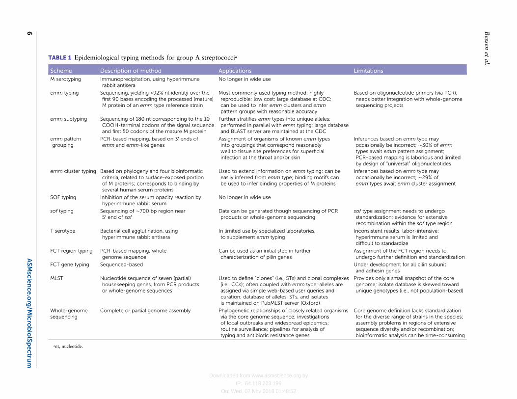
Numerous typing schemes have been used to aid inunderstanding the epidemiology and underlying orga-nization of the S. pyogenes species. A summary of typing(and related) schemes, and their applications and limita-tions, is provided in Table 1. Each epidemiological mol-ecular marker is discussed in detail below.
M ProteinDuring the 1920s, work began that aimed at under-standing the basis for protective immunity to GAS infec-tion. Antibodies raised to extractable surface antigens,calledM proteins, led to opsonophagocytosis of the strainfrom which the M protein was derived (52). However,antibody directed to the M protein of one organism oftenfailed to confer protection against many other GAS iso-lates tested. A serological typing scheme arose throughthe development of antibodies directed to M proteinsof different isolates. More than 80 distinct M types wereidentified, and protective immunity to GAS infection is, inlarge part, M type specific.
Copurifying with the M type-specific material wasM-associated protein (MAP), which has two major an-tigenic forms (53, 54). The MAP I antigen was largelyassociated with ARF isolates, and MAP II was highlyprevalent among serum opacity factor (SOF)-producingorganisms. The MAP antigens were subsequently iden-tified as part of the conserved C repeat region (CRR) ofM protein (55), which occupies the COOH-terminal halfof the surface-exposed portion of M protein. The CRRhas twomajor antigenic forms known as class I and classII (56). The structure of M protein is depicted in Fig. 2A.
emm typesA nucleotide sequence-based emm typing scheme thatclosely parallels theM serotype schemewas developed∼2decades ago (57). emm typing is based on the extensive
ASMscience.org/MicrobiolSpectrum 5
Group A Streptococci
Downloaded from www.asmscience.org by
IP: 64.118.223.196
On: Wed, 07 Nov 2018 01:48:52
TABLE 1 Epidemiological typing methods for group A streptococcia
Scheme Description of method Applications Limitations
M serotyping Immunoprecipitation, using hyperimmunerabbit antisera
No longer in wide use
emm typing Sequencing, yielding >92% nt identity over thefirst 90 bases encoding the processed (mature)M protein of an emm type reference strain
Most commonly used typing method; highlyreproducible; low cost; large database at CDC;can be used to infer emm clusters and emmpattern groups with reasonable accuracy
Based on oligonucleotide primers (via PCR);needs better integration with whole-genomesequencing projects
emm subtyping Sequencing of 180 nt corresponding to the 10COOH-terminal codons of the signal sequenceand first 50 codons of the mature M protein
Further stratifies emm types into unique alleles;performed in parallel with emm typing; large databaseand BLAST server are maintained at the CDC
emm patterngrouping
PCR-based mapping, based on 3′ ends ofemm and emm-like genes
Assignment of organisms of known emm typesinto groupings that correspond reasonablywell to tissue site preferences for superficialinfection at the throat and/or skin
Inferences based on emm type mayoccasionally be incorrect; ∼30% of emmtypes await emm pattern assignment;PCR-based mapping is laborious and limitedby design of “universal” oligonucleotides
emm cluster typing Based on phylogeny and four bioinformaticcriteria, related to surface-exposed portionof M proteins; corresponds to binding byseveral human serum proteins
Used to extend information on emm typing; can beeasily inferred from emm type; binding motifs canbe used to infer binding properties of M proteins
Inferences based on emm type mayoccasionally be incorrect; ∼29% ofemm types await emm cluster assignment
SOF typing Inhibition of the serum opacity reaction byhyperimmune rabbit serum
No longer in wide use
sof typing Sequencing of ∼700 bp region near5′ end of sof
Data can be generated though sequencing of PCRproducts or whole-genome sequencing
sof type assignment needs to undergostandardization; evidence for extensiverecombination within the sof type region
T serotype Bacterial cell agglutination, usinghyperimmune rabbit antisera
In limited use by specialized laboratories,to supplement emm typing
Inconsistent results; labor-intensive;hyperimmune serum is limited anddifficult to standardize
FCT region typing PCR-based mapping; wholegenome sequence
Can be used as an initial step in furthercharacterization of pilin genes
Assignment of the FCT region needs toundergo further definition and standardization
FCT gene typing Sequenced-based Under development for all pilin subunitand adhesin genes
MLST Nucleotide sequence of seven (partial)housekeeping genes, from PCR productsor whole-genome sequences
Used to define “clones” (i.e., STs) and clonal complexes(i.e., CCs); often coupled with emm type; alleles areassigned via simple web-based user queries andcuration; database of alleles, STs, and isolatesis maintained on PubMLST server (Oxford)
Provides only a small snapshot of the coregenome; isolate database is skewed towardunique genotypes (i.e., not population-based)
Whole-genomesequencing
Complete or partial genome assembly Phylogenetic relationships of closely related organismsvia the core genome sequence; investigationsof local outbreaks and widespread epidemics;routine surveillance; pipelines for analysis oftyping and antibiotic resistance genes
Core genome definition lacks standardizationfor the diverse range of strains in the species;assembly problems in regions of extensivesequence diversity and/or recombination;bioinformatic analysis can be time-consuming
ant, nucleotide.
6ASMscie
nce
.org/M
icrobiolSpectru
m
Bessen
etal.

Downloaded from www.asmscience.org by
IP: 64.118.223.196
On: Wed, 07 Nov 2018 01:48:52
nucleotide sequence differences at the 5′ end of the emmgene, whereby a unique emm type is defined as having<92% nucleotide identity to any other emm type, overthe sequence corresponding to the first 30 codons of themature M protein (Fig. 2A). Virtually all contemporaryepidemiological studies define GAS isolates according totheir emm type. Currently, there are ∼243 recognizedemm types (58).
Identification of the emm gene is not always obvious,and the problem can be due to paralogous emm-likegenes mapping immediately upstream (mrp) or down-stream (enn) of emm in numerous GAS strains (see nextsection; Fig. 2B). Confusion about emm type assign-ments has arisen wherein enn is directly fused to the 5′end region of emm as the result of DNA excision arisingfrom recombination between the emm and enn genes.
FIGURE 2 M protein structure and emm pattern gene arrangements. (A) Key features of Mprotein are shown, including the type-specific determinants, cell wall-spanning domain,and C repeat region. (B) Chromosomal arrangement of emm and the flanking emm-likegenes (mrp, enn) gives rise to five emm patterns, which form three main groupings (A-C,D, E). Transcription of emm and emm-like genes is positively regulated by Mga, which isencoded by mga, which lies immediately upstream of the emm region.
ASMscience.org/MicrobiolSpectrum 7
Group A Streptococci

Downloaded from www.asmscience.org by
IP: 64.118.223.196
On: Wed, 07 Nov 2018 01:48:52
Among the∼243 emm types recognized to date, thereare∼1,632 distinct allelic forms of the emm type-specificregion, known as emm subtypes (58). The emm subtypealleles are based on sequence differences relative to thetyping reference, within a 180-bp segment correspond-ing to the last 10 codons of the predicted signal peptidecoding region and the first 50 codons corresponding tothe NH2-terminus of the mature M protein molecule(59).
emm patternsMany strains of GAS have emm-like genes (mrp, enn)positioned immediately upstream and/or downstream ofemm on the chromosome (60–63). Near the 3′ end ofeach emm and emm-like gene lies a region that encodesone of four divergent cell wall-spanning domains thatcan be distinguished by oligonucleotides (60, 64). Usinga PCR-based mapping approach, the content of emmand emm-like genes and their arrangement on thechromosome was determined for GAS isolates of manydifferent emm types. The resulting emm region maps arereferred to as “emm patterns”; three major emm patterngroupings are recognized (Fig. 2B). M proteins of emmpattern E tend to be class II, whereas the emm patternsA-C and D groupings largely overlap with the class ICRR (60).
To date, the emm pattern genotype has been ascer-tained for GAS isolates of 170 of the ∼243 recognizedemm types (Table 2). With only two exceptions un-covered thus far (emm54, emm218), multiple isolatessharing the same emm type also share the same emmpattern grouping. The majority of emm types are re-presented by emm pattern groups D and E, accountingfor >75% of the emm types analyzed.
emm clades and clustersAn emm cluster typing system was recently developed,based on the entire surface-exposed portion of M pro-tein and its capacity to bind six human serum pro-teins (65). Phylogenetic analysis of >1,000 emm genescorresponding to 174 emm types reveals 2 main clades
(X and Y), and 16 well-supported clusters, accountingfor 82% of the emm types analyzed. The relationshipsbetween emm clades, clusters, and patterns for 174 emmtypes are illustrated in Table 3. Nearly all pattern E emmtypes (98%) belong to clade X, whereas 92% of patternA-C emm types are clade Y.
In contrast to patterns A-C and E emm types, thepattern D emm types are present in both clades X and Y(Table 3) (65). The highly specialized plasminogen-binding emm cluster D4 of clade Y represents the largestemm pattern D grouping (n = 30 emm types). Withinclade X, emm clusters E5 and E6 constitute a secondgroup of pattern D emm types (n = 14); these two clus-ters include emm types of other pattern groups as well.Most of the remaining pattern D emm types are inter-spersed throughout clade Y (n = 16). Overall, the emmcluster scheme, based on the phylogeny of the entiresurface-exposed portion of M protein, is highly consis-tent with the emm pattern scheme that is based on theCOOH-terminal peptidoglycan-spanning domain ofemm and emm-like flanking genes.
The emm cluster typing system is easy to imple-ment since the emm cluster can be inferred from the emmtype (66–69). Importantly, the emm type→ emm clusterscheme is also highly informative of the functional ca-pacity of the M protein molecule, based on consensusbinding motifs for numerous human host plasma pro-teins (65).
M protein functional domainsAs summarized in recent reviews (63, 70), the surface-exposed non-type-specific portion of M proteins (i.e.,central region) contains functional domains that bindhuman host proteins such as IgG (various subclasses),IgA, plasminogen, and fibrinogen. The M type-specificdeterminants at the NH2-terminus bind two comple-ment regulators, C4b-binding protein (C4BP) and factorH-like 1.
Binding capacities strongly correlate with emm cluster(65). For example, binding of human IgG is mostly re-stricted to M proteins of emm clusters E1 through E6.Similarly, binding of C4BP is limited to M proteins ofemm clusters E1, E3, E4, and E6. IgA binding is evidentonly in emm clusters E1, E4, and E6 products, whereasfibrinogen binding is restricted to M proteins of clade Y,and plasminogen binding is restricted emm cluster D4(51, 65).
Although much less is known about the M-like pro-teins Mrp and Enn (Fig. 2B), binding properties sharedwith M protein have been described (reviewed in refer-ence 63), and in some instances, they may use similar
TABLE 2 Distribution of emm types among emm patterngroups for 170 emm types.
emm pattern groupNo. of emm typesrepresented % of total
A–C 35 20.6
D 64 37.6
E 67 39.4
Rearranged (REA) 2 1.2
A–C and D 2 1.2
8 ASMscience.org/MicrobiolSpectrum
Bessen et al.

Downloaded from www.asmscience.org by
IP: 64.118.223.196
On: Wed, 07 Nov 2018 01:48:52
binding motifs. Additional investigation of the structureand functional binding properties of the M-like proteins,and structure of the mrp and enn genes, may eventuallylead to a higher-resolution typing scheme that includesthe entirety of the emm chromosomal region.
Serum Opacity FactorSOF promotes the opacification of serum, wherein theserum opacity reaction can be neutralized by hyper-immune antiserum produced in animals (71). Not allGAS strains produce SOF, but SOF typing was oftenused as an adjunct for strains that posed challenges forM typing. Of note, SOF producers have a strong asso-ciation with strains that harbor the MAP II antigen (72).In addition, SOF-producing strains tend to exhibit di-minished beta-hemolysis (73).
More recent studies show SOF to be a proteinaceousvirulence factor that is either bound to the GAS cell sur-face or secreted. SOF has an enzymatic domain thatinteracts with high-density lipoprotein, leading to theopacification of serum, plus a fibronectin-binding do-main (74–77). The genes encodingM protein (emm) andSOF (sof) map ∼15 kb apart on the GAS chromosome;as with emm and emm-like genes, transcription of sof ispositively regulated by the Mga regulon (78, 79). softypes have been defined based on ∼600 to 750 bp po-sitioned near the 5′ end of the sof gene (80). However,
sof typing is far less developed than emm typing, andattempts to generate a well-supported phylogeny for thesof type-specific determinants have largely failed due toextensive intragenic recombination (81).
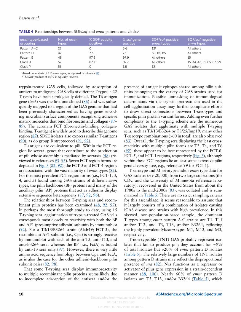
The relationships between the presence or absence ofSOF activity, or the sof gene, and the emm pattern andemm clade as inferred from the emm type, are summa-rized in Table 4. Based on the strong associations be-tween theMAP II material, class II M proteins, and emmpattern E (60), it is not surprising that there are strongcorrelations between emm pattern E and the presence ofthe sof gene and/or a positive serum opacification reac-tion (50, 80, 82, 83). Exceptions include several sof-positive strains within emm cluster E6 that are assignedto the emm pattern D grouping via PCR-based mapping(emm59, emm81, emm85); emm12 isolates (patternA-C) have an inactive sof gene. Despite the close genomemap positions of sof and emm, a single emm type can befound in association with more than one sof type andvice versa (80), indicative of HGT of emm or sof to newgenetic backgrounds and intraspecies recombinationwithin the greater emm region.
T-AntigensT-typing is based on agglutination of trypsin-treatedGAS cells with hyperimmune rabbit serum (84, 85). TheT-typing antisera is produced by immunization with
TABLE 3 Distribution of emm clusters relative to emm pattern groups, for 174 emm types
emm cluster emm cladeNo. ofemm types
No. ofemm types:pattern A–Ca
No. ofemm types:pattern D
No. ofemm types:pattern E
Other emm pattern(no. of emm types)b
E1 X 5 0 0 5
E2 X 15 0 0 15
E3 X 19 0 0 18 ND (1)
E4 X 17 0 0 17
E5 X 7 2 3 0 REA (2)
E6 X 18 1 11 6
singletons X 4 0 1 3
D1 Y 3 0 2 0 A–C and D (1)
D2 Y 5 0 5 0
D3 Y 2 0 2 0
D4 Y 31 0 30 0 ND (1)
D5 Y 3 1 2 0
A-C1 Y 2 2 0 0
A-C2 Y 2 2 0 0
A-C3 Y 5 5 0 0
A-C4 Y 5 5 0 0
A-C5 Y 3 1 0 0 ND (2)
Singletons Y 22 15 5 0 A–C and D (1); ND (1).
Outliers Outlier 6 2 3 1
aemm pattern assignments are based on references 65, 126, and 127.bND, not determined; REA, rearranged.
ASMscience.org/MicrobiolSpectrum 9
Group A Streptococci
Downloaded from www.asmscience.org by
IP: 64.118.223.196
On: Wed, 07 Nov 2018 01:48:52
trypsin-treated GAS cells, followed by adsorption ofantisera to undigested GAS cells of different T types;∼22T types have been serologically defined. The T6 antigengene (tee6) was the first one cloned (86) and was subse-quently mapped to a region of the GAS genome that hadbeen previously characterized as having genes encod-ing microbial surface components recognizing adhesivematrix molecules that bind fibronectin and collagen (87–89). The acronym FCT (fibronectin-binding, collagen-binding, T-antigen) is widely used to describe this genomeregion (87). SDSE isolates also express similar T-antigens(90), as do group B streptococci (91, 92).
T-antigens are equivalent to pili. Within the FCT re-gion lie several genes that contribute to the productionof pili whose assembly is mediated by sortases (48) (re-viewed in references 93–95). Seven FCT region forms aredepicted in Fig. 3 (82, 92); the FCT-3 and FCT-4 regionsare associated with the vast majority of emm types (82).For the most prevalent FCT region forms (i.e., FCT-1, 3,4, and 5) found among GAS strains of different emmtypes, the pilin backbone (BP) proteins and many of theancillary pilin (AP) proteins that act as adhesins displayextensive sequence heterogeneity (87, 92, 96).
The relationships between T-typing sera and recom-binant pilin proteins has been examined (48, 92, 97).In perhaps the most thorough study to date, using 15T-typing sera, agglutination of trypsin-treated GAS cellscorresponds most closely to reactivity with both the BPand AP1 (presumptive adhesin) subunits by immunoblot(92). For a T3/13/B3264 strain (Alab49; FCT-3), therecombinant AP1 subunit (i.e., Cpa) is strongly reactiveby immunoblot with each of the anti-T3, anti-T13, andanti-B3264 sera, whereas the BP (i.e., FctA) is boundby anti-T3 sera only (97). However, there is very littleamino acid sequence homology between Cpa and FctA,as is also the case for the other adhesin-backbone pilinsubunit pairs (82, 98).
That some T-typing sera display immunoreactivityto multiple recombinant pilin proteins seems likely dueto incomplete adsorption of the antisera and/or the
presence of antigenic epitopes shared among pilin sub-units belonging to the variety of GAS strains used forimmunization. Possible unmasking of immunologicaldeterminants via the trypsin pretreatment used in thecell agglutination assay may further complicate effortsto draw direct connections between T-serotypes andspecific pilin protein variant forms. Adding even furthercomplexity to the T-typing scheme are the numerousGAS isolates that agglutinate with multiple T-typingsera, such as T3/13/B3264 or T8/25/Imp19; many otherT-serotype combinations (>60 in total) are also observed(83). Overall, the T-typing sera displaying the least cross-reactivity with multiple pilin forms are T2, T4, and T6(92); these appear to be best represented by the FCT-6,FCT-5, and FCT-1 regions, respectively (Fig. 3), althoughwithin these FCT regions lie at least some extensive pilingene diversity (see, e.g., reference 99 for FCT-1).
T-serotype and M-serotype and/or emm-type data forGAS isolates (n > 20,000) from two large collections (theCDC and the University of Minnesota reference labo-ratory), recovered in the United States from about the1980s to the mid-2000s (83), was collated and is sum-marized in Table 5. There are no strict sampling criteriafor this assemblage; it seems reasonable to assume thatit largely consists of a combination of isolates causingiGAS disease and strains with high prevalence. In thisskewed, non-population-based sample, the dominantT types among emm pattern A-C strains are T1, T11and/or T12, and T3, T13, and/or B3264, reflectingthe highly prevalent M/emm types M1, M12, and M3,respectively.
T-non-typeable (TNT) GAS probably represent iso-lates that fail to produce pili; they account for ∼9%of total isolates but >20% of emm pattern D isolates(Table 5). The relatively large numbers of TNT isolatesamong pattern D strains may reflect the disproportionalpresence of nra (82); Nra functions as a repressor oractivator of pilus gene expression in a strain-dependentmanner (88, 100). Nearly 60% of emm pattern Disolates are T3, T13, and/or B3264 (Table 5), which
TABLE 4 Relationships between SOF/sof and emm patterns and cladesa
emm type-basedgrouping
No. of emmtypes
% SOF activitypositive
% sof genepositive
SOF/sof positiveemm types
SOF/sof negativeemm types
Pattern A–C 22 0 5.6 12b All others
Pattern D 42 7.3 7.1 59, 81, 85 All others
Pattern E 48 97.9 97.9 All others 15
Clade X 57 87.7 87.7 All others 15, 34, 42, 51, 65, 67, 99
Clade Y 56 0 1.8 12 All others
aBased on analysis of 113 emm types, as reported in reference 83.bThe SOF product of sof12 is typically inactive.
10 ASMscience.org/MicrobiolSpectrum
Bessen et al.

Downloaded from www.asmscience.org by
IP: 64.118.223.196
On: Wed, 07 Nov 2018 01:48:52
appears to be largely represented by the FCT-3 regionform (82, 87, 92, 97). The most frequent M/emm typesamong pattern E isolates of this collection are M4 andM28 (83), which largely reflects the dominance of theT4 and/or T28 serotype grouping (Table 5). Severalcorrelations between individual loci within the FCT re-gion and emm pattern and/or emm type have been noted(50, 82, 92, 101).
Within an emm cluster group, there can be a verywide array of T types (based on Table 5; detailed datanot shown) (83). For example, the emm cluster E3isolates (13 emm types) are found in association with T5,T27, and/or T44 (28%); TNT (16%); T8, T25, and/orImp19 (11%); T14 (11%); T9 (9%); T4 and/or T28(9%); T3, T13, and/or B3264 (6%); and T11 and/orT12 (4%), among others. In contrast, cluster D4 isolates(21 emm types, all encoding plasminogen-binding M
proteins) are far more homogeneous, with 72% associ-ated with T3, T13, and/or B3264 (another 16% areTNT). Although emm genes within a cluster are moreclosely related to each another than to emm of otherclusters, the extent to which surface pili contribute to theoverall antigenic cell surface repertoire appears to varywidely by cluster.
For GAS sharing the same emm type, individual iso-lates often differ in their T-serotype (80, 83). For exam-ple, while a substantial majority ofM/emm type 4 isolatesfrom the Table 5 study are T4, there are other M/emm4isolates that have T types T4/28, T8/25/Imp19, T3/13,and TNT (83) (detailed data not shown). M/emm4isolates typically have sof4, but there are exceptions hereas well (80). The unique combinations of M/emm, SOF/sof, and T types is indicative of HGT of the epidemio-logical marker genes among GAS.
FIGURE 3 Structure of the pilus-encoding FCT region. Seven forms of the FCT region areshown. For pilus structural protein genes: bp, tee, and fctA, backbone pilins; ap1, ap2, fctX,cpa, and fctB, ancillary pilins. In FCT-2, ap1, bp, and ap2 have sequence homology withcpa, fctA, and fctB, respectively, although divergence is high (82). Pilin adhesins includeFctX from FCT-1, AP1 from FCT-2, and Cpa from FCT-3 and FCT-4. Pilin subunits thatanchor the pilus shaft to the cell wall include AP2 and FctB (249). Fibronectin-bindingprotein genes include prtF1 (sometimes designated sfbI), prtF2 (sometimes designatedpfbp), and possibly others that remain to be classified.
ASMscience.org/MicrobiolSpectrum 11
Group A Streptococci

Downloaded from www.asmscience.org by
IP: 64.118.223.196
On: Wed, 07 Nov 2018 01:48:52
Unlike M type-specific antibodies, there is no knownhuman host protective effect mediated via T type-specificserum; this may be a consequence of immunoreactivitydirected toward trypsin-treated target antigens thatare not encountered during natural infection. However,antiserum raised to recombinant pilin subunits or intactpili can be protective in experimental assays (48, 102).Shortcomings of the T-typing cell agglutination assayinclude a general lack of good resolution, the time-con-suming process, and the requirement for standardizedrabbit serum. Because T-typing sera display high levelsof immunological cross-reactivity to different recombi-nant pilin polypeptides, and many GAS isolates agglu-tinate with multiple T-typing sera, it can be difficult topredict the FCT region genotype form based solely onthe T-agglutination pattern.
At present, there is no single widely recognized geno-typing scheme for the FCT region form and pilin genes(35, 82, 92, 96), but the need for one seems quite com-pelling given the importance of the FCT region in straindiversity as well as virulence.
Multilocus Sequence Typing andCore Housekeeping GenesThe first study to examine the population structure ofGAS utilized multilocus enzyme electrophoresis (103).Isolates recovered from cases of severe iGAS diseasecorrespond to several distinct lineages as defined byelectrophoretic type. The multilocus enzyme electro-phoresis approach was replaced by the more standard-ized multilocus sequence typing (MLST). For GAS,
MLST is based on the nucleotide sequences of seven(partial) core housekeeping genes (104). MLST data aremaintained at a user-interactive website (www.pubmlst.org/spyogenes) and currently lists 966 distinct sequencetypes (STs) (105); numerous investigators from through-out the world have generously contributed to this richdata set. Using goeBURST (106), 133 clonal complexes(CC) as defined by single locus variants (SLVs) have beendetected. Figure 4 provides a chromosomal map of therelative positions of the core housekeeping genes and theemm and FCT regions.
FIGURE 4 Chromosomal map of the epidemiological markergenes of GAS. Open circles represent the seven core house-keeping genes used in MLST (104). The sof locus, whenpresent, lies ∼15 kb upstream of emm. The FCT region, whichencodes pilus structural proteins and pilus biosyntheticenzymes plus other adhesins and transcriptional regulators,ranges in size from ∼11 to 16 kb. The FCT region lies ∼250 to300 kb from the emm region, on the opposite side of theorigin of replication (ori). GAS genomes range in size from∼1.8 to 1.9 Mb.
TABLE 5 Relationships between emm pattern and T types for >20,000 GAS isolatesa
T type % Total % Pattern A–C % Pattern D % Pattern E
T1 18.73 34.64 0.93 0.14
T2 3.30 0.00 0.00 7.85
T6 6.19 11.09 1.97 0.34
T9 0.82 0.00 1.39 1.81
T14 0.88 0.11 4.29 1.55
T15 0.00 0.00 0.00 0.00
T18 0.59 1.10 0.00 0.00
T22 0.02 0.00 0.00 0.04
T23 0.07 0.12 0.00 0.00
T3, 13, and/or B3264 16.10 17.14 58.63 10.75
T4 and/or 28 15.88 0.15 3.59 37.24
T5, 27, and/or 44 4.36 4.90 0.70 4.03
T8, 25, and/or Imp19 6.60 1.13 2.90 13.82
T11 and/or 12 18.17 20.80 4.52 16.17
Nontypeable 8.29 8.83 21.09 6.25
aBased on analysis of T-serotyping data (83) according to emm pattern groups or clades inferred from M or emm type (65, 126, 127), for 21,231 isolates, whereby 271isolates had >1 grouping and were counted more than once. M or emm types ranged from types 1 through 124.
12 ASMscience.org/MicrobiolSpectrum
Bessen et al.

Downloaded from www.asmscience.org by
IP: 64.118.223.196
On: Wed, 07 Nov 2018 01:48:52
A given pattern A-C emm type has a high tendencyto be associated with a single ST or CC (104, 107, 108).However, the relationships between emm type and STare not strict, and emm types of pattern groups D andE are often found in association with distant STs (see“LateralMovement of emm” below). These data parallelfindings on the complex relationships between M/emmtypes, sof types, and T types (80, 83) and, taken together,provide supporting evidence for the HGT of emm fromone genetic background to another.
Whole-Genome SequencesThe first completely assembled GAS genome was that ofan emm1 isolate (109). In the years that followed, nu-merous complete GAS genomes were reported for manyof the most prevalent emm types found in resource-richnations (updated listings are presented in recent reviews[50, 110]). Via next-generation sequencing technologies,many thousands of genomes of GAS isolates have un-dergone nucleotide sequence determination (e.g., 24, 30,111–115). In nearly all studies, core genome sequencesare compared for isolates sharing the same emm type,and numerous key insights on genetic changes that un-derlie enhanced transmission and/or virulence have beenuncovered. The core genome data can be used to buildevolutionary models of outbreaks and epidemics (see“Epidemiology: Building Evolutionary Models” and“Evolution: Short-Term” below).
A core genome MLST has not yet been developed forGAS. Although it is a reasonable concept for somebacterial species, its utility for GAS would probably berather limited due to the ultrafine resolution this ap-proach would provide for GAS, coupled with the ex-tensive genetic recombination that characterizes thisspecies. Core genome-based phylogenies of GAS of dif-ferent emm types and/or CCs tend to yield lineages withdeep branches, indicative of large genetic distances (50,110, 111, 116).
The use of genome sequence data for surveillanceof GAS has begun (35, 117–120). The CDC recentlyimplemented a whole-genome sequence-based typingscheme for population-based iGAS strain surveillanceconducted through the Active Bacterial Core program(35). For large-scale strain surveillance, the ability toobtain detailed information on typing and resistancefeatures within a single output, from a single raw datainput source, has largely precluded the need for multipletyping protocols and antibiotic susceptibility testing.The GAS typing pipeline automatically restricts an emmtype to the sequence linked to emm consensus primer 1(121) to improve accurate identification of emm and
assigns an emm subtype (i.e., allele) as well. Partialsequences of BP genes and sof are also captured. In ad-dition to emm subtype, the emm-like genes and MLSTalleles and STs are identified, as is gacI, which is specificto CHO-A biosynthesis. Among numerous other genes,antimicrobial resistance determinants are also assessed.
A key feature of genome sequence-based active iGASsurveillance is the capacity to detect temporally andgeographically related GAS clusters. For GAS isolatesthat are nearly identical at the single nucleotide poly-morphism (SNP) level, the circumstances of where andwhen the isolates were recovered can be examined tohelp establish whether there exist disease clusters thatmay be well suited for an intervention (e.g., health carefacilities, vulnerable patient populations such as thehomeless, and IVDUs). Thus, next-generation sequenc-ing coupled with an appropriate bioinformatics pipelinecan be used as an autodetection tool to help guide publichealth interventions.
EPIDEMIOLOGY: GLOBAL TRENDSInfection at the Epitheliumof the Throat and SkinPast studies show that GAS strains bearing certain Mtypes have a strong tendency to cause pharyngitis butnot impetigo. Similarly, there are M types often recov-ered from impetigo skin lesions but rarely from cases oftonsillopharyngitis (8, 122, 123). This observation gaverise to the important concept of classical throat and skinstrains that are largely distinct from one another in theirepidemiological and disease associations.
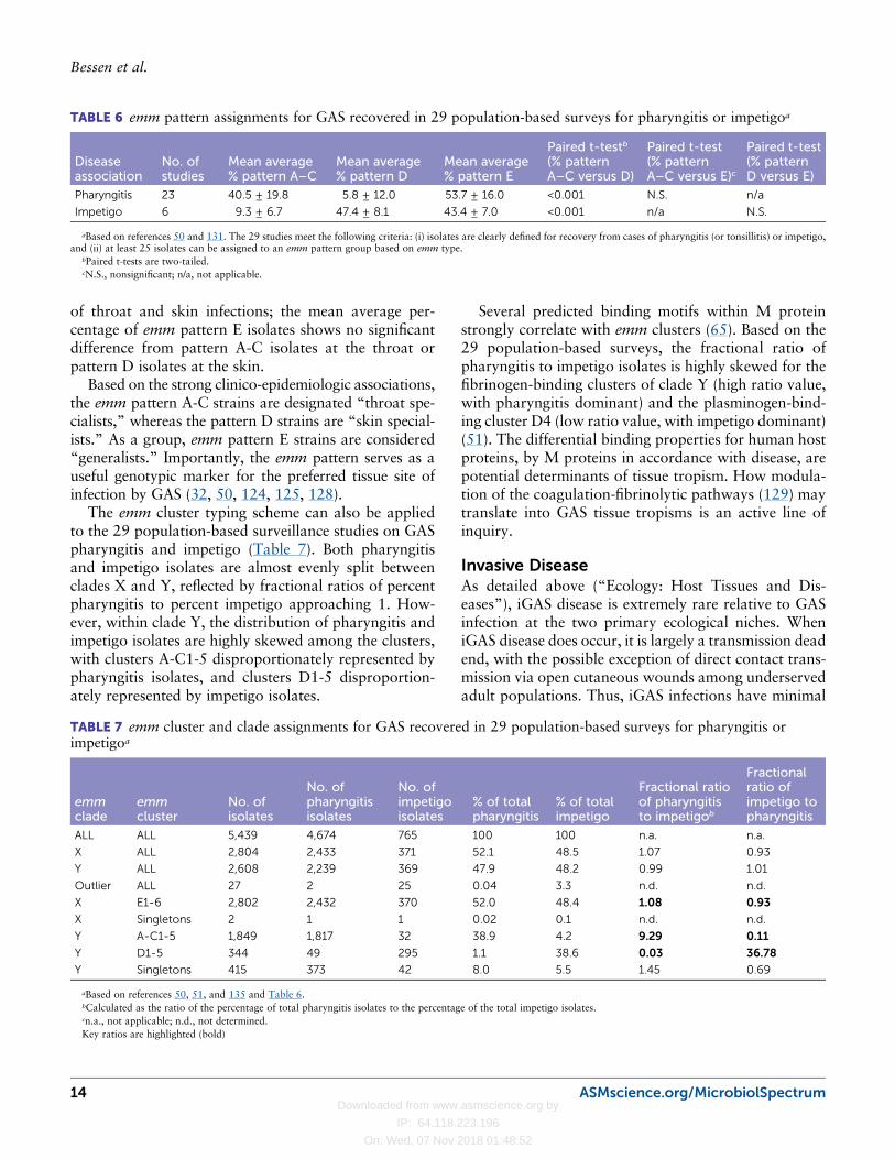
There are strong correlations between emm patterngroups A-C and D and recovery from cases of pharyn-gitis versus impetigo, respectively (50, 124, 125). Sincethe emm type is highly predictive of the emm patterngroup for nearly all emm types examined (126, 127),reasonable inferences can be made about emm patterngrouping based on knowledge of emm types. Such in-ferences were made on an expanded scale for >5,000GAS isolates derived from cases of pharyngitis or im-petigo in 29 population-based surveillance studies fromthroughout the world (Table 6). In 23 studies on phar-yngitis, emm pattern A-C and D isolates constituteda mean average of 41% and 6%, respectively. In sixstudies of impetigo, the mean average for emm patternA-C and D isolates was reversed, at 9% and 47%.For pharyngitis versus impetigo groups, the differencebetween the fraction of A-C and D isolates in eachsurveillance study is highly significant (Table 6). emmpattern E isolates account for almost equal fractions
ASMscience.org/MicrobiolSpectrum 13
Group A Streptococci
Downloaded from www.asmscience.org by
IP: 64.118.223.196
On: Wed, 07 Nov 2018 01:48:52
of throat and skin infections; the mean average per-centage of emm pattern E isolates shows no significantdifference from pattern A-C isolates at the throat orpattern D isolates at the skin.
Based on the strong clinico-epidemiologic associations,the emm pattern A-C strains are designated “throat spe-cialists,” whereas the pattern D strains are “skin special-ists.” As a group, emm pattern E strains are considered“generalists.” Importantly, the emm pattern serves as auseful genotypic marker for the preferred tissue site ofinfection by GAS (32, 50, 124, 125, 128).
The emm cluster typing scheme can also be appliedto the 29 population-based surveillance studies on GASpharyngitis and impetigo (Table 7). Both pharyngitisand impetigo isolates are almost evenly split betweenclades X and Y, reflected by fractional ratios of percentpharyngitis to percent impetigo approaching 1. How-ever, within clade Y, the distribution of pharyngitis andimpetigo isolates are highly skewed among the clusters,with clusters A-C1-5 disproportionately represented bypharyngitis isolates, and clusters D1-5 disproportion-ately represented by impetigo isolates.
Several predicted binding motifs within M proteinstrongly correlate with emm clusters (65). Based on the29 population-based surveys, the fractional ratio ofpharyngitis to impetigo isolates is highly skewed for thefibrinogen-binding clusters of clade Y (high ratio value,with pharyngitis dominant) and the plasminogen-bind-ing cluster D4 (low ratio value, with impetigo dominant)(51). The differential binding properties for human hostproteins, by M proteins in accordance with disease, arepotential determinants of tissue tropism. How modula-tion of the coagulation-fibrinolytic pathways (129) maytranslate into GAS tissue tropisms is an active line ofinquiry.
Invasive DiseaseAs detailed above (“Ecology: Host Tissues and Dis-eases”), iGAS disease is extremely rare relative to GASinfection at the two primary ecological niches. WheniGAS disease does occur, it is largely a transmission deadend, with the possible exception of direct contact trans-mission via open cutaneous wounds among underservedadult populations. Thus, iGAS infections have minimal
TABLE 7 emm cluster and clade assignments for GAS recovered in 29 population-based surveys for pharyngitis orimpetigoa
emmclade
emmcluster
No. ofisolates
No. ofpharyngitisisolates
No. ofimpetigoisolates
% of totalpharyngitis
% of totalimpetigo
Fractional ratioof pharyngitisto impetigob
Fractionalratio ofimpetigo topharyngitis
ALL ALL 5,439 4,674 765 100 100 n.a. n.a.
X ALL 2,804 2,433 371 52.1 48.5 1.07 0.93
Y ALL 2,608 2,239 369 47.9 48.2 0.99 1.01
Outlier ALL 27 2 25 0.04 3.3 n.d. n.d.
X E1-6 2,802 2,432 370 52.0 48.4 1.08 0.93
X Singletons 2 1 1 0.02 0.1 n.d. n.d.
Y A-C1-5 1,849 1,817 32 38.9 4.2 9.29 0.11
Y D1-5 344 49 295 1.1 38.6 0.03 36.78
Y Singletons 415 373 42 8.0 5.5 1.45 0.69
aBased on references 50, 51, and 135 and Table 6.bCalculated as the ratio of the percentage of total pharyngitis isolates to the percentage of the total impetigo isolates.cn.a., not applicable; n.d., not determined.Key ratios are highlighted (bold)
TABLE 6 emm pattern assignments for GAS recovered in 29 population-based surveys for pharyngitis or impetigoa
Diseaseassociation
No. ofstudies
Mean average% pattern A–C
Mean average% pattern D
Mean average% pattern E
Paired t-testb
(% patternA–C versus D)
Paired t-test(% patternA–C versus E)c
Paired t-test(% patternD versus E)
Pharyngitis 23 40.5 ± 19.8 5.8 ± 12.0 53.7 ± 16.0 <0.001 N.S. n/a
Impetigo 6 9.3 ± 6.7 47.4 ± 8.1 43.4 ± 7.0 <0.001 n/a N.S.
aBased on references 50 and 131. The 29 studies meet the following criteria: (i) isolates are clearly defined for recovery from cases of pharyngitis (or tonsillitis) or impetigo,and (ii) at least 25 isolates can be assigned to an emm pattern group based on emm type.
bPaired t-tests are two-tailed.cN.S., nonsignificant; n/a, not applicable.
14 ASMscience.org/MicrobiolSpectrum
Bessen et al.

Downloaded from www.asmscience.org by
IP: 64.118.223.196
On: Wed, 07 Nov 2018 01:48:52
impact on shaping the long-term evolution of GAS. Theoccurrence of iGAS disease is strongly affected by hostrisk factors and to a lesser extent by the genotype of theinfecting GAS strain.
In the United States and Europe, most iGAS dis-ease depends on transmission of GAS via a respiratoryroute (Fig. 1). Whole-genome sequencing of hundreds ofGAS isolates has provided important details on geneticchanges that enhance transmission and/or invasiveness(see “Evolution: Short-Term” below). The invasive in-dex value provides insight on whether a GAS strain hasan inherent ability to better gain access to deep tissuerelative to its overall prevalence in the host population(22, 130). The emm types associated with iGAS diseasehave been intensively studied and are well described (e.g.,131–133).
Nonbullous impetigo caused by GAS usually arisesin young children in tropical or subtropical climates.GAS can also cause slightly deeper cutaneous infectionsthat are considered “pyoderma,” and while they are notconsidered part of the iGAS disease spectrum, thesecutaneous infections are distinct from typical impetigoin terms of patient demographics (i.e., adult, temperateregions) and host risk factors (e.g., IVDU, homeless,military) (23, 24, 134–136). Of interest is the strongassociation between certain pattern D emm types (spe-cifically, emm59 and emm81) and recent infections andoutbreaks of deeper cutaneous infections. The emm59and emm81 types are notable for their grouping incluster E6 and tend to harbor sof as well; conceivably,these are “super-specialist” strains containing a hybridmix of virulence factors that shift the modes of trans-mission and GAS disease course. Most other GAS-causing skin infection outbreaks among IVDUs aresof-positive emm pattern E strains (137, 138).
Acute Rheumatic FeverAcute rheumatic fever (ARF) follows an inadequatelytreated GAS infection of the URT in a genetically pre-disposed host by ∼3 to 6 weeks and can lead to rheu-matic heart disease (RHD) via autoimmune attack of theheart valves (139–143). With >500,000 annual deaths,half attributed to RHD, GAS ranks high among all in-fectious causes of morbidity and mortality. Recent esti-mates of the prevalence of RHD are ≥15 to 30 millioncases worldwide (15, 144–146); RHD is the leadingcause of mortality from acquired heart disease in indi-viduals <50 years old (147) and of all cardiovasculardisease in children in resource-poor nations (148). To-day, ARF and RHD are largely diseases of poverty, withan occasional resurgence in disease elsewhere (149).
Early epidemiological studies of ARF outbreaks ininstitutional settings showed that only a subset of GASstrains trigger ARF (11, 55, 140, 150–156). Historically,“rheumatogenic” GAS strains were referred to by theirM protein serotypes, with M3, M5, M6, andM18 beinghighly prevalent. The concept of “nonrheumatogenic”strains is exemplified by the RHD patients of IrvingtonHouse during the preantibiotic era, who experienced aGAS outbreak (M type 4) in the absence of recurrentARF attacks (155). Of ten M types often associated withARF (M types 1, 3, 5, 6, 14, 18, 19, 24, 27, and 29[157]), nine are emm pattern A-C (one is pattern E);of five relatively common M types that are rarely asso-ciated with ARF (M types 2, 4, 12, 22, and 28 [157]),four are emm pattern E (one is emm pattern A-C) (126,127).
ARF-associated isolates recovered in Hawaii differfrom the classic rheumatogenic emm types (158).Atypical emm types are also found in tropical Australia,where the prevalence of RHD is among the highest in theworld (159). Over time, it has become much less clearwhich emm types are best assigned the “rheumatogenic”label, even though it is widely accepted that not all GASstrains trigger ARF in a genetically susceptible host (150,155).
The potential role of GAS skin infection in trigger-ing ARF and RHD in certain settings (e.g., noninstitu-tional, tropical) has been an ongoing debate since the1960s (160). For example, the indigenous populationof Australia suffers from a high incidence of ARF andRHD, yet GAS is far more frequently recovered fromimpetigo lesions than from the throat. Moreover, thestrains recovered from the URT in this host popula-tion often belong to the emm pattern D skin specialistgroup or pattern E generalists. The epidemiological co-occurrence of GAS skin infections and RHD supportsthe idea that impetigo may contribute to this chronicdisease. More extensive studies are needed to better un-derstand skin infections and their potential role in ARFand RHD. The possibility that the closely related spe-cies SDSE might also trigger episodes of ARF or worsenRHD deserves deeper consideration as well (161, 162).
EPIDEMIOLOGY: DIVERSITY WITHINHOST COMMUNITIESWithin a human host community over a narrow timeframe, numerous GAS strains are in circulation. For the29 population-based collections of GAS recovered fromcases of pharyngitis and impetigo, the number of emmtypes and the Simpsons diversity index was calculated
ASMscience.org/MicrobiolSpectrum 15
Group A Streptococci

Downloaded from www.asmscience.org by
IP: 64.118.223.196
On: Wed, 07 Nov 2018 01:48:52
(Table 8). The number of emm types for the impetigocollections averaged nearly twice that of the pharyn-gitis collections (t < 0.01, unpaired t-test, 2-tailed). TheSimpson’s diversity index (D), which incorporates thenumber of recovered organisms into the calculation, alsoshows highly significant differences between the impe-tigo and pharyngitis collections, wherein the impetigogroup has a higher mean average D value (a D valueof 1 reflects maximal diversity). An analysis that in-cludes all GAS, irrespective of clinical association, alsoshows fewer numbers of emm types accounting for alarge proportion of isolates in resource-rich countries,where URT infections tend to predominate (131). Thelarge numbers of circulating emm types is likely theconsequence of expansive GAS strain migration, eveninto geographically remote communities (108). The bi-ological basis for the difference in diversity among GASin host populations experiencing pharyngitis versus im-petigo may be tied to the mechanisms for transmission:respiratory droplets versus direct contact. It may be thatthe respiratory route is more highly efficient, such thata single infected individual can infect many more newhosts (i.e., higher basic reproductive rate), and this inturn leads to dominance by fewer strains, resulting in alower D value.
EPIDEMIOLOGY: EVOLUTIONOF MARKERS (emm)Although emm type diversity within a host community isrelatively low for the emm pattern A-C throat specialistscompared to the pattern D skin specialists (Table 8),there is relatively high diversity among emm pattern A-Corganisms in terms of the number of emm subtypes (i.e.,alleles based on the 5′ end of the emm gene) that evolvedworldwide (Table 9). The average mean number of emmsubtypes identified for pattern A-C strains is 22.3 sub-types per emm type, compared to only 4.0 and 6.9 forpattern D and E strains, respectively, based on currentdata available in the CDC online database (58), whichreceives numerous deposits from investigators through-out the world.
The >5-fold difference in the number of emm subtypesfor throat specialists versus skin specialists may be partlydue to sampling bias that skews in favor of GAS invasiveand pharyngitis isolates in resource-rich nations, such asthe highly prevalent emm1 type (pattern A-C). In addi-tion, the tandem sequence repeats that overlap the emmsubtype-determining region of emm5 and emm6 genes(pattern A-C) are a mutational hotspot due to DNAslipped-strand mispairing (163). However, biologicalfactors such as host immune selection promoting im-mune escape (164) may also play a role.
Nucleotide sequence alignment of emm subtypealleles for each emm type-specific region, derived from>500 global GAS isolates (n = 105 emm types), revealsaverage ratios of nonsynonymous substitutions per non-synonymous site (Ka) and synonymous substitutions persynonymous site (Ks) of 4.9, 1.5, and 1.3 for emm typesof the emm pattern A-C, D, and E groups, respectively(107). Thus, diversifying (positive) selection acting onthe emm type-specific sequence appears to be strongestfor emm pattern A-C strains.
A likely source of diversifying selection acting on theemm type region is the protective host immune response,which can drive immune escape. Conceivably, the im-munological pressures at the oropharynx (pattern A-Cthroat specialists) are more intense than those at the skin(pattern D skin specialists), although an underlyingmolecular mechanism is not presently obvious. Perhapsherd immunity builds upmore quickly with a respiratorymode of transmission compared to direct skin con-tact; as herd immunity builds, the number of susceptiblehosts dwindles, leading to more frequent occurrencesof immune escape. In further support of a role for herdimmunity in shaping the emm type content of GASpopulations, newly acquired URT infections in olderchildren are skewed toward less common emm types,consistent with exposure and protective immunity to themore prevalent emm types in early childhood (165).
The collection of M protein types assigned to asingle emm cluster have high sequence similarity relativeto M proteins of other clusters (65). Indeed, an M type-specific region sharing the same cluster as other M types
TABLE 8 emm type diversity within communities for the 29 population-based surveys for pharyngitis and impetigoa
Disease association No. of studies Mean average no. of emm types Mean average D indexb
Pharyngitis 23 17.4 ± 7.8 0.873 ± 0.065
Impetigo 6 33.8 ± 8.1 0.959 ± 0.012
Unpaired t-test, 2-tailed t = 0.0049 t < 0.001
aBased on references50, 51, and 135 and Tables 6 and 7.bSimpson’s diversity index (D) (248) is based on emm types.
16 ASMscience.org/MicrobiolSpectrum
Bessen et al.

Downloaded from www.asmscience.org by
IP: 64.118.223.196
On: Wed, 07 Nov 2018 01:48:52
often elicits cross-protective antibodies (see “Epidemi-ology: Strategies for M Protein-Based Vaccines” below).Ultimately, this cross-protective effect may aid in thedesign of highly efficacious vaccines that have broadcoverage but contain only a limited selection of type-specific epitopes. Whereas 90% of pattern D and E emmtypes belong to 1 of the 16 emm clusters, nearly halfof pattern A-C emm types are standalone and do notcluster with any other emm type, highlighting once againa distinct dynamic for the evolution of many pattern A-Cemm types.
Another possible factor at play in shaping the evolu-tion of emm type-specific sequences is functional con-straints, such as those imposed by binding C4BP via thetype-specific region of numerous M proteins (166–168).Whole GAS organisms that bind C4BP tend to harborM types associated with emm pattern groups D and E(167). Thus, there may be limits to diversification withinM proteins that bind C4BP for the GAS strains thatutilize this virulence strategy to enhance survival. Also,
potentially related to selection pressures acting on GASis the finding that engineered mutants with emm genedeletions have a (near) complete loss in the antiphago-cytic effect for pattern A-C strains, whereas the loss isonly partial for pattern D and E strains (169).
Overall, the throat specialists are characterized byfewer distinct emm types within a human host com-munity but high diversification worldwide within thetype-specific region (i.e., more emm subtypes). The skinspecialists have about twice the number of emm typeswithin a community, but diversification within the type-specific region is ∼5-fold lower based on the numberof emm subtypes or Ka/Ks ratios. The emm pattern Egeneralists are most similar to the skin specialists in thesevalues.
The “emm type” and “emm cluster” definitions arecategorical, and the noted differences between throatand skin specialists may partly reflect the placement ofthe categorical “cutoff” value along a continuum of se-quence divergence. Factors that shape the GAS popula-
TABLE 9 Relationships between emm pattern or cluster and emm type and subtype
emm groupingcategory Name of group
No. ofemm typesrepresented
No. of emmsubtypesrepresenteda
Average no. ofemm subtypesper emm type
Pattern A-C 35 781 22.31
Pattern D 64 253 3.95
Pattern E 67 460 6.85
Clade X 85 518 6.09
Clade Y 89 1,014 11.39
Cluster E1 5 40 8.0
Cluster E2 15 84 5.6
Cluster E3 19 119 6.3
cluster E4 17 150 8.3
Cluster E5 7 15 2.1
Cluster E6 18 98 5.4
Cluster Clade X singletons 4 12 3.0
Cluster A-C1 2 7 3.5
Cluster A-C2 2 26 13.0
Cluster A-C3 5 118 23.6
Cluster A-C4 5 111 22.2
Cluster A-C5 3 140 46.7
Cluster D1 3 13 4.3
Cluster D2 5 22 4.4
Cluster D3 2 4 2.0
Cluster D4 31 144 4.6
Cluster D5 3 6 2.0
Cluster Clade Y singletons 28 423 15.1
Clade Y singletons Pattern A-C 16 385 24.1
Clade Y singletons Pattern D 8 16 2.0
Clade X singletons Pattern E 2 6 3.0
aBased on the CDC online database (58).
ASMscience.org/MicrobiolSpectrum 17
Group A Streptococci
Downloaded from www.asmscience.org by
IP: 64.118.223.196
On: Wed, 07 Nov 2018 01:48:52
tion structure, as defined by emm type, likely includethe route of transmission, environmental risk factors,the host immune response, tissue-specific host factors,binding functions of M protein, and intrinsic GASfactors that influence mechanisms for genetic change.
EPIDEMIOLOGY: STRATEGIES FORM PROTEIN-BASED VACCINESNumerous vaccine strategies for protection against GASinfections were recently reviewed (170–172). Briefly,vaccine strategies can be divided into M protein andnon-M protein target antigens, with the latter includ-ing streptolysin O, C5a peptidase, CHO-A, pili, and theenzyme arginine deiminase (102, 173–178). However,only the vaccine candidates that incorporate M proteinas the target antigen have attained human clinical trialstatus (179).
M protein vaccines based on the conserved antigensof the CRR (Fig. 2A) have undergone extensive investi-gation (49, 180–185). Since all M proteins have a CRR,a CRR-based vaccine has the theoretical advantage ofproviding broad coverage against all GAS strains; sev-eral formulations are currently, or will soon be, in clin-ical trials. Potential drawbacks of a CRR-based vaccineinclude immune escape. The CRR region of M pro-tein, which includes MAP I/II and the class I/II antigenicepitopes, is not 100% conserved. For example, among175 emm types from a worldwide strain collection, thereare 22 alleles of the CRR-based J8 vaccine candidate(65). Importantly, some of the amino acid residueswithin the CRR of the M protein display signaturesof diversifying selection. Immune escape is a possibilityfor any vaccine target, and current knowledge indicatesthat it deserves careful consideration for a CRR-basedvaccine.
The second category of M protein-based vaccine can-didates targets the NH2-terminal type-specific determi-nants (179, 186). Pioneering work in the 1940s and1950s showed that type-specific serum is responsible forprotective immunity against the homologous emm type(187–190); in contrast, minimal to no protective effectswere observed for infection by heterologous GAS types.A 26-valent M type-specific recombinant polypeptidevaccine is the only GAS vaccine to have reached phase IIclinical trials, whereupon it was found to be safe andhighly immunogenic (179). The “type-specific protec-tion” paradigm of GAS immunity has led to concernsabout the potential for low coverage by the multivalentvaccine, especially in tropical settings, where the numberof GAS emm types can be very high (131, 191–195).
Recent data indicate that cross-protective immunitybetween closely related emm types can induce broaderprotection via multivalent vaccines than type-specificimmunity might otherwise predict. An initial hypothesison cross-protective immunity was raised following ge-netic analysis of GAS strains collected in Brazil (196,197). Cross-protective opsonization by hyperimmunerabbit antiserum generated via immunization with Mtype-based vaccines was subsequently demonstratedusing a 30-valent vaccine (186) and an experimental4-valent vaccine designed to protect against the 17 emmtypes belonging to emm cluster E4 (198).
Superficial skin infection can stimulate a robust an-tibody response. A recent study of humoral immunityacquired after impetigo infection in Fijian schoolchildrendemonstrates that 38% of subjects had a ≥4-fold in-crease in anti-M-type-specific IgG titers that match theM type of the infecting strain (199). Importantly, cross-protective emm cluster-specific immunity was evidentfor clusters E4, E6, and D4, although the levels of cross-protection were variable depending on both the patientand emm cluster. That the cross-reactive immune re-sponses frequently align with emm cluster groups holdspromise for the design of M type-based vaccines havingbroad coverage. More data are needed to further solidifythe clinical and experimental findings obtained to date;identifying immunoassays to help define correlates ofprotection (CoPs) is a high-priority goal (200). Serotypereplacement may potentially be an issue for GAS fol-lowing widespread immunization with M type-basedvaccines, as is the case for Streptococcus pneumoniaeand serotype-based capsule vaccines (201, 202).
EPIDEMIOLOGY: BUILDINGEVOLUTIONARY MODELSMLST data can be used to build models of evolutionarydescent via eBURST or goeBURST, which are clusteringalgorithms based on SLVs (and/or double locus variants)among the seven core housekeeping genes (106, 203).The single mismatched allele that defines an SLV paircan arise by either point mutation or homologous re-combination, whereby the latter may introduce >1 SNP.
For GAS, the combined total length of the sevenhousekeeping (partial) alleles is 3,134 bp. However,3,134 bp represents only a fraction of the GAS coregenome; for example, emm3 type iGAS have a core ge-nome of ∼1,645 kb (115), which is ∼524-fold longerin sequence than the MLST alleles combined. Based onthese values, it can be estimated that an MLST-definedSLV pair of GAS would differ by an average minimum
18 ASMscience.org/MicrobiolSpectrum
Bessen et al.

Downloaded from www.asmscience.org by
IP: 64.118.223.196
On: Wed, 07 Nov 2018 01:48:52
of >500 SNPs within its core genome if genetic changewere strictly due to point mutation or intraspecies re-combination between very closely related organisms.However, if there was extensive HGT and recombi-nation between more distally related GAS, the numberof SNPs that differentiate the SLV pair is expected tobe even higher. Given that the core genome of emm3type iGAS strains has an estimated 29 to 133 SNPsper isolate (i.e., short-term evolution) (115), the modelsfor evolutionary descent based on the SLVs of MLSTusing only seven core housekeeping genes may bestcapture intermediate stages of evolution, which can bequite informative.
MLST data in combination with epidemiologicalmarker genotypes can be compared to whole-genomesequence data. A good example for comparison is theemm89 strains causing iGAS disease in Europe and theUnited States, which underwent a marked increase inprevalence during the 2010s (30, 31, 111, 204). Usingthe ST and GAS isolate data deposited at www.pubmlst.org/spyogenes (105), a goeBURST plot that includesthe recent epidemic emm89 strains is shown in Fig. 5A.The newly emerged epidemic clade of emm89 corres-ponds to ST101 (and its descendants). Based on demo-graphic information (i.e., dates of recovery), it seemsreasonable to assume that ST407 is ancestral to ST101.Notably, ST407 is recovered in association with two dis-tinct emm types: at least one other ST407 GAS isolate inthe PubMLST database has emm78 in place of emm89,as do several other SLVs and double locus variants ofST407. Alignment of the 180-bp emm subtype regions ofemm78.0 (ST253) and emm89.0 (ST407, ST101) reveals<60% identity for both nucleotide and amino acid se-quences, indicative of substantial divergence (ClustalW;data not shown) and supporting a model whereinemm78 and/or emm89 underwent HGT and recombi-nation onto an ST407-related genetic background. Bothemm78 and emm89 are emm pattern E but belong todifferent emm clusters (E1 and E4, respectively).
Examination of the FCT regions of ST407 and ST101emm89 type GAS isolates reveals highly divergent Cpaand FctA proteins, corresponding to the AP1 and BPsubunits of pili, respectively (Fig. 5B). The distinct Cpaand FctA forms appear to coincide with T serotypes T11(ST407) and T13 (ST101) (35). The housekeeping genelocus that distinguishes ST407 and ST101 is yqiL, whichlies adjacent to the FCT region (Fig. 4); yqiL01 (ST401)differs from yqiL03 (ST101) by three nucleotide sub-stitutions, highly suggestive of diversification via recom-bination. In contrast to the eBURST analysis of MLSTdata, whole-genome sequencing and analysis of the core
genomes of emm89 isolates provides a much higher levelof resolution (30, 35, 111, 112). Importantly, severalSNPs and indels that distinguish the emm89 clade 2 →clade 3 transition lead to alteration of virulence factorexpression: loss of the hyaluronic acid capsule (ΔhasA)and transcriptional upregulation of toxin genes encod-ing Nga and streptolysin O.
The genetic relationships among GAS strains har-boring emm89 and ∼20 additional emm types wereinferred based on ∼75,000 SNPs of the core genome(111). The phylogeny shows that the emm89 isolates ofthe three clades detailed above, and corresponding toCC407 (ST101, ST407), cluster within the same majorlineage; ST142 isolates (double locus variants of ST407;Fig. 5A) also cluster on that same deep branch. How-ever, GAS of other emm types, plus emm89-ST380 iso-lates belonging to another CC (204), each cluster alongdistinct lineages with deep branches (111). ST380 differsfrom both ST101 and ST407 at all seven housekeepingalleles, indicative of HGT of emm89 to a distant geneticbackground.
The MLST data for emm89 strains captures twodifferent modalities of HGT involving emm: replace-ment of a nascent emm by a new emm type on the origi-nal genetic background (i.e., recipient-new clone pairs,with two emm types and one ST) and lateral movementof an emm type to a new genetic background (i.e., do-nor-new clone pairs, with one emm type and two STs)(107). Within CC407 (recipient) there is evidence for theformer process, involving replacement of emm78 withemm89 or vice versa (Fig. 5A). A possible donor ofemm89 to CC407 is emm89-ST380 (latter scenario);alternatively, ST380 may have been the recipient ofemm89 from a CC407 donor. Comparisons of com-pletely assembled genomes of possible donor-recipient-new clone pairings may aid in establishing the direc-tionality of horizontal emm gene movements.
Based on the connections within CC407, the sevenhousekeeping genes of MLST appear to recapitulate themore expansive core genome sequence data reasonablywell. However, MLST fails to capture SNPs arising incore virulence-related genes that may alter the trajectoryof GAS disease in a host population. What MLST maybe best tuned to, when combined with typing data onemm, pilin genes, and/or sof, are HGT events wherein atyping gene is present on distant STs/CCs or multipletyping genes are recovered in association with the sameST/CC (such as divergent Cpa-FctA in emm89-CC407;Fig. 5B). For GAS, MLST relationships can provide anintermediary link between short- and long-term evolu-tion andmay help delineate the genetic roots of the emer-
ASMscience.org/MicrobiolSpectrum 19
Group A Streptococci

Downloaded from www.asmscience.org by
IP: 64.118.223.196
On: Wed, 07 Nov 2018 01:48:52
FIGURE 5 Model for evolutionary descent (CC407). (A) goeBURST diagram of CC407,which includes single representatives of 41 STs and is based on data from www.pubmlst.org/spyogenes (105). Unless noted otherwise (orange shaded boxes), all isolates (n = 114)are emm89 and were recovered from (i) the 1990s through 2010s, (ii) Europe or NorthAmerica, and (iii) URT infections or iGAS disease. For evolutionary analysis of emm89(formerly PT4245 [250]) isolates based on the core genome sequence (31, 111), clade 1strain MGAS11027 is ST407, whereas clade 2 and epidemic clade 3 strains (MGAS23530and MGAS27061, respectively) are ST101; the epidemic emm89 strain H293 from theUnited Kingdom is also ST101 (30). Note that ST407 corresponds to GAS with eitheremm89 or emm78 (105). (B) Amino acid sequence identities among predicted FCT regiongene products of ST407 (MGAS11027) and ST101 (H293, MGAS23530) strains (all emm89)via ClustalW alignments.
20 ASMscience.org/MicrobiolSpectrum
Bessen et al.

Downloaded from www.asmscience.org by
IP: 64.118.223.196
On: Wed, 07 Nov 2018 01:48:52
gence of new strains that have unique repertoires of cellsurface antigens and their linked virulence functions.
EVOLUTION: MOLECULAR MECHANISMSOF GENETIC CHANGECore genes of GAS contain a historical record of nu-merous homologous recombination events (111, 205–208). Intraspecific gene exchange between GAS strainsmost likely occurs during cocolonization of the URT orcoinfection of impetigo lesions with multiple strains(209). GAS can grow as biofilms or form microcoloniesduring infection within both tissue niches (210, 211),and the high bacterial density that is attained likely fa-cilitates HGT. There is also ample evolutionary evidencefor gene exchange between GAS and other species, suchas SDSE (212–218).
A major unresolved puzzle of GAS biology is themolecular mechanisms that govern HGT, leading to thehigh rates of homologous recombination that is inferredfrom its evolutionary history. In principle, nonspecificpackaging of chromosomal fragments into bacterio-phage capsids could provide a vehicle for HGT of bac-terial DNA (i.e., generalized transduction). In additionto bacteriophage, GAS are also rich in integrative andconjugative elements (91). The degree to which randomDNA fragments may slip through during the conjuga-tive process remains unclear. A conserved RNA uracilmethyltransferase (rum) gene provides an integrationhotspot for both bacteriophage and integrative and con-jugative elements in GAS, as well as other related species(219). Natural transformation via high-frequency up-take of naked DNA has not been demonstrated forGAS, although competence gene homologues are presentin the GAS genome and (very) low efficiency transfor-mation is detectable in biofilms (220–222). However,GAS secrete numerous DNases that degrade extracellu-lar DNA, which may render the transforming substanceunavailable for uptake by a competent cell.
A mutator phenotype that results in an increasedmutation rate has been characterized for GAS. This de-fect in DNA mismatch repair is controlled by excisionand reintegration of a streptococcal phage-like chro-mosomal island (SpyCI) at a site between the mutS andmutL loci (223) and is present in the genomes of nu-merous GAS strains (50). SpyCI yields an ∼100-foldincrease in the rate of spontaneous mutation, to as highas ∼10–7 to 10–8 mutations per generation. A possiblerelationship between the presence of SpyCI and CCswith a relatively low ratio of recombinational to muta-tional events remains to be explored.
EVOLUTION: LONG TERMCore Housekeeping GenesAs previously stated, core genome-based phylogeniesof GAS of different emm types and/or CCs tend to yieldlineages with deep branches, indicative of large geneticdistances (50, 110, 111, 116). Whole-core genomephylogenies depicted as SplitsTree graphs (neighbor-netanalysis) show recombination between GAS of differentemm types located deep within the network. Further-more, there is no obvious clustering of the major lineagesin accordance with emm pattern grouping or by anyother recognized parameter.
Numerous methods have been used to assess the rel-ative amount of recombination versus mutation thatcontributes to genetic change in GAS, based on the sevencore housekeeping genes used in MLST (205–208). To-gether, the findings support the notion that GAS exhibitrelatively high levels of recombination which, at leastby some measures, is roughly comparable to the levelsobserved for the pneumococcus.
Using MLST data, the relative amount of recombi-nation versus mutation among the emm pattern-definedsubgroups of GAS was examined (107, 224). Findingsbased on several methodologies all point to the samegeneral trend, wherein the pattern A-C throat specialistshave the highest tendency to diversify by mutation, andpattern D skin specialists and pattern E generalists havea greater tendency to diversify by recombination. Thedata on a larger role for mutation, relative to recombi-nation, in genetic diversification among the classicalthroat strains is consistent with the molecular evolu-tionary analysis of emm subtypes (see “Epidemiology:Evolution of Markers (emm)” above; Table 9) (107).Differences among the emm pattern-defined groups ofGAS in terms of their mechanisms for genetic changeunderscore different dynamics in shaping their long-term population structures.
GAS strains may encounter ecological separation dueto their tissue site preferences for infection (throat versusskin), geographic partitioning (temperate versus tropi-cal) and temporal distances (winter versus summer).Ecological barriers can give rise to allopatric speciation,and therefore, signatures of early stages of speciationwithin the GAS population may be evident. However,analyses of housekeeping alleles provide strong sup-port for highly fluid HGT and homologous recombi-nation of housekeeping genes among all three emmpattern groupings (101, 128, 224). These findings ex-tend to isolates known to be recovered from the URTversus impetigo lesions, regardless of emm pattern group(224).
ASMscience.org/MicrobiolSpectrum 21
Group A Streptococci

Downloaded from www.asmscience.org by
IP: 64.118.223.196
On: Wed, 07 Nov 2018 01:48:52
In summary, despite wide spatial-temporal distancesbetween many GAS strains, there is no clear evidencefor diminished opportunities for HGT of core house-keeping genes between organisms of the different emmpattern groups. Numerous statistical measures all pointto a population genetic structure for the S. pyogenesspecies that is highly nonclonal (205–208). Against abackground of highly random genetic change in corehousekeeping genes, loci that exhibit strong linkage withemm pattern genotypes are reasonably good candidatesfor playing a direct role in the adaptations leading tothroat versus skin infection (e.g., 50, 101).
Lateral Movement of emmFor the same global set of >500 GAS isolates used tocalculate Ka/Ks ratios within the emm type-specific re-gion (see “Epidemiology: Evolution of Markers (emm)”above), the number of HGT events as a function of emmtype was measured (107). Movement of emm to a distalST is quantified as recovery of an emm type in associa-tion with >1 ST differing by ≥5 of the 7 housekeepingalleles; data show an average of 0.17, 0.60, and 0.75lateral transfer events per emm type for the pattern A-C,D, and E groups, respectively. This finding supports thenotion that for the classical throat strains, but less so forskin strains or generalists, emm type is often a reason-ably accurate marker for ST or CC (104, 107, 108). Inanother measure of HGT and recombination, the num-ber of emm types associated with emm type-variableSTs (i.e., STs recovered in association with >1 emm type)was calculated; the number of emm type replacementsper ST was 0.12, 0.15, and 0.02 for pattern A-C, D, andE isolates, respectively (107). Overall, there were threetimes as many events captured for HGT of emm to adistant ST (n = 63) than for replacement of emm on thesame ST (n = 21).
Taken together, these data shed light on GAS straincompetition surrounding the narrow time-space whereinthe donor, recipient, and newly emerged clone all coexist.Strain competition can be mediated through host/herdimmunity (225) and perhaps via other host-pathogeninteractions as well. For the throat specialist strains,competition appears to be most intense between strainssharing the same emm type, because organisms of theoriginal ST that served as the (presumptive) emm donorare rarely recovered. In contrast, for generalists, com-petition at the level of emm type appears to be consid-erably weaker, as evidenced by the frequent recovery ofthe same emm type in association with multiple distantST backgrounds. How other cell surface antigens, suchas SOF and/or pili, might contribute to the competitive
sphere will require study of lateral movements of theirgenes and alleles within the GAS population.
Full Complement of EpidemiologicalMarkers ConsideredHow do the host’s specific immune defenses perceive theincoming antigenic repertoire present on the GAS cellsurface? Except during carriage, the Mga regulon thatupregulates the expression of emm and emm-like genesand sof is typically active. The pilus T-antigens appearto be expressed by >90% of the GAS population. Theexpression/nonexpression of the hyaluronic acid capsulecan potentially mask/unmask some or all the GAS cellsurface proteins. Setting aside the variance in the tran-scriptional program, which can have a profound effecton transmission and virulence (see “Evolution: ShortTerm” below), the collection of epidemiological markersprovides a useful construct for defining GAS “strains.”
Figure 6 summarizes the relative diversity or presence/absence of each major epidemiological marker in accor-dance with the emm pattern-defined groupings of throatspecialists, skin specialists, and generalists. Importantly,emm type is rarely recovered in association with multipleemm pattern groupings (Table 2), thereby allowingreasonably valid inferences to be drawn. The pattern-and cluster-defined emm subtype alleles reveal stark dif-ferences between the throat specialist, skin specialist, andgeneralist groupings regarding diversification (Table 9).Major distinctions also lie in the relative distributions ofthe sof locus (Table 4), emm-like genes (see Fig. 2B), andFCT region forms (Fig. 3) (82).
Do highly diverse emm subtypes compensate for thedearth of emm-like genes and sof among the throatspecialists—and vice versa for the generalists? The di-versity among mrp and enn alleles is modest, but theytoo have undergone intraspecies HGT (226); selectionmay be tied to functional binding attributes of the Mrpor Enn proteins (e.g., IgA, IgG) (63, 227–230). The sofgene also undergoes rather extensive intraspecies HGT(80); however, sof gene movement is nonrandom inthat it is largely restricted to the emm pattern E gen-eralists (Table 4). Yet the relationships between indi-vidual emm types and sof types are highly varied, withmany unique combinations arising via HGT to distantgenetic backgrounds (35).
Among the throat specialist group is a wide varietyof FCT region forms (Fig. 6) (82). The FCT-1 region isthe most prevalent form among representative patternA-C organisms of unique emm types; FCT-1 is charac-terized by an AP1 adhesin (FctX) and major BP (Tee)that are markedly divergent from the highly variable
22 ASMscience.org/MicrobiolSpectrum
Bessen et al.

Downloaded from www.asmscience.org by
IP: 64.118.223.196
On: Wed, 07 Nov 2018 01:48:52
Cpa and FctA pilins of FCT-3 and FCT-4 (82, 92). Notshown in Fig. 6 is FCT-2, which is (almost) exclusive tothe highly prevalent emm1 organisms; the AP1 and BPpilins of FCT-2 are akin to a distant lineage of Cpa andFctA, respectively (82). Thus, the unusually high success(i.e., fitness) of emm1 strains may be due, in part, to itsantigenically unique pilus structure. Overall, the absenceof sof and emm-like genes among the pattern A-C straingrouping may be compensated for by the antigenic andstructural diversity among its pili.
The skin specialist group appears to be the most ho-mogeneous regarding epidemiological marker loci con-tent, largely lacking sof but having both mrp and enn,and an FCT-3 region skewed heavily toward T3/13/B3264 (Table 5); this genotype is coupled to the lowestdiversity among emm subtypes (Table 9). Also, the skinspecialist group is dominated by emm cluster D4, en-coding the plasminogen-binding M protein (51, 65).However, past lateral movement of emm type to distantgenetic backgrounds is rather abundant among the skinspecialists and is ∼2-fold greater than that of emm typereplacement on the same ST (107) (see “Lateral Move-ment of emm” above); there is also ample HGT ofhousekeeping alleles. Conceivably, the emm pattern Dskin specialists represent a more recent subpopulation ofGAS (i.e., younger) that has less diversity within any onegene, but the genes themselves are highly recombina-torial. Alternatively, the cell surface protein repertoire ofskin specialists may be subject to less intense pressuresfor diversifying selection, perhaps due to its microenvi-ronment and/or the host immune response at the skin.
The relative lack of pattern D and E emm types re-covered in association with FCT-1 or FCT-2 regionsmay be indicative of functional constraints that wouldotherwise lower fitness, such as inadvertent adherenceto a suboptimal tissue via specific host receptors. Asa group, the emm pattern E generalists display all theemm region epidemiological markers and tend to haveeither the large FCT-4 region or FCT-5 (Fig. 6), both ofwhich harbor the prtF1 gene that encodes a fibronectin-binding protein (Fig. 3). Pattern E strains are alsocharacterized by relatively high levels of recombination,involving both emm and the seven core housekeep-ing genes of MLST (50, 231, 232). Conceivably, withinthe generalist grouping are discrete throat or skin spe-cialist strains whose phenotype is masked by the orga-nizational framework (i.e., emm pattern group) that isimposed.
Historically GAS “strains” have been defined (pri-marily) by M serotype and secondarily by T and/orSOF serotype in combination with M type. Over thepast 2 decades, emm typing has largely replaced GASserotyping. The multitude of combinations of emm typeand ST underscores the HGT of emm type, which is alsoconsistent with the diverse array of M, T, and SOF se-rotype combinations observed. Even within a CC, STsare found in association with multiple emm types and/or divergent pilin genes (e.g., Fig. 5). Therein lies a co-nundrum for studying the population biology of GAS:How does one best define “strain?” This may or shouldbe one of the next new challenges to be addressed by theGAS field.
FIGURE 6 Trends in the distribution of epidemiological markers. An overview of thediversity and/or occurrence of epidemiological marker genes among three clinico-epidemiologic phenotype groupings of GAS. The relative distribution of FCT region formsis in accordance with one representative isolate for 94 unique emm types, as reported inreference 82; however, given the extensive HGT of emm and/or FCT genes to distantgenetic backgrounds, the present analysis of the FCT region distribution may be far fromcomplete.
ASMscience.org/MicrobiolSpectrum 23
Group A Streptococci
Downloaded from www.asmscience.org by
IP: 64.118.223.196
On: Wed, 07 Nov 2018 01:48:52
EVOLUTION: SHORT-TERMThe monophyletic grouping of GAS strains in accor-dance with emm type and/or MLST-defined CC readilyallows for inference of ancestral-descendent pairs withina branch, especially when coupled to epidemiologicalcollections of strains with known dates of recovery.Whole-genome sequences of hundreds or thousands ofGAS isolates sharing the same emm type and collectedover a narrow spatial-temporal distance have capturedepidemic waves of clones that are distinct from pre-sumptive ancestral clones (24, 30, 111, 113–115, 233).An important finding is that within an emm type (spe-cifically, emm1 and emm3), iGAS isolates arise from allmajor lineages occupied by URT isolates and are ge-netically similar to the circulating pharyngitis strainsfrom which they evolved (114, 234).
Comparative genomics of GAS recovered within anarrow spatial-temporal distance, and in associationwith a defined bacteria-host ecology (i.e., pharyngitis,asymptomatic carriage, and/or iGAS disease), have re-vealed high levels of diversifying selection at certainloci, particularly those involved in regulating the phe-notypic expression of virulence factors (24, 30, 44,111, 113–115, 233–240). Affected loci include hasABC(hyaluronic acid capsule biosynthesis), covRS (two-component system that acts as a global regulator), rgg/ropB (standalone response regulator), mga (responseregulator and activator of emm transcription), lia (sen-sor kinase), and nga-slo (exotoxins). There are somerecurrent themes, wherein downregulation of emm en-hances URT carriage, downregulation of speB enhancesiGAS disease, and upregulation of nga-slo enhancesURT transmission. The contribution of capsule to GASecology seems more complex, and perhaps it is GASstrain dependent. In fact, in general terms, all mutationsthat alter fitness are context dependent and, accordingly,may be strain dependent. In addition to SNPs in criticalvirulence loci, GAS genomes are profoundly shaped bymobile genetic elements (which are often not included aspart of the core genome); bacteriophage and integrativeand conjugative elements can harbor critical virulencegenes as well (e.g., 114).
EVOLUTION (OR NOT):RESURGENCE OF SCARLET FEVERScarlet fever (SF) was a dreaded GAS disease through theearly 1900s; it manifests as a URT infection coupled to apathognomonic scarlatina rash triggered by the action oferythrogenic toxin, which was subsequently character-ized as a T-lymphocyte superantigen. The occurrence of
SF in the United States and Europe declined mid-centuryand was largely sporadic in recent decades. However, in2011 there was an alarming increase in the number ofreported cases of SF in Hong Kong and in China soonthereafter (241–243). This upsurge was largely attrib-uted to emm12 GAS organisms that were resistant tomacrolides and tetracycline. Comparative genome se-quence analysis with other emm12 strains revealed anovel prophage harboring the superantigen genes speCand ssa (113, 244). However, the Hong Kong SF out-break was multiclonal in origin.
A resurgence of SF cases also occurred in the UnitedKingdom, beginning in ∼2013 (245). Comparative ge-nomics failed to reveal a single clone or a single gene(e.g., a superantigen gene on a mobile genetic element)associated with those disease cases. As in Asia, the in-crease in SF incidence was associated with multiplelineages of GAS (246, 247).
Taken together, the recent upsurge in SF appearsto be multifactorial and likely includes host-related fac-tors. Numerous GAS lineages are responsible for SF,although resistance to antibiotics may have fueled thespread of some clones. Possible shifts in the content ofsuperantigen genes may collectively have had an effect.The two cohorts—children in Asia and Europe andlargely nonrural—possibly had a history of past expo-sure to a similar set of prevalent emm types and strains(131), and their herd immunity may be similar despitethe geographic distance. Conceivably, an unknowncopathogen may affect host susceptibility to SF and de-velopment of the scarlatina rash. Heightened clinicianawareness and better reporting and surveillance, and/orchanges in clinical practice, can also contribute to ahigher disease incidence. The mysterious underpinningsof the recent upsurge in SF cases in Asia and Europeunderscore the contributory role of factors other thangenetic changes in GAS in shaping the epidemiology ofGAS disease.
EVOLUTION AND POPULATION DYNAMICSA central problem concerning GAS is the difficulty inconnecting the evolutionary histories of different emmtype-defined strains. There are large genetic gaps be-tween extant organisms—but also rampant gene flow.So, what became of the evolutionary intermediates?Was there an extended series of population bottleneckswhereby new clones outcompeted their progenitors?And perhaps each successful new clone bore a new emmsubtype plus changes in other genes, until a very uniqueand highly fit organism with a far more divergent emm
24 ASMscience.org/MicrobiolSpectrum
Bessen et al.

Downloaded from www.asmscience.org by
IP: 64.118.223.196
On: Wed, 07 Nov 2018 01:48:52
type emerged and gained a strong foothold in the humanhost population. Perhaps the genetic intermediates wentextinct due to low fitness in the context of shifting host-environmental conditions, or perhaps they remain ex-tant, lurking in low numbers in a semiquiescent state andhave yet to be sampled.
When did GAS emerge as a human pathogen?Was theGAS progenitor a human commensal? Or was the pro-genitor an animal pathogen that has yet to be recoveredvia sampling? Did the new GAS pathogen acquire keygenes from a trading partner as a pivotal step in itsemergence? What are the genotypes that confer adapta-tion of GAS to the primary ecological niches of the throatand skin? Are GAS commensals at heart, and should oneview their ecology primarily through that lens?
What was the structure of the human population atthe time GAS emerged as a human pathogen—highlymigratory with intermingling or isolated tribes? Whatis the minimum host population size necessary to sus-tain GAS transmission? What becomes of a GAS strainif/when it runs out of susceptible hosts? Does the or-ganism persist in an altered carrier state? Why do thereexist ∼243 emm types? And why are there even farlarger numbers of unique combinations of M, SOF, andT antigens? Is the number of unique combinations of cellsurface proteins related to characteristics of the humanhost population? If so, how?
What are the selective pressures shaping the GASpopulation of today? Is the ability to cause a clinicallyinapparent infection a more recent adaptation, drivenby social behavior in the form of seeking antibioticswhen ill, thereby giving the stealth invader a survivaladvantage? Why did SF recently re-emerge in Asia andthe United Kingdom? Why did acute rheumatic feverundergo a renaissance in the United States during the1980s? How might a future vaccine alter its evolutionarytrajectory? A better understanding of the ecology andevolution of GAS, and its population genetic structure,may ultimately lead to interventions that limit its impacton human health.
ACKNOWLEDGMENTSFunding support for this work is from the NIH (grant AI-117088to D.E.B.) and FNRS research grants (PDR T.0255.16 and CDRJ.0019.17 to P.R.S.).The authors dedicate this work to Susan K. Hollingshead (1953-
2017), a coauthor of earlier editions of this chapter and a pioneerin the field of group A streptococcal genetics and evolution.
REFERENCES1. García A, Paul K, Beall B, McClure H. 2006. Toxic shock due toStreptococcus pyogenes in a rhesus monkey (Macacamulatta). J AmAssocLab Anim Sci 45:79–82.
2. Hook EW, Wagner RR, Lancefield RC. 1960. An epizootic in Swissmice caused by a group A Streptococcus, newly designated type 50. Am JHyg 72:111–119.3. Hysmith ND, Kaplan EL, Cleary PP, Johnson DR, Penfound TA,Dale JB. 2017. Prospective longitudinal analysis of immune responsesin pediatric subjects after pharyngeal acquisition of group A streptococci.J Pediatric Infect Dis Soc 6:187–196 http://dx.doi.org/10.1093/jpids/piw070.4. Kaplan EL. 1980. The group A streptococcal upper respiratory tractcarrier state: an enigma. J Pediatr 97:337–345 http://dx.doi.org/10.1016/S0022-3476(80)80178-8.5. Cleary PP, McLandsborough L, Ikeda L, Cue D, Krawczak J, Lam H.1998. High-frequency intracellular infection and erythrogenic toxin A ex-pression undergo phase variation inM1 group A streptococci.Mol Micro-biol 28:157–167 http://dx.doi.org/10.1046/j.1365-2958.1998.00786.x.6. Fiedler T, Köller T, Kreikemeyer B. 2015. Streptococcus pyogenes bio-films: formation, biology, and clinical relevance. Front Cell Infect Micro-biol 5:15 http://dx.doi.org/10.3389/fcimb.2015.00015.
7. Ferrieri P, Dajani AS, Wannamaker LW, Chapman SS. 1972. Naturalhistory of impetigo. I. Site sequence of acquisition and familial patterns ofspread of cutaneous streptococci. J Clin Invest 51:2851–2862 http://dx.doi.org/10.1172/JCI107108.
8. Anthony BF, Kaplan EL, Wannamaker LW, Chapman SS. 1976. Thedynamics of streptococcal infections in a defined population of children:serotypes associated with skin and respiratory infections. Am J Epidemiol104:652–666 http://dx.doi.org/10.1093/oxfordjournals.aje.a112344.
9. Leyden JJ, Stewart R, Kligman AM. 1980. Experimental infections withgroup A streptococci in humans. J Invest Dermatol 75:196–201 http://dx.doi.org/10.1111/1523-1747.ep12522655.
10. Martin DR, Sriprakash KS. 1996. Epidemiology of group A strep-tococcal disease in Australia and New Zealand. Rec Adv Microbiol 4:1–40.
11. Potter EV, Svartman M, Mohammed I, Cox R, Poon-King T, EarleDP. 1978. Tropical acute rheumatic fever and associated streptococcalinfections compared with concurrent acute glomerulonephritis. J Pediatr92:325–333 http://dx.doi.org/10.1016/S0022-3476(78)80036-5.
12. Swe PM, Christian LD, Lu HC, Sriprakash KS, Fischer K. 2017.Complement inhibition by Sarcoptes scabiei protects Streptococcuspyogenes: an in vitro study to unravel the molecular mechanisms behindthe poorly understood predilection of S. pyogenes to infect mite-inducedskin lesions. PLoS Negl Trop Dis 11:e0005437 http://dx.doi.org/10.1371/journal.pntd.0005437.
13. Bowen AC, Tong SY, Chatfield MD, Carapetis JR. 2014. Themicrobiology of impetigo in indigenous children: associations betweenStreptococcus pyogenes, Staphylococcus aureus, scabies, and nasal car-riage. BMC Infect Dis 14:727 http://dx.doi.org/10.1186/s12879-014-0727-5.
14. Dajani AS, Ferrieri P, Wannamaker LW. 1972. Natural history ofimpetigo. II. Etiologic agents and bacterial interactions. J Clin Invest51:2863–2871 http://dx.doi.org/10.1172/JCI107109.
15. Carapetis JR, Steer AC, Mulholland EK, Weber M. 2005. The globalburden of group A streptococcal diseases. Lancet Infect Dis 5:685–694http://dx.doi.org/10.1016/S1473-3099(05)70267-X.
16. Bowen AC, Mahé A, Hay RJ, Andrews RM, Steer AC, Tong SY,Carapetis JR. 2015. The global epidemiology of impetigo: a systematicreview of the population prevalence of impetigo and pyoderma. PLoSOne10:e0136789 http://dx.doi.org/10.1371/journal.pone.0136789.
17. Breese BB, Hall CB. 1978. Beta Hemolytic Streptococcal Diseases.Houghton Mifflin, Boston, MA.
18. Martin J. 2016. The Streptococcus pyogenes carrier state. In FerrettiJJ, Stevens DL, Fischetti VA (ed), Streptococcus pyogenes: Basic Biologyto Clinical Manifestations. University of Oklahoma Health SciencesCenter, Oklahoma City, OK.
ASMscience.org/MicrobiolSpectrum 25
Group A Streptococci

Downloaded from www.asmscience.org by
IP: 64.118.223.196
On: Wed, 07 Nov 2018 01:48:52
19. Nelson GE, Pondo T, Toews KA, FarleyMM, LindegrenML, LynfieldR, Aragon D, Zansky SM, Watt JP, Cieslak PR, Angeles K, Harrison LH,Petit S, Beall B, Van Beneden CA. 2016. Epidemiology of invasive group Astreptococcal infections in the United States, 2005-2012. Clin Infect Dis63:478–486 http://dx.doi.org/10.1093/cid/ciw248.20. Van Beneden C. 2017. Recent outbreaks of group A streptococcalinfection in North America: challenges and new opportunities., The 20thLancefield International Symposium on Streptococci and StreptococcalDiseases, Denarau Island, Fiji.21. Fiorentino TR, Beall B, Mshar P, Bessen DE. 1997. A genetic-basedevaluation of the principal tissue reservoir for group A streptococci iso-lated from normally sterile sites. J Infect Dis 176:177–182 http://dx.doi.org/10.1086/514020.
22. Shulman ST, Tanz RR, KabatW, Kabat K, Cederlund E, Patel D, Li Z,Sakota V, Dale JB, Beall B, US Streptococcal Pharyngitis SurveillanceGroup. 2004. Group A streptococcal pharyngitis serotype surveillance inNorth America, 2000-2002. Clin Infect Dis 39:325–332 http://dx.doi.org/10.1086/421949.
23. Tyrrell GJ, Lovgren M, St Jean T, Hoang L, Patrick DM, Horsman G,Van Caeseele P, Sieswerda LE, McGeer A, Laurence RA, Bourgault AM,Low DE. 2010. Epidemic of group A Streptococcus M/emm59 causinginvasive disease in Canada. Clin Infect Dis 51:1290–1297 http://dx.doi.org/10.1086/657068.
24. Fittipaldi N, Beres SB, Olsen RJ, Kapur V, Shea PR, Watkins ME,Cantu CC, Laucirica DR, Jenkins L, Flores AR, Lovgren M, Ardanuy C,Liñares J, Low DE, Tyrrell GJ, Musser JM. 2012. Full-genome dissectionof an epidemic of severe invasive disease caused by a hypervirulent, re-cently emerged clone of group A Streptococcus. Am J Pathol 180:1522–1534 http://dx.doi.org/10.1016/j.ajpath.2011.12.037.
25. Wessels MR, Bronze MS. 1994. Critical role of the group A strepto-coccal capsule in pharyngeal colonization and infection in mice. Proc NatlAcad Sci USA 91:12238–12242 http://dx.doi.org/10.1073/pnas.91.25.12238.
26. Cywes C, Stamenkovic I, Wessels MR. 2000. CD44 as a receptorfor colonization of the pharynx by group A Streptococcus. J Clin Invest106:995–1002 http://dx.doi.org/10.1172/JCI10195.
27. Manetti AG, Zingaretti C, Falugi F, Capo S, Bombaci M, Bagnoli F,Gambellini G, Bensi G, Mora M, Edwards AM, Musser JM, Graviss EA,Telford JL, Grandi G, Margarit I. 2007. Streptococcus pyogenes pilipromote pharyngeal cell adhesion and biofilm formation. Mol Microbiol64:968–983 http://dx.doi.org/10.1111/j.1365-2958.2007.05704.x.
28. Abbot EL, Smith WD, Siou GP, Chiriboga C, Smith RJ, Wilson JA,Hirst BH, Kehoe MA. 2007. Pili mediate specific adhesion of Strepto-coccus pyogenes to human tonsil and skin. Cell Microbiol 9:1822–1833http://dx.doi.org/10.1111/j.1462-5822.2007.00918.x.
29. Brouwer S, Barnett TC, Rivera-Hernandez T, Rohde M, Walker MJ.2016. Streptococcus pyogenes adhesion and colonization. FEBS Lett590:3739–3757 http://dx.doi.org/10.1002/1873-3468.12254.
30. Turner CE, Abbott J, Lamagni T, Holden MT, David S, Jones MD,Game L, Efstratiou A, Sriskandan S. 2015. Emergence of a new highlysuccessful acapsular group A Streptococcus clade of genotype emm89 inthe United Kingdom. MBio 6:e00622 http://dx.doi.org/10.1128/mBio.00622-15.
31. Zhu L, Olsen RJ, Nasser W, de la Riva Morales I, Musser JM. 2015.Trading capsule for increased cytotoxin production: contribution to vir-ulence of a newly emerged clade of emm89 Streptococcus pyogenes.MBio6:e01378–e15 http://dx.doi.org/10.1128/mBio.01378-15.
32. Bessen DE, Lizano S. 2010. Tissue tropisms in group A streptococcal in-fections. Future Microbiol 5:623–638 http://dx.doi.org/10.2217/fmb.10.28.
33. Shimomura Y, Okumura K,Murayama SY, Yagi J, Ubukata K, KirikaeT, Miyoshi-Akiyama T. 2011. Complete genome sequencing and analysisof a Lancefield group G Streptococcus dysgalactiae subsp. equisimilis straincausing streptococcal toxic shock syndrome (STSS). BMCGenomics 12:17http://dx.doi.org/10.1186/1471-2164-12-17.
34. Davies MR, McMillan DJ, Beiko RG, Barroso V, Geffers R,Sriprakash KS, Chhatwal GS. 2007. Virulence profiling of Strepto-coccus dysgalactiae subspecies equisimilis isolated from infected hu-mans reveals two distinct genetic lineages which do not segregate withtheir phenotypes or propensity to cause diseases. Clin Infect Dis 44:1142–1154.35. Chochua S, Metcalf BJ, Li Z, Rivers J, Mathis S, Jackson D, GertzRE Jr, Srinivasan V, Lynfield R, Van Beneden C, McGee L, Beall B. 2017.Population and whole genome sequence based characterization of invasivegroup A streptococci recovered in the United States during 2015. MBio8:8 http://dx.doi.org/10.1128/mBio.01422-17.36. Brandt CM, Spellerberg B. 2009. Human infections due to Strepto-coccus dysgalactiae subspecies equisimilis. Clin Infect Dis 49:766–772http://dx.doi.org/10.1086/605085.37. Broyles LN, Van Beneden C, Beall B, Facklam R, Shewmaker PL,Malpiedi P, Daily P, Reingold A, Farley MM. 2009. Population-basedstudy of invasive disease due to beta-hemolytic streptococci of groupsother than A and B. Clin Infect Dis 48:706–712 http://dx.doi.org/10.1086/597035.38. Shelburne SA III, Sumby P, Sitkiewicz I, Okorafor N, Granville C,Patel P, Voyich J, Hull R, DeLeo FR, Musser JM. 2006. Maltodextrinutilization plays a key role in the ability of group A Streptococcus tocolonize the oropharynx. Infect Immun 74:4605–4614 http://dx.doi.org/10.1128/IAI.00477-06.39. Cook LC, LaSarre B, Federle MJ. 2013. Interspecies communicationamong commensal and pathogenic streptococci. MBio 4:4 http://dx.doi.org/10.1128/mBio.00382-13.40. Wescombe PA, Heng NC, Burton JP, Tagg JR. 2010. Something oldand something new: an update on the amazing repertoire of bacteriocinsproduced by Streptococcus salivarius. Probiotics Antimicrob Proteins2:37–45 http://dx.doi.org/10.1007/s12602-009-9026-7.41. Loughman JA, CaparonM. 2006. Regulation of SpeB in Streptococcuspyogenes by pH andNaCl: a model for in vivo gene expression. J Bacteriol188:399–408 http://dx.doi.org/10.1128/JB.188.2.399-408.2006.
42. Henningham A, Döhrmann S, Nizet V, Cole JN. 2015. Mechanismsof group A Streptococcus resistance to reactive oxygen species. FEMSMicrobiol Rev 39:488–508 http://dx.doi.org/10.1093/femsre/fuu009.
43. Kreikemeyer B, McIver KS, Podbielski A. 2003. Virulence fac-tor regulation and regulatory networks in Streptococcus pyogenes andtheir impact on pathogen-host interactions. TrendsMicrobiol 11:224–232http://dx.doi.org/10.1016/S0966-842X(03)00098-2.
44. Flores AR, Jewell BE, Olsen RJ, Shelburne SA III, Fittipaldi N,Beres SB, Musser JM. 2014. Asymptomatic carriage of group A Strepto-coccus is associated with elimination of capsule production. Infect Immun82:3958–3967 http://dx.doi.org/10.1128/IAI.01788-14.
45. Flores AR, Olsen RJ, Wunsche A, Kumaraswami M, Shelburne SA III,Carroll RK, Musser JM. 2013. Natural variation in the promoter ofthe gene encoding the Mga regulator alters host-pathogen interactionsin group A Streptococcus carrier strains. Infect Immun 81:4128–4138http://dx.doi.org/10.1128/IAI.00405-13.
46. Anderson R, May R. 1991. Infectious Diseases of Humans: Dynamicsand Control. Oxford University Press, Oxford, UK.
47. Lancefield RC. 1959. Persistence of type-specific antibodies in manfollowing infection with group A streptococci. J Exp Med 110:271–292http://dx.doi.org/10.1084/jem.110.2.271.
48. Mora M, Bensi G, Capo S, Falugi F, Zingaretti C, Manetti AGO,Maggi T, Taddei AR, Grandi G, Telford JL. 2005. Group A Streptococcusproduce pilus-like structures containing protective antigens and LancefieldT antigens. Proc Natl Acad Sci U S A 102:15641–15646 http://dx.doi.org/10.1073/pnas.0507808102.
49. Bessen D, Fischetti VA. 1990. Synthetic peptide vaccine against mu-cosal colonization by group A streptococci. I. Protection against a heter-ologous M serotype with shared C repeat region epitopes. J Immunol145:1251–1256.
26 ASMscience.org/MicrobiolSpectrum
Bessen et al.

Downloaded from www.asmscience.org by
IP: 64.118.223.196
On: Wed, 07 Nov 2018 01:48:52
50. Bessen DE, McShan WM, Nguyen SV, Shetty A, Agrawal S, TettelinH. 2015. Molecular epidemiology and genomics of group A Strepto-coccus. Infect Genet Evol 33:393–418 http://dx.doi.org/10.1016/j.meegid.2014.10.011.51. Bessen DE. 2016. Tissue tropisms in group A Streptococcus: what vir-ulence factors distinguish pharyngitis from impetigo strains? Curr Opin In-fect Dis 29:295–303 http://dx.doi.org/10.1097/QCO.0000000000000262.52. Lancefield RC. 1962. Current knowledge of type-specific M antigensof group A streptococci. J Immunol 89:307–313.53. Widdowson JP, Maxted WR, Pinney AM. 1971. An M-associatedprotein antigen (MAP) of group A streptococci. J Hyg (Lond) 69:553–564http://dx.doi.org/10.1017/S0022172400021823.54. Widdowson JP, Maxted WR, Pinney AM. 1976. Immunological het-erogeneity among the M-associated protein antigens of group-A strepto-cocci. J Med Microbiol 9:73–88 http://dx.doi.org/10.1099/00222615-9-1-73.55. Bessen D, Jones KF, Fischetti VA. 1989. Evidence for two distinctclasses of streptococcal M protein and their relationship to rheumaticfever. J Exp Med 169:269–283 http://dx.doi.org/10.1084/jem.169.1.269.56. Bessen DE, Fischetti VA. 1990. Differentiation between two biologi-cally distinct classes of group A streptococci by limited substitutions ofamino acids within the shared region of M protein-like molecules. J ExpMed 172:1757–1764 http://dx.doi.org/10.1084/jem.172.6.1757.57. Beall B, Facklam R, Thompson T. 1996. Sequencing emm-specificPCR products for routine and accurate typing of group A streptococci.J Clin Microbiol 34:953–958.58. Beall B. 2017. https://www.cdc.gov/streplab/groupa-strep/index.html.Accessed July 10, 2018.59. Li Z, Sakota V, Jackson D, Franklin AR, Beall B, Active Bacterial CoreSurveillance/Emerging Infections Program Network. 2003. Array of Mprotein gene subtypes in 1064 recent invasive group A streptococcusisolates recovered from the active bacterial core surveillance. J Infect Dis188:1587–1592 http://dx.doi.org/10.1086/379050.60. Hollingshead SK, Readdy TL, Yung DL, Bessen DE. 1993. Structuralheterogeneity of the emm gene cluster in group A streptococci. Mol Mi-crobiol 8:707–717 http://dx.doi.org/10.1111/j.1365-2958.1993.tb01614.x.61. Podbielski A. 1993. Three different types of organization of the virregulon in group A streptococci. Mol Gen Genet 237:287–300.62. Haanes EJ, Heath DG, Cleary PP. 1992. Architecture of the virregulons of group A streptococci parallels opacity factor phenotype andMprotein class. J Bacteriol 174:4967–4976 http://dx.doi.org/10.1128/jb.174.15.4967-4976.1992.63. Frost H, Sanderson-Smith M, Walker M, Botteaux A, Smeesters P.2017. Group A streptococcal M-like proteins: from pathogenesis to vac-cine potential. FEMS Microbiol Rev 42:193–204.64. Hollingshead SK, Arnold J, Readdy TL, Bessen DE. 1994. Molecularevolution of a multigene family in group A streptococci. Mol Biol Evol11:208–219.65. Sanderson-Smith M, De Oliveira DM, Guglielmini J, McMillan DJ,Vu T, Holien JK, Henningham A, Steer AC, Bessen DE, Dale JB, Curtis N,Beall BW, Walker MJ, Parker MW, Carapetis JR, Van Melderen L,Sriprakash KS, Smeesters PR, M Protein Study Group. 2014. A systematicand functional classification of Streptococcus pyogenes that serves as anew tool for molecular typing and vaccine development. J Infect Dis210:1325–1338 http://dx.doi.org/10.1093/infdis/jiu260.66. Baroux N, D’Ortenzio E, Amédéo N, Baker C, Ali Alsuwayyid B,Dupont-Rouzeyrol M, O’Connor O, Steer A, Smeesters PR. 2014. Theemm-cluster typing system for group A Streptococcus identifies epidemi-ologic similarities across the Pacific region. Clin Infect Dis 59:e84–e92http://dx.doi.org/10.1093/cid/ciu490.67. Shulman ST, Tanz RR, Dale JB, Steer AC, Smeesters PR. 2014. Addedvalue of the emm-cluster typing system to analyze group A Streptococcusepidemiology in high-income settings. Clin Infect Dis 59:1651–1652http://dx.doi.org/10.1093/cid/ciu649.
68. Seale AC, Davies MR, Anampiu K, Morpeth SC, Nyongesa S,Mwarumba S, Smeesters PR, Efstratiou A, Karugutu R,Mturi N,WilliamsTN, Scott JA, Kariuki S, Dougan G, Berkley JA. 2016. Invasive group AStreptococcus infection among children, rural Kenya. Emerg Infect Dis22:224–232 http://dx.doi.org/10.3201/eid2202.151358.69. Smeesters PR, Laho D, Beall B, Steer AC, Van Beneden CA. 2017.Seasonal, geographic, and temporal trends of emm clusters associated withinvasive group A streptococcal infections in US multistate surveillance.Clin Infect Dis 64:694–695 http://dx.doi.org/10.1093/cid/ciw807.70. Smeesters PR, McMillan DJ, Sriprakash KS. 2010. The streptococcalM protein: a highly versatile molecule. Trends Microbiol 18:275–282http://dx.doi.org/10.1016/j.tim.2010.02.007.71. Maxted WR, Widdowson JP, Fraser CAM, Ball LC, Bassett DCJ.1973. The use of the serum opacity reaction in the typing of group-Astreptococci. J Med Microbiol 6:83–90 http://dx.doi.org/10.1099/00222615-6-1-83.72. Widdowson JP, Maxted WR, Grant DL, Pinney AM. 1971. The re-lationship between M-antigen and opacity factor in group A streptococci.J Gen Microbiol 65:69–80 http://dx.doi.org/10.1099/00221287-65-1-69.73. Pinney AM, Widdowson JP, Maxted WR. 1977. Inhibition of beta-haemolysis by opacity factor in group A streptococci. J Hyg (Lond)78:355–362 http://dx.doi.org/10.1017/S0022172400056254.74. Courtney HS, Hasty DL, Li Y, ChiangHC, Thacker JL, Dale JB. 1999.Serum opacity factor is a major fibronectin-binding protein and a viru-lence determinant of M type 2 Streptococcus pyogenes. Mol Microbiol32:89–98 http://dx.doi.org/10.1046/j.1365-2958.1999.01328.x.75. Timmer AM, Kristian SA, Datta V, Jeng A, Gillen CM, Walker MJ,Beall B, Nizet V. 2006. Serum opacity factor promotes group A strepto-coccal epithelial cell invasion and virulence. Mol Microbiol 62:15–25http://dx.doi.org/10.1111/j.1365-2958.2006.05337.x.76. Zhu L, Olsen RJ, Musser JM. 2017. Opacification domain of serumopacity factor inhibits beta-hemolysis and contributes to virulence ofStreptococcus pyogenes. MSphere 2:e00147-17 http://dx.doi.org/10.1128/mSphereDirect.00147-17.77. Courtney HS, Pownall HJ. 2010. The structure and function of serumopacity factor: a unique streptococcal virulence determinant that targetshigh-density lipoproteins. J Biomed Biotechnol 2010:956071 http://dx.doi.org/10.1155/2010/956071.78. Rakonjac JV, Robbins JC, Fischetti VA. 1995. DNA sequence of theserum opacity factor of group A streptococci: identification of a fibro-nectin-binding repeat domain. Infect Immun 63:622–631.79. Jeng A, Sakota V, Li Z, Datta V, Beall B, Nizet V. 2003. Moleculargenetic analysis of a group A Streptococcus operon encoding serumopacity factor and a novel fibronectin-binding protein, SfbX. J Bacteriol185:1208–1217 http://dx.doi.org/10.1128/JB.185.4.1208-1217.2003.80. Beall B, Gherardi G, Lovgren M, Facklam RR, Forwick BA, TyrrellGJ. 2000. emm and sof gene sequence variation in relation to serologicaltyping of opacity-factor-positive group A streptococci. Microbiology146:1195–1209 http://dx.doi.org/10.1099/00221287-146-5-1195.
81. Wertz JE, McGregor KF, Bessen DE. 2007. Detecting key struc-tural features within highly recombined genes. PLOS Comput Biol 3:e14http://dx.doi.org/10.1371/journal.pcbi.0030014.
82. Kratovac Z, Manoharan A, Luo F, Lizano S, Bessen DE. 2007.Population genetics and linkage analysis of loci within the FCT regionof Streptococcus pyogenes. J Bacteriol 189:1299–1310 http://dx.doi.org/10.1128/JB.01301-06.
83. Johnson DR, Kaplan EL, VanGheem A, Facklam RR, Beall B.2006. Characterization of group A streptococci (Streptococcus pyogenes):correlation of M-protein and emm-gene type with T-protein agglutinationpattern and serum opacity factor. J Med Microbiol 55:157–164 http://dx.doi.org/10.1099/jmm.0.46224-0.
84. Griffith F. 1934. The serological classification of Streptococcuspyogenes. J Hyg (Lond) 34:542–584 http://dx.doi.org/10.1017/S0022172400043308.
ASMscience.org/MicrobiolSpectrum 27
Group A Streptococci

Downloaded from www.asmscience.org by
IP: 64.118.223.196
On: Wed, 07 Nov 2018 01:48:52
85. Johnson D, Kaplan E, Sramek J, Bicova R, Havlicek J, Havlickova H,Motlova J, Kriz P. 1996. Laboratory Diagnosis of Group A StreptococcalInfections. World Health Organization, Geneva, Switzerland.86. Schneewind O, Jones KF, Fischetti VA. 1990. Sequence and struc-tural characteristics of the trypsin-resistant T6 surface protein of group Astreptococci. J Bacteriol 172:3310–3317 http://dx.doi.org/10.1128/jb.172.6.3310-3317.1990.87. Bessen DE, Kalia A. 2002. Genomic localization of a T serotype locusto a recombinatorial zone encoding extracellular matrix-binding proteinsin Streptococcus pyogenes. Infect Immun 70:1159–1167 http://dx.doi.org/10.1128/IAI.70.3.1159-1167.2002.88. Podbielski A, Woischnik M, Leonard BA, Schmidt KH. 1999. Char-acterization of nra, a global negative regulator gene in group A strepto-cocci. Mol Microbiol 31:1051–1064 http://dx.doi.org/10.1046/j.1365-2958.1999.01241.x.89. Hanski E, Caparon M. 1992. Protein F, a fibronectin-binding protein,is an adhesin of the group A streptococcus Streptococcus pyogenes. ProcNatl Acad Sci U S A 89:6172–6176 http://dx.doi.org/10.1073/pnas.89.13.6172.90. Efstratiou A. 1983. The serotyping of hospital strains of streptococcibelonging to Lancefield group C and G. J Hyg (Camb) 90:71–80.91. Beres SB, Musser JM. 2007. Contribution of exogenous geneticelements to the group A Streptococcus metagenome. PLoS One 2:e800http://dx.doi.org/10.1371/journal.pone.0000800.92. Falugi F, Zingaretti C, Pinto V, Mariani M, Amodeo L, Manetti AG,Capo S,Musser JM,Orefici G,Margarit I, Telford JL, Grandi G,MoraM.2008. Sequence variation in group A Streptococcus pili and association ofpilus backbone types with lancefield T serotypes. J Infect Dis 198:1834–1841 http://dx.doi.org/10.1086/593176.93. Telford JL, Barocchi MA, Margarit I, Rappuoli R, Grandi G. 2006.Pili in Gram-positive pathogens. Nat Rev Microbiol 4:509–519 http://dx.doi.org/10.1038/nrmicro1443.94. Scott JR, Zähner D. 2006. Pili with strong attachments: Gram-positivebacteria do it differently. Mol Microbiol 62:320–330 http://dx.doi.org/10.1111/j.1365-2958.2006.05279.x.95. Margarit y Ros I. 2016. Streptococcus pyogenes pili. In Ferretti JJ,Stevens DL, Fischetti VA (ed), Streptococcus pyogenes: Basic Biology toClinical Manifestations. University of Oklahoma Health Sciences Center,Oklahoma City, OK.96. Steemson JD,MorelandNJ,Williamson D,Morgan J, Carter PE, ProftT. 2014. Survey of the bp/tee genes from clinical group A streptococcusisolates in New Zealand: implications for vaccine development. J MedMicrobiol 63:1670–1678 http://dx.doi.org/10.1099/jmm.0.080804-0.97. Lizano S, Luo F, Bessen DE. 2007. Role of streptococcal T antigens insuperficial skin infection. J Bacteriol 189:1426–1434 http://dx.doi.org/10.1128/JB.01179-06.98. Zingaretti C, Falugi F, Nardi-Dei V, Pietrocola G, Mariani M,Liberatori S, GallottaM, TontiniM, Tani C, Speziale P, Grandi G,MargaritI. 2010. Streptococcus pyogenes SpyCEP: a chemokine-inactivating proteasewith unique structural and biochemical features. FASEB J 24:2839–2848http://dx.doi.org/10.1096/fj.09-145631.99. Bao Y, Liang Z, Booyjzsen C, Mayfield JA, Li Y, Lee SW, Ploplis VA,Song H, Castellino FJ. 2014. Unique genomic arrangements in an invasiveserotype M23 strain of Streptococcus pyogenes identify genes that inducehypervirulence. J Bacteriol 196:4089–4102 http://dx.doi.org/10.1128/JB.02131-14.100. Luo F, Lizano S, Bessen DE. 2008.Heterogeneity in the polarity ofNraregulatory effects on streptococcal pilus gene transcription and virulence.Infect Immun 76:2490–2497 http://dx.doi.org/10.1128/IAI.01567-07.101. Bessen DE, Kumar N, Hall GS, Riley DR, Luo F, Lizano S, Ford CN,McShanWM,Nguyen SV, DunningHotopp JC, Tettelin H. 2011.Whole-genome association study on tissue tropism phenotypes in group AStreptococcus. J Bacteriol 193:6651–6663 http://dx.doi.org/10.1128/JB.05263-11.
102. Loh JMS, Lorenz N, Tsai CJ, Khemlani AHJ, Proft T. 2017.Mucosalvaccination with pili from group A Streptococcus expressed on Lacto-coccus lactis generates protective immune responses. Sci Rep 7:7174http://dx.doi.org/10.1038/s41598-017-07602-0.103.Musser JM, Hauser AR, KimMH, Schlievert PM,Nelson K, SelanderRK. 1991. Streptococcus pyogenes causing toxic-shock-like syndromeand other invasive diseases: clonal diversity and pyrogenic exotoxinexpression. Proc Natl Acad Sci U S A 88:2668–2672 http://dx.doi.org/10.1073/pnas.88.7.2668.
104. Enright MC, Spratt BG, Kalia A, Cross JH, Bessen DE. 2001.Multilocus sequence typing of Streptococcus pyogenes and the relation-ships between emm type and clone. Infect Immun 69:2416–2427 http://dx.doi.org/10.1128/IAI.69.4.2416-2427.2001.
105. Jolley K. 2017. Streptococcus pyogenes MLST databases. http://pubmlst.org/spyogenes. Accessed Decmber 2017.
106. Francisco AP, Vaz C, Monteiro PT, Melo-Cristino J, Ramirez M,Carriço JA. 2012. PHYLOViZ: phylogenetic inference and data visuali-zation for sequence based typing methods. BMC Bioinformatics 13:87http://dx.doi.org/10.1186/1471-2105-13-87.
107. Bessen DE, McGregor KF, Whatmore AM. 2008. Relationshipsbetween emm and multilocus sequence types within a global collection ofStreptococcus pyogenes. BMC Microbiol 8:59 http://dx.doi.org/10.1186/1471-2180-8-59.
108. McGregor KF, Bilek N, Bennett A, Kalia A, Beall B, Carapetis JR,Currie BJ, Sriprakash KS, Spratt BG, Bessen DE. 2004. Group A strep-tococci from a remote community have novel multilocus genotypes butshare emm types and housekeeping alleles with isolates from worldwidesources. J Infect Dis 189:717–723 http://dx.doi.org/10.1086/381452.
109. Ferretti JJ, McShan WM, Ajdic D, Savic DJ, Savic G, Lyon K,Primeaux C, Sezate S, Suvorov AN, Kenton S, Lai HS, Lin SP, Qian Y,Jia HG, Najar FZ, Ren Q, Zhu H, Song L, White J, Yuan X, Clifton SW,Roe BA, McLaughlin R. 2001. Complete genome sequence of an M1strain of Streptococcus pyogenes. Proc Natl Acad Sci U S A 98:4658–4663http://dx.doi.org/10.1073/pnas.071559398.
110. Maruyama F, Watanabe T, Nakagawa I. 2016. Streptococcuspyogenes genomics. In Ferretti JJ, Stevens DL, Fischetti VA (ed), Strep-tococcus pyogenes: Basic Biology to Clinical Manifestations. University ofOklahoma Health Sciences Center, Oklahoma City, OK.
111. Beres SB, Kachroo P, Nasser W, Olsen RJ, Zhu L, Flores AR,de la Riva I, Paez-Mayorga J, Jimenez FE, Cantu C, Vuopio J, Jalava J,Kristinsson KG, Gottfredsson M, Corander J, Fittipaldi N, Di LucaMC, Petrelli D, Vitali LA, Raiford A, Jenkins L, Musser JM. 2016.Transcriptome remodeling contributes to epidemic disease caused by thehuman pathogen Streptococcus pyogenes. MBio 7: e00403-16 http://dx.doi.org/10.1128/mBio.00403-16.
112. Zhu L, Olsen RJ, Nasser W, Beres SB, Vuopio J, Kristinsson KG,Gottfredsson M, Porter AR, DeLeo FR, Musser JM. 2015. A moleculartrigger for intercontinental epidemics of group A Streptococcus. J ClinInvest 125:3545–3559 http://dx.doi.org/10.1172/JCI82478.
113. Davies MR, Holden MT, Coupland P, Chen JH, Venturini C,Barnett TC, Zakour NL, Tse H, Dougan G, Yuen KY, Walker MJ. 2015.Emergence of scarlet fever Streptococcus pyogenes emm12 clones inHong Kong is associated with toxin acquisition and multidrug resistance.Nat Genet 47:84–87 http://dx.doi.org/10.1038/ng.3147.
114. Nasser W, Beres SB, Olsen RJ, Dean MA, Rice KA, Long SW,Kristinsson KG, Gottfredsson M, Vuopio J, Raisanen K, Caugant DA,Steinbakk M, Low DE, McGeer A, Darenberg J, Henriques-Normark B,Van Beneden CA, Hoffmann S, Musser JM. 2014. Evolutionary pathwayto increased virulence and epidemic group A Streptococcus disease derivedfrom 3,615 genome sequences. Proc Natl Acad Sci USA 111:E1768–E1776 http://dx.doi.org/10.1073/pnas.1403138111.
115. Beres SB, Carroll RK, Shea PR, Sitkiewicz I, Martinez-Gutierrez JC,Low DE, McGeer A, Willey BM, Green K, Tyrrell GJ, Goldman TD,Feldgarden M, Birren BW, Fofanov Y, Boos J, Wheaton WD, Honisch C,
28 ASMscience.org/MicrobiolSpectrum
Bessen et al.

Downloaded from www.asmscience.org by
IP: 64.118.223.196
On: Wed, 07 Nov 2018 01:48:52
Musser JM. 2010. Molecular complexity of successive bacterial epidemicsdeconvoluted by comparative pathogenomics. Proc Natl Acad Sci USA107:4371–4376 http://dx.doi.org/10.1073/pnas.0911295107.116. Bao YJ, Liang Z, Mayfield JA, Donahue DL, Carothers KE, Lee SW,Ploplis VA, Castellino FJ. 2016. Genomic characterization of a pattern DStreptococcus pyogenes emm53 isolate reveals a genetic rationale for in-vasive skin tropicity. J Bacteriol 198:1712–1724 http://dx.doi.org/10.1128/JB.01019-15.117. Kapatai G, Coelho J, Platt S, Chalker VJ. 2017. Whole genomesequencing of group A Streptococcus: development and evaluation of anautomated pipeline for emm gene typing. Peer J 5:e3226 http://dx.doi.org/10.7717/peerj.3226.118. Athey TB, Teatero S, Li A, Marchand-Austin A, Beall BW, FittipaldiN. 2014. Deriving group A Streptococcus typing information from short-read whole-genome sequencing data. J Clin Microbiol 52:1871–1876http://dx.doi.org/10.1128/JCM.00029-14.119. Tagini F, Aubert B, Troillet N, Pillonel T, Praz G, Crisinel PA,Prod’hom G, Asner S, Greub G. 2017. Importance of whole genomesequencing for the assessment of outbreaks in diagnostic laboratories:analysis of a case series of invasive Streptococcus pyogenes infections. EurJ Clin Microbiol Infect Dis 36:1173–1180 http://dx.doi.org/10.1007/s10096-017-2905-z.120. Turner CE, Bedford L, Brown NM, Judge K, Török ME, Parkhill J,Peacock SJ. 2017. Community outbreaks of group A Streptococcus re-vealed by genome sequencing. Sci Rep 7:8554 http://dx.doi.org/10.1038/s41598-017-08914-x.121.Whatmore AM, Kapur V, Sullivan DJ,Musser JM, KehoeMA. 1994.Non-congruent relationships between variation in emm gene sequencesand the population genetic structure of group A streptococci. Mol Micro-biol 14:619–631 http://dx.doi.org/10.1111/j.1365-2958.1994.tb01301.x.122. Wannamaker LW. 1970. Differences between streptococcalinfections of the throat and of the skin. I. N Engl J Med 282:23–31 http://dx.doi.org/10.1056/NEJM197001012820106.123. Bisno AL, Stevens DL. 2010. Streptococcus pyogenes. In MandellGL, Douglas RG, Dolin R (ed), Principles and Practice of InfectiousDiseases, 7th ed. Churchill Livingstone, Philadelphia, PA. http://dx.doi.org/10.1016/B978-0-443-06839-3.00198-3.124. Bessen DE, Sotir CM, Readdy TL, Hollingshead SK. 1996. Geneticcorrelates of throat and skin isolates of group A streptococci. J Infect Dis173:896–900 http://dx.doi.org/10.1093/infdis/173.4.896.125. Bessen DE, Carapetis JR, Beall B, Katz R, Hibble M, Currie BJ,Collingridge T, Izzo MW, Scaramuzzino DA, Sriprakash KS. 2000.Contrasting molecular epidemiology of group A streptococci causingtropical and nontropical infections of the skin and throat. J Infect Dis182:1109–1116 http://dx.doi.org/10.1086/315842.126. McGregor KF, Spratt BG, Kalia A, Bennett A, Bilek N, Beall B,Bessen DE. 2004. Multilocus sequence typing of Streptococcus pyogenesrepresenting most known emm types and distinctions among subpopula-tion genetic structures. J Bacteriol 186:4285–4294 http://dx.doi.org/10.1128/JB.186.13.4285-4294.2004.
127. McMillan DJ, Drèze PA, Vu T, Bessen DE, Guglielmini J,Steer AC, Carapetis JR, Van Melderen L, Sriprakash KS, Smeesters PR.2013. Updated model of group A Streptococcus M proteins based on acomprehensive worldwide study. Clin Microbiol Infect 19:E222–E229http://dx.doi.org/10.1111/1469-0691.12134.
128. Bessen DE. 2009. Population biology of the human restricted path-ogen, Streptococcus pyogenes. Infect Genet Evol 9:581–593 http://dx.doi.org/10.1016/j.meegid.2009.03.002.
129. Loof TG, Deicke C, Medina E. 2014. The role of coagulation/fibrinolysis during Streptococcus pyogenes infection. Front Cell InfectMicrobiol 4:128 http://dx.doi.org/10.3389/fcimb.2014.00128.
130. Rogers S, Commons R, DanchinMH, Selvaraj G, Kelpie L, Curtis N,Robins-Browne R, Carapetis JR. 2007. Strain prevalence, rather thaninnate virulence potential, is the major factor responsible for an increase in
serious group A streptococcus infections. J Infect Dis 195:1625–1633http://dx.doi.org/10.1086/513875.131. Steer AC, Law I, Matatolu L, Beall BW, Carapetis JR. 2009.Global emm type distribution of group A streptococci: systematic reviewand implications for vaccine development. Lancet Infect Dis 9:611–616http://dx.doi.org/10.1016/S1473-3099(09)70178-1.
132. Efstratiou A, Lamagni T. 2016. Epidemiology of Streptococcuspyogenes. In Ferretti JJ, Stevens DL, Fischetti VA (ed), Streptococcuspyogenes: Basic Biology to Clinical Manifestations. University ofOklahoma Health Sciences Center, Oklahoma City, OK.
133. van Beneden C. 2017. Active bacterial core surveillance (ABCs).https://www.cdc.gov/abcs/reports-findings/surv-reports.html. AccessedDecember 2017.
134. McGregor KF, Spratt BG. 2005. Identity and prevalence of multi-locus sequence typing-defined clones of group A streptococci within ahospital setting. J ClinMicrobiol 43:1963–1967 http://dx.doi.org/10.1128/JCM.43.4.1963-1967.2005.
135. Engelthaler DM, Valentine M, Bowers J, Pistole J, Driebe EM,Terriquez J, Nienstadt L, Carroll M, Schumacher M, Ormsby ME, BradyS, Livar E, Yazzie D, Waddell V, Peoples M, Komatsu K, Keim P. 2016.Hypervirulent emm59 clone in invasive group A Streptococcus outbreak,southwestern United States. Emerg Infect Dis 22:734–738 http://dx.doi.org/10.3201/eid2204.151582.
136. Wasserzug O, Valinsky L, Klement E, Bar-Zeev Y, Davidovitch N,Orr N, Korenman Z, Kayouf R, Sela T, Ambar R, Derazne E, Dagan R,Zarka S. 2009. A cluster of ecthyma outbreaks caused by a single clone ofinvasive and highly infective Streptococcus pyogenes. Clin Infect Dis48:1213–1219 http://dx.doi.org/10.1086/597770.
137. Kwiatkowska RM, Manley P, Sims B, Lamagni T, Ready D, CoelhoJ, Alsaffar L, Beck CR, Neely F, Outbreak Control Team. 2017. Outbreakof group A Streptococcus emm94.0 affecting people who inject drugs insouthwest England, April 2017. Am J Infect Control 46:238–240.
138. Bundle N, Bubba L, Coelho J, Kwiatkowska R, Cloke R, King S,Rajan-Iyer J, Courtney-PillingerM, Beck CR, Hope V, Lamagni T, BrownCS, Jermacane D, Glass R, Desai M, Gobin M, Balasegaram S, AndersonC. 2017. Ongoing outbreak of invasive and non-invasive disease due togroup A Streptococcus (GAS) type emm66 among homeless and people whoinject drugs in England andWales, January toDecember 2016.Euro Surveill22:30446 http://dx.doi.org/10.2807/1560-7917.ES.2017.22.3.30446.
139. Veasy LG, Tani LY. 2005. A new look at acute rheumatic mitralregurgitation. Cardiol Young 15:568–577 http://dx.doi.org/10.1017/S104795110500171X.
140. Bisno AL. 2010. Nonsuppurative poststreptococcal sequelae: rheu-matic fever and glomerulonephritis. In Mandell GL, Bennett JE, Dolin R(ed), Principles and Practice of Infectious Diseases, 7th ed. ChurchillLivingstone, Philadelphia, PA. http://dx.doi.org/10.1016/B978-0-443-06839-3.00199-5
141. Zühlke LJ, Beaton A, Engel ME, Hugo-Hamman CT, KarthikeyanG, Katzenellenbogen JM, Ntusi N, Ralph AP, Saxena A, Smeesters PR,Watkins D, Zilla P, Carapetis J. 2017. Group A Streptococcus, acuterheumatic fever and rheumatic heart disease: epidemiology and clinicalconsiderations. Curr Treat Options Cardiovasc Med 19:15 http://dx.doi.org/10.1007/s11936-017-0513-y.
142. Martin WJ, Steer AC, Smeesters PR, Keeble J, Inouye M, CarapetisJ, Wicks IP. 2015. Post-infectious group A streptococcal autoimmunesyndromes and the heart. Autoimmun Rev 14:710–725 http://dx.doi.org/10.1016/j.autrev.2015.04.005.
143. Parks T, Mirabel MM, Kado J, Auckland K, Nowak J, Rautanen A,Mentzer AJ, Marijon E, Jouven X, Perman ML, Cua T, Kauwe JK, AllenJB, Taylor H, Robson KJ, Deane CM, Steer AC, Hill AVS, Allen L, AllenM, Braunstein C, Colquhoun SM, Jewine A, Ah Kee M, Kumar R, JohnMartin W, Mataika R, Nadra M, Nadu S, Naseri T, Noël B, Simon N,Ward B, Pacific Islands Rheumatic Heart Disease Genetics Network.2017. Association between a common immunoglobulin heavy chain allele
ASMscience.org/MicrobiolSpectrum 29
Group A Streptococci

Downloaded from www.asmscience.org by
IP: 64.118.223.196
On: Wed, 07 Nov 2018 01:48:52
and rheumatic heart disease risk in Oceania. Nat Commun 8:14946http://dx.doi.org/10.1038/ncomms14946.144. Carapetis JR. 2007. Rheumatic heart disease in developing countries.N Engl J Med 357:439–441 http://dx.doi.org/10.1056/NEJMp078039.145. Agarwal BL. 1981. Rheumatic heart disease unabated in developingcountries. Lancet 2:910–911 http://dx.doi.org/10.1016/S0140-6736(81)91400-8.146. Watkins DA, Johnson CO, Colquhoun SM, Karthikeyan G, BeatonA, BukhmanG, ForouzanfarMH, Longenecker CT,Mayosi BM,MensahGA, Nascimento BR, Ribeiro ALP, Sable CA, Steer AC, Naghavi M,Mokdad AH, Murray CJL, Vos T, Carapetis JR, Roth GA. 2017. Global,regional, and national burden of rheumatic heart disease, 1990-2015.N Engl J Med 377:713–722 http://dx.doi.org/10.1056/NEJMoa1603693.147. Martins TB, Veasy LG, Hill HR. 2006. Antibody responses to groupA streptococcal infections in acute rheumatic fever. Pediatr Infect Dis J25:832–837 http://dx.doi.org/10.1097/01.inf.0000232634.55883.d0.148. Soler-Soler J, Galve E. 2000. Worldwide perspective of valve disease.Heart 83:721–725 http://dx.doi.org/10.1136/heart.83.6.721.149. Veasy LG, Wiedmeier SE, Orsmond GS, Ruttenberg HD, BoucekMM, Roth SJ, Tait VF, Thompson JA, Daly JA, Kaplan EL, Hill HR.1987. Resurgence of acute rheumatic fever in the intermountain area ofthe United States. N Engl J Med 316:421–427 http://dx.doi.org/10.1056/NEJM198702193160801.150. Stollerman GH. 1991. Rheumatogenic streptococci and autoimmu-nity.Clin Immunol Immunopathol 61:131–142 http://dx.doi.org/10.1016/S0090-1229(05)80019-4.151. Bisno AL, Pearce IA, Stollerman GH. 1977. Streptococcal infectionsthat fail to cause recurrences of rheumatic fever. J Infect Dis 136:278–285http://dx.doi.org/10.1093/infdis/136.2.278.152. Stollerman GH. 1971. Rheumatogenic and nephritogenic strepto-cocci.Circulation 43:915–921 http://dx.doi.org/10.1161/01.CIR.43.6.915.153. Bisno AL. 1980. The concept of rheumatogenic and nonrheu-matogenic group A streptococci, p 789–803. In Read SE, Zabriskie JB(ed), Streptococcal Diseases and the Immune Response. Academic Press,New York, NY.154. Johnson DR, Stevens DL, Kaplan EL. 1992. Epidemiologic analysisof group A streptococcal serotypes associated with severe systemicinfections, rheumatic fever, or uncomplicated pharyngitis. J Infect Dis166:374–382 http://dx.doi.org/10.1093/infdis/166.2.374.
155. Kuttner AG, Krumwiede E. 1941. Observations on the effect ofstreptococcal upper respiratory infections on rheumatic children: a threeyear study. J Clin Invest 20:273–287 http://dx.doi.org/10.1172/JCI101220.
156. Bisno AL, Pearce IA, Wall HP, Moody MD, Stollerman GH.1970. Contrasting epidemiology of acute rheumatic fever and acute glo-merulonephritis. N Engl J Med 283:561–565 http://dx.doi.org/10.1056/NEJM197009102831103.
157. Shulman ST, Stollerman G, Beall B, Dale JB, Tanz RR. 2006.Temporal changes in streptococcal M protein types and the near-disap-pearance of acute rheumatic fever in the United States. Clin Infect Dis42:441–447 http://dx.doi.org/10.1086/499812.
158. Erdem G, Mizumoto C, Esaki D, Reddy V, Kurahara D, Yamaga K,Abe L, Johnson D, Yamamoto K, Kaplan EL. 2007. Group A strepto-coccal isolates temporally associated with acute rheumatic fever inHawaii: differences from the continental United States. Clin Infect Dis 45:e20–e24 http://dx.doi.org/10.1086/519384.
159. McDonald M, Currie BJ, Carapetis JR. 2004. Acute rheumatic fever:a chink in the chain that links the heart to the throat? Lancet Infect Dis4:240–245 http://dx.doi.org/10.1016/S1473-3099(04)00975-2.
160. Parks T, Smeesters PR, Steer AC. 2012. Streptococcal skin infectionand rheumatic heart disease. Curr Opin Infect Dis 25:145–153 http://dx.doi.org/10.1097/QCO.0b013e3283511d27.
161. Williamson DA, Smeesters PR, Steer AC, Steemson JD, Ng AC,Proft T, Fraser JD, Baker MG, Morgan J, Carter PE, Moreland NJ. 2015.
M-protein analysis of Streptococcus pyogenes isolates associated withacute rheumatic fever in New Zealand. J Clin Microbiol 53:3618–3620http://dx.doi.org/10.1128/JCM.02129-15.162. O’Sullivan L, Moreland NJ, Webb RH, Upton A, Wilson NJ. 2017.Acute rheumatic fever after group A Streptococcus pyoderma and group GStreptococcus pharyngitis. Pediatr Infect Dis J 36:692–694 http://dx.doi.org/10.1097/INF.0000000000001558.163. Hollingshead SK, Fischetti VA, Scott JR. 1987. Size variation ingroup A streptococcal M protein is generated by homologous recombi-nation between intragenic repeats.Mol Gen Genet 207:196–203 http://dx.doi.org/10.1007/BF00331578.164. Lannergård J, Gustafsson MC,Waldemarsson J, Norrby-Teglund A,Stålhammar-CarlemalmM, Lindahl G. 2011. The hypervariable region ofStreptococcus pyogenes M protein escapes antibody attack by antigenicvariation and weak immunogenicity. Cell Host Microbe 10:147–157http://dx.doi.org/10.1016/j.chom.2011.06.011.165. Jaggi P, Tanz RR, Beall B, Shulman ST. 2005. Age influences theemm type distribution of pediatric group A streptococcal pharyngealisolates. Pediatr Infect Dis J 24:1089–1092 http://dx.doi.org/10.1097/01.inf.0000190023.89759.96.166. Stewart CM, Buffalo CZ, Valderrama JA, Henningham A, Cole JN,Nizet V, Ghosh P. 2016. Coiled-coil destabilizing residues in the group AStreptococcus M1 protein are required for functional interaction. ProcNatl Acad Sci U S A 113:9515–9520 http://dx.doi.org/10.1073/pnas.1606160113.167. Persson J, Beall B, Linse S, Lindahl G. 2006. Extreme sequencedivergence but conserved ligand-binding specificity in Streptococcus pyo-genes M protein. PLoS Pathog 2:e47 http://dx.doi.org/10.1371/journal.ppat.0020047.168. Buffalo CZ, Bahn-Suh AJ, Hirakis SP, Biswas T, Amaro RE, Nizet V,Ghosh P. 2016. Conserved patterns hidden within group A StreptococcusM protein hypervariability recognize human C4b-binding protein. NatMicrobiol 1:16155 http://dx.doi.org/10.1038/nmicrobiol.2016.155.169. Rouchon CN, Ly AT, Noto JP, Luo F, Lizano S, Bessen DE. 2017.Incremental contributions of FbaA and other impetigo-associated sur-face proteins to fitness and virulence of a classical group A streptococcalskin strain. Infect Immun 85:e00374–e17 http://dx.doi.org/10.1128/IAI.00374-17.170. Steer AC, Carapetis JR, Dale JB, Fraser JD, Good MF, Guilherme L,Moreland NJ, Mulholland EK, Schodel F, Smeesters PR. 2016. Statusof research and development of vaccines for Streptococcus pyogenes.Vaccine 34:2953–2958 http://dx.doi.org/10.1016/j.vaccine.2016.03.073.171. Moreland NJ, Waddington CS, Williamson DA, Sriskandan S,Smeesters PR, Proft T, Steer AC, Walker MJ, Baker EN, Baker MG,Lennon D, Dunbar R, Carapetis J, Fraser JD. 2014. Working towards agroup A streptococcal vaccine: report of a collaborative Trans-Tasmanworkshop. Vaccine 32:3713–3720 http://dx.doi.org/10.1016/j.vaccine.2014.05.017.
172. Schödel F, Moreland NJ, Wittes JT, Mulholland K, Frazer I,Steer AC, Fraser JD, Carapetis J. 2017. Clinical development strategyfor a candidate group A streptococcal vaccine. Vaccine 35:2007–2014http://dx.doi.org/10.1016/j.vaccine.2017.02.060.
173. Steer AC, Dale JB, Carapetis JR. 2013. Progress toward a globalgroup a streptococcal vaccine. Pediatr Infect Dis J 32:180–182 http://dx.doi.org/10.1097/INF.0b013e318281da11.
174. Dale JB, Fischetti VA, Carapetis JR, Steer AC, Sow S, Kumar R,Mayosi BM, Rubin FA, Mulholland K, Hombach JM, Schödel F, Henao-Restrepo AM. 2013. Group A streptococcal vaccines: paving a path foraccelerated development. Vaccine 31(Suppl 2):B216–B222 http://dx.doi.org/10.1016/j.vaccine.2012.09.045.
175. Pandey M, Batzloff MR, Good MF. 2012. Vaccination againstrheumatic heart disease: a review of current research strategies andchallenges. Curr Infect Dis Rep 14:381–390 http://dx.doi.org/10.1007/s11908-012-0263-7.
30 ASMscience.org/MicrobiolSpectrum
Bessen et al.
Downloaded from www.asmscience.org by
IP: 64.118.223.196
On: Wed, 07 Nov 2018 01:48:52
176. Good MF, Batzloff MR, Pandey M. 2013. Strategies in the devel-opment of vaccines to prevent infections with groupA streptococcus.HumVaccin Immunother 9:2393–2397 http://dx.doi.org/10.4161/hv.25506.177. Henningham A, Gillen CM, Walker MJ. 2013. Group a strepto-coccal vaccine candidates: potential for the development of a humanvaccine. Curr Top Microbiol Immunol 368:207–242 http://dx.doi.org/10.1007/82_2012_284.178. Henningham A, Ericsson DJ, Langer K, Casey LW, Jovcevski B,Chhatwal GS, Aquilina JA, Batzloff MR, Kobe B, Walker MJ. 2013.Structure-informed design of an enzymatically inactive vaccine componentfor group A Streptococcus. MBio 4:e00509-13 http://dx.doi.org/10.1128/mBio.00509-13.179. McNeil SA, Halperin SA, Langley JM, Smith B, Warren A, SharrattGP, Baxendale DM, Reddish MA, HuMC, Stroop SD, Linden J, Fries LF,Vink PE, Dale JB. 2005. Safety and immunogenicity of 26-valent group Astreptococcus vaccine in healthy adult volunteers. Clin Infect Dis41:1114–1122 http://dx.doi.org/10.1086/444458.180. Pandey M, Wykes MN, Hartas J, Good MF, Batzloff MR. 2013.Long-term antibody memory induced by synthetic peptide vaccination isprotective against Streptococcus pyogenes infection and is independent ofmemory T cell help. J Immunol 190:2692–2701 http://dx.doi.org/10.4049/jimmunol.1202333.181. Bauer MJ, Georgousakis MM, Vu T, Henningham A, Hofmann A,Rettel M, Hafner LM, Sriprakash KS, McMillan DJ. 2012. Evaluation ofnovel Streptococcus pyogenes vaccine candidates incorporating multipleconserved sequences from the C-repeat region of the M-protein. Vaccine30:2197–2205 http://dx.doi.org/10.1016/j.vaccine.2011.12.115.182. McNeilly C, Cosh S, Vu T, Nichols J, Henningham A, Hofmann A,Fane A, Smeesters PR, Rush CM, Hafner LM, Ketheesan N, SriprakashKS, McMillan DJ. 2016. Predicted coverage and immuno-safety of a re-combinant C-repeat region based Streptococcus pyogenes vaccine candi-date. PLoS One 11:e0156639 http://dx.doi.org/10.1371/journal.pone.0156639.183. Bessen D, Fischetti VA. 1988. Influence of intranasal immunizationwith synthetic peptides corresponding to conserved epitopes of M proteinon mucosal colonization by group A streptococci. Infect Immun 56:2666–2672.184. De Amicis KM, Freschi de Barros S, Alencar RE, Postol E, MartinsCD, Arcuri HA, Goulart C, Kalil J, Guilherme L. 2013. Analysis of thecoverage capacity of the StreptInCor candidate vaccine against Strepto-coccus pyogenes. Vaccine 32:4104–4110.185. Fischetti VA, Hodges WM, Hruby DE. 1989. Protection againststreptococcal pharyngeal colonization with a vaccinia: M protein re-combinant. Science 244:1487–1490 http://dx.doi.org/10.1126/science.2660266.186. Dale JB, Penfound TA, Chiang EY, Walton WJ. 2011. New 30-valent M protein-based vaccine evokes cross-opsonic antibodies againstnon-vaccine serotypes of group A streptococci. Vaccine 29:8175–8178http://dx.doi.org/10.1016/j.vaccine.2011.09.005.187. Denny FW Jr, Perry WD, Wannamaker LW. 1957. Type-specificstreptococcal antibody. J Clin Invest 36:1092–1100 http://dx.doi.org/10.1172/JCI103504.188. Wannamaker LW, Denny FW, Perry WD, Siegel AC, RammelkampCH Jr. 1953. Studies on immunity to streptococcal infections in man.AMA Am J Dis Child 86:347–348.189. Watson RF, Rothbard S, Swift HF. 1946. Type-specific protectionand immunity following intranasal inoculation of monkeys with group Ahemolytic streptococci. J Exp Med 84:127–142 http://dx.doi.org/10.1084/jem.84.2.127.190. Lancefield RC. 1959. Persistence of type-specific antibodies in manfollowing infection with group A streptococci. J Exp Med 110:271–292http://dx.doi.org/10.1084/jem.110.2.271.191. Smeesters PR, Vergison A, Campos D, de Aguiar E, Miendje DeyiVY, Van Melderen L. 2006. Differences between Belgian and Brazilian
group A Streptococcus epidemiologic landscape. PLoS One 1:e10 http://dx.doi.org/10.1371/journal.pone.0000010.192. Smeesters PR, McMillan DJ, Sriprakash KS, Georgousakis MM.2009. Differences among group A streptococcus epidemiological land-scapes: consequences for M protein-based vaccines? Expert Rev Vaccines8:1705–1720 http://dx.doi.org/10.1586/erv.09.133.193. Tewodros W, Kronvall G. 2005. M protein gene (emm type) analysisof group A beta-hemolytic streptococci from Ethiopia reveals uniquepatterns. J Clin Microbiol 43:4369–4376 http://dx.doi.org/10.1128/JCM.43.9.4369-4376.2005.194. Dey N, McMillan DJ, Yarwood PJ, Joshi RM, Kumar R, Good MF,Sriprakash KS, Vohra H. 2005. High diversity of group A streptococcalemm types in an Indian community: the need to tailor multivalentvaccines. Clin Infect Dis 40:46–51 http://dx.doi.org/10.1086/426443.195. Steer AC, Magor G, Jenney AW, Kado J, Good MF, McMillan D,Batzloff M, Carapetis JR. 2009. emm and C-repeat region moleculartyping of beta-hemolytic streptococci in a tropical country: implicationsfor vaccine development. J Clin Microbiol 47:2502–2509 http://dx.doi.org/10.1128/JCM.00312-09.196. Smeesters PR, Mardulyn P, Vergison A, Leplae R, Van Melderen L.2008. Genetic diversity of group A Streptococcus M protein: implicationsfor typing and vaccine development. Vaccine 26:5835–5842 http://dx.doi.org/10.1016/j.vaccine.2008.08.037.197. Smeesters PR, Dramaix M, Van Melderen L. 2010. The emm-typediversity does not always reflect the M protein genetic diversity: is there acase for designer vaccine against GAS. Vaccine 28:883–885 http://dx.doi.org/10.1016/j.vaccine.2009.10.137.198. Dale JB, Smeesters PR, Courtney HS, Penfound TA, Hohn CM,Smith JC, Baudry JY. 2017. Structure-based design of broadly protec-tive group a streptococcal M protein-based vaccines. Vaccine 35:19–26http://dx.doi.org/10.1016/j.vaccine.2016.11.065.199. Frost HR, Laho D, Sanderson-Smith ML, Licciardi P, Donath S,Curtis N, Kado J, Dale JB, Steer AC, Smeesters PR. 2017. Immune cross-opsonization within emm clusters following group A Streptococcus skininfection: broadening the scope of type-specific immunity. Clin Infect Dis65:1523–1531 http://dx.doi.org/10.1093/cid/cix599.200. Tsoi SK, Smeesters PR, Frost HR, Licciardi P, Steer AC. 2015.Correlates of protection for M protein-based vaccines against group AStreptococcus. J Immunol Res 2015:167089 http://dx.doi.org/10.1155/2015/167089.201. Hausdorff WP, Van Dyke MK, Van Effelterre T. 2012. Serotypereplacement after pneumococcal vaccination. Lancet 379:1387–1388, au-thor reply 1388–1389 http://dx.doi.org/10.1016/S0140-6736(12)60589-3.202. Lipsitch M. 1997. Vaccination against colonizing bacteria withmultiple serotypes. Proc Natl Acad Sci USA 94:6571–6576 http://dx.doi.org/10.1073/pnas.94.12.6571.203. Feil EJ, Li BC, Aanensen DM, Hanage WP, Spratt BG. 2004.eBURST: inferring patterns of evolutionary descent among clusters ofrelated bacterial genotypes from multilocus sequence typing data. J Bac-teriol 186:1518–1530 http://dx.doi.org/10.1128/JB.186.5.1518-1530.2004.204. Friães A, Machado MP, Pato C, Carriço J, Melo-Cristino J, RamirezM. 2015. Emergence of the same successful clade among distinct pop-ulations of emm89 Streptococcus pyogenes in multiple geographic re-gions. MBio 6:e01780–e15 http://dx.doi.org/10.1128/mBio.01780-15.205. Turner KM, Hanage WP, Fraser C, Connor TR, Spratt BG. 2007.Assessing the reliability of eBURST using simulated populations withknown ancestry. BMC Microbiol 7:30 http://dx.doi.org/10.1186/1471-2180-7-30.206. Feil EJ, Holmes EC, Bessen DE, Chan M-S, Day NPJ, Enright MC,Goldstein R, Hood DW, Kalia A, Moore CE, Zhou J, Spratt BG. 2001.Recombination within natural populations of pathogenic bacteria: short-term empirical estimates and long-term phylogenetic consequences. ProcNatl Acad Sci USA 98:182–187 http://dx.doi.org/10.1073/pnas.98.1.182.
ASMscience.org/MicrobiolSpectrum 31
Group A Streptococci

Downloaded from www.asmscience.org by
IP: 64.118.223.196
On: Wed, 07 Nov 2018 01:48:52
207. Hanage WP, Fraser C, Spratt BG. 2006. The impact of homologousrecombination on the generation of diversity in bacteria. J Theor Biol239:210–219 http://dx.doi.org/10.1016/j.jtbi.2005.08.035.208. Vos M, Didelot X. 2009. A comparison of homologous recombi-nation rates in bacteria and archaea. ISME J 3:199–208 http://dx.doi.org/10.1038/ismej.2008.93.209. Carapetis J, Gardiner D, Currie B, Mathews JD. 1995. Multiplestrains of Streptococcus pyogenes in skin sores of aboriginal Australians.J Clin Microbiol 33:1471–1472.210. Roberts AL, Connolly KL, Kirse DJ, Evans AK, Poehling KA, PetersTR, Reid SD. 2012. Detection of group A Streptococcus in tonsils frompediatric patients reveals high rate of asymptomatic streptococcal car-riage. BMC Pediatr 12:3 http://dx.doi.org/10.1186/1471-2431-12-3.211. Akiyama H, Morizane S, Yamasaki O, Oono T, Iwatsuki K. 2003.Assessment of Streptococcus pyogenes microcolony formation in infectedskin by confocal laser scanning microscopy. J Dermatol Sci 32:193–199http://dx.doi.org/10.1016/S0923-1811(03)00096-3.212. Choi SC, RasmussenMD,HubiszMJ, Gronau I, StanhopeMJ, SiepelA. 2012. Replacing and additive horizontal gene transfer in Streptococcus.Mol Biol Evol 29:3309–3320 http://dx.doi.org/10.1093/molbev/mss138.213. Davies MR, McMillan DJ, Beiko RG, Barroso V, Geffers R,Sriprakash KS, Chhatwal GS. 2007. Virulence profiling of Streptococcusdysgalactiae subspecies equisimilis isolated from infected humans reveals2 distinct genetic lineages that do not segregate with their phenotypes orpropensity to cause diseases. Clin Infect Dis 44:1442–1454 http://dx.doi.org/10.1086/516780.214. McMillan DJ, Bessen DE, Pinho M, Ford C, Hall GS, Melo-CristinoJ, Ramirez M. 2010. Population genetics of Streptococcus dysgalactiaesubspecies equisimilis reveals widely dispersed clones and extensive re-combination.PLoS One 5:e11741 http://dx.doi.org/10.1371/journal.pone.0011741.215. Ahmad Y, Gertz RE Jr, Li Z, Sakota V, Broyles LN, Van Beneden C,Facklam R, Shewmaker PL, Reingold A, Farley MM, Beall BW. 2009.Genetic relationships deduced from emm and multilocus sequence typingof invasive Streptococcus dysgalactiae subsp. equisimilis and S. canisrecovered from isolates collected in the United States. J Clin Microbiol47:2046–2054 http://dx.doi.org/10.1128/JCM.00246-09.216. Towers RJ, Gal D, McMillan D, Sriprakash KS, Currie BJ, WalkerMJ, Chhatwal GS, Fagan PK. 2004. Fibronectin-binding protein generecombination and horizontal transfer between group A and G strepto-cocci. J Clin Microbiol 42:5357–5361 http://dx.doi.org/10.1128/JCM.42.11.5357-5361.2004.217. Bessen DE, Manoharan A, Luo F, Wertz JE, Robinson DA. 2005.Evolution of transcription regulatory genes is linked to niche special-ization in the bacterial pathogen Streptococcus pyogenes. J Bacteriol187:4163–4172 http://dx.doi.org/10.1128/JB.187.12.4163-4172.2005.218. Kalia A, Bessen DE. 2004. Natural selection and evolution of strep-tococcal virulence genes involved in tissue-specific adaptations. J Bacteriol186:110–121 http://dx.doi.org/10.1128/JB.186.1.110-121.2004.219. Giovanetti E, Brenciani A, Morroni G, Tiberi E, Pasquaroli S,Mingoia M, Varaldo PE. 2014. Transduction of the Streptococcus pyo-genes bacteriophage Φm46.1, carrying resistance genesmef(A) and tet(O),to other Streptococcus species. Front Microbiol 5:746.220. Mashburn-Warren L, Morrison DA, Federle MJ. 2012. The crypticcompetence pathway in Streptococcus pyogenes is controlled by a pep-tide pheromone. J Bacteriol 194:4589–4600 http://dx.doi.org/10.1128/JB.00830-12.221. Woodbury RL, Wang X, Moran CP Jr. 2006. Sigma X inducescompetence gene expression in Streptococcus pyogenes. Res Microbiol157:851–856 http://dx.doi.org/10.1016/j.resmic.2006.07.002.222. Marks LR, Mashburn-Warren L, Federle MJ, Hakansson AP. 2014.Streptococcus pyogenes biofilm growth in vitro and in vivo and its rolein colonization, virulence, and genetic exchange. J Infect Dis 210:25–34http://dx.doi.org/10.1093/infdis/jiu058.
223. Scott J, Thompson-Mayberry P, Lahmamsi S, King CJ, McShanWM. 2008. Phage-associated mutator phenotype in group A streptococ-cus. J Bacteriol 190:6290–6301 http://dx.doi.org/10.1128/JB.01569-07.224. Kalia A, Spratt BG, Enright MC, Bessen DE. 2002. Influence ofrecombination and niche separation on the population genetic structureof the pathogen Streptococcus pyogenes. Infect Immun 70:1971–1983http://dx.doi.org/10.1128/IAI.70.4.1971-1983.2002.225. Gupta S, Maiden MC, Feavers IM, Nee S, May RM, Anderson RM.1996. The maintenance of strain structure in populations of recom-bining infectious agents. Nat Med 2:437–442 http://dx.doi.org/10.1038/nm0496-437.226. Bessen DE, Hollingshead SK. 1994. Allelic polymorphism of emmloci provides evidence for horizontal gene spread in group A streptococci.Proc Natl Acad Sci USA 91:3280–3284 http://dx.doi.org/10.1073/pnas.91.8.3280.227. Bessen DE, Izzo MW, McCabe EJ, Sotir CM. 1997. Two-domain motif for IgG-binding activity by group A streptococcal emm geneproducts. Gene 196:75–82 http://dx.doi.org/10.1016/S0378-1119(97)00201-1.228. Bessen DE. 1994. Localization of immunoglobulin A-binding siteswithin M or M-like proteins of group A streptococci. Infect Immun 62:1968–1974.229. O’Toole PW, Stenberg L, Rissler M, Lindahl G. 1992. Two majorclasses in theM protein family in group A streptococci. Proc Natl Acad SciUSA 89:8661–8665 http://dx.doi.org/10.1073/pnas.89.18.8661.230. Johnsson E, Andersson G, Lindahl G, Hedén L-O. 1994. Identifica-tion of the IgA-binding region in streptococcal protein Arp. J Immunol153:3557–3564.231. Bessen DE, McGregor KF, Whatmore AM. 2008. Relationshipsbetween emm and multilocus sequence types within a global collection ofStreptococcus pyogenes. BMC Microbiol 8:59 http://dx.doi.org/10.1186/1471-2180-8-59.232. Kalia A, Spratt BG, Enright MC, Bessen DE. 2002. Influence ofrecombination and niche separation on the population genetic structureof the pathogen Streptococcus pyogenes. Infect Immun 70:1971–1983http://dx.doi.org/10.1128/IAI.70.4.1971-1983.2002.233. Beres SB, Sylva GL, Sturdevant DE, Granville CN, Liu M, RicklefsSM, Whitney AR, Parkins LD, Hoe NP, Adams GJ, Low DE, DeLeo FR,McGeer A, Musser JM. 2004. Genome-wide molecular dissection of se-rotype M3 group A Streptococcus strains causing two epidemics of in-vasive infections. Proc Natl Acad Sci U S A 101:11833–11838 http://dx.doi.org/10.1073/pnas.0404163101.
234. Shea PR, Beres SB, Flores AR, Ewbank AL, Gonzalez-Lugo JH,Martagon-Rosado AJ, Martinez-Gutierrez JC, Rehman HA, Serrano-Gonzalez M, Fittipaldi N, Ayers SD, Webb P, Willey BM, Low DE,Musser JM. 2011. Distinct signatures of diversifying selection revealed bygenome analysis of respiratory tract and invasive bacterial populations.Proc Natl Acad Sci U S A 108:5039–5044 http://dx.doi.org/10.1073/pnas.1016282108.
235. Olsen RJ, Laucirica DR, Watkins ME, Feske ML, Garcia-BustillosJR, Vu C, Cantu C, Shelburne SA III, Fittipaldi N, Kumaraswami M,Shea PR, Flores AR, Beres SB, Lovgren M, Tyrrell GJ, Efstratiou A, LowDE, Van Beneden CA, Musser JM. 2012. Polymorphisms in regulator ofprotease B (RopB) alter disease phenotype and strain virulence of serotypeM3 group A Streptococcus. J Infect Dis 205:1719–1729 http://dx.doi.org/10.1093/infdis/jir825.
236. Ikebe T, Ato M, Matsumura T, Hasegawa H, Sata T, Kobayashi K,Watanabe H. 2010. Highly frequent mutations in negative regulators ofmultiple virulence genes in group A streptococcal toxic shock syndromeisolates. PLoS Pathog 6:e1000832 http://dx.doi.org/10.1371/journal.ppat.1000832.
237. Riddle DJ, Bessen DE, Caparon MG. 2010. Variation in Strepto-coccus pyogenes NAD+ glycohydrolase is associated with tissue tropism.J Bacteriol 192:3735–3746 http://dx.doi.org/10.1128/JB.00234-10.
32 ASMscience.org/MicrobiolSpectrum
Bessen et al.

Downloaded from www.asmscience.org by
IP: 64.118.223.196
On: Wed, 07 Nov 2018 01:48:52
238. Sanson M, O’Neill BE, Kachroo P, Anderson JR, Flores AR, ValsonC, Cantu CC, Makthal N, Karmonik C, Fittipaldi N, Kumaraswami M,Musser JM, Olsen RJ. 2015. A naturally occurring single amino acidreplacement in multiple gene regulator of group A Streptococcus signifi-cantly increases virulence. Am J Pathol 185:462–471 http://dx.doi.org/10.1016/j.ajpath.2014.10.018.239. Walker MJ, Hollands A, Sanderson-Smith ML, Cole JN, KirkJK, Henningham A, McArthur JD, Dinkla K, Aziz RK, Kansal RG,Simpson AJ, Buchanan JT, Chhatwal GS, Kotb M, Nizet V. 2007.DNase Sda1 provides selection pressure for a switch to invasive group Astreptococcal infection. Nat Med 13:981–985 http://dx.doi.org/10.1038/nm1612.240. Flores AR, Jewell BE, Yelamanchili D, Olsen RJ, Musser JM. 2015.A single amino acid replacement in the sensor kinase LiaS contributes to acarrier phenotype in group A Streptococcus. Infect Immun 83:4237–4246http://dx.doi.org/10.1128/IAI.00656-15.241. Lau EH, Nishiura H, Cowling BJ, Ip DK, Wu JT. 2012. Scarlet feveroutbreak, Hong Kong, 2011. Emerg Infect Dis 18:1700–1702 http://dx.doi.org/10.3201/eid1810.120062.242. Luk EY, Lo JY, Li AZ, LauMC, Cheung TK,Wong AY,WongMM,Wong CW, Chuang SK, Tsang T. 2012. Scarlet fever epidemic, HongKong, 2011. Emerg Infect Dis 18:1658–1661 http://dx.doi.org/10.3201/eid1810.111900.243. Hsieh YC, Huang YC. 2011. Scarlet fever outbreak in Hong Kong,2011. J Microbiol Immunol Infect 44:409–411 http://dx.doi.org/10.1016/j.jmii.2011.07.003.244. Tse H, Bao JY, Davies MR, Maamary P, Tsoi HW, Tong AH, HoTC, Lin CH, Gillen CM, Barnett TC, Chen JH, Lee M, Yam WC, WongCK, Ong CL, Chan YW, Wu CW, Ng T, Lim WW, Tsang TH, Tse CW,Dougan G, Walker MJ, Lok S, Yuen KY. 2012. Molecular characteriza-
tion of the 2011 Hong Kong scarlet fever outbreak. J Infect Dis 206:341–351 http://dx.doi.org/10.1093/infdis/jis362.245. Lamagni T, Guy R, Chand M, Henderson KL, Chalker V, Lewis J,Saliba V, Elliot AJ, Smith GE, Rushton S, Sheridan EA, Ramsay M,Johnson AP. 2017. Resurgence of scarlet fever in England, 2014-16: apopulation-based surveillance study. Lancet Infect Dis 10.1016/S1473-3099(17)30693-X.246. Chalker V, Jironkin A, Coelho J, Al-Shahib A, Platt S, Kapatai G,Daniel R, Dhami C, Laranjeira M, Chambers T, Guy R, Lamagni T,Harrison T, Chand M, Johnson AP, Underwood A, Scarlet Fever IncidentManagement Team. 2017. Genome analysis following a national increasein scarlet fever in England 2014. BMCGenomics 18:224 http://dx.doi.org/10.1186/s12864-017-3603-z.247. Turner CE, Pyzio M, Song B, Lamagni T, Meltzer M, Chow JY,Efstratiou A, Curtis S, Sriskandan S. 2016. Scarlet fever upsurge in Englandandmolecular-genetic analysis in North-West London, 2014. Emerg InfectDis 22:1075–1078 http://dx.doi.org/10.3201/eid2206.151726.248. Grundmann H, Hori S, Tanner G. 2001. Determining confidenceintervals when measuring genetic diversity and the discriminatory abilitiesof typing methods for microorganisms. J Clin Microbiol 39:4190–4192http://dx.doi.org/10.1128/JCM.39.11.4190-4192.2001.249. Linke C, Young PG, KangHJ, Bunker RD,MiddleditchMJ, Caradoc-Davies TT, Proft T, Baker EN. 2010. Crystal structure of the minor pilinFctB reveals determinants of group A streptococcal pilus anchoring. J BiolChem 285:20381–20389 http://dx.doi.org/10.1074/jbc.M109.089680.250. Facklam R, Beall B, Efstratiou A, Fischetti V, Johnson D, Kaplan E,Kriz P, Lovgren M, Martin D, Schwartz B, Totolian A, Bessen D,Hollingshead S, Rubin F, Scott J, Tyrrell G. 1999. emm typing and vali-dation of provisional M types for group A streptococci. Emerg Infect Dis5:247–253 http://dx.doi.org/10.3201/eid0502.990209.
ASMscience.org/MicrobiolSpectrum 33
Group A Streptococci