
Ecografía del ier trimestre 2016x
19
ECOGRAFÍA DEL IER TRIMESTRE Y. ADELITA HÍJAR SIFUENTES GINECÓLOGA – OBSTETRA FELLOW BARCELONA 2016: MEDICINA FETAL MIEMBRO DE LA INTERNATIONAL SOCIETY OF ULTRASOUND IN OBSTETRICS AND GYNECOLOGY (ISUOG) MIEMBRO DE LA FETAL MEDICINE FOUNDATION (FMF) MIEMBRO FUNDADOR DE LA ASOCIACIÓN DE DIAGNÓSTICO Y TERAPIA FETAL SERVICIO DE MEDICINA FETAL HONADOMANI SAN BARTOLOMÉ
-
Upload
y-adelita-hijar-sifuentes -
Category
Health & Medicine
-
view
207 -
download
0
Transcript of Ecografía del ier trimestre 2016x
ECOGRAFÍA DEL IER TRIMESTRE
Y. ADELITA HÍJAR SIFUENTESGINECÓLOGA – OBSTETRA
FELLOW BARCELONA 2016: MEDICINA FETAL
MIEMBRO DE LA INTERNATIONAL SOCIETY OF ULTRASOUND IN OBSTETRICS AND GYNECOLOGY (ISUOG)
MIEMBRO DE LA FETAL MEDICINE FOUNDATION (FMF)
MIEMBRO FUNDADOR DE LA ASOCIACIÓN DE DIAGNÓSTICO Y TERAPIA FETAL
SERVICIO DE MEDICINA FETAL HONADOMANI SAN BARTOLOMÉ
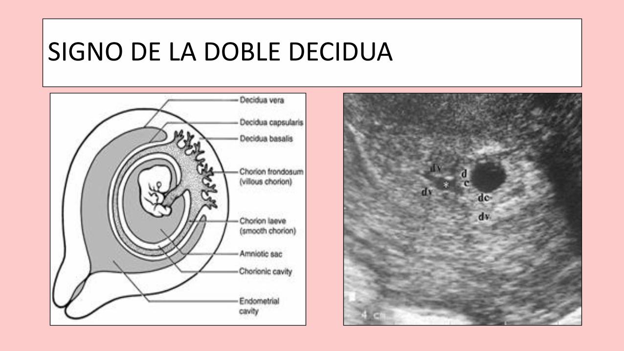
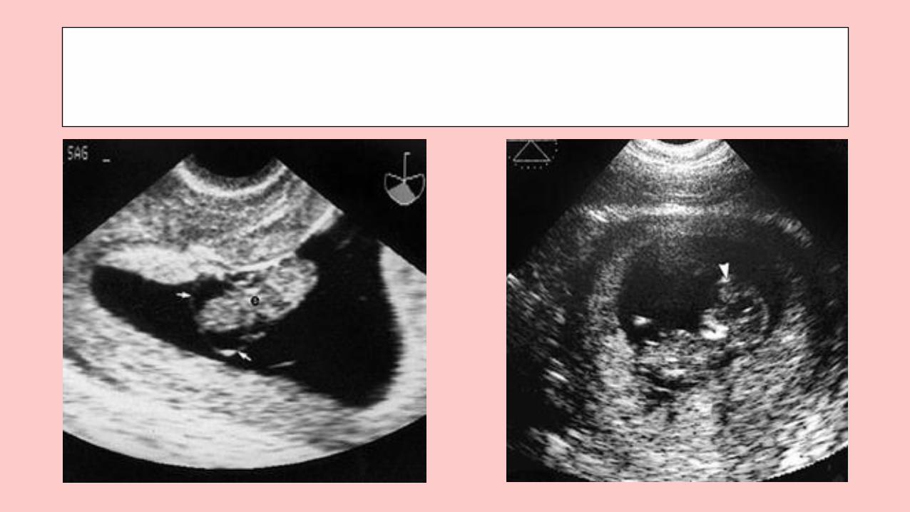
SIGNO DE LA DOBLE DECIDUA
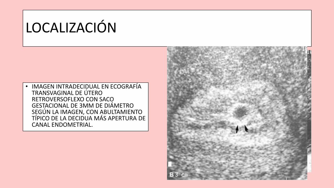
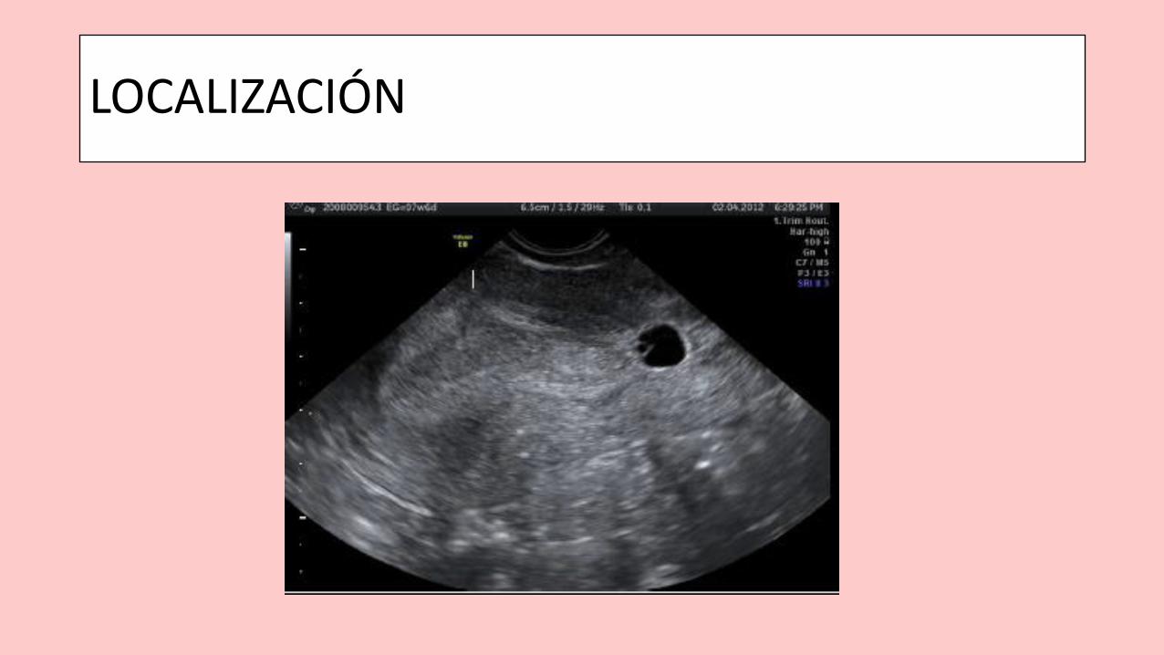
LOCALIZACIÓN
• IMAGEN INTRADECIDUAL EN ECOGRAFÍA TRANSVAGINAL DE ÚTERO RETROVERSOFLEXO CON SACO GESTACIONAL DE 3MM DE DIÁMETRO SEGÚN LA IMAGEN, CON ABULTAMIENTO TÍPICO DE LA DECIDUA MÁS APERTURA DE CANAL ENDOMETRIAL.
LOCALIZACIÓN
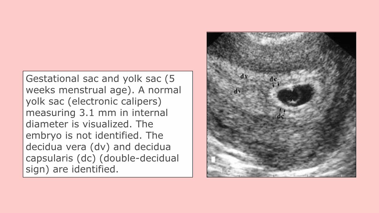
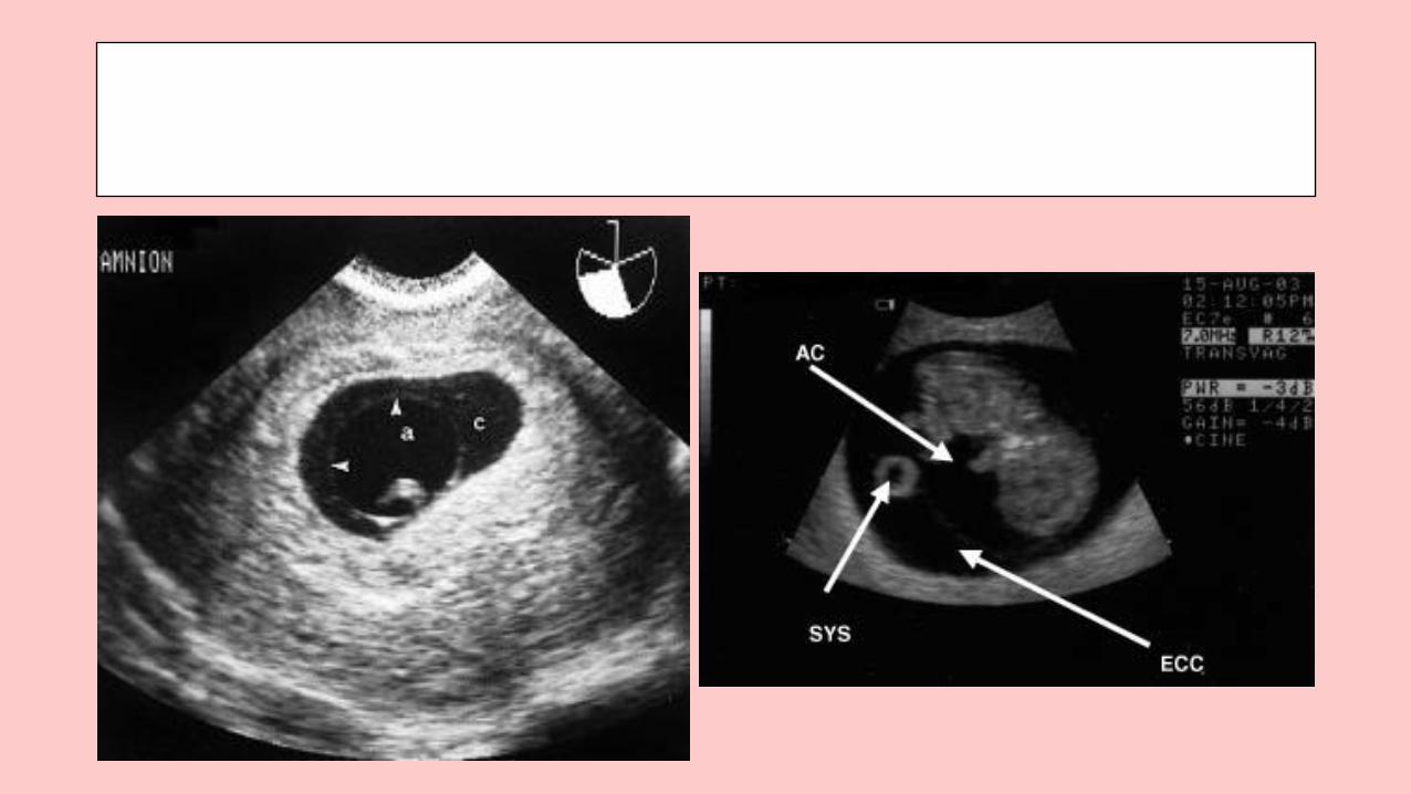
Gestational sac and yolk sac (5 weeks menstrual age). A normal yolk sac (electronic calipers) measuring 3.1 mm in internal diameter is visualized. The embryo is not identified. The decidua vera (dv) and decidua capsularis (dc) (double-decidualsign) are identified.
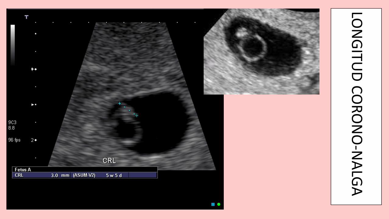
LON
GITU
D C
OR
ON
O-N
ALG
A
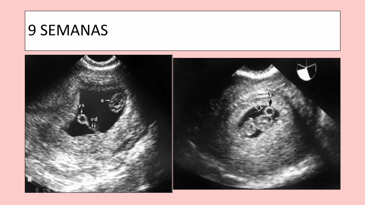
9 SEMANAS
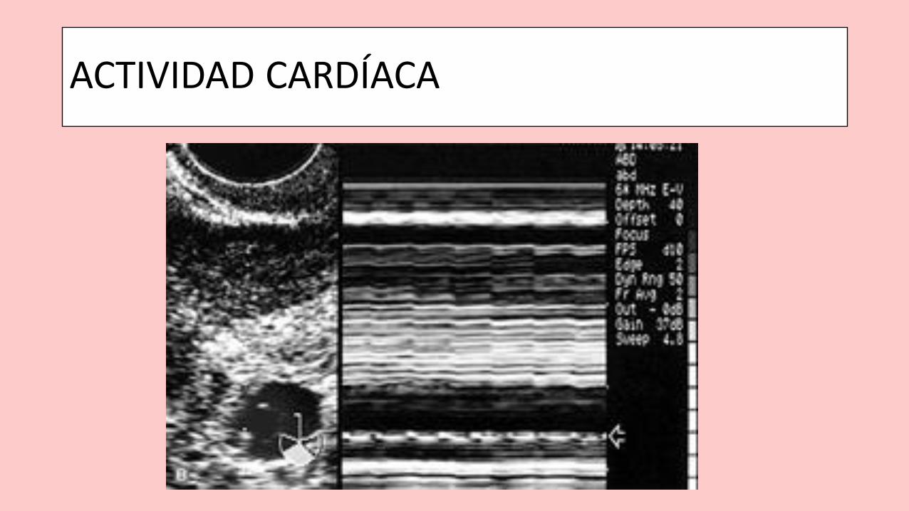
ACTIVIDAD CARDÍACA
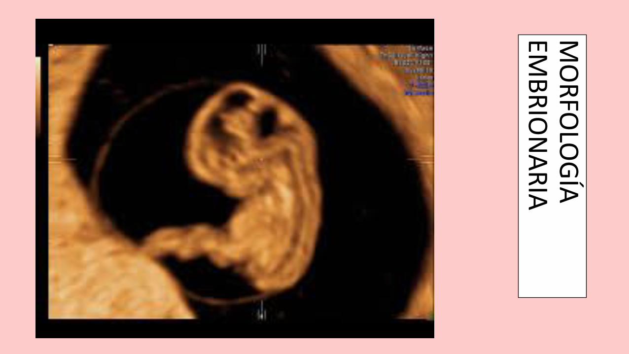
MO
RFO
LOG
ÍA
EMB
RIO
NA
RIA
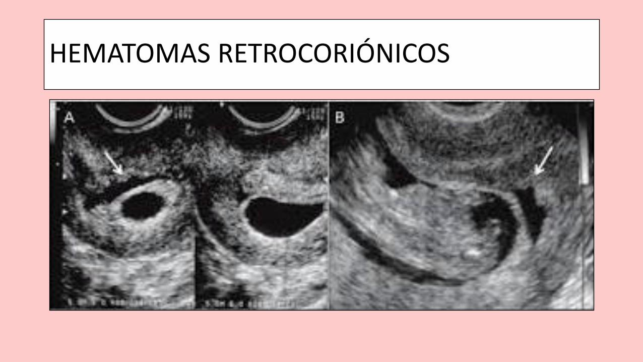
HEMATOMAS RETROCORIÓNICOS
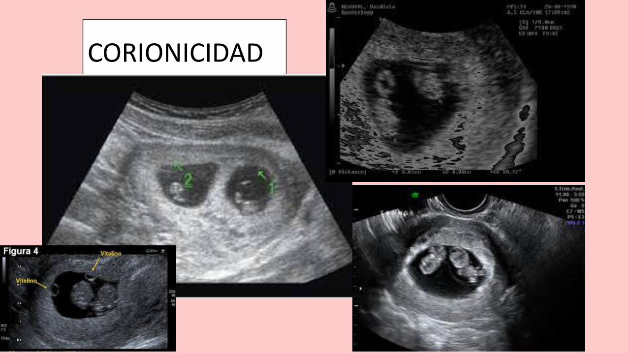
CORIONICIDAD
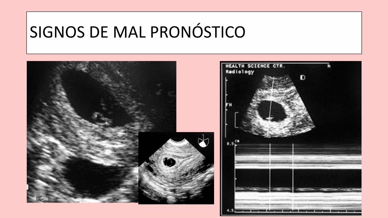
SIGNOS DE MAL PRONÓSTICO
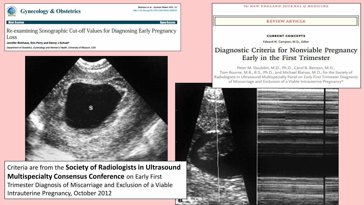
Criteria are from the Society of Radiologists in Ultrasound Multispecialty Consensus Conference on Early First Trimester Diagnosis of Miscarriage and Exclusion of a Viable Intrauterine Pregnancy, October 2012
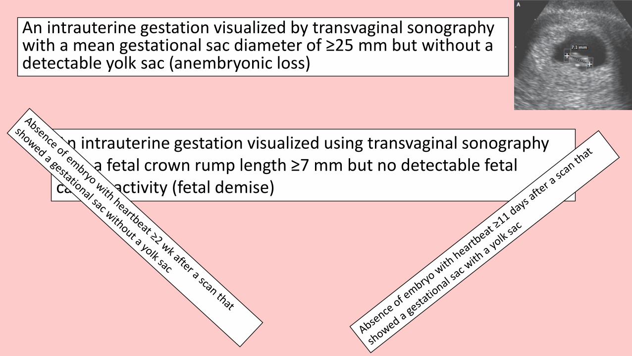
An intrauterine gestation visualized by transvaginal sonography with a mean gestational sac diameter of ≥25 mm but without a detectable yolk sac (anembryonic loss)
An intrauterine gestation visualized using transvaginal sonography with a fetal crown rump length ≥7 mm but no detectable fetal cardiac activity (fetal demise)
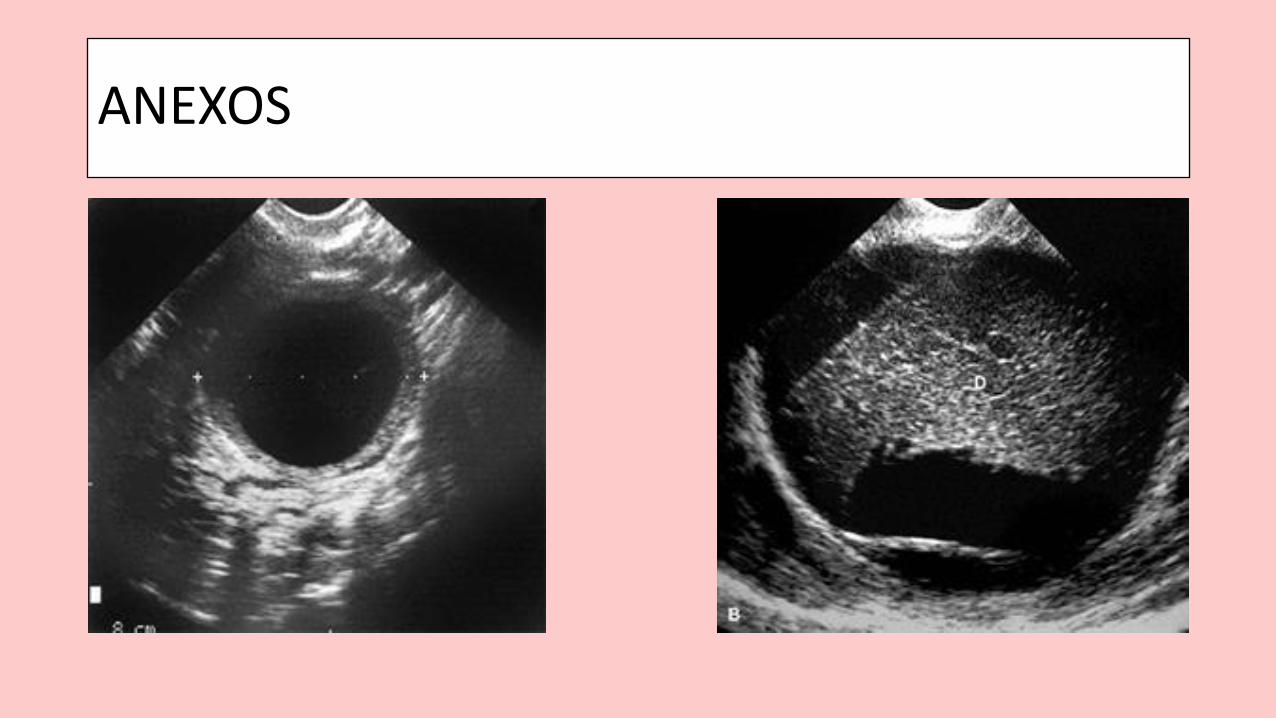
ANEXOS
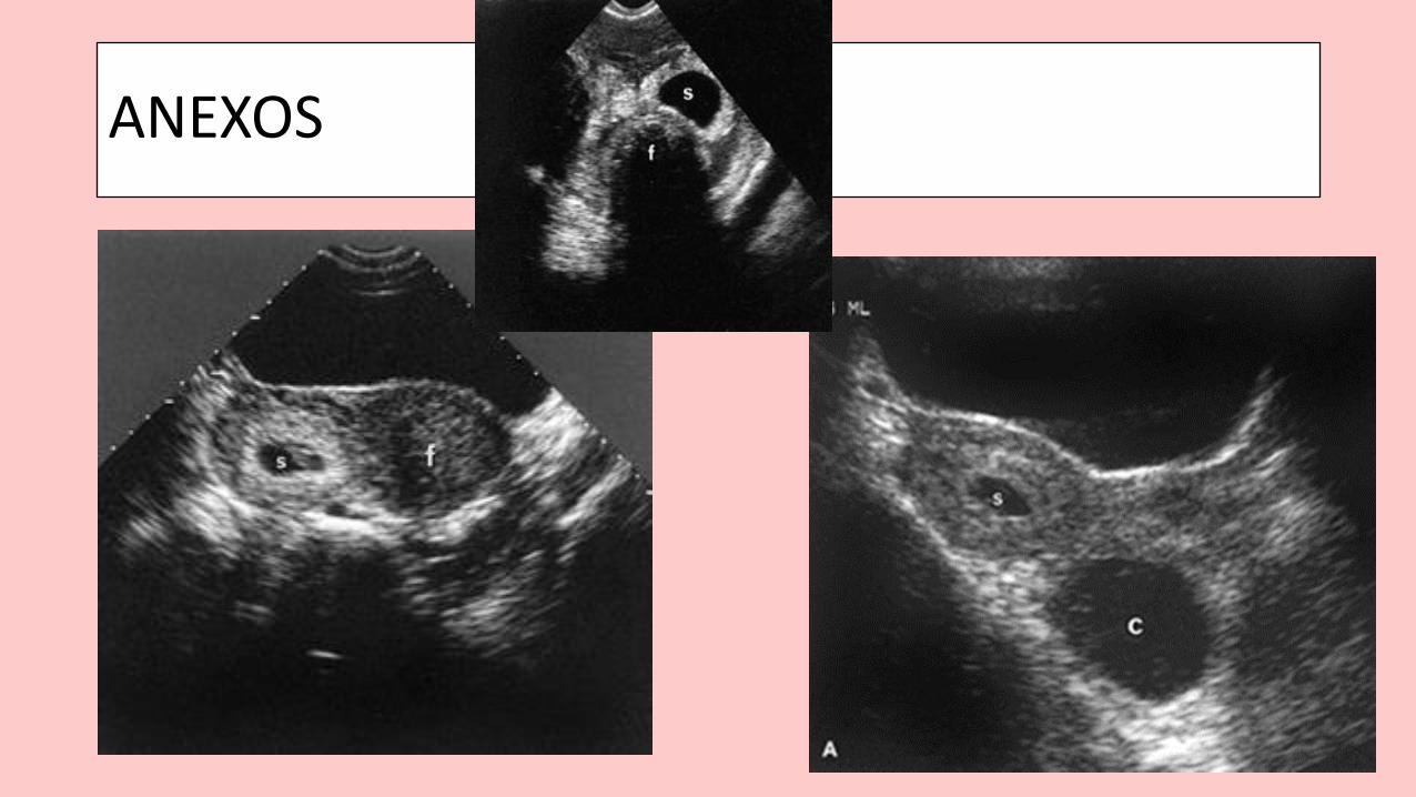
ANEXOS