E-Gel Imager - Thermo Fisher Scientific · The E-Gel Imager System makes agarose gel documentation...
4
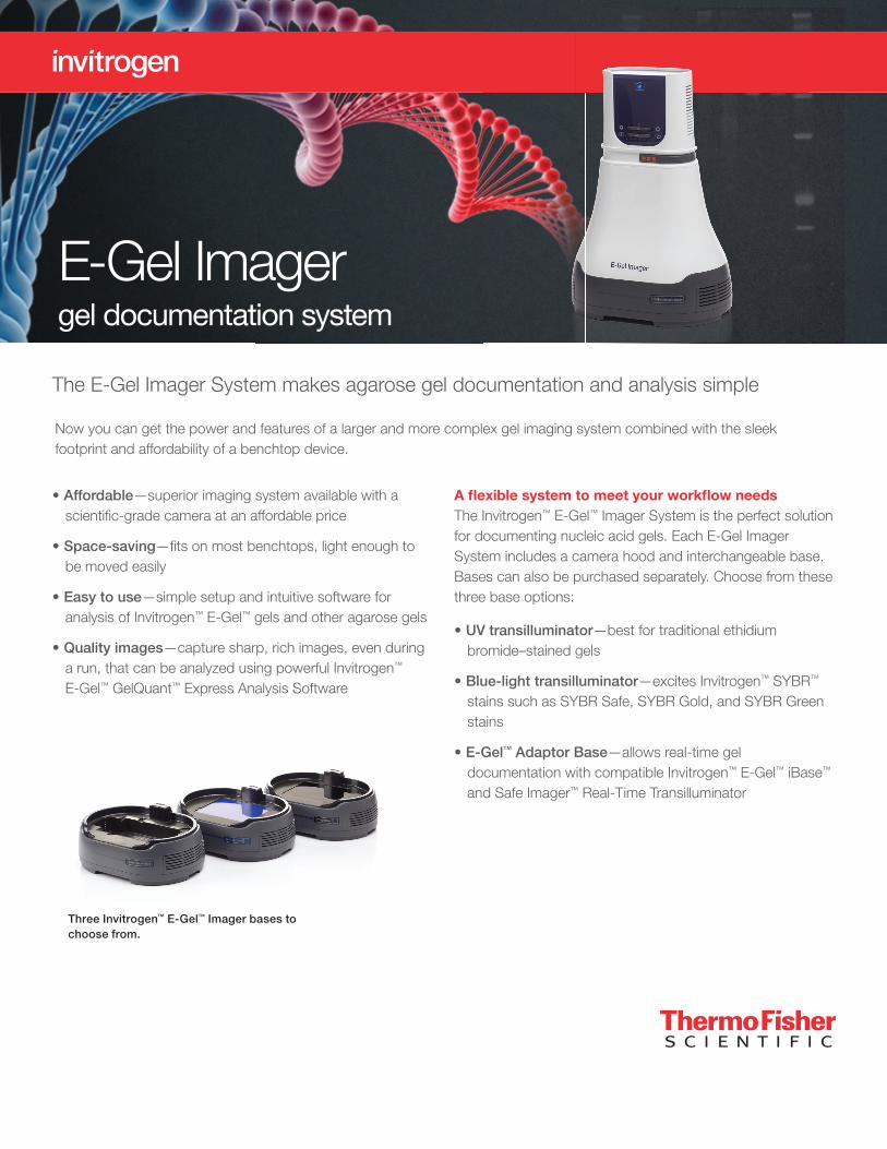
The E-Gel Imager System makes agarose gel documentation and analysis simple A flexible system to meet your workflow needs The Invitrogen ™ E-Gel ™ Imager System is the perfect solution for documenting nucleic acid gels. Each E-Gel Imager System includes a camera hood and interchangeable base. Bases can also be purchased separately. Choose from these three base options: • UV transilluminator—best for traditional ethidium bromide–stained gels • Blue-light transilluminator—excites Invitrogen ™ SYBR ™ stains such as SYBR Safe, SYBR Gold, and SYBR Green stains • E-Gel ™ Adaptor Base—allows real-time gel documentation with compatible Invitrogen ™ E-Gel ™ iBase ™ and Safe Imager ™ Real-Time Transilluminator Now you can get the power and features of a larger and more complex gel imaging system combined with the sleek footprint and affordability of a benchtop device. • Affordable—superior imaging system available with a scientific-grade camera at an affordable price • Space-saving—fits on most benchtops, light enough to be moved easily • Easy to use—simple setup and intuitive software for analysis of Invitrogen ™ E-Gel ™ gels and other agarose gels • Quality images—capture sharp, rich images, even during a run, that can be analyzed using powerful Invitrogen ™ E-Gel ™ GelQuant ™ Express Analysis Software Three Invitrogen ™ E-Gel ™ Imager bases to choose from. E-Gel Imager gel documentation system
Transcript of E-Gel Imager - Thermo Fisher Scientific · The E-Gel Imager System makes agarose gel documentation...
The E-Gel Imager System makes agarose gel documentation and analysis simple
A fl exible system to meet your workfl ow needsThe Invitrogen™ E-Gel™ Imager System is the perfect solution for documenting nucleic acid gels. Each E-Gel Imager System includes a camera hood and interchangeable base. Bases can also be purchased separately. Choose from these three base options:
• UV transilluminator—best for traditional ethidium bromide–stained gels
• Blue-light transilluminator—excites Invitrogen™ SYBR™ stains such as SYBR Safe, SYBR Gold, and SYBR Green stains
• E-Gel™ Adaptor Base—allows real-time gel documentation with compatible Invitrogen™ E-Gel™ iBase™ and Safe Imager™ Real-Time Transilluminator
Now you can get the power and features of a larger and more complex gel imaging system combined with the sleek footprint and affordability of a benchtop device.
• Affordable—superior imaging system available with a scientifi c-grade camera at an affordable price
• Space-saving—fi ts on most benchtops, light enough to be moved easily
• Easy to use—simple setup and intuitive software for analysis of Invitrogen™ E-Gel™ gels and other agarose gels
• Quality images—capture sharp, rich images, even during a run, that can be analyzed using powerful Invitrogen™ E-Gel™ GelQuant™ Express Analysis Software
Three Invitrogen™ E-Gel™ Imager bases to choose from.
E-Gel Imager gel documentation system
Easy-to-use softwareGelQuant Express Analysis Software is a Windows™
application for the analysis of 1D gels. This easy-to-use, high-precision software enables accurate, consistent, and fast analysis of all gel images.
• Easy and fast quantitation: automatic detection and analysis of bands with just one click. Band boundaries are automatically and accurately detected using an innovative boundary detection algorithm. Even distorted gel images can be analyzed, and molecular weight calculated, based on marker lane standards.
• Accurate analysis: automatic deduction of background noise allows accurate quantitation and normalization of bands
• Flexible analysis tools: dynamic graphic intensity analysis tools allow easy adjustment to band and lane boundaries for complex results (e.g., degraded, doublet bands)
• One-click data export: customize results with one-click export to an Excel™ worksheet or as a JPEG image
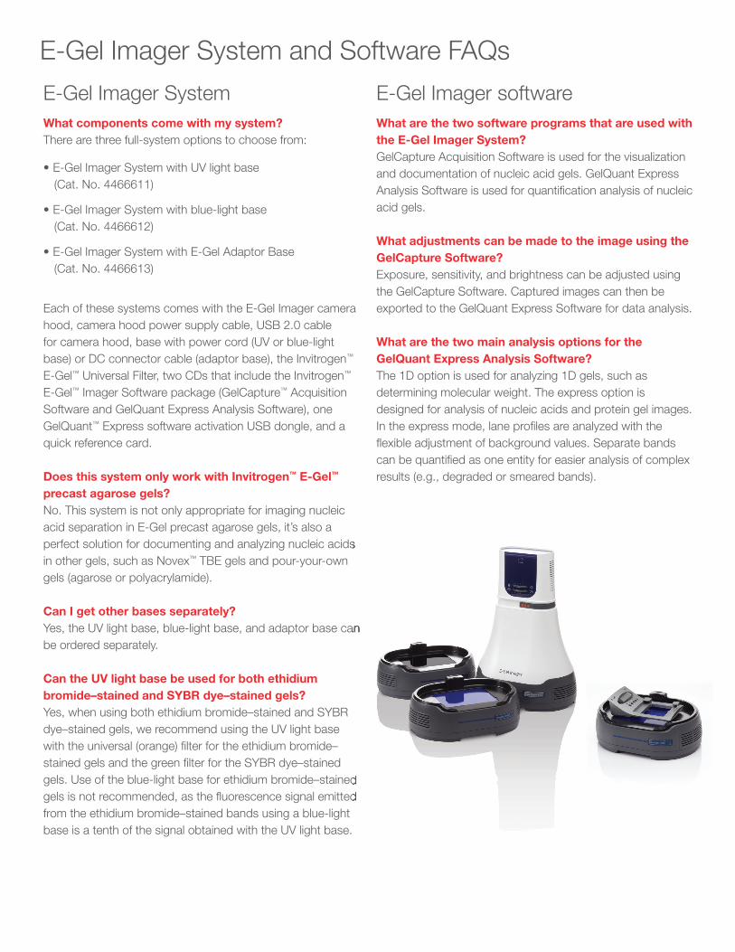
Preview your image and adjust brightness, sensitivity, and exposure time on the main imaging screen. In this example, an E-Gel EX 2% gel is being imaged using blue-light transillumination.
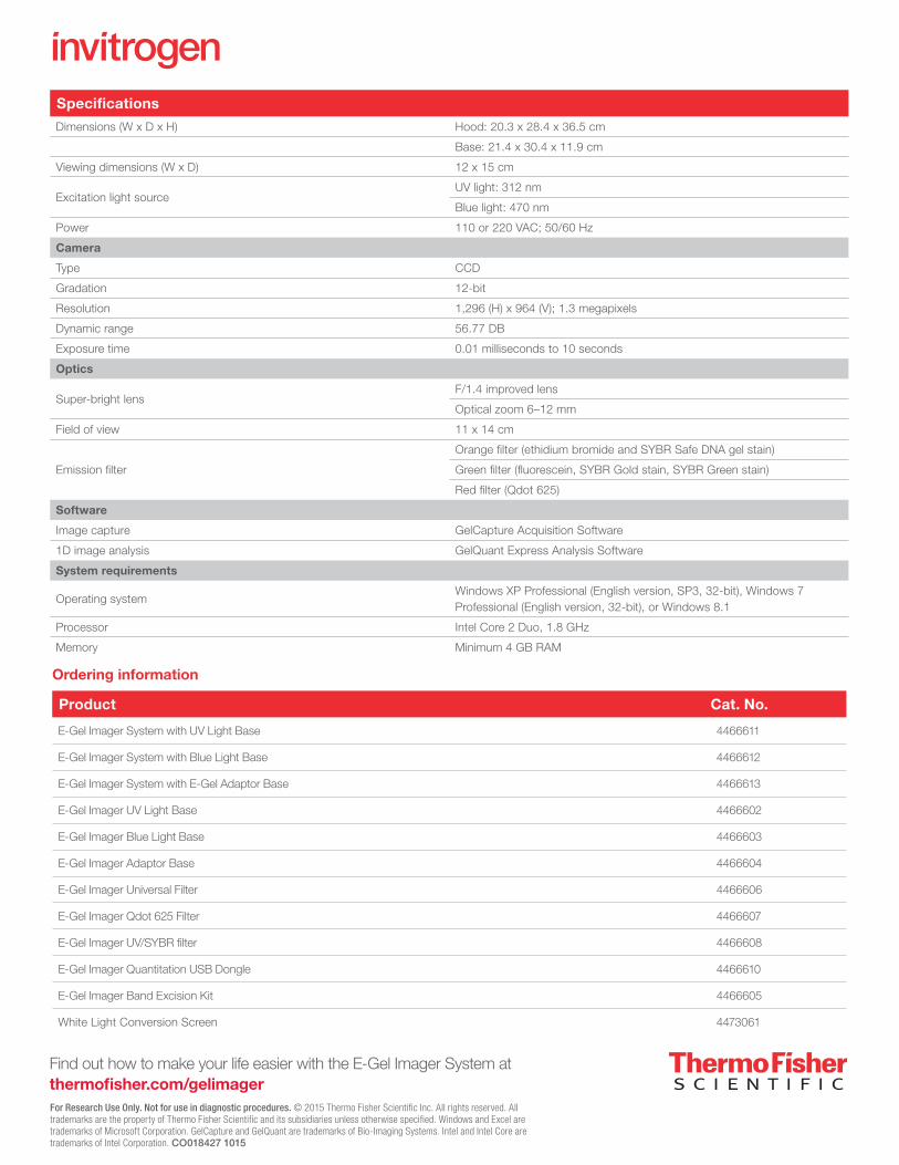
The Edit Image screen is where post-capture manipulations canbe performed.
Do a quick check or more in-depth analysisIt’s easy to quickly capture an image any time you run a gel. For many applications, estimating the size and quantity of nucleic acid in a certain band in a gel is important for downstream steps. The GelQuant Express Software was designed for just this type of image analysis after capture. Use this full-featured yet uncomplicated software to document, quantitate, and analyze your results.
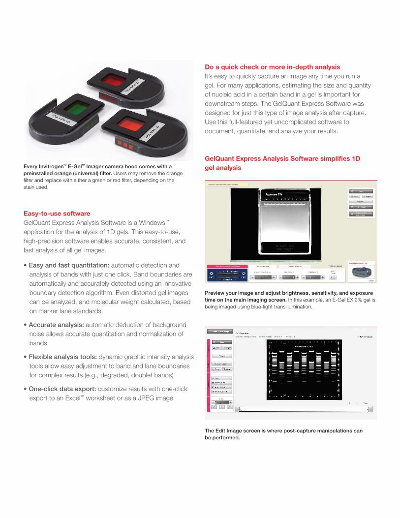
GelQuant Express Analysis Software simplifi es 1D gel analysisEvery Invitrogen™ E-Gel™ Imager camera hood comes with a
preinstalled orange (universal) fi lter. Users may remove the orangefi lter and replace with either a green or red fi lter, depending on thestain used.
E-Gel Imager System and Software FAQs
E-Gel Imager SystemWhat components come with my system?There are three full-system options to choose from:
• E-Gel Imager System with UV light base(Cat. No. 4466611)
• E-Gel Imager System with blue-light base(Cat. No. 4466612)
• E-Gel Imager System with E-Gel Adaptor Base(Cat. No. 4466613)
Each of these systems comes with the E-Gel Imager camera hood, camera hood power supply cable, USB 2.0 cable for camera hood, base with power cord (UV or blue-light base) or DC connector cable (adaptor base), the Invitrogen™
E-Gel™ Universal Filter, two CDs that include the Invitrogen™
E-Gel™ Imager Software package (GelCapture™ Acquisition Software and GelQuant Express Analysis Software), one GelQuant™ Express software activation USB dongle, and a quick reference card.
Does this system only work with Invitrogen™ E-Gel™
precast agarose gels?No. This system is not only appropriate for imaging nucleic acid separation in E-Gel precast agarose gels, it’s also a perfect solution for documenting and analyzing nucleic acids in other gels, such as Novex™ TBE gels and pour-your-own gels (agarose or polyacrylamide).
Can I get other bases separately? Yes, the UV light base, blue-light base, and adaptor base can be ordered separately.
Can the UV light base be used for both ethidium bromide–stained and SYBR dye–stained gels? Yes, when using both ethidium bromide–stained and SYBR dye–stained gels, we recommend using the UV light base with the universal (orange) fi lter for the ethidium bromide–stained gels and the green fi lter for the SYBR dye–stained gels. Use of the blue-light base for ethidium bromide–stained gels is not recommended, as the fl uorescence signal emitted from the ethidium bromide–stained bands using a blue-light base is a tenth of the signal obtained with the UV light base.
E-Gel Imager softwareWhat are the two software programs that are used with the E-Gel Imager System? GelCapture Acquisition Software is used for the visualization and documentation of nucleic acid gels. GelQuant Express Analysis Software is used for quantifi cation analysis of nucleic acid gels.
What adjustments can be made to the image using the GelCapture Software? Exposure, sensitivity, and brightness can be adjusted using the GelCapture Software. Captured images can then be exported to the GelQuant Express Software for data analysis.
What are the two main analysis options for the GelQuant Express Analysis Software?The 1D option is used for analyzing 1D gels, such as determining molecular weight. The express option is designed for analysis of nucleic acids and protein gel images. In the express mode, lane profi les are analyzed with the fl exible adjustment of background values. Separate bands can be quantifi ed as one entity for easier analysis of complex results (e.g., degraded or smeared bands).
perfect solution for documenting and analyzing nucleic acids
Yes, the UV light base, blue-light base, and adaptor base can
gels. Use of the blue-light base for ethidium bromide–stained gels is not recommended, as the fl uorescence signal emitted
Find out how to make your life easier with the E-Gel Imager System at thermofisher.com/gelimagerFor Research Use Only. Not for use in diagnostic procedures. © 2015 Thermo Fisher Scientific Inc. All rights reserved. All trademarks are the property of Thermo Fisher Scientific and its subsidiaries unless otherwise specified. Windows and Excel are trademarks of Microsoft Corporation. GelCapture and GelQuant are trademarks of Bio-Imaging Systems. Intel and Intel Core are trademarks of Intel Corporation. CO018427 1015
Specifications
Dimensions (W x D x H) Hood: 20.3 x 28.4 x 36.5 cm
Base: 21.4 x 30.4 x 11.9 cm
Viewing dimensions (W x D) 12 x 15 cm
Excitation light sourceUV light: 312 nm
Blue light: 470 nm
Power 110 or 220 VAC; 50/60 Hz
Camera
Type CCD
Gradation 12-bit
Resolution 1,296 (H) x 964 (V); 1.3 megapixels
Dynamic range 56.77 DB
Exposure time 0.01 milliseconds to 10 seconds
Optics
Super-bright lensF/1.4 improved lens
Optical zoom 6–12 mm
Field of view 11 x 14 cm
Emission filter
Orange filter (ethidium bromide and SYBR Safe DNA gel stain)
Green filter (fluorescein, SYBR Gold stain, SYBR Green stain)
Red filter (Qdot 625)
Software
Image capture GelCapture Acquisition Software
1D image analysis GelQuant Express Analysis Software
System requirements
Operating systemWindows XP Professional (English version, SP3, 32-bit), Windows 7 Professional (English version, 32-bit), or Windows 8.1
Processor Intel Core 2 Duo, 1.8 GHz
Memory Minimum 4 GB RAM
Ordering information
Product Cat. No.
E-Gel Imager System with UV Light Base 4466611
E-Gel Imager System with Blue Light Base 4466612
E-Gel Imager System with E-Gel Adaptor Base 4466613
E-Gel Imager UV Light Base 4466602
E-Gel Imager Blue Light Base 4466603
E-Gel Imager Adaptor Base 4466604
E-Gel Imager Universal Filter 4466606
E-Gel Imager Qdot 625 Filter 4466607
E-Gel Imager UV/SYBR filter 4466608
E-Gel Imager Quantitation USB Dongle 4466610
E-Gel Imager Band Excision Kit 4466605
White Light Conversion Screen 4473061