DRAFT KEY EVENT BASED TEST GUIDELINES 442D TG 442D_21Dec_clean.pdf · DRAFT KEY EVENT BASED TEST...
46
Revised TG 442D – 21 Dec 2017 General Introduction DRAFT KEY EVENT BASED TEST GUIDELINES 442D In Vitro Skin Sensitisation assays addressing the AOP Key Event on: Keratinocyte activation GENERAL INTRODUCTION Keratinocyte activation Key Event based Test Guideline 1. A skin sensitiser refers to a substance that will lead to an allergic response following skin contact as defined by the United Nations Globally Harmonized System of Classification and Labelling of Chemicals (UN GHS) (1). There is general agreement on the key biological events underlying skin sensitisation. The current knowledge of the chemical and biological mechanisms associated with skin sensitisation has been summarised as an Adverse Outcome Pathway (AOP) (2), starting with the molecular initiating event through intermediate events to the adverse effect, namely allergic contact dermatitis. In this instance, the molecular initiating event (i.e. the first key event) is the covalent binding of electrophilic substances to nucleophilic centres in skin proteins. The second key event in this AOP takes place in the keratinocytes and includes inflammatory responses as well as changes in gene expression associated with specific cell signalling pathways such as the antioxidant/electrophile response element (ARE)-dependent pathways. The third key event is the activation of dendritic cells, typically assessed by expression of specific cell surface markers, chemokines and cytokines. The fourth key event is T-cell proliferation (3). 2. This Test Guideline describes in vitro assays that address mechanisms described under the second Key Event of the AOP for skin sensitisation, namely keratinocyte activation (2). The Test Guideline comprises test methods to be used for supporting the discrimination between skin sensitisers and non-sensitisers in accordance with the UN GHS (1). The test methods currently described in this Test Guideline are: - The ARE-Nrf2 luciferase KeratinoSens TM test method (Appendix IA), and - The ARE-Nrf2 luciferase LuSens test method (Appendix IB). 3. These two in vitro ARE-Nrf2 luciferase test methods have been considered scientifically valid. The KeratinoSens TM test method first underwent a validation study followed by an independent peer-review by EURL ECVAM Scientific Advisory Committee (ESAC) and positive recommendations by EURL ECVAM, and is considered the validated reference method (VRM) (3) (4) (5) (6). The LuSens test method later underwent a Performance Standard-based validation study based on which it was also reviewed and received positive opinion by ESAC (7) (8) (9) (10). 4. The test methods included in this Test Guideline may differ in relation to the procedure used to generate the data and the readouts measured but can be used indiscriminately to address countries’ requirements for test results on the keratinocytes activation Key Event of the AOP for skin sensitisation while benefiting from the Mutual Acceptance of Data.
Transcript of DRAFT KEY EVENT BASED TEST GUIDELINES 442D TG 442D_21Dec_clean.pdf · DRAFT KEY EVENT BASED TEST...
Revised TG 442D – 21 Dec 2017 General Introduction
DRAFT KEY EVENT BASED TEST GUIDELINES 442D
In Vitro Skin Sensitisation assays addressing the AOP Key Event on:
Keratinocyte activation
GENERAL INTRODUCTION
Keratinocyte activation Key Event based Test Guideline
1. A skin sensitiser refers to a substance that will lead to an allergic response following skin
contact as defined by the United Nations Globally Harmonized System of Classification and Labelling
of Chemicals (UN GHS) (1). There is general agreement on the key biological events underlying skin
sensitisation. The current knowledge of the chemical and biological mechanisms associated with skin
sensitisation has been summarised as an Adverse Outcome Pathway (AOP) (2), starting with the
molecular initiating event through intermediate events to the adverse effect, namely allergic contact
dermatitis. In this instance, the molecular initiating event (i.e. the first key event) is the covalent
binding of electrophilic substances to nucleophilic centres in skin proteins. The second key event in
this AOP takes place in the keratinocytes and includes inflammatory responses as well as changes in
gene expression associated with specific cell signalling pathways such as the antioxidant/electrophile
response element (ARE)-dependent pathways. The third key event is the activation of dendritic cells,
typically assessed by expression of specific cell surface markers, chemokines and cytokines. The
fourth key event is T-cell proliferation (3).
2. This Test Guideline describes in vitro assays that address mechanisms described under the
second Key Event of the AOP for skin sensitisation, namely keratinocyte activation (2). The Test
Guideline comprises test methods to be used for supporting the discrimination between skin sensitisers
and non-sensitisers in accordance with the UN GHS (1). The test methods currently described in this
Test Guideline are:
- The ARE-Nrf2 luciferase KeratinoSensTM
test method (Appendix IA), and
- The ARE-Nrf2 luciferase LuSens test method (Appendix IB).
3. These two in vitro ARE-Nrf2 luciferase test methods have been considered scientifically
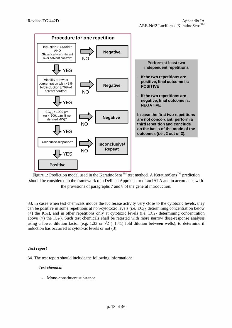
valid. The KeratinoSensTM
test method first underwent a validation study followed by an independent
peer-review by EURL ECVAM Scientific Advisory Committee (ESAC) and positive
recommendations by EURL ECVAM, and is considered the validated reference method (VRM) (3) (4)
(5) (6). The LuSens test method later underwent a Performance Standard-based validation study based
on which it was also reviewed and received positive opinion by ESAC (7) (8) (9) (10).
4. The test methods included in this Test Guideline may differ in relation to the procedure
used to generate the data and the readouts measured but can be used indiscriminately to address
countries’ requirements for test results on the keratinocytes activation Key Event of the AOP for skin
sensitisation while benefiting from the Mutual Acceptance of Data.
Revised TG 442D – 21 Dec 2017 General Introduction
Background and principles of the test methods included in the Key Event based Test Guidelines
5. The assessment of skin sensitisation has typically involved the use of laboratory animals.
The classical methods that use guinea-pigs, the Guinea Pig Maximisation Test (GPMT) of Magnusson
and Kligman and the Buehler Test (OECD TG 406) (11), assess both the induction and elicitation
phases of skin sensitisation. The murine tests, the LLNA (OECD TG 429) (12) and its three non-
radioactive modifications, LLNA: DA (OECD TG 442A) (13) as well as LLNA: BrdU-ELISA and
BrdU-FCM1 (OECD TG 442B) (14), all assess the induction response exclusively, and have gained
acceptance since they provide an advantage over the guinea pig tests in terms of animal welfare
together with an objective measurement of the induction phase of skin sensitisation.
6. Mechanistically-based in chemico and in vitro test methods addressing the first three key
events of the skin sensitisation AOP have been adopted for contributing to the evaluation of the skin
sensitisation hazard potential of chemicals: the OECD TG 442C describes the Direct Peptide
Reactivity Assay (15) addressing the first key event; the present Test Guideline assesses keratinocyte
activation addressing the second key event and the OECD TG 442E addresses the activation of
dendritic cells, the third key event of the skin sensitisation AOP (16). Finally, the fourth key event
representing T-cell proliferation is indirectly assessed in the murine Local Lymph Node Assay
(LLNA) (12)..
7. As keratinocyte activation represents only one key event of the skin sensitisation AOP (2) (17),
information generated with test methods developed to address this specific key event may not be
sufficient to conclude on the presence or absence of skin sensitisation potential of chemicals.
Therefore data generated with the test methods described in this Test Guideline are proposed to
support the discrimination between skin sensitisers (i.e. UN GHS Category 1) and non-sensitisers
when used within Integrated Approaches to Testing and Assessment (IATA), together with other
relevant complementary information, e.g. derived from in vitro assays addressing other key events of
the skin sensitisation AOP as well as non-testing methods, including read-across from chemical
analogues (17). Examples on the use of data generated with these methods within Defined
Approaches, i.e. approaches standardised both in relation to the set of information sources used and in
the procedure applied to derive predictions have been published (17) and can be employed as useful
elements within IATA.
8. The test methods described in this Test Guideline cannot be used on their own, neither to
sub-categorise skin sensitisers into subcategories 1A and 1B as defined by UN GHS (1), for authorities
implementing these two optional subcategories, nor to predict potency for safety assessment decisions.
However, depending on the regulatory framework, positive results generated with these methods may
be used on their own to classify a chemical into UN GHS category 1.
9. The term "test chemical" is used in this Test Guideline to refer to what is being tested2
and is not
related to the applicability of the test methods to the testing of mono-constituent substances, multi-
constituent substances and/or mixtures. When testing in submerged cultures, it should be verified that
the test chemical is dissolved in the exposure medium or at least forms a stable dispersion (e.g. by
visual inspection of the test chemical dissolved/prepared at the maximal final test concentration in the
exposure medium, showing that no undissolved residues remain and that no precipitate or phase
separation forms if the solution is left to settle for several hours).
1 To be confirmed upon adoption of revised TG 442B with BrDU-FCM
2 In June 2013, the Joint Meeting agreed that where possible, a more consistent use of the term "test chemical"
describing what is being tested should be applied in new and updated Test Guidelines.
General Introduction
p. 3 of 46
10. Limited information is currently available on the applicability of the test methods to multi-
constituent substances/mixtures (18) (19). Although not evaluated in the validation studies, the test
methods may nevertheless be technically applicable to the testing of multi-constituent substances and
mixtures. However, before use of this Test Guideline on a mixture for generating data for an intended
regulatory purpose, it should be considered whether, and if so why, it may provide adequate results for
that purpose3. Such considerations are not needed when there is a regulatory requirement for the testing
of the mixture. Moreover, when testing multi-constituent substances or mixtures, consideration should
be given to possible interference of cytotoxic constituents with the observed responses (i.e. the presence
of a high content of non-sensitising cytotoxic constituents may mask the response of weakly sensitising
components or sensitising components present at low concentration). It might be scientifically justified
to test either single main constituents or several fractions of the mixture to conclude on the sensitisation
risk of the complex mixture.
LITERATURE
1. United Nations (UN) (2015). Globally Harmonized System of Classification and Labelling
of Chemicals (GHS). Sixth revised edition, New York and Geneva, United Nations
Publications. Available at:
[https://www.unece.org/trans/danger/publi/ghs/ghs_rev06/06files_e.html]
2. OECD (2012). Series on Testing and Assessment No. 168. The Adverse Outcome Pathway
for Skin Sensitisation Initiated by Covalent Binding to Proteins. Part 1: Scientific Evidence.
Organisation for Economic Cooperation and Development, Paris. Available at:
http://www.oecd.org/officialdocuments/publicdisplaydocumentpdf/?cote=ENV/JM/MONO(
2012)10/PART1&docLanguage=En
3. Emter R., Ellis G., Natsch A.(2010). Performance of a novel keratinocyte-based reporter cell
line to screen skin sensitizers in vitro. Toxicology and Applied Pharmacology 245, 281-290.
4. Natsch A., Bauch C., Foertsch L., Gerberick F., Normann K., Hilberer A., Inglis H.,
Landsiedel R., Onken S., Reuter H., Schepky A., Emter R. (2011). The intra- and inter-
laboratory reproducibility and predictivity of the KeratinoSens assay to predict skin
sensitizers in vitro: results of a ring-study in five laboratories. Toxicol. In Vitro 25, 733-744.
5. Natsch A., Ryan C.A., Foertsch L., Emter R., Jaworska J., Gerberick G.F., Kern P. (2013). A
dataset on 145 chemicals tested in alternative assays for skin sensitization undergoing
prevalidation. Journal of Applied Toxicology 33, 1337-1352.
6. EURL-ECVAM (2014). Recommendation on the KeratinoSensTM
assay for skin sensitisation
testing, 42 pp. Available at: [http://ihcp.jrc.ec.europa.eu/our_labs/eurl-ecvam/eurl-ecvam-
recommendations/recommendation-keratinosens-skin-sensitisation].
7. Ramirez T., Mehling A., Kolle S.N., Wruck C.J., Teubner W., Eltze T., Aumann A., Urbisch
D., van Ravenzwaay B., Landsiedel R. (2014). LuSens: a keratinocyte based ARE reporter
gene assay for use in integrated testing strategies for skin sensitization hazard identification.
Toxicol In Vitro 28, 1482-1497.
8. Ramirez T., Stein N., Aumann A., Remus T., Edwards A., Norman K.G., Ryan C., Bader
J.E., Fehr M., Burleson F., Foertsch L., Wang X., Gerberick F., Beilstein P., Hoffmann S.,
3 This sentence was proposed and agreed at the April 2014 WNT meeting
General Introduction
p. 4 of 46
Mehling A., van Ravenzwaay B., Landsiedel R. (2016). Intra- and inter-laboratory
reproducibility and accuracy of the LuSens assay: A reporter gene-cell line to detect
keratinocyte activation by skin sensitizers. Toxicol In Vitro 32, 278-286.
9. ESAC (2016). ESAC opinion on the BASF-coordinated Performance Standards-based
validation of the LuSens test method for skin sensitisation testing. Available at:
[http://publications.jrc.ec.europa.eu/repository/bitstream/JRC103706/esac_opinion_2016-
04_lusens_final.pdf].
10. OECD (2015). Guidance Document No 213. Performance Standards for assessment of
proposed similar or modified in vitro skin sensitisation ARE-NrF2 luciferase test methods.
Series on Testing and Assessment. Available at: http://www.oecd.org/env/ehs/testing/series-
testing-assessment-publications-number.htm
11. OECD (1992). OECD Guidelines for the Testing of Chemicals No. 406. Skin Sensitisation.
Organisation for Economic Cooperation and Development, Paris. Available at:
[http://www.oecd.org/env/testguidelines].
12. OECD (2010). OECD Guidelines for Chemical Testing No. 429. Skin sensitization: Local
Lymph Node assay. Organisation for Economic Cooperation and Development, Paris.
Available at: [http://www.oecd.org/env/testguidelines].
13. OECD (2010). OECD Guidelines for Chemical Testing No. 442A.Skin sensitization: Local
Lymph Node assay: DA. Organisation for Economic Cooperation and Development, Paris.
Available at: [http://www.oecd.org/env/testguidelines].
14. OECD (2010). OECD Guidelines for Chemical Testing No. 442B. Skin sensitization: Local
Lymph Node assay: BrdU-ELISA. Organisation for Economic Cooperation and
Development, Paris. Available at: [http://www.oecd.org/env/testguidelines].
15. OECD (2015). OECD Guideline for the Testing of Chemicals No. 442C: In Chemico Skin
Sensitisation: Direct Peptide Reactivity Assay (DPRA). Organisation for Economic
Cooperation and Development, Paris. Available at: [http://www.oecd.org/env/testguidelines].
16. OECD (2017). OECD Guideline for the Testing of Chemicals No. 442E: In Vitro Skin
Sensitisation assays addressing the AOP Key Event on Activation of Dendritic Cells.
Organisation for Economic Cooperation and Development, Paris. Available at:
[http://www.oecd.org/env/testguidelines].
17. OECD (2016). Series on Testing & Assessment No. 256: Guidance Document On The
Reporting Of Defined Approaches And Individual Information Sources To Be Used Within
Integrated Approaches To Testing And Assessment (IATA) For Skin Sensitisation, Annex 1
and Annex 2. ENV/JM/HA(2016)29. Organisation for Economic Cooperation and
Development, Paris. Available at: [https://community.oecd.org/community/iatass].
18. Andres E., Sa-Rocha V.M., Barrichello C., Haupt T., Ellis G., Natsch A. (2013). The
sensitivity of the KeratinoSensTM assay to evaluate plant extracts: A pilot study. Toxicology
In Vitro 27, 1220-1225.
19. Kolle, S.N., Mehling A., Teubner W., van Ravenzwaay B., Landsiedel R. (2013). Alternative
method in practice: Post-validation experience of the skin sensitization in vitro test strategy.
Toxicology Letters 221, S204.
20. OECD (2005). Guidance Document the Validation and International Acceptance of New or
Updated Test Methods for Hazard Assessment. OECD Environment, Health and Safety
General Introduction
p. 5 of 46
publications, OECD Series on Testing and Assessment No.34. Organisation for Economic
Cooperation and Development, Paris.
21. Natsch A. (2010). The Nrf2-Keap1-ARE Toxicity Pathway as a Cellular Sensor for Skin
Sensitizers-Functional Relevance and Hypothesis on Innate Reactions to Skin Sensitizers.
Toxicological Sciences 113, 284-292.
22. Dinkova-Kostova A.T., Holtzclaw W.D., Kensler T.W. (2005). The role of Keap1 in cellular
protective responses. Chem. Res. Toxicol. 18, 1779-1791.
23. Kansanen E., Kuosmanen S.M., Leinonen H., Levonen A.L. (2013). The Keap1-Nrf2
pathway: Mechanisms of activation and dysregulation in cancer. Redox Biol. 1, 45-49.
General Introduction
p. 6 of 46
ANNEX
DEFINITIONS
Accuracy: The closeness of agreement between test method results and accepted reference values. It
is a measure of test method performance and one aspect of “relevance.” The term is often used
interchangeably with “concordance”, to mean the proportion of correct outcomes of a test method
(20).
Animal-product-free: absence of non-human animal-derived components, including serum, in the
test system.
AOP (Adverse Outcome Pathway): sequence of events from the chemical structure of a target
chemical or group of similar chemicals through the molecular initiating event to an in vivo outcome of
interest (2).
ARE: Antioxidant response element (also called EpRE, electrophile response element), is a response
element found in the upstream promoter region of many cytoprotective and phase II genes. When
activated by Nfr2, it mediates the transcriptional induction of these genes.
CV: Cell viability
Coefficient of variation: a measure of variability that is calculated for a group of replicate data by
dividing the standard deviation by the mean. It can be multiplied by 100 for expression as a
percentage.
CV75: The estimated concentration resulting in 75% cell viability.
EC1.5: Interpolated concentration resulting in a 1.5 fold luciferase induction.
Fold luciferase activity induction: Represents the ratio of luminescence of treated cells (minus
blank) over the luminescence of the cells exposed to the concurrent solvent/vehicle control (minus
blank).
IC30: Concentration effecting a reduction of cellular viability by 30%.
IC50: Concentration effecting a reduction of cellular viability by 50%.
Hazard: Inherent property of an agent or situation having the potential to cause adverse effects when
an organism, system or (sub) population is exposed to that agent.
IATA (Integrated Approach to Testing and Assessment): A structured approach used for hazard
identification (potential), hazard characterisation (potency) and/or safety assessment (potential/potency
and exposure) of a chemical or group of chemicals, which strategically integrates and weights all
relevant data to inform regulatory decision regarding potential hazard and/or risk and/or the need for
further targeted and therefore minimal testing.
Imax: Maximal induction factor of luciferase activity compared to the solvent (negative) control
measured at any test chemical concentration.
General Introduction
p. 7 of 46
Keap1: Kelch-like ECH-associated protein 1, is a sensor protein that can regulate the Nrf2 activity.
Under un-induced conditions the Keap1 sensor protein targets the Nrf2 transcription factor for
ubiquitinylation and proteolytic degradation in the proteasome. Covalent modification of the reactive
cysteine residues of Keap 1 by small molecules can lead to dissociation of Nrf2 from Keap1 (21) (22)
(23).
Mixture: A mixture or a solution composed of two or more substances in which they do not react (1).
Mono-constituent substance: A substance, defined by its quantitative composition, in which one
main constituent is present to at least 80% (w/w).
Multi-constituent substance: A substance, defined by its quantitative composition, in which more
than one main constituent is present in a concentration ≥ 10% (w/w) and < 80% (w/w). A multi-
constituent substance is the result of a manufacturing process. The difference between mixture and
multi-constituent substance is that a mixture is obtained by blending of two or more substances
without chemical reaction. A multi-constituent substance is the result of a chemical reaction.
Negative control: A sample containing all components of a test system and treated with a substance
known not to induce a positive response in the test system. This sample is processed with test
chemical-treated samples and other control samples.
Nrf2: nuclear factor (erythroid-derived 2)-like 2, is a transcription factor involved in the antioxidant
response pathway. When Nrf2 is not ubiquitinylated, it builds up in the cytoplasm and translocates
into the nucleus, where it combines to the ARE in the upstream promoter region of many
cytoprotective genes, initiating their transcription (21) (22) (23).
Performance standards: Standards, based on a validated test method, that provide a basis for
evaluating the comparability of a proposed test method that is mechanistically and functionally
similar. Included are (i) essential test method components; (ii) a minimum list of reference chemicals
selected from among the chemicals used to demonstrate the acceptable performance of the validated
test method; and (iii) the comparable levels of accuracy and reliability, based on what was obtained for
the validated test method, that the proposed test method should demonstrate when evaluated using the
minimum list of reference chemicals (20).
Positive control: A replicate containing all components of a test system and treated with a substance
known to induce a positive response. To ensure that variability in the positive control response across
time can be assessed, the magnitude of the positive response should not be excessive.
Proficiency chemicals (substances): A subset of the Reference Chemicals included in the
Performance Standards that can be used by laboratories to demonstrate technical competence with a
standardised test method. Selection criteria for these substances typically include that they represent
the range of responses, are commercially available, and have high quality reference data available.
Reference chemicals (substances): A set of chemicals to be used to demonstrate the ability of a new
test method to meet the acceptability criteria demonstrated by the validated reference test method(s).
These chemicals should be representative of the classes of chemicals for which the test method is
General Introduction
p. 8 of 46
expected to be used, and should represent the full range of responses that may be expected from the
chemicals for which it may be used, from strong, to weak, to negative.
Relevance: Description of relationship of the test to the effect of interest and whether it is meaningful
and useful for a particular purpose. It is the extent to which the test correctly measures or predicts the
biological effect of interest. Relevance incorporates consideration of the accuracy (concordance) of a
test method (20).
Reliability: Measures of the extent that a test method can be performed reproducibly within and
between laboratories over time, when performed using the same protocol. It is assessed by calculating
intra- and inter-laboratory reproducibility and intra-laboratory repeatability (20).
Reproducibility: The agreement among results obtained from testing the same substance using the
same test protocol (see reliability) (20).
Sensitivity: The proportion of all positive / active chemicals that are correctly classified by the test
method. It is a measure of accuracy for a test method that produces categorical results, and is an
important consideration in assessing the relevance of a test method (20).
Solvent/vehicle control: A replicate containing all components of a test system except of the test
chemical, but including the solvent that is used. It is used to establish the baseline response for the
samples treated with the test chemical dissolved in the same solvent.
Specificity: The proportion of all negative / inactive chemicals that are correctly classified by the test
method. It is a measure of accuracy for a test method that produces categorical results and is an
important consideration in assessing the relevance of a test method (20).
Substance: Chemical elements and their compounds in the natural state or obtained by any production
process, including any additive necessary to preserve the stability of the product and any impurities
deriving from the process used, but excluding any solvent which may be separated without affecting
the stability of the substance or changing its composition (1).
Test chemical: The term "test chemical" is used to refer to what is being tested.
United Nations Globally Harmonized System of Classification and Labelling of Chemicals (UN
GHS): A system proposing the classification of chemicals (substances and mixtures) according to
standardised types and levels of physical, health and environmental hazards, and addressing
corresponding communication elements, such as pictograms, signal words, hazard statements,
precautionary statements and safety data sheets, so that to convey information on their adverse effects
with a view to protect people (including employers, workers, transporters, consumers and emergency
responders) and the environment (1).
UVCB: substances of unknown or variable composition, complex reaction products or biological
materials.
Validated Reference Method (VRM): the first method(s) endorsed as scientific valid and used as a
reference for performance-based validation studies.

General Introduction
p. 9 of 46
Valid test method: A test method considered to have sufficient relevance and reliability for a specific
purpose and which is based on scientifically sound principles. A test method is never valid in an
absolute sense, but only in relation to a defined purpose (20).
Revised TG 442D Appendix IA
ARE-Nrf2 Luciferase KeratinoSensTM
Appendix IA: In Vitro Skin Sensitisation:
The ARE-Nrf2 Luciferase KeratinoSensTM
Test Method
INITIAL CONSIDERATIONS, APPLICABILITY AND LIMITATIONS
1. The test method described in this Appendix to Test Guideline 442D addresses the second
key event of the skin sensitisation AOP (1), namely keratinocytes activation, by assessing with the
help of luciferase, the Nrf2-mediated activation of antioxidant response element (ARE)-dependent
genes. Skin sensitisers have been reported to induce genes that are regulated by the ARE (2) (3). Small
electrophilic substances such as skin sensitisers can act on the sensor protein Keap1 (Kelch-like ECH-
associated protein 1), by e.g. covalent modification of its cysteine residue, resulting in its dissociation
from the transcription factor Nrf2 (nuclear factor-erythroid 2-related factor 2). The dissociated Nrf2
can then activate ARE-dependent genes such as those coding for phase II detoxifying enzymes (2) (4)
(5).
2. The in vitro ARE-Nrf2 luciferase KeratinoSensTM
test method (hereafter called the
KeratinoSensTM
test method) underwent validation studies (3) (6) (7) followed by an independent peer
review conducted by the European Union Reference Laboratory for Alternatives to Animal Testing
(EURL ECVAM) (8). The KeratinoSensTM
test method was considered scientifically valid to be used
as part of an IATA, to support the discrimination between skin sensitisers and non-sensitisers for the
purpose of hazard identification (8).
3. Based on the dataset from the validation study and in-house testing used for the
independent peer-review of the test method, the KeratinoSensTM
test method proved to be transferable
to laboratories experienced in cell culture techniques (8). The level of reproducibility in predictions
that can be expected from the KeratinoSensTM
test method is in the order of 85% within and between
laboratories (8). The accuracy (77% - 155/201), sensitivity (78% - 71/91) and specificity (76% -
84/110) of the KeratinoSensTM
test method for discriminating skin sensitisers (i.e. UN GHS Cat. 1)
from non-sensitisers when compared to LLNA results were calculated by considering all of the data
submitted to EURL ECVAM for evaluation and peer-review of the test method (8). These figures are
similar to those published based on in-house testing of about 145 test substances (77% accuracy, 79%
sensitivity, 72% specificity) (7). This information indicates the usefulness of the KeratinoSensTM
test
method to contribute to the identification of skin sensitisation hazard. However, the accuracy values
given here for KeratinoSensTM
test method as a stand-alone test method, are only indicative since the
test method should be considered in combination with other sources of information in the context of a
Defined Approach or an IATA and in accordance with the provisions of paragraphs 7 and 8 in the
General Introduction of this Test Guideline. Furthermore when evaluating non-animal methods for
skin sensitisation, it should be kept in mind that the LLNA test as well as other animal tests may not
fully reflect the situation in humans.
4. On the basis of the current data available, the KeratinoSensTM
test method was shown to be
applicable to test chemicals covering a variety of organic functional groups, reaction mechanisms, skin
sensitisation potency (as determined with in vivo studies) and physico-chemical properties (3) (6) (7)
(8). The test method is applicable to test chemicals soluble or that form a stable dispersion in the
exposure medium (i.e. a colloid or suspension in which the test chemical does not settle or separate
from the solvent into different phases) Test chemicals that do not fulfil these conditions at the highest
final required concentration of 2000 µM may still be tested at lower concentrations. In such a case,
results fulfilling the criteria for positivity could still be used to support the identification of the test
chemical as a skin sensitiser. In cases where a negative result is obtained in a test with a maximal
Revised TG 442D Appendix IA
ARE-Nrf2 Luciferase KeratinoSensTM
p. 11 of 46
concentrations < 1000 µM and no cytotoxicityis reached, the result should be considered as
inconclusive (see prediction model in paragraph 32). If cytotoxicity (<70% viability) is reached at a
maximal soluble test concentration < 1000 µM, criteria for negativity could still be applied. In general
mono constituent substances with a LogP above 7 may be insoluble in the exposure medium, however,
if solubility or stable dispersion can be obtained and documented, testing may still be conducted.
5. Negative results should be interpreted with caution as substances with an exclusive
reactivity towards lysine-residues can be detected as negative by the test method as the key mechanism
leading to the activation of the Keap1-Nrf2-ARE pathway appears to be the electrophilic reaction of
stressors with nucleophilic thiols (cysteine sulfhydryl groups) of Keap-1. Complementary information
from peptide reactivity assays may help addressing this uncertainty, in particular assays able to
distinguish between cysteine and lysine reactivity. Furthermore, because of the limited metabolic
capability of the cell line used (10) and because of the experimental conditions, pro-haptens (i.e.
chemicals requiring enzymatic activation for example via P450 enzymes) and pre-haptens (i.e.
chemicals activated by auto-oxidation) in particular with a slow oxidation rate may also provide
negative results. However, it has been shown that the majority of pre-haptens (i.e. chemicals activated
by auto-oxidation) and pro-haptens (i.e. chemicals requiring enzymatic activation for example via
P450 enzymes) are sufficiently well identified by a combination of test methods covering key events
1, 2 and 3 on the AOP so that negative results can in general be used to support classification (12) (20)
(34). On the other hand, test chemicals that do not act as a sensitiser but are nevertheless chemical
stressors may lead to false positive results (8). Finally, test chemicals that interfere with the luciferase
enzyme can confound the activity of luciferase in cell-based assays causing either apparent inhibition
or increased luminescence (13). For example, phytoestrogen concentrations higher than 1 M were
reported to interfere with the luminescence signals in other luciferase-based reporter gene assays due
to over-activation of the luciferase reporter gene (14) As a consequence, luciferase expression
obtained at high concentrations of phytoestrogens or similar compounds suspected of producing
phytoestrogen-like over-activation of the luciferase reporter gene needs to be examined carefully (14).
In cases where evidence can be demonstrated on the non-applicability of the KeratinoSensTM
test
method to other specific categories of test chemicals, the test method should not be used for those
specific categories.
6. In addition to supporting discrimination between skin sensitisers (i.e. UN GHS Category 1)
and non-sensitisers, the KeratinoSensTM
test method also provides concentration-response information
that may potentially contribute to the assessment of sensitising potency when used in integrated
approaches such as IATA (11) (15). Examples on how to use the KeratinoSensTM
test method results
in combination with other information sources are reported in the literature (7) (11) (16) (17) (18) (19)
(20). Specifically, the use of KeratinoSensTM
test method dose-response data along with quantitative
peptide reactivity data to assess potency in the LLNA and in human tests has been described (21) and
has been used in Bayesian integrated testing strategies on LLNA potency (11) (22). Furthermore,
evaluation has been conducted on how to specifically address potency in humans (23). Finally, the use
of KeratinoSensTM
test method to assess potency of specific chemical classes has also been described
(21) (24).
7. Definitions are provided in the Annex 1 of the General Introduction.
PRINCIPLE OF THE TEST
8 The KeratinoSensTM
test method makes use of an immortalised adherent cell line derived
from human keratinocytes stably harbouring a luciferase reporter gene under the control of the
antioxidant response element of the human AKR1C2 gene (25). This gene is known to be up-regulated
by skin sensitisers (26) (27). The cell line contains the luciferase gene under the transcriptional control
of a constitutive promoter fused with the ARE element. The luciferase signal reflects the activation by
Revised TG 442D Appendix IA
ARE-Nrf2 Luciferase KeratinoSensTM
p. 12 of 46
sensitisers of endogenous Nrf2 dependent genes, and the dependence of the luciferase signal in the
recombinant cell line on Nrf2 has been demonstrated (28). This allows quantitative measurement (by
luminescence detection) of luciferase gene induction, using well established light producing luciferase
substrates, as an indicator of the activity of the Nrf2 transcription factor in cells following exposure to
electrophilic test substances.
9 Test chemicals are considered positive in the KeratinoSens™ test method if they induce a
statistically significant induction of the luciferase activity above a given threshold (i.e. 1.5 fold, or
50% increase), below a defined concentration which does not significantly affect cell viability (i.e.
below 1000 M and at a concentration at which the cellular viability is above 70% (3) (6). For this
purpose, the maximal fold induction of the luciferase activity over solvent (negative) control (Imax) is
determined. Furthermore, since cells are exposed to series of concentrations of the test chemicals, the
concentration needed for a statistically significant induction of luciferase activity above the threshold
(i.e. EC1.5 value) should be interpolated from the dose-response curve obtained from the series of
tested concentrations of the test chemical (see paragraph 26 for calculations). Finally, parallel
cytotoxicity measurements should be conducted to assess whether luciferase induction occurs at sub-
cytotoxic concentrations.
10. Prior to routine use of the KeratinoSens™ test method that adheres to this Test Guideline,
laboratories should demonstrate technical proficiency, using the ten Proficiency Substances listed in
Annex 1 of this Appendix.
11. Performance standards (PS) (29) are available to facilitate the validation of new or modified
in vitro ARE-Nrf2 luciferase test methods similar to the KeratinoSens™ VRM and allow for timely
amendment of this Test Guideline for their inclusion. Mutual Acceptance of Data (MAD) will only be
guaranteed for test methods validated according to the PS, if these test methods have been reviewed
and included in this Test Guideline by the OECD.
PROCEDURE
12. A DB-ALM protocol for the KeratinoSensTM
test method is available and should be employed
when implementing and using the test method in the laboratory (9). Laboratories implementing the test
method can obtain the recombinant cell line used in the KeratinoSensTM
test method by signing a
standard agreement with the test method developer 4 which includes the licence for the commercial
use of the luciferase gene. The luciferase reporter gene assay is also subject to a Promega limited use
licence that requires the use of luminescent assay reagents purchased from Promega. The following
paragraphs provide with a description of the main components and procedures of the KeratinoSensTM
test method. Furthermore, an adaptation of the KeratinoSensTM
test method to animal-product-free
culture conditions using human reagents is described in Annex 2 of this Appendix (33).
Preparation of the keratinocyte cultures
13. The KeratinoSensTM
transgenic cell line having a stable insertion of the luciferase reporter
gene under the control of the ARE-element should be used. Upon receipt, KeratinoSens™ cells are
propagated as defined by the test method protocol (e.g. 2 to 4 passages) and stored frozen as a
homogeneous stock. Cells from this original stock can be propagated up to maximum 25 passages and
are employed for routine testing using the maintenance/growth medium (Dulbecco's Modified Eagle's
4 Givaudan Schweiz AG, CH-8310 Kemptthal, Andreas Natsch; [email protected]
Revised TG 442D Appendix IA
ARE-Nrf2 Luciferase KeratinoSensTM
p. 13 of 46
medium (DMEM) containing serum and Geneticin to allow maintaining the gene) as described within
the test method’s SOP (9).
14. For testing, cells should be 80-90% confluent, and care should be taken to ensure that cells are
never grown to full confluence. One day prior to testing cells are harvested, and distributed into 96-
well plates at a cell density of 10,000 cells/well. Attention should be paid to avoid sedimentation of
the cells during seeding to ensure homogeneous cell number distribution across wells. If this is not the
case, this step may give rise to high well-to-well variability. For each repetition, three replicates are
used for the luciferase activity measurements, and at least one parallel replicate is used for the cell
viability assay.
Preparation of the test chemical and control substances
15. The test chemical and control substances are prepared on the day of testing. Test chemicals
are dissolved in dimethyl sulfoxide (DMSO, CAS No. 67-68-5, 99% purity) to the final desired
concentration (e.g. 200 mM). The DMSO solutions can be considered self-sterilising, so that no sterile
filtration is needed. Test chemicals not soluble in DMSO are dissolved in sterile water or culture
medium, and the solutions sterilised by e.g. filtration. For a test chemical which has no defined
molecular weight (MW), a stock solution is prepared to the default concentration of 40 mg/mL or 4%
(w/v). In case solvents other than DMSO, water or the culture medium are used, appropriate scientific
rationale should be provided.
16. Based on the stock solutions of the test chemical, serial dilutions are made using DMSO or a
suitable solvent (i.e. sterile water or culture medium) to obtain 12 master concentrations of the
chemical to be tested (from 0.098 to 200 mM). Independent of the solvent used, the master
concentrations, are then further diluted 25 fold into culture medium containing serum, and finally used
for treatment with a further 4 fold dilution factor so that the final concentrations of the tested chemical
range from 0.98 to 2000 M (based on a dilution factor of 2). Alternative concentrations may be used
upon justification (e.g. in case of cytotoxicity or poor solubility). For a test chemical which has no
defined MW, serial dilutions are made using DMSO or a suitable solvent to obtain the desired final
concentrations of the test chemical (e.g., 12 concentrations ranging from 0.196 to 400 g/ml).
17. A concurrent solvent/vehicle control should be tested within each repetition (i.e. DMSO), for
which a sufficient number of wells should be prepared per plate (i.e., six). The solvent/vehicle control
undergoes the same dilutions as described for the master concentrations in paragraph 16, so that the
final solvent/vehicle control concentration is 1%, known not to affect cell viability and corresponding
to the same concentration of DMSO found in the tested chemical and in the positive control. For a test
chemical not soluble in DMSO, for which the dilutions were made in water, the DMSO level in all
wells of the final test solution must be adjusted to 1% as for the other test chemicals and control
substances. This solvent/vehicle control (i.e., DMSO) also represents the negative control for the
KeratinoSensTM
test method.
18. A concurrent positive control should also be tested in a sufficient number of wells within
each repetition as described within the SOP (9)to demonstrate appropriate response of the test system.
For example, five concentrations of cinnamic aldehyde (CAS No. 14371-10-9, 98% purity) are used
within each replicate in the KeratinoSensTM
test method, for which a series of 5 master concentrations
ranging from 0.4 to 6.4 mM are prepared in DMSO (from a 6.4 mM stock solution) and diluted as
described for the master concentrations in paragraph 16, so that the final concentration of the positive
control range from 4 to 64 M. Other suitable positive controls, preferentially providing EC1.5 values
in the mid-range, may be used if historical data are available to derive comparable run acceptance
criteria.
Revised TG 442D Appendix IA
ARE-Nrf2 Luciferase KeratinoSensTM
p. 14 of 46
Application of the test chemical and control substances
19. For each test chemical and positive control substance, one experiment is needed to derive a
prediction (positive or negative), consisting of at least two independent repetitions containing each
three replicates (i.e. n=6). In case of discordant results between the two independent repetitions, a third
repetition containing three replicates should be performed (i.e. n=9). Each independent repetition is
performed on a different day with fresh stock solution of test chemicals and independently harvested
cells. Cells may come from the same passage however.
20. After seeding as described in paragraph 14, cells are grown for 24 hours in the 96-wells
microtiter plates. The medium is then removed and replaced with fresh culture medium (150 µl culture
medium containing serum but without Geneticin as described within the SOP (9)) to which 50 µl of
the 25 fold diluted test chemical and control substances are added. At least one well per plate should
be left empty (no cells and no treatment) to assess background values.
21. The treated plates are then incubated for about 48 hours at 37±1oC in the presence of 5%
CO2. Care should be taken to avoid evaporation of volatile test chemicals and cross-contamination
between wells by test chemicals by e.g. covering the plates with a foil during incubation with the test
chemicals.
Luciferase activity measurements
22. Three factors are critical to ensure appropriate luminescence readings:
- the choice of a sensitive luminometer,
- the use of a plate format with sufficient height to avoid light-cross-contamination; and
- the use of a luciferase substrate with sufficient light output to ensure sufficient sensitivity and
low variability.
Prior to testing, a control experiment setup as described in Annex 3 of this Appendix should be carried
out to ensure that these three points are met.
23. After the 48 hour exposure time with the test chemical and control substances, cells are
washed with a phosphate buffered saline, and the relevant lysis buffer for luminescence readings
added to each well for a sufficient time (e.g., 20 min at room temperature).
24. Plates with the cell lysate are then placed in the luminometer for reading which is
programmed to: (i) add the luciferase substrate to each well (i.e. 50 l), (ii) wait for 1 second, and (iii)
integrate the luciferase activity for 2 seconds. In case alternative settings are used, e.g. depending on
the model of luminometer used, these should be justified. Furthermore, a glow substrate may also be
used provided that the quality control experiment of Annex 3 of this Appendix is successfully fulfilled.
Cytotoxicity Assessment
25. For the KeratinoSensTM
cell viability assay, medium is replaced after the 48 hour exposure
time with fresh medium containing 5 mg/ml MTT (3-(4,5-Dimethylthiazol-2-yl)-2,5-
diphenyltetrazolium bromide, Thiazolyl blue tetrazolium bromide; CAS No. 298-93-1) and cells are
incubated for 4 hours at 37±1oC in the presence of 5% CO2. The MTT medium is then removed and
cells are lysed by using an appropriate lysing agent for a sufficient amount of time (e.g. 10% SDS
overnight). After shaking, the absorption is then measured at i.e. 600 nm with a photometer as
described in the test method protocols (9).
Revised TG 442D Appendix IA
ARE-Nrf2 Luciferase KeratinoSensTM
p. 15 of 46
DATA AND REPORTING
Data evaluation
26. The following parameters are calculated in the KeratinoSensTM
test method:
- the maximal average fold induction of luciferase activity (Imax) value observed at any
concentration of the tested chemical and positive control;
- the EC1.5 value representing the concentration for which induction of luciferase activity is
above the 1.5 fold threshold (i.e. 50% enhanced luciferase activity) was obtained; and
- the IC50 and IC30 concentration values for which 50% and 30% reduction of cellular viability
occur respectively.
Fold luciferase activity induction is calculated by Equation 1, and the overall maximal fold induction
(Imax) is calculated as the average of the individual repetitions.
Equation 1: 𝐹𝑜𝑙𝑑 𝑖𝑛𝑑𝑢𝑐𝑡𝑖𝑜𝑛 = (𝐿𝑠𝑎𝑚𝑝𝑙𝑒−𝐿𝑏𝑙𝑎𝑛𝑘)
(𝐿𝑠𝑜𝑙𝑣𝑒𝑛𝑡−𝐿𝑏𝑙𝑎𝑛𝑘)
where
Lsample is the luminescence reading in the test chemical well
Lblank is the luminescence reading in the blank well containing no cells and no treatment
Lsolvent is the average luminescence reading in the wells containing cells and solvent (negative)
control
EC1.5 is calculated by linear interpolation according to Equation 2, and the overall EC1.5 is calculated
as the geometric mean of the individual repetitions.
Equation 2: 𝐸𝐶1.5 = (𝐶𝑏 − 𝐶𝑎) × (1.5− 𝐼𝑎
𝐼𝑏− 𝐼𝑎) + 𝐶𝑎
where
Ca is the lowest concentration in µM with > 1.5 fold induction
Cb is the highest concentration in µM with < 1.5 fold induction
Ia is the fold induction measured at the lowest concentration with > 1.5 fold induction (mean of
three replicate wells)
Ib is the fold induction at the highest concentration with < 1.5 fold induction (mean of three
replicate wells)
Viability is calculated by Equation 3:
Equation 3: 𝑉𝑖𝑎𝑏𝑖𝑙𝑖𝑡𝑦 = (𝑉𝑠𝑎𝑚𝑝𝑙𝑒−𝑉𝑏𝑙𝑎𝑛𝑘)
(𝑉𝑠𝑜𝑙𝑣𝑒𝑛𝑡−𝑉𝑏𝑙𝑎𝑛𝑘)× 100
where
Vsample is the MTT-absorbance reading in the test chemical well
Vblank is the MTT-absorbance reading in the blank well containing no cells and no treatment
Vsolvent is the average MTT-absorbance reading in the wells containing cells and solvent (negative)
control
IC50 and IC30 are calculated by linear interpolation according to Equation 4, and the overall IC50 and
IC30 are calculated as the geometric mean of the individual repetitions.
Equation 4: 𝐼𝐶𝑥 = (𝐶𝑏 − 𝐶𝑎) × ((100−𝑥)− 𝑉𝑎
𝑉𝑏− 𝑉𝑎) + 𝐶𝑎
Revised TG 442D Appendix IA
ARE-Nrf2 Luciferase KeratinoSensTM
p. 16 of 46
where
X is the % reduction at the concentration to be calculated (50 and 30 for IC50 and IC30)
Ca is the lowest concentration in µM with > x% reduction in viability
Cb is the highest concentration in µM with < x% reduction in viability
Va is the % viability at the lowest concentration with > x% reduction in viability
Vb is the % viability at the highest concentration with < x% reduction in viability
27. For each concentration showing a luciferase activity induction equal or higher () than 1.5 fold,
statistical significance is determined (e.g. using a two-tailed Student’s t-test) by comparing the
luminescence values of the three replicate samples with the luminescence values in the solvent/vehicle
control wells to assess whether the luciferase activity induction is statistically significant (p <0.05).
Furthermore, it should be checked that no significant cytotoxic effects occur at the lowest
concentration leading to 1.5 fold luciferase induction and that this concentrations is below the IC30
value, indicating that there is less than or equal to 30% reduction in cellular viability. In addition, at
least two consecutive concentrations should have > 70% viability, otherwise the concentration range
should be adjusted.
28. It is recommended that data are visually checked with the help of graphs. If no clear dose-
response curve is observed, or if the dose-response curve obtained is biphasic (i.e. crossing the
threshold of 1.5 twice), the experiment should be repeated to verify whether this is specific to the test
chemical or due to an experimental artefact. In case the biphasic response is reproducible in an
independent experiment, the lower concentration, i.e. when the threshold of 1.5 is crossed the first
time should be reported.
29. In the KeratinoSensTM
test method, in the rare cases where a statistically non-significant
luciferase induction equal or above 1.5 fold is observed followed by a higher concentration with a
statistically significant induction, results from this repetition are only considered as valid and positive
if the statistically significant induction equal or above the threshold of 1.5 was obtained for a non-
cytotoxic concentration.
30. Finally, for test chemicals generating in the KeratinoSensTM
test method a 1.5 fold or higher
induction already at the lowest tested concentration (i.e. 0.98 µM), the EC1.5 value of <0.98 is set
based on visual inspection of the dose-response curve.
Acceptance criteria
31. The following acceptance criteria should be met when using the KeratinoSensTM
test method.
- The luciferase activity induction obtained with the positive control, cinnamic aldehyde,
should be statistically significant above the threshold of 1.5 (e.g. using a t-test) in at least one
of the tested concentrations (4 to 64 M).
- The EC1.5 value of the positive control should be within two standard deviations of the
historical mean of the testing facility (e.g. between 7 µM and 30 µM based on the validation
dataset) which should be regularly updated. In addition, the average induction in the three
replicates for cinnamic aldehyde at 64 µM should be between 2 and 8. If the latter criterion is
not fulfilled, the dose-response of cinnamic aldehyde should be carefully checked, and tests
may be accepted only if there is a clear dose-response with increasing luciferase activity
induction at increasing concentrations for the positive control.
- The average coefficient of variation of the luminescence reading for the solvent/vehicle
control (i.e. DMSO) should be below 20% in each repetition, excluding eventual outliers. If
the variability is higher, results should be discarded.
Revised TG 442D Appendix IA
ARE-Nrf2 Luciferase KeratinoSensTM
p. 17 of 46
Interpretation of results and prediction model
32. A KeratinoSensTM
prediction is considered positive if the following 4 conditions are all met
in 2 of 2 or in the same 2 of 3 repetitions, otherwise the KeratinoSensTM
prediction is considered
negative (Figure 1):
• the Imax is equal or higher than () 1.5 fold and statistically significantly different as
compared to the solvent/vehicle control (as determined by a two-tailed, unpaired Student’s
T-test);
• the cellular viability is higher than (>) 70% at the lowest concentration with induction of
luciferase activity 1.5 fold (i.e. at the EC1.5 determining concentration);
• the EC1.5 value is less than (<) 1000 µM (or < 200 g/mL for test chemicals with no defined
MW);
• there is adose-dependent increase in luciferase induction (or a biphasic response as
mentioned under paragraph 28).
If in a given repetition, all of the three first conditions are met but a clear dose-dependent increase in
luciferase induction cannot be observed, then the result of that repetition should be considered
inconclusive and further testing may be required (Figure 1). In addition, a negative result obtained
with test chemicals tested at a maximal test concentration < 1000 µM (or 200 µg/mL for test chemicals
with no defined MW) and which do not reach cytotoxicity (< 70% viability) at the maximal tested
concentration should also be considered as inconclusive (see paragraph 4).
Revised TG 442D Appendix IA
ARE-Nrf2 Luciferase KeratinoSensTM
p. 18 of 46
Figure 1: Prediction model used in the KeratinoSens
TM test method. A KeratinoSens
TM prediction
should be considered in the framework of a Defined Approach or of an IATA and in accordance with
the provisions of paragraphs 7 and 8 of the general introduction.
33. In cases when test chemicals induce the luciferase activity very close to the cytotoxic levels, they
can be positive in some repetitions at non-cytotoxic levels (i.e. EC1.5 determining concentration below
(<) the IC30), and in other repetitions only at cytotoxic levels (i.e. EC1.5 determining concentration
above (>) the IC30). Such test chemicals shall be retested with more narrow dose-response analysis
using a lower dilution factor (e.g. 1.33 or 2 (=1.41) fold dilution between wells), to determine if
induction has occurred at cytotoxic levels or not (3).
Test report
34. The test report should include the following information:
Test chemical
- Mono-constituent substance
Induction 1.5 fold ?
AND
Statistically significant
over solvent control?
Negative
Viability at lowest
concentration with > 1.5-
fold induction 70% of
solvent control?
EC1.5 < 1000 µM
(or < 200g/ml if no
defined MW)?
YES
NO
YES
Negative
NO
YES
YES
Positive
Negative
NO
Inconclusive/
RepeatNO
Perform at least two
independent repetitions
- If the two repetitions are
positive, final outcome is:
POSITIVE
- If the two repetitions are
negative, final outcome is:
NEGATIVE
In case the first two repetitions
are not concordant, perform a
third repetition and conclude
on the basis of the mode of the
outcomes (i.e., 2 out of 3).
Clear dose-response?
Procedure for one repetition
Revised TG 442D Appendix IA
ARE-Nrf2 Luciferase KeratinoSensTM
p. 19 of 46
Chemical identification, such as IUPAC or CAS name(s), CAS number(s), SMILES or
InChI code, structural formula, and/or other identifiers;
Physical appearance, water solubility, DMSO solubility, molecular weight, and
additional relevant physicochemical properties, to the extent available;
Statement on (in) solubility or stable dispersion in exposure media;
Purity, chemical identity of impurities as appropriate and practically feasible, etc;
Treatment prior to testing, if applicable (e.g. warming, grinding);
Concentration(s) tested;
Storage conditions and stability to the extent available.
- Multi-constituent substance, UVCB and mixture:
Characterisation as far as possible by e.g. chemical identity (see above), purity,
quantitative occurrence and relevant physicochemical properties (see above) of the
constituents, to the extent available;
Physical appearance, water solubility, DMSO solubility and additional relevant
physicochemical properties, to the extent available;
Molecular weight or apparent molecular weight in case of mixtures/polymers of known
compositions or other information relevant for the conduct of the study;
Statement on (in)solubility or stable dispersion in exposure media;
Treatment prior to testing, if applicable (e.g. warming, grinding);
Concentration(s) tested;
Storage conditions and stability to the extent available.
Controls
- Positive control
Chemical identification, such as IUPAC or CAS name(s), CAS number(s), SMILES or
InChI code, structural formula, and/or other identifiers;
Physical appearance, water solubility, DMSO solubility, molecular weight, and
additional relevant physicochemical properties, to the extent available and where
applicable;
Purity, chemical identity of impurities as appropriate and practically feasible, etc;
Treatment prior to testing, if applicable (e.g. warming, grinding);
Concentration(s) tested;
Storage conditions and stability to the extent available;
Reference to historical positive control results demonstrating suitable run acceptance
criteria, if applicable.
- Solvent/vehicle/negative control
Revised TG 442D Appendix IA
ARE-Nrf2 Luciferase KeratinoSensTM
p. 20 of 46
Chemical identification, such as IUPAC or CAS name(s), CAS number(s), and/or other
identifiers;
Purity, chemical identity of impurities as appropriate and practically feasible, etc;
Physical appearance, molecular weight, and additional relevant physicochemical
properties in the case other solvents/vehicles /negative controls than those mentioned in
this Appendix are used and to the extent available;
Storage conditions and stability to the extent available;
Justification for choice of solvent/vehicle for each test chemical.
Test method conditions
- Name and address of the sponsor, test facility and study director;
- Description of test method used;
- Cell line used, its storage conditions and source (e.g. the facility from which they were
obtained);
- Passage number and level of confluence of cells used for testing;
- Cell counting method used for seeding prior to testing and measures taken to ensure
homogeneous cell number distribution (cf. paragraph 14);
- Luminometer used (e.g. model), including instrument settings, luciferase substrate used,
and demonstration of appropriate luminescence measurements based on the control test
described in Annex 3 of this Appendix;
- The procedure used to demonstrate proficiency of the laboratory in performing the test
method (e.g. by testing of proficiency substances) or to demonstrate reproducible
performance of the test method over time.
Test procedure
- Number of repetitions and replicates used;
- Test chemical concentrations, application procedure and exposure time used (if different
than the one recommended)
- Description of evaluation and decision criteria used;
- Description of study acceptance criteria used;
- Description of any modifications of the test procedure.
Results
- Tabulation of Imax, EC1.5 and viability values (i.e. IC50, IC30) obtained for the test chemical
and for the positive control for each repetition as well as the mean values (Imax: average;
EC1.5 and viability values: geometric mean) and SD calculated using data from all
individual repetitions and an indication of the rating of the test chemical according to the
prediction model;
- Coefficient of variation obtained with the luminescence readings for the
solvent/vehicle/negative control for each experiment;
- A graph depicting dose-response curves for induction of luciferase activity and viability;
- Description of any other relevant observations, if applicable.
Revised TG 442D Appendix IA
ARE-Nrf2 Luciferase KeratinoSensTM
p. 21 of 46
Discussion of the results
- Discussion of the results obtained with the KeratinoSensTM
test method;
- Consideration of the test method results within the context of an IATA, if other relevant
information is available.
Conclusion
LITERATURE
1. OECD (2012). Series on Testing and Assessment No. 168. The Adverse Outcome Pathway
for Skin Sensitisation Initiated by Covalent Binding to Proteins. Part 1: Scientific Evidence.
Organisation for Economic Cooperation and Development, Paris. Available at:
http://www.oecd.org/officialdocuments/publicdisplaydocumentpdf/?cote=ENV/JM/MONO(
2012)10/PART1&docLanguage=En
2. Natsch A. (2010). The Nrf2-Keap1-ARE Toxicity Pathway as a Cellular Sensor for Skin
Sensitizers-Functional Relevance and Hypothesis on Innate Reactions to Skin Sensitizers.
Toxicological Sciences 113, 284-292.
3. Emter R., Ellis G., Natsch A.(2010). Performance of a novel keratinocyte-based reporter cell
line to screen skin sensitizers in vitro. Toxicology and Applied Pharmacology 245, 281-290.
4. Dinkova-Kostova A.T., Holtzclaw W.D., Kensler T.W. (2005). The role of Keap1 in cellular
protective responses. Chem. Res. Toxicol. 18, 1779-1791.
5. Kansanen E., Kuosmanen S.M., Leinonen H., Levonen A.L. (2013). The Keap1-Nrf2
pathway: Mechanisms of activation and dysregulation in cancer. Redox Biol. 1, 45-49.
6. Natsch A., Bauch C., Foertsch L., Gerberick F., Normann K., Hilberer A., Inglis H.,
Landsiedel R., Onken S., Reuter H., Schepky A., Emter R. (2011). The intra- and inter-
laboratory reproducibility and predictivity of the KeratinoSens assay to predict skin
sensitizers in vitro: results of a ring-study in five laboratories. Toxicol. In Vitro 25, 733-744.
7. Natsch A., Ryan C.A., Foertsch L., Emter R., Jaworska J., Gerberick G.F., Kern P. (2013). A
dataset on 145 chemicals tested in alternative assays for skin sensitization undergoing
prevalidation. Journal of Applied Toxicology 33, 1337-1352.
8. EURL-ECVAM (2014). Recommendation on the KeratinoSensTM
assay for skin sensitisation
testing, 42 pp. Available at: http://ihcp.jrc.ec.europa.eu/our_labs/eurl-ecvam/eurl-ecvam-
recommendations/recommendation-keratinosens-skin-sensitisation.
9. DB-ALM (INVITTOX) (2013) Protocol 155: KeratinoSensTM.
, 17 pp. Available:
[http://ecvam-dbalm.jrc.ec.europa.eu/].
10. Fabian E., Vogel D., Blatz V., Ramirez T., Kolle S., Eltze T., van Ravenzwaay B., Oesch F.,
Landsiedel R. (2013). Xenobiotic metabolizin enzyme activities in cells used for testing skin
sensitization in vitro. Arch Toxicol 87, 1683-1969.
Revised TG 442D Appendix IA
ARE-Nrf2 Luciferase KeratinoSensTM
p. 22 of 46
11. Jaworska J., Dancik Y., Kern P., Gerberick F., Natsch A. (2013). Bayesian integrated testing
strategy to assess skin sensitization potency: from theory to practice. J. Appl. Toxicol. 33,
1353-1364.
12. Casati S., Aschberger K., Asturiol D., Basketter D., Dimitrov S., Dumont C., Karlberg A.T.,
Lepoittevin J.P., Patlewicz G., Roberts D.W., Worth A. (2016). Ability of non-animal
methods for skin sensitisation to detect pre- and pro-haptens: Report and recommendations
of an EURL ECVAM expert meeting. EUR 27752 EN. Available at : https://eurl-
ecvam.jrc.ec.europa.eu/eurl-ecvam-status-reports/pre-prohapten-workshop-report.
13. Thorne N., Inglese J., Auld D.S. (2010). Illuminating Insights into Firefly Luciferase and
Other Bioluminescent Reporters Used in Chemical Biology. Chemistry and Biology 17, 646-
657.
14. OECD (2012). BG1Luc Estrogen Receptor Transactivation Test Method for Identifying
Estrogen Receptor Agonists and Antagonists. OECD Guidelines for Chemical Testing No.
457. Organisation for Economic Cooperation and Development, Paris. Available at:
[http://www.oecd.org/env/testguidelines].
15. ECETOC (2003). Contact sensitization: Classification according to potency. European
Centre for Ecotoxicology & Toxicology of Chemicals (Technical Report No. 87).
16. Bauch C., Kolle S.N., Ramirez T., Eltze T., Fabian E., Mehling A., Teubner W., van
Ravenzwaay B., Landsiedel R. (2012). Putting the parts together: combining in vitro
methods to test for skin sensitizing potentials. Regul. Toxicol. Pharmacol. 63, 489-504.
17. Natsch A., Emter R., Ellis G. (2009). Filling the concept with data: integrating data from
different in vitro and in silico assays on skin sensitizers to explore the battery approach for
animal-free skin sensitization testing. Toxicol. Sci. 107, 106-121.
18. Ball N., Cagen S., Carrillo J.C., Certa H., Eigler D., Emter R., Faulhammer F., Garcia C.,
Graham C., Haux C., Kolle S.N., Kreiling R., Natsch A., Mehling A. (2011). Evaluating the
sensitization potential of surfactants: integrating data from the local lymph node assay,
guinea pig maximization test, and in vitro methods in a weight-of-evidence approach. Regul
Toxicol. Pharmacol. 60, 389-400.
19. Urbisch D., Mehling A., Guth K., Ramirez T., Honarvar N., Kolle S., Landsiedel R.,
Jaworska J., Kern P.S., Gerberick F., Natsch A., Emter R., Ashikaga T., Miyazawa M.,
Sakaguchi H. (2015). Assessing skin sensitization hazard in mice and men using non-animal
test methods. Regul. Toxicol. Pharmacol. 71, 337–351.
20. Urbisch D., Becker M., Honarvar N., Kolle S.N., Mehling A., Teubner W., Wareing B.,
Landsiedel R. (2016). Assessment of Pre- and Pro-haptens Using Non-animal Test Methods
for Skin Sensitization. Chem. Res. Toxicol. 29, 901-13.
21. Natsch A, Emter R, Gfeller H, Haupt T, Ellis G (2015). Predicting Skin Sensitizer Potency
Based on In Vitro Data from KeratinoSens and Kinetic Peptide Binding: Global Versus
Domain-Based Assessment. Toxicol Sci 143, 319-332.
22. Jaworska JS, Natsch A, Ryan C, Strickland J, Ashikaga T, Miyazawa M (2015). Bayesian
integrated testing strategy (ITS) for skin sensitization potency assessment: a decision support
system for quantitative weight of evidence and adaptive testing strategy. Arch Toxicol 89,
2355-2383.
Revised TG 442D Appendix IA
ARE-Nrf2 Luciferase KeratinoSensTM
p. 23 of 46
23. Strickland J, Zang Q, Paris M, Lehmann DM, Allen D, Choksi N, Matheson J, Jacobs A,
Casey W, Kleinstreuer N (2017). Multivariate models for prediction of human skin
sensitization hazard. J Appl Toxicol 37, 347-360.
24. Delaine T, Ponting DJ, Niklasson IB, Emter R, Hagvall L, Norrby PO, Natsch A, Luthman
K, Karlberg AT (2014). Epoxyalcohols: bioactivation and conjugation required for skin
sensitization. Chem Res Toxicol 27, 1860-1870.
25. Wasserman W.W., Fahl W.E. (1997). Comprehensive analysis of proteins which interact
with the antioxidant responsive element: correlation of ARE-BP-1 with the chemoprotective
induction response. Arch. Biochem. Biophys. 344, 387-396.
26. Gildea L.A., Ryan C.A., Foertsch L.M., Kennedy J.M., Dearman R.J., Kimber I., Gerberick
G.F. (2006). Identification of gene expression changes induced by chemical allergens in
dendritic cells: opportunities for skin sensitization testing. J. Invest. Dermatol. 126, 1813-
1822.
27. Ryan C.A., Gildea L.A., Hulette B.C., Dearman R.J., Kimber I. And Gerberick G.F. (2004).
Gene expressing changes in peripheral blood-derived dendritic cells following exposure to a
contact allergen. Toxicol. Lett. 150, 301-316.
28. Emter R., van der Veen J.W., Adamson G., Ezendam J., van Loveren H., Natsch A. (2013).
Gene expression changes induced by skin sensitizers in the KeratinoSens™ cell line:
Discriminating Nrf2-dependent and Nrf2-independent events. Toxicol. In Vitro 27, 2225-
2232.
29. OECD (2015). Guidance Document No 213. Performance Standards for assessment of
proposed similar or modified in vitro skin sensitisation ARE-NrF2 luciferase test methods.
Series on Testing and Assessment. Available at: http://www.oecd.org/env/ehs/testing/series-
testing-assessment-publications-number.htm
30. OECD (2005). Guidance Document the Validation and International Acceptance of New or
Updated Test Methods for Hazard Assessment. OECD Environment, Health and Safety
publications, OECD Series on Testing and Assessment No.34. Organisation for Economic
Cooperation and Development, Paris.
31. United nations (UN) (2015). Globally Harmonized System of Classification and Labelling of
Chemicals (GHS). Sixth revised edition, New York and Geneva, United Nations
Publications. Available at:
[https://www.unece.org/trans/danger/publi/ghs/ghs_rev06/06files_e.html]
32. Basketter D.A., Alépée N., Ashikaga T., Barroso J., Gilmour N., Goebel C., Hibatallah J.,
Hoffmann S., Kern P., Martinozzi-Teissier S., Maxwell G., Reisinger K., Sakaguchi H.,
Schepky A., Tailhardat M., Templier M. (2014). Categorization of chemicals according to
their relative human skin sensitizing potency. Dermatitis 25, 11-21.
33. Belot N, Sim B, Longmore CL, Roscoe L, Treasure C. (2017) Adaptation of the
KeratinoSens™ skin sensitisation test to animal-product-free cell culture.ALTEX. 2017 Mar
16. doi: 10.14573/altex.1701311
https://www.ncbi.nlm.nih.gov/pubmed/?term=Ashikaga%20T%5BAuthor%5D&cauthor=true&cauthor_uid=24407057
Revised TG 442D Appendix IA
ARE-Nrf2 Luciferase KeratinoSensTM
p. 24 of 46
34. Patlewicz G, Casati S, Basketter DA, Asturiol D, Roberts DW, Lepoittevin J-P, Worth A and
Aschberger K (2016) Can currently available non-animal methods detect pre and pro haptens
relevant for skin sensitisation? Regul Toxicol Pharmacol, 82, 147-155.
Revised TG 442D Appendix IA
ARE-Nrf2 Luciferase KeratinoSensTM
p. 25 of 46
APPENDIX IA - ANNEX 1
PROFICIENCY SUBSTANCES
In Vitro Skin Sensitisation: The ARE-Nrf2 Luciferase KeratinoSensTM
Test Method
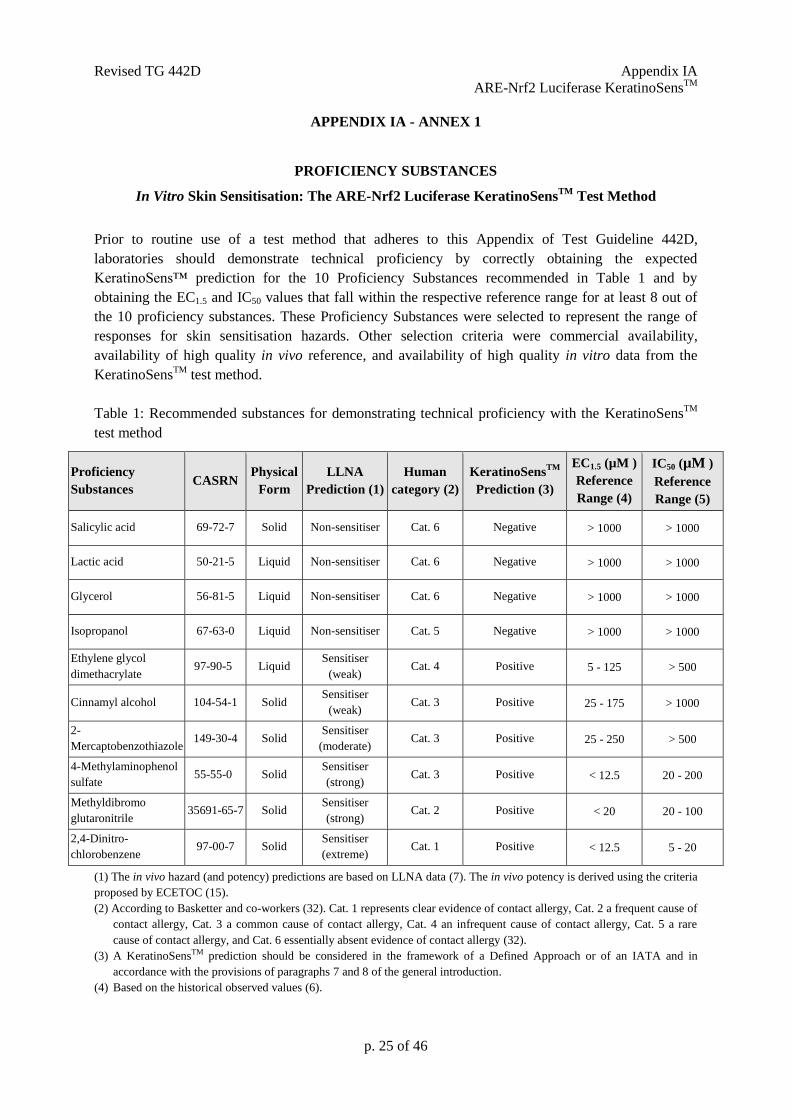
Prior to routine use of a test method that adheres to this Appendix of Test Guideline 442D,
laboratories should demonstrate technical proficiency by correctly obtaining the expected
KeratinoSens™ prediction for the 10 Proficiency Substances recommended in Table 1 and by
obtaining the EC1.5 and IC50 values that fall within the respective reference range for at least 8 out of
the 10 proficiency substances. These Proficiency Substances were selected to represent the range of
responses for skin sensitisation hazards. Other selection criteria were commercial availability,
availability of high quality in vivo reference, and availability of high quality in vitro data from the
KeratinoSensTM
test method.
Table 1: Recommended substances for demonstrating technical proficiency with the KeratinoSensTM
test method
Proficiency
Substances CASRN
Physical
Form
LLNA
Prediction (1)
Human
category (2)
KeratinoSensTM
Prediction (3)
EC1.5 (µM )
Reference
Range (4)
IC50 (µM )
Reference
Range (5)
Salicylic acid 69-72-7 Solid Non-sensitiser Cat. 6 Negative > 1000 > 1000
Lactic acid 50-21-5 Liquid Non-sensitiser Cat. 6 Negative > 1000 > 1000
Glycerol 56-81-5 Liquid Non-sensitiser Cat. 6 Negative > 1000 > 1000
Isopropanol 67-63-0 Liquid Non-sensitiser Cat. 5 Negative > 1000 > 1000
Ethylene glycol
dimethacrylate 97-90-5 Liquid
Sensitiser
(weak) Cat. 4 Positive 5 - 125 > 500
Cinnamyl alcohol 104-54-1 Solid Sensitiser
(weak) Cat. 3 Positive 25 - 175 > 1000
2-
Mercaptobenzothiazole 149-30-4 Solid
Sensitiser
(moderate) Cat. 3 Positive 25 - 250 > 500
4-Methylaminophenol
sulfate 55-55-0 Solid
Sensitiser
(strong) Cat. 3 Positive < 12.5 20 - 200
Methyldibromo
glutaronitrile 35691-65-7 Solid
Sensitiser
(strong) Cat. 2 Positive < 20 20 - 100
2,4-Dinitro-
chlorobenzene 97-00-7 Solid
Sensitiser
(extreme) Cat. 1 Positive < 12.5 5 - 20
(1) The in vivo hazard (and potency) predictions are based on LLNA data (7). The in vivo potency is derived using the criteria
proposed by ECETOC (15).
(2) According to Basketter and co-workers (32). Cat. 1 represents clear evidence of contact allergy, Cat. 2 a frequent cause of
contact allergy, Cat. 3 a common cause of contact allergy, Cat. 4 an infrequent cause of contact allergy, Cat. 5 a rare
cause of contact allergy, and Cat. 6 essentially absent evidence of contact allergy (32).
(3) A KeratinoSensTM prediction should be considered in the framework of a Defined Approach or of an IATA and in
accordance with the provisions of paragraphs 7 and 8 of the general introduction.
(4) Based on the historical observed values (6).
Revised TG 442D Appendix IA
ARE-Nrf2 Luciferase KeratinoSensTM
p. 26 of 46
APPENDIX IA - ANNEX 2
ADAPTATION OF THE KERATINOSENSTM
TEST METHOD USING HUMAN
REAGENTS TO ACHIEVE ANIMAL-PRODUCT-FREE CELL CULTURE
The following adaptation to the KeratinoSensTM
test method may be performed using human reagents
(human serum and recombinant human trypsin) to achieve animal-product-free cell culture, subject to
demonstration of technical proficiency (as described in Annex 1) using the adapted method (33).
Prior to use for testing purposes, the KeratinoSensTM
cell line should be adapted to routine culture
using 10% human serum. Human serum (from pooled donors) should be obtained from a reputable
commercial source, with appropriate donor consent and QC testing for cell culture applications. As
with any type of serum, when a new batch is used, an internal validation of the batch including cell
morphology, growth rates and Imax / EC1.5 values with at least the positive control, and preferably
representative reference chemicals (at least one sensitiser and one non sensitiser) should be conducted,
with subsequent reservation of successfully performing batches for long term use. If the cells have
previously been cultured in foetal calf serum, they should be weaned into human serum over at least 3
passages. Provided that the cells are showing healthy morphology and comparable growth rates with
those in foetal calf serum, a cell bank should then be created for future use. With reference to
paragraph 13, it should be noted that the KeratinoSensTM
cell line, when cultured in human serum,
should be cultured up to a maximum passage number of 22 for optimal performance. To achieve fully
animal-product-free cell culture, a non-animal source of recombinant trypsin (for example,
TrypzeanTM
) should be used to harvest the cells during sub-culture. In all other respects, the cells
should be cultured in the same way as described in this Appendix to Test Guideline 442D and the
Standard Operating Procedure (SOP) (9) (33) for the reference KeratinoSensTM
cell line
With reference to paragraph 18, the animal-product-free adaptation of the KeratinoSensTM
test method
using human reagents has been optimised using cinnamic aldehyde (CAS No. 14371-10-9, >98%
purity) as a positive control, at a final concentration range from 8 to 128µM. Other positive controls,
preferentially providing EC1.5 values in the mid-range, may be used if historical data are available to
derive comparable run acceptance criteria (33).
With reference to paragraph 25, the animal-product-free adaptation of the KeratinoSensTM
test method
using human reagents has been optimised using the following method for cytotoxicity assessment.
Medium is replaced after the 48 hour exposure time with fresh medium containing MTT (3-(4,5-
dimethyl2-thiazolyl)-2,5-diphenyl-2H-tetrazolium bromide; CAS No. 298-93-1) at a concentration: of
1mg/ml, and cells incubated for 3 hours at 37±1oC in the presence of 5% CO2. The MTT medium is
then removed and cells are solubilised by the addition of isopropanol. After shaking for 30 minutes,
the absorption is measured at 570 nm with a spectrophotometer.
All other aspects the animal-product-free adaptation of the KeratinoSensTM
test method using human
reagents should be conducted in the same way as described for the standard method described in this
Appendix to Test Guideline 442D and the DB-ALM protocol (9) (33) for the reference
KeratinoSensTM
cell line.
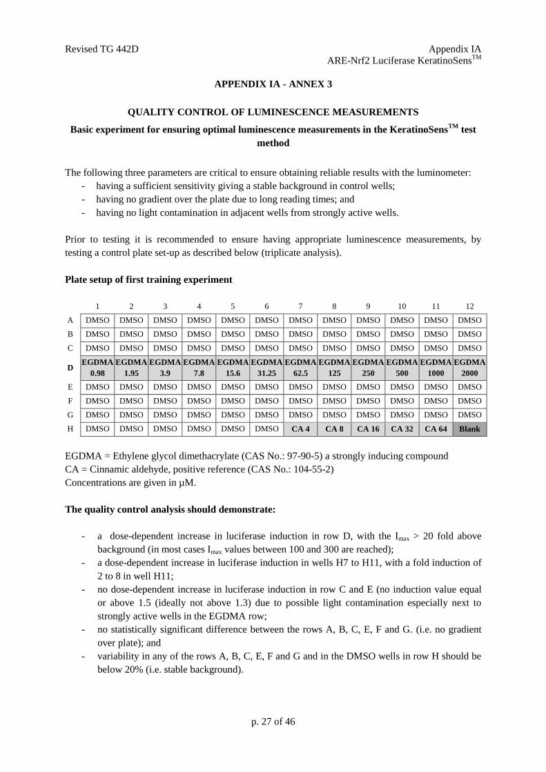
Revised TG 442D Appendix IA
ARE-Nrf2 Luciferase KeratinoSensTM
p. 27 of 46
APPENDIX IA - ANNEX 3
QUALITY CONTROL OF LUMINESCENCE MEASUREMENTS
Basic experiment for ensuring optimal luminescence measurements in the KeratinoSensTM
test
method
The following three parameters are critical to ensure obtaining reliable results with the luminometer:
- having a sufficient sensitivity giving a stable background in control wells;
- having no gradient over the plate due to long reading times; and
- having no light contamination in adjacent wells from strongly active wells.
Prior to testing it is recommended to ensure having appropriate luminescence measurements, by
testing a control plate set-up as described below (triplicate analysis).
Plate setup of first training experiment
1 2 3 4 5 6 7 8 9 10 11 12
A DMSO DMSO DMSO DMSO DMSO DMSO DMSO DMSO DMSO DMSO DMSO DMSO
B DMSO DMSO DMSO DMSO DMSO DMSO DMSO DMSO DMSO DMSO DMSO DMSO
C DMSO DMSO DMSO DMSO DMSO DMSO DMSO DMSO DMSO DMSO DMSO DMSO
D EGDMA
0.98
EGDMA
1.95
EGDMA
3.9
EGDMA
7.8
EGDMA
15.6
EGDMA
31.25
EGDMA
62.5
EGDMA
125
EGDMA
250
EGDMA
500
EGDMA
1000
EGDMA
2000
E DMSO DMSO DMSO DMSO DMSO DMSO DMSO DMSO DMSO DMSO DMSO DMSO
F DMSO DMSO DMSO DMSO DMSO DMSO DMSO DMSO DMSO DMSO DMSO DMSO
G DMSO DMSO DMSO DMSO DMSO DMSO DMSO DMSO DMSO DMSO DMSO DMSO
H DMSO DMSO DMSO DMSO DMSO DMSO CA 4 CA 8 CA 16 CA 32 CA 64 Blank
EGDMA = Ethylene glycol dimethacrylate (CAS No.: 97-90-5) a strongly inducing compound
CA = Cinnamic aldehyde, positive reference (CAS No.: 104-55-2)
Concentrations are given in µM.
The quality control analysis should demonstrate:
- a dose-dependent increase in luciferase induction in row D, with the Imax > 20 fold above
background (in most cases Imax values between 100 and 300 are reached);
- a dose-dependent increase in luciferase induction in wells H7 to H11, with a fold induction of
2 to 8 in well H11;
- no dose-dependent increase in luciferase induction in row C and E (no induction value equal
or above 1.5 (ideally not above 1.3) due to possible light contamination especially next to
strongly active wells in the EGDMA row;
- no statistically significant difference between the rows A, B, C, E, F and G. (i.e. no gradient
over plate); and
- variability in any of the rows A, B, C, E, F and G and in the DMSO wells in row H should be
below 20% (i.e. stable background).
Revised TG 442D Appendix IB
ARE-Nrf2 Luciferase LuSens
Appendix IB: In Vitro Skin Sensitisation:
The ARE-Nrf2 Luciferase LuSens Test Method
INITIAL CONSIDERATIONS, APPLICABILITY AND LIMITATIONS
1. The test method described in this Appendix to Test Guideline 442D addresses the second
key event of the skin sensitisation AOP (1), namely keratinocytes activation, by assessing with the
help of luciferase, the Nrf2-mediated activation of antioxidant response element (ARE)-dependent
genes. Skin sensitisers have been reported to induce genes that are regulated by the ARE (2) (3). Small
electrophilic substances such as skin sensitisers can act on the sensor protein Keap1 (Kelch-like ECH-
associated protein 1), by e.g. covalent modification of its cysteine residue, resulting in its dissociation
from the transcription factor Nrf2 (nuclear factor-erythroid 2-related factor 2). The dissociated Nrf2
can then activate ARE-dependent genes such as those coding for phase II detoxifying enzymes (2) (4)
(5).
2. The in vitro ARE-Nrf2 luciferase LuSens test method (hereafter called the LuSens test
method) underwent a Performance Standard-based validation study based on the KeratinoSensTM
Validated Reference Method (VRM) (6) (7) (8), followed by an independent peer review conducted by
the European Union Reference Laboratory for Alternatives to Animal Testing (EURL ECVAM) (9).
The LuSens test method was considered scientifically valid to be used as part of an IATA, to support
the discrimination between skin sensitisers and non-sensitisers for regulatory purposes (9).
3. The LuSens test method proved to be transferable to laboratories experienced in cell culture
techniques and met the reproducibility performance standards required both within and between
laboratories (9). Additional information from earlier in-house study on 72 test chemicals showed
similar predictive capacity as the VRM (74% accuracy, 74% sensitivity, and 74% specificity) for
discriminating skin sensitisers (i.e. UN GHS Cat. 1) from non-sensitisers when compared to LLNA
results (7) (9), indicating the usefulness of the LuSens test method to contribute to the identification of
skin sensitisation hazard. However, the accuracy values given here for LuSens test method as a stand-
alone test method, are only indicative since the test method should be considered in combination with
other sources of information in the context of a Defined Approach or an IATA and in accordance with
the provisions of paragraphs 7 and 8 in the General Introduction of this Test Guideline. Furthermore
when evaluating non-animal methods for skin sensitisation, it should be kept in mind that the LLNA
test as well as other animal tests may not fully reflect the situation in humans.
Revised TG 442D Appendix IB
ARE-Nrf2 Luciferase LuSens
p. 29 of 46
4. On the basis of the current data available, the LuSens test method was shown to be
applicable to test chemicals covering a variety of organic functional groups, reaction mechanisms, skin
sensitisation potency (as determined with in vivo studies) and physico-chemical properties (7) (8). The
test method is applicable to test chemicals soluble or that form a stable dispersion in the exposure
medium (i.e. a colloid or suspension in which the test chemical does not settle or separate from the
solvent into different phases). Test chemicals that do not fulfil these conditions at the highest final
required testing concentration (e.g. 2000 µM or 2000 µg/mL if no molecular weight is availble) may
still be tested at lower concentrations. In such a case, results fulfilling the criteria for positivity could
still be used to support the identification of the test chemical as a skin sensitiser, In cases where a
negative result is obtained in a test with a maximal concentrations < 2000 µM (or 2000 µg/mL if no
molecular weight is availble) and no cytotoxicity is observed, the result should be considered as
inconclusive (see prediction model in paragraph 32). If cytotoxicity (<70% viability) is reached at a
maximal soluble test concentration < 2000 µM (or 2000 µg/mL if no molecular weight is availble),
criteria for negativity could still be applied. In general mono constituent substances with a LogP above
7 may be insoluble in the exposure medium, however, if solubility or stable dispersion can be obtained
and documented, testing may still be conducted.
5. Negative results should be interpreted with caution as substances with an exclusive reactivity
towards lysine-residues can be detected as negative by the test method as the key mechanism leading
to the activation of the Keap1-Nrf2-ARE pathway appears to be the electrophilic reaction of stressors
with nucleophilic thiols (cysteine sulfhydryl groups) of Keap-1. Complementary information from
peptide reactivity assays may help addressing this uncertainty, in particular assays able to distinguish
between cysteine and lysine reactivity. Furthermore, because of the limited metabolic capability of the
cell line used (11) and because of the experimental conditions, pro-haptens (i.e. chemicals requiring
enzymatic activation for example via P450 enzymes) and pre-haptens (i.e. chemicals activated by
auto-oxidation) in particular with a slow oxidation rate may also provide negative results. However, it
has been shown that the majority of pre-haptens (i.e. chemicals activated by auto-oxidation) and pro-
haptens (i.e. chemicals requiring enzymatic activation for example via P450 enzymes) are sufficiently
well identified by a combination of test methods covering key events 1, 2 and 3 on the AOP so that
negative results can in general be used to support classification (12) (13) (18). On the other hand, test
chemicals that do not act as a sensitiser but are nevertheless chemical stressors may lead to false
positive results as shown with the VRM (10). Finally, test chemicals that interfere with the luciferase
enzyme can confound the activity of luciferase in cell-based assays causing either apparent inhibition
or increased luminescence (14). For example, phytoestrogen concentrations higher than 1 M were
reported to interfere with the luminescence signals in other luciferase-based reporter gene assays due
to over-activation of the luciferase reporter gene (15). As a consequence, luciferase expression
obtained at high concentrations of phytoestrogens or similar compounds suspected of producing
phytoestrogen-like over-activation of the luciferase reporter gene needs to be examined carefully (15).
In cases where evidence can be demonstrated on the non-applicability of the LuSens test method to
other specific categories of test chemicals, the test method should not be used for those specific
categories.
6. In addition to supporting discrimination between skin sensitisers (i.e. UN GHS Category 1)
and non-sensitisers, the LuSens test method also provides information (e.g., dose- response) that may
potentially contribute to the assessment of sensitising potency when used in integrated approaches
such as IATA such as described for the VRM (12). However, further work, preferably based on human
data, is required to determine how the LuSens test method results can contribute to potency
assessment, especially in the context of an IATA (16). Examples on how to use the ARE-Nrf2
luciferase test methods in combination with other information are reported in literature (17) (18).
7. Definitions are provided in Annex 1 of the General Introduction.
Revised TG 442D Appendix IB
ARE-Nrf2 Luciferase LuSens
p. 30 of 46
PRINCIPLE OF THE TEST
8 The LuSens test method makes use of an immortalised adherent cell line derived from
human keratinocytes stably harbouring a luciferase reporter gene under the control of the antioxidant
response element of the rat NQO1 gene (19). Genes dependent on the ARE such as NQO1 are known
to be up-regulated by contact sensitisers (20) (20) (21). The cell line contains the luciferase gene under
the transcriptional control of a promoter fused with the ARE element (7). The luciferase signal reflects
the activation by sensitisers of endogenous Nrf2 dependent genes, and the dependence of the luciferase
signal in the recombinant cell line on Nrf2 has been directly demonstrated for the VMR (22), and
indirectly demonstrated for the LuSens (7). This allows quantitative measurement (by luminescence
detection) of luciferase gene induction, using well established light producing luciferase substrates, as
an indicator of the activity of the Nrf2 transcription factor in cells following exposure to electrophilic
test substances.
9. Test chemicals are considered positive in the LuSens test method if they induce a statistically
significant induction of the luciferase activity above a given threshold (i.e. 1.5 fold, or 50% increase)
in at least two consecutive concentrations which do not significantly affect cell viability (i.e. at which
the cellular viability is above 70%) (7) (8). For this purpose, induction of the luciferase activity over
solvent/vehicle control is determined. Furthermore, parallel cytotoxicity measurements should be
conducted to assess whether luciferase activity induction levels occur at sub-cytotoxic concentrations.
10. Prior to routine use of the LuSens test method that adheres to this Test Guideline,
laboratories should demonstrate technical proficiency, using the ten Proficiency Substances listed in
Annex 1 of this Appendix.
PROCEDURE
11. A DB-ALM protocol for the LuSens test method is available and should be employed when
implementing and using the test method in the laboratory (23). A summary of the main protocol steps
of the LuSens test method as compared to the VRM is given in Annex 2 of this Appendix.
Laboratories implementing this Test Guideline can obtain the recombinant cell line used in the test
method by requests to the test developers 5. The luciferase reporter gene assay is subject to a Promega
limited use licence requiring i) the use of luminescent assay reagents purchased from Promega; or ii)
to contact Promega to obtain a free license for commercial use. The following paragraphs provide with
a description of the main components and procedures of the LuSens test method.
Preparation of the keratinocyte cultures
12. The LuSens transgenic cell line having a stable insertion of the luciferase reporter gene under the
control of the ARE-element should be used. Upon receipt, cells are propagated as defined by the test
method protocol (e.g. 1 to 3 passages) and stored frozen as a homogeneous stock. Cells from this
original stock can be propagated up to a maximum of 20 passage number and are employed for routine
testing using the appropriate maintenance/growth medium (e.g. Dulbecco's Modified Eagle's medium
(DMEM) containing serum and antibiotics such as puromycin in the maintenance medium (for
selection) and penicillin/streptomycin (to prevent contamination)) as described within the test
method’s protocol (23). No antibiotics are added however to the medium during testing.
13. For testing, cells should be 80-90% confluent, and care should be taken to ensure that cells
are never grown to full confluence. One day prior to testing cells are harvested, and distributed into
5 BASF SE, 67056 Ludwigshafen, Germany.
Revised TG 442D Appendix IB
ARE-Nrf2 Luciferase LuSens
p. 31 of 46
96-well plates at the appropriate cell density (i.e. 10,000 cells/well). Attention should be paid to avoid
sedimentation of the cells during seeding to ensure homogeneous cell number distribution across
wells. If this is not the case, this step may result in high well-to-well variability. For each repetition of
the main luciferase test for each test chemical concentration, three replicates are used for the luciferase
activity measurements, and three replicates used for the cell viability assay.
Preparation of the test chemical and control substances
14. The test chemical and control substances are prepared (or thawed in case of stable frozen
solutions) on the day of testing. Test chemicals are dissolved in a suitable solvent, e.g. dimethyl
sulfoxide (DMSO, CAS No. 67-68-5, 99% purity) to the final concentration that allows reaching the
maximum concentration tested (e.g. 200 mM). The DMSO solutions can be considered self-sterilising,
so that no sterile filtration is needed. Test chemicals not soluble in DMSO are dissolved in sterile
water or culture medium, in which the appropriate measures should be taken to ensure that the final
solutions are sterile. For a test chemical which has no defined molecular weight (MW), a stock
solution is prepared to a default concentration of 200 mg/mL or 20% (w/v). In case solvents other than
DMSO, water or the culture medium are used, appropriate scientific rationale should be provided.
15. Based on the stock solutions of the test chemical, serial dilutions are made using DMSO or
for test chemicals not soluble in DMSO, using sterile water or culture medium, to obtain master
concentrations of the chemical to be tested (e.g. 12 concentrations ranging from 0.098 to 200 mM).
Independent of the solvent used, the master concentrations, are then further diluted 25 fold into culture
medium containing serum, and finally used for treatment with a further 4 fold dilution factor so that
the final concentrations of the tested chemical are reached (e.g., ranging from 0.98 to 2000 M based
on a dilution factor of 2). For a test chemical which has no defined MW, serial dilutions are made
using DMSO or a suitable solvent to obtain the desired final concentrations of the test chemical (e.g.,
from 0.98 to 2000 g/ml).
16. A cytotoxicity pre-range dose finding test is first performed, based e.g. on the above
concentrations, to determine the concentration at which cell viability is reduced to 75% (CV75). The
CV75 is then used as a basis for determining the concentrations to be tested in the main luciferase test
and the parallel cytotoxicity test (e.g., one concentration above CV75, the CV75 and four concentrations
below CV75 using a serial dilution factor of 1.2 resulting in the concentrations CV75/2.07, CV75/1.73,
CV75/1.44, CV75/1.2, CV75 and CV75x1.2 μM). Alternative concentrations may be used upon
justification (e.g. in case of too low or too high cytotoxicity or poor solubility) (23).
17. A concurrent solvent/vehicle control should be tested within each repetition (e.g. DMSO),
for which a sufficient number of wells should be prepared per plate (e.g., 12 for the cytotoxicity pre-
range dose finding test and 24 for the main luciferase test as described in the protocol (23)). The
solvent/vehicle control undergoes the same dilutions as described for the master concentrations in
paragraph 15, so that the final solvent/vehicle control concentration should correspond to the same
concentration as in the tested chemicals and in the positive control (i.e., 1%), and should not
significantly affect cell viability. For a test chemical not soluble in the used solvent (e.g. DMSO), for
which the dilutions were made in water, the solvent level in all wells of the final test solution of this
test chemical must be adjusted to be equal to the solvent concentration used for the other test
chemicals and control substances (i.e. 1%).
18. A concurrent negative control should also be tested within each repetition, for which a
sufficient number of wells should be prepared per plate (e.g. 3 for the cytotoxicity pre-range dose
finding test and 6 for the main luciferase test as described in the protocol (23)). In the LuSens test
method, the concurrent negative control tested is 5000 μM (or 450 μg/mL) DL-Lactic acid (CAS No.
50-21-5, ≥99% purity), known to be a non-sensitiser and to result in a negative prediction with the
LuSens test method. Other suitable negative controls may be used if historical data are available to
derive comparable run acceptance criteria. Furthermore, in the LuSens test method a sufficient number
Revised TG 442D Appendix IB
ARE-Nrf2 Luciferase LuSens
p. 32 of 46
of wells (e.g. 6 for the cytotoxicity pre-range dose finding test and 12 for the main luciferase test as
described in the protocol (23)) containing blank medium controls are prepared consisting of untreated
cells and culture medium only.
19. A concurrent positive control should also be tested in a sufficient number of wells within
each repetition to demonstrate appropriate response of the test system (e.g. 2 for the cytotoxicity pre-
range dose finding test and 5 for the main luciferase test as described in the protocol (23)). For the
LuSens test method, 120 µM Ethylene Glycol Dimethacrylate (EGMDA, CAS No. 97-90-5, ≥ 99%
purity) is used. The positive control is prepared using the same dilution steps as described for the
master concentrations in paragraph 14 and as described in the test method’s protocols (23). If the
positive control concentration of 120 μM is too toxic or not able to induce luciferase ≥ 2.5 (see
paragraph 31) due to e.g. a new laboratory facility or a new batch of EGMDA, the performing
laboratory may run a range finder experiment with EGDMA (confirmed in at least two more runs) in
order to set the concentration at which luciferase induction is ≥ 2.5 folds compared to solvent/vehicle
control, and for which cell viability is 70%. Finally, other suitable positive controls, preferentially
providing EC1.5 values in the mid-range, may be used if historical data are available to derive
comparable run acceptance criteria.
Application of the test chemical and control substances
20. For each test chemical and positive control substance, one experiment is needed to derive a
prediction (positive or negative), consisting of at least two independent repetitions containing each
three replicates (i.e. n=6). In case of discordant results between the two independent repetitions, a third
repetition containing three replicates should be performed (i.e. n=9). Each independent repetition is
performed on a different day with fresh stock solution of test chemicals and independently harvested
cells. Cells may come from the same passage however.
21. After seeding as described in paragraph 13, cells are grown for 24 hours in the 96-wells
microtiter plates. The medium is then removed and replaced with fresh culture medium (i.e., 150 µl
DMEM containing serum but without antibiotics as described within the method’s protocol (23)) to
which 50 µl of the 25 fold diluted test chemical and control substances are added. At least one well per
plate should be left empty (no cells and no treatment) to assess background values.
22. The treated plates are then incubated for about 48 hours at 37±1oC in the presence of 5%
CO2. Care should be taken to avoid evaporation of volatile test chemicals and cross-contamination
between wells by test chemicals by e.g. covering the plates with a foil during incubation with the test
chemicals.
Luciferase activity measurements
23. Three factors are critical to ensure appropriate luminescence readings:
- the choice of a sensitive luminometer,
- the use of a plate format with sufficient height to avoid light-cross-contamination; and
- the use of a luciferase substrate with sufficient light output to ensure sufficient sensitivity and
low variability.
Prior to testing, a control experiment setup as described in Annex 3 of this Appendix should be carried
out to ensure that these three points are met.
24. After the 48 hour exposure time with the test chemical and control substances, cells are
washed with a phosphate buffered saline, and the relevant lysis buffer for luminescence readings
added to each well for a sufficient time (e.g. 5-10 min in dark).
Revised TG 442D Appendix IB
ARE-Nrf2 Luciferase LuSens
p. 33 of 46
25. Plates with the cell lysate are then placed in the luminometer for reading using the specific
program prescribed within the test method’s protocol (23). In case alternative settings are used, e.g.
depending on the model of luminometer used, these should be justified. Furthermore, a glow substrate
may also be used provided that the quality control experiment of Annex 3 of this Appendix is
successfully fulfilled.
Cytotoxicity Assessment
26. For the LuSens cell viability assay, medium is replaced after the 48 hour exposure time with
fresh medium containing 0.5 mg/ml MTT (3-(4,5-Dimethylthiazol-2-yl)-2,5-diphenyltetrazolium
bromide, Thiazolyl blue tetrazolium bromide; CAS No. 298-93-1) and cells are incubated for 2 hours
at 37±1oC in the presence of 5% CO2. The MTT medium is then removed and cells are lysed by using
an appropriate lysing agent for a sufficient amount of time (e.g. 10 % (w/v) SDS and 0.4% (v/v) acetic
acid solution in DMSO for 5 min). After shaking, the absorption is then measured using the
parameters described in the test method protocol (23).
DATA AND REPORTING
Data evaluation
27. The following parameters are calculated in the LuSens test method (see Annex 4 of this
Appendix for the detailed equations):
- Fold luciferase activity induction at all concentrations of the tested chemical, positive control
and negative control.
- Cellular viability (CV) at all concentrations of the tested chemical and for all controls to
determine (by interpolation) the concentration value at which 75% of cell viability occurs
(CV75).
28. For each concentration showing a luciferase activity induction equal or higher () than 1.5,
statistical significance is determined (e.g. using a two-tailed Student’s t-test) by comparing the
luminescence values of the three replicate samples with the luminescence values in the solvent/vehicle
control wells to assess whether the luciferase activity induction is statistically significant (p <0.05).
Furthermore, it should be checked that no significant cytotoxic effects occur at these concentrations
(i.e., that the cell viability is 70% at the concentrations leading to 1.5 fold luciferase induction).
29. It is recommended that data are visually checked with the help of graphs. If no clear dose-
response curve is observed, or if the dose-response curve obtained is biphasic (i.e. crossing the
threshold of 1.5 twice), the experiment should be repeated to verify whether this is specific to the test
chemical or due to an experimental artefact. In case the biphasic response is reproducible in an
independent experiment, the lower concentration, i.e., when the threshold of 1.5 is crossed the first
time should be reported. However, a concentration delivering an EC1.5 is not a requirement.
30. Finally, when in the LuSens test method a 1.5 fold luciferase activity induction is observed
only at the lowest tested concentration (e.g., CV75/2.07), re-testing should be conducted using at least
one additional lower concentration.
Revised TG 442D Appendix IB
ARE-Nrf2 Luciferase LuSens
p. 34 of 46
Acceptance criteria
31. The following acceptance criteria should be met when using the LuSens test method. If any
of the criteria listed below is not met, the data should be discarded and a new repetition should be
performed.
- The average luciferase activity induction obtained with the positive control, 120 µM
EGDMA (or comparable concentration – see paragraph 19) should be ≥ 2.5, and the positive
control should have a relative cell viability 70% as compared to the solvent/vehicle
control.
- The average luciferase activity induction obtained with the negative control, i.e., 5000 μM
DL-Lactic acid, as well as the basal expression of untreated cells should be <1.5 fold as
compared to the average solvent/vehicle control.
- The average coefficient of variation of the luminescence reading for the solvent/vehicle
controls (e.g. DMSO) should be below 20% in each repetition.
- At least three test concentrations should have cell viability of at least 70% relative to the
solvent/vehicle controls. Moreover, in case a result is to be considered negative, at least one
concentration should be cytotoxic, i.e. have a cell viability <70%, or the maximum
concentration of 2000 μM (or 2000 µg/ mL for substances with no defined MW) should have
been tested.
In some cases, test chemicals may induce no cytotoxicity, in which cases the maximum
concentration tested should be 2000 μM (or 2000 µg/ mL for test chemicals having undefined
MW). If in the main luciferase test no concentration is cytotoxic, i.e. has a cell viability <70%,
and no luciferase induction is observed, then a second repetition should be performed using e.g.
a 1.44 serial dilution factor based on the CV75 (i.e., starting with 1.44xCV75) instead of the 1.2
serial dilution factor used in the main luciferase test. If in the second repetition cytotoxicity and
luciferase induction are still not observed, a third repetition should be run with the maximum
concentration of 2000 μM (or 2000 µg/ mL for substances with undefined MW). This repetition
should then be confirmed by performing a fourth repetition.
Interpretation of results and prediction model
32. A LuSens prediction is considered positive if the following conditions are met in 2 of 2 or in the
same 2 of 3 repetitions, otherwise the LuSens prediction is considered negative (Figure 1):
• the luciferase induction is above or equal to () 1.5 fold and is statistically significant
compared to the solvent control in at least 2 consecutive non-cytotoxic tested concentrations
(i.e. cellular viability is equal or higher than () 70%), whereby at least three tested
concentrations should be non-cytotoxic (cellular viability equal or higher than () 70%).
In addition, a negative result obtained with test chemicals that do not form a stable dispersion and
were not tested up to 2000 µM (or 2000 µg/mL for test chemicals with no defined MW) and for which
no cytotoxicity is observed in any of the tested concentration (see paragraph 31) should also be
considered as inconclusive (see paragraph 4).
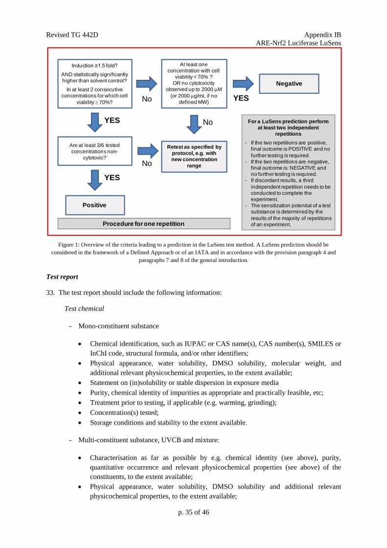
Revised TG 442D Appendix IB
ARE-Nrf2 Luciferase LuSens
p. 35 of 46
Figure 1: Overview of the criteria leading to a prediction in the LuSens test method. A LuSens prediction should be
considered in the framework of a Defined Approach or of an IATA and in accordance with the provision paragraph 4 and
paragraphs 7 and 8 of the general introduction.
Test report
33. The test report should include the following information:
Test chemical
- Mono-constituent substance
Chemical identification, such as IUPAC or CAS name(s), CAS number(s), SMILES or
InChI code, structural formula, and/or other identifiers;
Physical appearance, water solubility, DMSO solubility, molecular weight, and
additional relevant physicochemical properties, to the extent available;
Statement on (in)solubility or stable dispersion in exposure media
Purity, chemical identity of impurities as appropriate and practically feasible, etc;
Treatment prior to testing, if applicable (e.g. warming, grinding);
Concentration(s) tested;
Storage conditions and stability to the extent available.
- Multi-constituent substance, UVCB and mixture:
Characterisation as far as possible by e.g. chemical identity (see above), purity,
quantitative occurrence and relevant physicochemical properties (see above) of the
constituents, to the extent available;
Physical appearance, water solubility, DMSO solubility and additional relevant
physicochemical properties, to the extent available;
Induction ≥1.5 fold?
AND statistically significantly
higher than solvent control?
In at least 2 consecutive
concentrations for which cell
viability 70%? No
YES
Negative
Are at least 3/6 tested
concentrations non-
cytotoxic?
No
YES
Retest as specified by
protocol, e.g. with
new concentration
range
Positive
For a LuSens prediction perform
at least two independent
repetitions
- If the two repetitions are positive,
final outcome is POSITIVE and no
further testing is required.
- If the two repetitions are negative,
final outcome is: NEGATIVE and
no further testing is required.
- If discordant results, a third
independent repetition needs to be
conducted to complete the
experiment.
- The sensitization potential of a test
substance is determined by the
results of the majority of repetitions
of an experiment.
At least one
concentration with cell
viability < 70% ?
OR no cytotoxicity
observed up to 2000 M
(or 2000 g/mL if no
defined MW) YES
No
Procedure for one repetition
Revised TG 442D Appendix IB
ARE-Nrf2 Luciferase LuSens
p. 36 of 46
Statement of (in)solubility or stable dispersion in exposure media
Molecular weight or apparent molecular weight in case of mixtures/polymers of known
compositions or other information relevant for the conduct of the study;
Treatment prior to testing, if applicable (e.g. warming, grinding);
Concentration(s) tested;
Storage conditions and stability to the extent available.
Controls
- Positive control
Chemical identification, such as IUPAC or CAS name(s), CAS number(s), SMILES or
InChI code, structural formula, and/or other identifiers;
Physical appearance, water solubility, DMSO solubility, molecular weight, and
additional relevant physicochemical properties, to the extent available and where
applicable;
Purity, chemical identity of impurities as appropriate and practically feasible, etc;
Treatment prior to testing, if applicable (e.g. warming, grinding);
Concentration(s) tested;
Storage conditions and stability to the extent available;
Reference to historical positive control results demonstrating suitable run acceptance
criteria, if applicable.
- Solvent/vehicle control
Chemical identification, such as IUPAC or CAS name(s), CAS number(s), and/or other
identifiers;
Purity, chemical identity of impurities as appropriate and practically feasible, etc;
Physical appearance, molecular weight, and additional relevant physicochemical
properties in the case other solvents/vehicles than those mentioned in this Appendix are
used and to the extent available;
Storage conditions and stability to the extent available;
Justification for choice of solvent/vehicle for each test chemical.
- Negative control
Chemical identification, such as IUPAC or CAS name(s), CAS number(s), and/or other
identifiers;
Purity, chemical identity of impurities as appropriate and practically feasible, etc;
Physical appearance, molecular weight, and additional relevant physicochemical
properties in the case other negative controls than those mentioned in this Appendix are
used and to the extent available;
Storage conditions and stability to the extent available;
Justification for choice of the negative control in the case other negative controls than
those mentioned in the Test Guideline are used.
Revised TG 442D Appendix IB
ARE-Nrf2 Luciferase LuSens
p. 37 of 46
Test method conditions
- Name and address of the sponsor, test facility and study director;
- Description of test method used;
- Cell line used, its storage conditions and source (e.g. the facility from which they were
obtained);
- Passage number and level of confluence of cells used for testing;
- Cell counting method used for seeding prior to testing and measures taken to ensure
homogeneous cell number distribution (cf. paragraph 13);
- Luminometer used (e.g. model), including instrument settings, luciferase substrate used,
and demonstration of appropriate luminescence measurements based on the control test
described in Annex 3 of this Appendix;
- The procedure used to demonstrate proficiency of the laboratory in performing the test
method (e.g. by testing of proficiency substances) or to demonstrate reproducible
performance of the test method over time.
Test procedure
- Number of repetitions and replicates used;
- Test chemical concentrations, application procedure and exposure time used (if different
than the one recommended)
- Description of evaluation and decision criteria used;
- Description of study acceptance criteria used;
- Description of any modifications of the test procedure.
Results
- Tabulation of fold luciferase induction activity and viability values (i.e.CV75 for the
LuSens test method) obtained for the test chemical and for the positive control for each
repetition;
- The mean values (i.e. arithmetic means of cell viability and luciferase activity induction)
and SD calculated using data from all individual repetitions;
- An indication of the rating of the test chemical according to the prediction model;
- Coefficient of variation obtained with the luminescence readings for the solvent/vehicle
control for each experiment;
- A graph depicting dose-response curves for induction of luciferase activity and viability;
- Description of any other relevant observations, if applicable.
Revised TG 442D Appendix IB
ARE-Nrf2 Luciferase LuSens
p. 38 of 46
Discussion of the results
- Discussion of the results obtained with the LuSens test method;
- Consideration of the test method results within the context of an IATA, if other relevant
information is available.
Conclusion
LITERATURE
1. OECD (2012). Series on Testing and Assessment No. 168. The Adverse Outcome Pathway
for Skin Sensitisation Initiated by Covalent Binding to Proteins. Part 1: Scientific Evidence.
Organisation for Economic Cooperation and Development, Paris. Available at:
http://www.oecd.org/officialdocuments/publicdisplaydocumentpdf/?cote=ENV/JM/MONO(
2012)10/PART1&docLanguage=En
2. Natsch A. (2010). The Nrf2-Keap1-ARE Toxicity Pathway as a Cellular Sensor for Skin
Sensitizers-Functional Relevance and Hypothesis on Innate Reactions to Skin Sensitizers.
Toxicological Sciences 113, 284-292.
3. Emter R., Ellis G., Natsch A.(2010). Performance of a novel keratinocyte-based reporter cell
line to screen skin sensitizers in vitro. Toxicology and Applied Pharmacology 245, 281-290.
4. Dinkova-Kostova A.T., Holtzclaw W.D., Kensler T.W. (2005). The role of Keap1 in cellular
protective responses. Chem. Res. Toxicol. 18, 1779-1791.
5. Kansanen E., Kuosmanen S.M., Leinonen H., Levonen A.L. (2013). The Keap1-Nrf2
pathway: Mechanisms of activation and dysregulation in cancer. Redox Biol. 1, 45-49.
6. OECD (2015). Guidance Document No 213. Performance Standards for assessment of
proposed similar or modified in vitro skin sensitisation ARE-NrF2 luciferase test methods.
Series on Testing and Assessment. Available at: http://www.oecd.org/env/ehs/testing/series-
testing-assessment-publications-number.htm
7. Ramirez T., Mehling A., Kolle S.N., Wruck C.J., Teubner W., Eltze T., Aumann A., Urbisch
D., van Ravenzwaay B., Landsiedel R. (2014). LuSens: a keratinocyte based ARE reporter
gene assay for use in integrated testing strategies for skin sensitization hazard identification.
Toxicol. In Vitro 28, 1482-1497.
8. Ramirez T., Stein N., Aumann A., Remus T., Edwards A., Norman K.G., Ryan C., Bader
J.E., Fehr M., Burleson F., Foertsch L., Wang X., Gerberick F., Beilstein P., Hoffmann S.,
Mehling A., van Ravenzwaay B., Landsiedel R. (2016). Intra- and inter-laboratory
reproducibility and accuracy of the LuSens assay: A reporter gene-cell line to detect
keratinocyte activation by skin sensitizers. Toxicol. In Vitro 32, 278-286.
9. ESAC (2016). ESAC opinion on the BASF-coordinated Performance Standards-based
validation of the LuSens test method for skin sensitisation testing. Available at:
http://publications.jrc.ec.europa.eu/repository/bitstream/JRC103706/esac_opinion_2016-
04_lusens_final.pdf.
Revised TG 442D Appendix IB
ARE-Nrf2 Luciferase LuSens
p. 39 of 46
10. EURL-ECVAM (2014). Recommendation on the KeratinoSensTM
assay for skin sensitisation
testing, 42 pp. Available at: http://ihcp.jrc.ec.europa.eu/our_labs/eurl-ecvam/eurl-ecvam-
recommendations/recommendation-keratinosens-skin-sensitisation.
11. Fabian E., Vogel D., Blatz V., Ramirez T., Kolle S., Eltze T., van Ravenzwaay B., Oesch F.,
Landsiedel R. (2013). Xenobiotic metabolizin enzyme activities in cells used for testing skin
sensitization in vitro. Arch Toxicol 87, 1683-1969.
12. Jaworska J., Dancik Y., Kern P., Gerberick F., Natsch A. (2013). Bayesian integrated testing
strategy to assess skin sensitization potency: from theory to practice. J. Appl. Toxicol. 33,
1353-1364.
13. Casati S., Aschberger K., Asturiol D., Basketter D., Dimitrov S., Dumont C., Karlberg A.T.,
Lepoittevin J.P., Patlewicz G., Roberts D.W., Worth A. (2016). Ability of non-animal
methods for skin sensitisation to detect pre- and pro-haptens: Report and recommendations
of an EURL ECVAM expert meeting. EUR 27752 EN. Available at : https://eurl-
ecvam.jrc.ec.europa.eu/eurl-ecvam-status-reports/pre-prohapten-workshop-report.
14. Thorne N., Inglese J., Auld D.S. (2010). Illuminating Insights into Firefly Luciferase and
Other Bioluminescent Reporters Used in Chemical Biology. Chemistry and Biology 17, 646-
657.
15. OECD (2012). BG1Luc Estrogen Receptor Transactivation Test Method for Identifying
Estrogen Receptor Agonists and Antagonists. OECD Guidelines for Chemical Testing No.
457. Organisation for Economic Cooperation and Development, Paris. Available at:
[http://www.oecd.org/env/testguidelines].
16. ECETOC (2003). Contact sensitization: Classification according to potency. European
Centre for Ecotoxicology & Toxicology of Chemicals (Technical Report No. 87).
17. Bauch C., Kolle S.N., Ramirez T., Eltze T., Fabian E., Mehling A., Teubner W., van
Ravenzwaay B., Landsiedel R. (2012). Putting the parts together: combining in vitro
methods to test for skin sensitizing potentials. Regul. Toxicol. Pharmacol. 63, 489-504.
18. Urbisch D., Becker M., Honarvar N., Kolle S.N., Mehling A., Teubner W., Wareing B.,
Landsiedel R. (2016). Assessment of Pre- and Pro-haptens Using Non-animal Test Methods
for Skin Sensitization. Chem. Res. Toxicol. 29, 901-13.
19. Wasserman W.W., Fahl W.E. (1997). Comprehensive analysis of proteins which interact
with the antioxidant responsive element: correlation of ARE-BP-1 with the chemoprotective
induction response. Arch. Biochem. Biophys. 344, 387-396.
20. Ade N, Leon F, Pallardy M, Peiffer JL, Kerdine-Romer S, Tissier MH, Bonnet PA, Fabre I,
Ourlin JC.(2009); HMOX1 and NQO1 genes are upregulated in response to contact
sensitizers in dendritic cells and THP-1 cell line: role of the Keap1/Nrf2 pathway. Toxicol
Sci. 2009 Feb;107(2):451-60.
21. Rob J. Vandebriel, Jeroen L. A. Pennings, Kirsten A. Baken, Tessa E. Pronk, Andre
Boorsma, Ralph Gottschalk, Henk Van Loveren; Keratinocyte Gene Expression Profiles
Discriminate Sensitizing and Irritating Compounds, Toxicological Sciences, Volume 117,
Issue 1, 1 September 2010, Pages 81–89, https://doi.org/10.1093/toxsci/kfq182
22. Emter R., van der Veen J.W., Adamson G., Ezendam J., van Loveren H., Natsch A. (2013).
Gene expression changes induced by skin sensitizers in the KeratinoSens™ cell line:
Revised TG 442D Appendix IB
ARE-Nrf2 Luciferase LuSens
p. 40 of 46
Discriminating Nrf2-dependent and Nrf2-independent events. Toxicol. In Vitro 27, 2225-
2232.
23. DB-ALM Protocol No. 184 (2017). LuSens Assay. Available at https://ecvam-
dbalm.jrc.ec.europa.eu
24. OECD (2005). Guidance Document the Validation and International Acceptance of New or
Updated Test Methods for Hazard Assessment. OECD Environment, Health and Safety
publications, OECD Series on Testing and Assessment No.34. Organisation for Economic
Cooperation and Development, Paris.
25. United nations (UN) (2015). Globally Harmonized System of Classification and Labelling of
Chemicals (GHS). Sixth revised edition, New York and Geneva, United Nations
Publications. Available at:
[https://www.unece.org/trans/danger/publi/ghs/ghs_rev06/06files_e.html]
26. Natsch A., Ryan C.A., Foertsch L., Emter R., Jaworska J., Gerberick G.F., Kern P. (2013). A
dataset on 145 chemicals tested in alternative assays for skin sensitization undergoing
prevalidation. Journal of Applied Toxicology 33, 1337-1352.
27. Urbisch D., Mehling A., Guth K., Ramirez T., Honarvar N., Kolle S., Landsiedel R.,
Jaworska J., Kern P.S., Gerberick F., Natsch A., Emter R., Ashikaga T., Miyazawa M.,
Sakaguchi H. (2015). Assessing skin sensitization hazard in mice and men using non-animal
test methods. Regul. Toxicol. Pharmacol. 71, 337–351.
28. Basketter D.A., Alépée N., Ashikaga T., Barroso J., Gilmour N., Goebel C., Hibatallah J.,
Hoffmann S., Kern P., Martinozzi-Teissier S., Maxwell G., Reisinger K., Sakaguchi H.,
Schepky A., Tailhardat M., Templier M. (2014). Categorization of chemicals according to
their relative human skin sensitizing potency. Dermatitis 25, 11-21.
29. Basketter DA, Whittle E, Griffiths HA, York M. (1994); The identification and classification
of skin irritation hazard by a human patch test. Food Chem Toxicol. 1994 Aug;32(8):769-75.
https://www.ncbi.nlm.nih.gov/pubmed/?term=Ashikaga%20T%5BAuthor%5D&cauthor=true&cauthor_uid=24407057
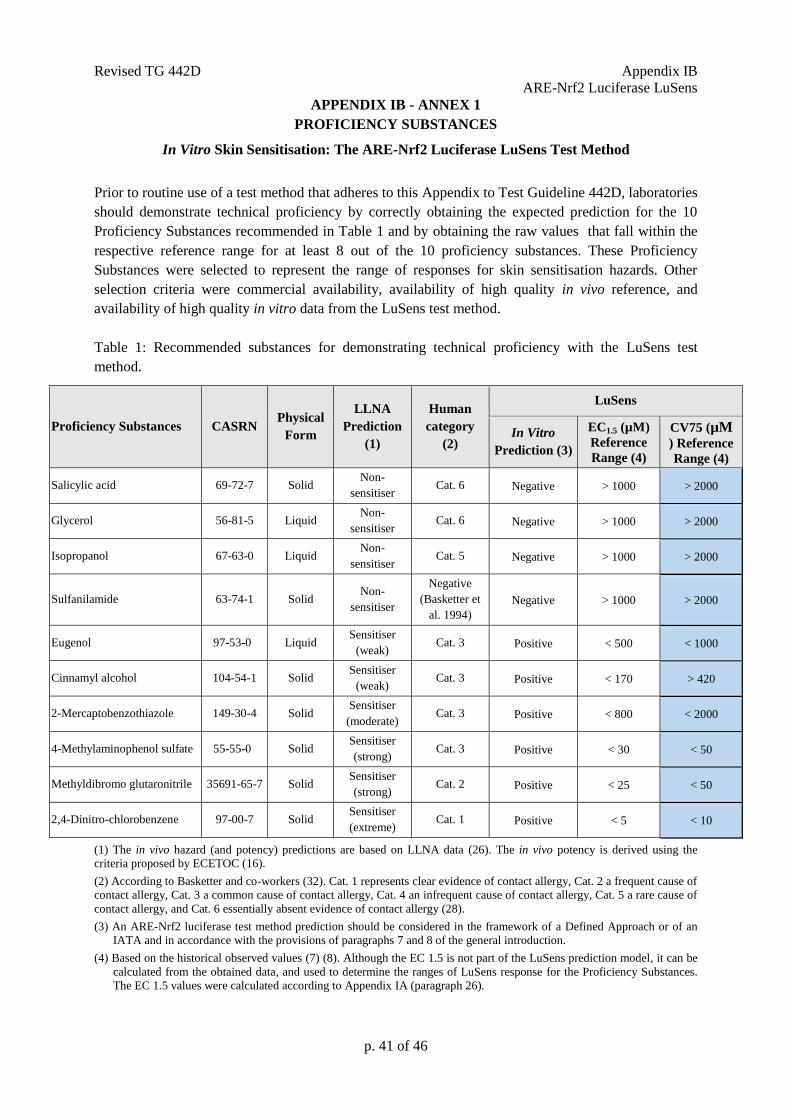
Revised TG 442D Appendix IB
ARE-Nrf2 Luciferase LuSens
p. 41 of 46
APPENDIX IB - ANNEX 1
PROFICIENCY SUBSTANCES
In Vitro Skin Sensitisation: The ARE-Nrf2 Luciferase LuSens Test Method
Prior to routine use of a test method that adheres to this Appendix to Test Guideline 442D, laboratories
should demonstrate technical proficiency by correctly obtaining the expected prediction for the 10
Proficiency Substances recommended in Table 1 and by obtaining the raw values that fall within the
respective reference range for at least 8 out of the 10 proficiency substances. These Proficiency
Substances were selected to represent the range of responses for skin sensitisation hazards. Other
selection criteria were commercial availability, availability of high quality in vivo reference, and
availability of high quality in vitro data from the LuSens test method.
Table 1: Recommended substances for demonstrating technical proficiency with the LuSens test
method.
Proficiency Substances CASRN Physical
Form
LLNA
Prediction
(1)
Human
category
(2)
LuSens
In Vitro
Prediction (3)
EC1.5 (µM)
Reference
Range (4)
CV75 (µM
) Reference
Range (4)
Salicylic acid 69-72-7 Solid Non-
sensitiser Cat. 6 Negative > 1000 > 2000
Glycerol 56-81-5 Liquid Non-
sensitiser Cat. 6 Negative > 1000 > 2000
Isopropanol 67-63-0 Liquid Non-
sensitiser Cat. 5 Negative > 1000 > 2000
Sulfanilamide 63-74-1 Solid Non-
sensitiser
Negative
(Basketter et
al. 1994)
Negative > 1000 > 2000
Eugenol 97-53-0 Liquid Sensitiser
(weak) Cat. 3 Positive < 500 < 1000
Cinnamyl alcohol 104-54-1 Solid Sensitiser
(weak) Cat. 3 Positive < 170 > 420
2-Mercaptobenzothiazole 149-30-4 Solid Sensitiser
(moderate) Cat. 3 Positive < 800 < 2000
4-Methylaminophenol sulfate 55-55-0 Solid Sensitiser
(strong) Cat. 3 Positive < 30 < 50
Methyldibromo glutaronitrile 35691-65-7 Solid Sensitiser
(strong) Cat. 2 Positive < 25 < 50
2,4-Dinitro-chlorobenzene 97-00-7 Solid Sensitiser
(extreme) Cat. 1 Positive < 5 < 10
(1) The in vivo hazard (and potency) predictions are based on LLNA data (26). The in vivo potency is derived using the
criteria proposed by ECETOC (16).
(2) According to Basketter and co-workers (32). Cat. 1 represents clear evidence of contact allergy, Cat. 2 a frequent cause of
contact allergy, Cat. 3 a common cause of contact allergy, Cat. 4 an infrequent cause of contact allergy, Cat. 5 a rare cause of
contact allergy, and Cat. 6 essentially absent evidence of contact allergy (28).
(3) An ARE-Nrf2 luciferase test method prediction should be considered in the framework of a Defined Approach or of an
IATA and in accordance with the provisions of paragraphs 7 and 8 of the general introduction.
(4) Based on the historical observed values (7) (8). Although the EC 1.5 is not part of the LuSens prediction model, it can be
calculated from the obtained data, and used to determine the ranges of LuSens response for the Proficiency Substances.
The EC 1.5 values were calculated according to Appendix IA (paragraph 26).
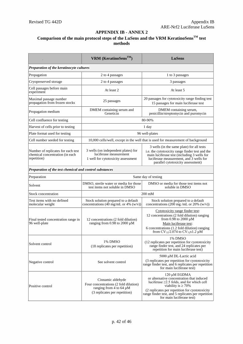
Revised TG 442D Appendix IB
ARE-Nrf2 Luciferase LuSens
p. 42 of 46
APPENDIX IB - ANNEX 2
Comparison of the main protocol steps of the LuSens and the VRM KeratinoSensTM
test
methods
VRM (KeratinoSensTM
) LuSens
Preparation of the keratinocyte cultures
Propagation 2 to 4 passages 1 to 3 passages
Cryopreserved storage 2 to 4 passages 3 passages
Cell passages before main experiment
At least 2 At least 5
Maximal passage number propagation from frozen stocks
25 passages 20 passages for cytotoxicity range finding test
15 passages for main luciferase test
Propagation medium DMEM containing serum and
Geneticin DMEM containing serum,
penicillin/streptomycin and puromycin
Cell confluence for testing 80-90%
Harvest of cells prior to testing 1 day
Plate format used for testing 96 well-plates
Cell number seeded for testing 10,000 cells/well, except in the well that is used for measurement of background
Number of replicates for each test chemical concentration (in each repetition)
3 wells (on independent plates) for luciferase measurement
1 well for cytotoxicity assessment
3 wells (in the same plate) for all tests
i.e. the cytotoxicity range finder test and the main luciferase test (including 3 wells for luciferase measurement, and 3 wells for
parallel cytotoxicity assessment)
Preparation of the test chemical and control substances
Preparation Same day of testing
Solvent DMSO, sterile water or media for those
test items not soluble in DMSO DMSO or media for those test items not
soluble in DMSO
Stock concentration 200 mM
Test items with no defined molecular weight
Stock solution prepared to a default concentrations (40 mg/mL or 4% (w/v))
Stock solution prepared to a default concentrations (200 mg /mL or 20% (w/v))
Final tested concentration range in 96 well-plate
12 concentrations (2 fold dilution) ranging from 0.98 to 2000 µM
Cytotoxicity range finder test:
12 concentrations (2 fold dilution) ranging from 0.98 to 2000 µM
Main luciferase test:
6 concentrations (1.2 fold dilution) ranging from CV75/2.074 to CV75x1.2 µM
Solvent control 1% DMSO
(18 replicates per repetition)
1% DMSO (12 replicates per repetition for cyototoxicity
range finder test, and 24 replicates per repetition for main luciferase test)
Negative control See solvent control
5000 μM DL-Lactic acid
(3 replicates per repetition for cyototoxicity range finder test, and 6 replicates per repetition
for main luciferase test)
Positive control
Cinnamic aldehyde
Four concentrations (2 fold dilution) ranging from 4 to 64 µM
(3 replicates per repetition)
120 μM EGDMA or alternative concentration that induced luciferase ≥2.5 folds, and for which cell
viability is 70%
(2 replicates per repetition for cyototoxicity range finder test, and 5 replicates per repetition
for main luciferase test)
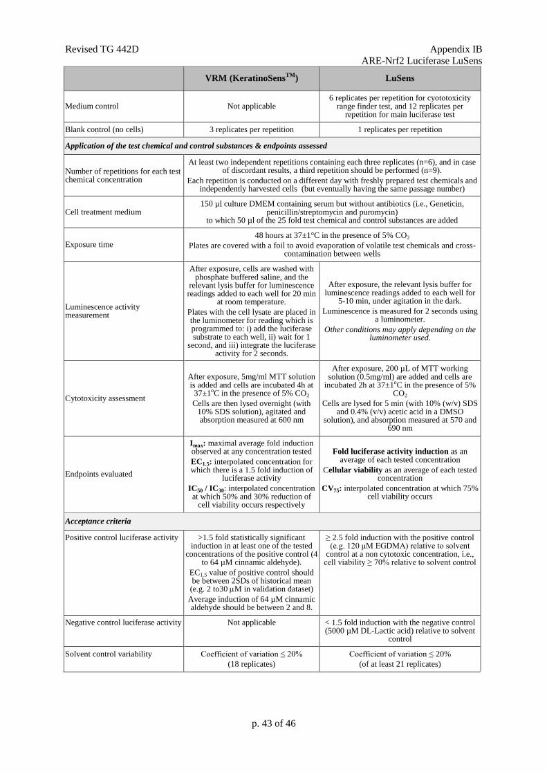
Revised TG 442D Appendix IB
ARE-Nrf2 Luciferase LuSens
p. 43 of 46
VRM (KeratinoSensTM
) LuSens
Medium control Not applicable 6 replicates per repetition for cyototoxicity
range finder test, and 12 replicates per repetition for main luciferase test
Blank control (no cells) 3 replicates per repetition 1 replicates per repetition
Application of the test chemical and control substances & endpoints assessed
Number of repetitions for each test chemical concentration
At least two independent repetitions containing each three replicates (n=6), and in case of discordant results, a third repetition should be performed (n=9).
Each repetition is conducted on a different day with freshly prepared test chemicals and independently harvested cells (but eventually having the same passage number)
Cell treatment medium 150 µl culture DMEM containing serum but without antibiotics (i.e., Geneticin,
penicillin/streptomycin and puromycin) to which 50 µl of the 25 fold test chemical and control substances are added
Exposure time 48 hours at 37±1°C in the presence of 5% CO2
Plates are covered with a foil to avoid evaporation of volatile test chemicals and cross-contamination between wells
Luminescence activity measurement
After exposure, cells are washed with phosphate buffered saline, and the
relevant lysis buffer for luminescence readings added to each well for 20 min
at room temperature.
Plates with the cell lysate are placed in the luminometer for reading which is programmed to: i) add the luciferase substrate to each well, ii) wait for 1
second, and iii) integrate the luciferase activity for 2 seconds.
After exposure, the relevant lysis buffer for luminescence readings added to each well for
5-10 min, under agitation in the dark.
Luminescence is measured for 2 seconds using a luminometer.
Other conditions may apply depending on the luminometer used.
Cytotoxicity assessment
After exposure, 5mg/ml MTT solution is added and cells are incubated 4h at 37±1oC in the presence of 5% CO2
Cells are then lysed overnight (with 10% SDS solution), agitated and absorption measured at 600 nm
After exposure, 200 µL of MTT working solution (0.5mg/ml) are added and cells are
incubated 2h at 37±1oC in the presence of 5% CO2
Cells are lysed for 5 min (with 10% (w/v) SDS and 0.4% (v/v) acetic acid in a DMSO
solution), and absorption measured at 570 and 690 nm
Endpoints evaluated
Imax: maximal average fold induction observed at any concentration tested
EC1.5: interpolated concentration for which there is a 1.5 fold induction of
luciferase activity
IC50 / IC30: interpolated concentration at which 50% and 30% reduction of
cell viability occurs respectively
Fold luciferase activity induction as an average of each tested concentration
Cellular viability as an average of each tested concentration
CV75: interpolated concentration at which 75% cell viability occurs
Acceptance criteria
Positive control luciferase activity >1.5 fold statistically significant induction in at least one of the tested
concentrations of the positive control (4 to 64 µM cinnamic aldehyde).
EC1.5 value of positive control should be between 2SDs of historical mean (e.g. 2 to30 M in validation dataset)
Average induction of 64 µM cinnamic aldehyde should be between 2 and 8.
≥ 2.5 fold induction with the positive control (e.g. 120 μM EGDMA) relative to solvent
control at a non cytotoxic concentration, i.e., cell viability ≥ 70% relative to solvent control
Negative control luciferase activity Not applicable < 1.5 fold induction with the negative control (5000 µM DL-Lactic acid) relative to solvent
control
Solvent control variability Coefficient of variation ≤ 20%
(18 replicates)
Coefficient of variation ≤ 20%
(of at least 21 replicates)
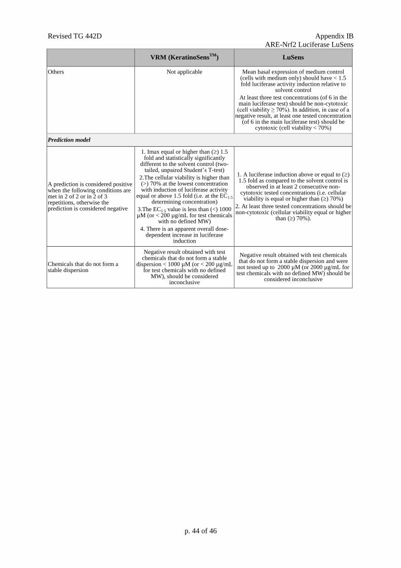
Revised TG 442D Appendix IB
ARE-Nrf2 Luciferase LuSens
p. 44 of 46
VRM (KeratinoSensTM
) LuSens
Others Not applicable Mean basal expression of medium control (cells with medium only) should have < 1.5 fold luciferase activity induction relative to
solvent control
At least three test concentrations (of 6 in the main luciferase test) should be non-cytotoxic (cell viability ≥ 70%). In addition, in case of a
negative result, at least one tested concentration (of 6 in the main luciferase test) should be
cytotoxic (cell viability < 70%)
Prediction model
A prediction is considered positive when the following conditions are met in 2 of 2 or in 2 of 3 repetitions, otherwise the prediction is considered negative
1. Imax equal or higher than () 1.5 fold and statistically significantly
different to the solvent control (two-tailed, unpaired Student’s T-test)
2.The cellular viability is higher than (>) 70% at the lowest concentration with induction of luciferase activity
equal or above 1.5 fold (i.e. at the EC1.5 determining concentration)
3.The EC1.5 value is less than (<) 1000 µM (or < 200 µg/mL for test chemicals
with no defined MW)
4. There is an apparent overall dose-dependent increase in luciferase
induction
1. A luciferase induction above or equal to () 1.5 fold as compared to the solvent control is
observed in at least 2 consecutive non-cytotoxic tested concentrations (i.e. cellular
viability is equal or higher than () 70%)
2. At least three tested concentrations should be non-cytotoxic (cellular viability equal or higher
than () 70%).
Chemicals that do not form a stable dispersion
Negative result obtained with test chemicals that do not form a stable
dispersion < 1000 µM (or < 200 µg/mL for test chemicals with no defined
MW), should be considered inconclusive
Negative result obtained with test chemicals that do not form a stable dispersion and were not tested up to 2000 µM (or 2000 µg/mL for test chemicals with no defined MW) should be
considered inconclusive
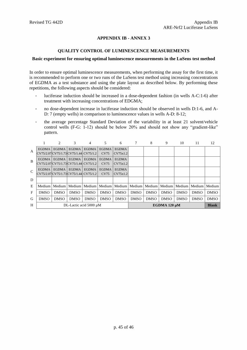
Revised TG 442D Appendix IB
ARE-Nrf2 Luciferase LuSens
p. 45 of 46
APPENDIX IB - ANNEX 3
QUALITY CONTROL OF LUMINESCENCE MEASUREMENTS
Basic experiment for ensuring optimal luminescence measurements in the LuSens test method
In order to ensure optimal luminescence measurements, when performing the assay for the first time, it
is recommended to perform one or two runs of the LuSens test method using increasing concentrations
of EGDMA as a test substance and using the plate layout as described below. By performing these
repetitions, the following aspects should be considered:
- luciferase induction should be increased in a dose-dependent fashion (in wells A-C:1-6) after
treatment with increasing concentrations of EDGMA;
- no dose-dependent increase in luciferase induction should be observed in wells D:1-6, and A-
D: 7 (empty wells) in comparison to luminescence values in wells A-D: 8-12;
- the average percentage Standard Deviation of the variability in at least 21 solvent/vehicle
control wells (F-G: 1-12) should be below 20% and should not show any “gradient-like”
pattern.
1 2 3 4 5 6 7 8 9 10 11 12
A EGDMA
CV75/2.07
EGDMA
CV75/1.73
EGDMA
CV75/1.44
EGDMA
CV75/1.2
EGDMA
CV75
EGDMA
CV75x1.2
B EGDMA
CV75/2.07
EGDMA
CV75/1.73
EGDMA
CV75/1.44
EGDMA
CV75/1.2
EGDMA
CV75
EGDMA
CV75x1.2
C EGDMA
CV75/2.07
EGDMA
CV75/1.73
EGDMA
CV75/1.44
EGDMA
CV75/1.2
EGDMA
CV75
EGDMA
CV75x1.2
D
E Medium Medium Medium Medium Medium Medium Medium Medium Medium Medium Medium Medium
F DMSO DMSO DMSO DMSO DMSO DMSO DMSO DMSO DMSO DMSO DMSO DMSO
G DMSO DMSO DMSO DMSO DMSO DMSO DMSO DMSO DMSO DMSO DMSO DMSO
H DL-Lactic acid 5000 µM EGDMA 120 µM Blank
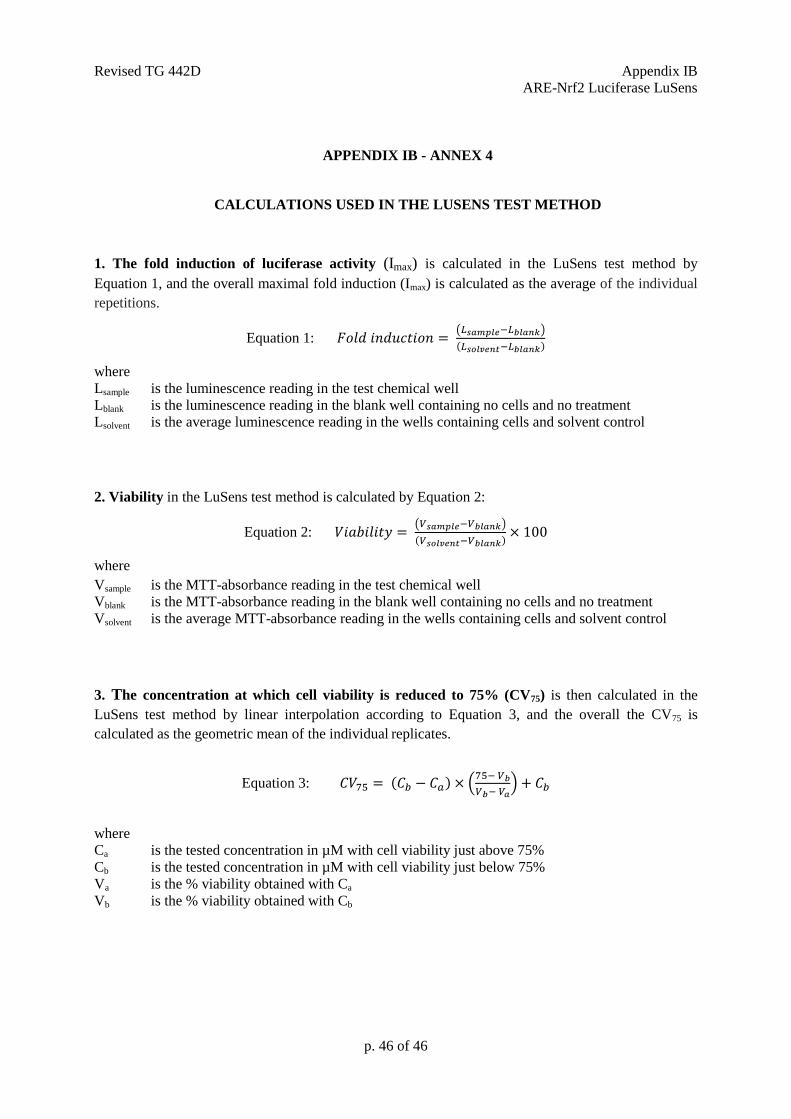
Revised TG 442D Appendix IB
ARE-Nrf2 Luciferase LuSens
p. 46 of 46
APPENDIX IB - ANNEX 4
CALCULATIONS USED IN THE LUSENS TEST METHOD
1. The fold induction of luciferase activity (Imax) is calculated in the LuSens test method by
Equation 1, and the overall maximal fold induction (Imax) is calculated as the average of the individual
repetitions.
Equation 1: 𝐹𝑜𝑙𝑑 𝑖𝑛𝑑𝑢𝑐𝑡𝑖𝑜𝑛 = (𝐿𝑠𝑎𝑚𝑝𝑙𝑒−𝐿𝑏𝑙𝑎𝑛𝑘)
(𝐿𝑠𝑜𝑙𝑣𝑒𝑛𝑡−𝐿𝑏𝑙𝑎𝑛𝑘)
where
Lsample is the luminescence reading in the test chemical well
Lblank is the luminescence reading in the blank well containing no cells and no treatment
Lsolvent is the average luminescence reading in the wells containing cells and solvent control
2. Viability in the LuSens test method is calculated by Equation 2:
Equation 2: 𝑉𝑖𝑎𝑏𝑖𝑙𝑖𝑡𝑦 = (𝑉𝑠𝑎𝑚𝑝𝑙𝑒−𝑉𝑏𝑙𝑎𝑛𝑘)
(𝑉𝑠𝑜𝑙𝑣𝑒𝑛𝑡−𝑉𝑏𝑙𝑎𝑛𝑘)× 100
where
Vsample is the MTT-absorbance reading in the test chemical well
Vblank is the MTT-absorbance reading in the blank well containing no cells and no treatment
Vsolvent is the average MTT-absorbance reading in the wells containing cells and solvent control
3. The concentration at which cell viability is reduced to 75% (CV75) is then calculated in the
LuSens test method by linear interpolation according to Equation 3, and the overall the CV75 is
calculated as the geometric mean of the individual replicates.
Equation 3: 𝐶𝑉75 = (𝐶𝑏 − 𝐶𝑎) × (75− 𝑉𝑏
𝑉𝑏− 𝑉𝑎) + 𝐶𝑏
where
Ca is the tested concentration in µM with cell viability just above 75%
Cb is the tested concentration in µM with cell viability just below 75%
Va is the % viability obtained with Ca
Vb is the % viability obtained with Cb