Differentiation of human alveolar epithelial cells in ... of human.pdf · culture of human alveolar...
15
Abstract Human alveolar type II cells were isolated from lung tissue and cultured for several days. The mor- phology of cells was investigated at different time points postseeding and the synthesis of alveolar cell-type spe- cific proteins was analyzed using different methods. The rationale of the study was to characterize a primary cell culture of human alveolar cells for the development of an in vitro model studying pulmonary drug delivery. In vitro test systems based on human cells are attracting in- creasing interest as important alternatives to animal- derived models because possible interspecies differences in alveolar cell biology and transport mechanisms cannot be excluded. In our study, both morphological character- ization and marker protein synthesis of human alveolar cells in culture indicate the differentiation of isolated al- veolar type II cells into epithelial monolayers consisting of alveolar type I-like and alveolar type II-like cells, which corresponds to the composition of the alveolar epithelium of the donor tissue. By using flow cytometry, immunofluorescence, immunoblotting and reverse tran- scriptase polymerase chain reaction (RT-PCR), we observed a shift in the synthesis of important marker proteins. Early cultures were characterized by low cave- olin-1 and high Sp-C levels. In comparison, the protein biosynthesis of alveolar cells switched with time of cul- ture to high caveolin-1 and low Sp-C levels. Based on the similarity between human alveolar epithelium and the development of our primary alveolar cell culture, we suggest that the culture may serve as a suitable model to study epithelial transport or cell biological processes in human alveolar cells. Keywords Human alveolar epithelial cells · Caveolin-1 · Caveolae · Surfactant protein C · Flow cytometry · Immunogold electron microscopy · Cell culture Introduction Lung alveolar epithelium in vivo is composed of two specialized epithelial cell types, the squamous alveolar epithelial type I (ATI) cell, which constitutes approxi- mately 93% of the alveolar epithelial surface area, and the surfactant-producing cuboidal alveolar epithelial type II (ATII) cell (Crapo et al. 1982). Current evidence supports the hypothesis that ATII cells serve as the sole progenitor for the ATI cells in vivo (reviewed in Uhal 1997; Fehrenbach 2001). Accordingly, isolated ATII cells in culture lose their characteristic phenotype and acquire over a 5- to 10-day period morphological and biochemical markers characteristic of ATI cells. Morphological changes during differentiation include the generation of monolayers with high transepithelial elec- trical resistance (>1,000 Ω cm 2 ) and a loss of microvilli, an increase in the cell surface area and the development of thin cytoplasmic attenuations extending away from a protruding nucleus (Cheek et al. 1989). Biochemical The Fonds der Chemischen Industrie and ZEBET (BgVV, Berlin) are thanked for their financial support. S. Fuchs · U.F. Schaefer · C.-M. Lehr ( ✉ ) Department of Biopharmaceutics and Pharmaceutical Technology, Saarland University, Saarbrücken, Germany e-mail: [email protected] Tel.: +49-681-3023039, Fax: +49-681-3024677 A.J. Hollins · M. Gumbleton Pharmaceutical Cell Biology, Welsh School of Pharmacy, Cardiff University, Wales, UK M. Laue Centre for Electron Microscopy, Department of Anatomy and Cell Biology, Medical Faculty, Saarland University, Homburg, Germany K. Roemer Department of Virology, Medical Faculty, Saarland University, Homburg, Germany C.-M. Lehr Department of Biopharmaceutics and Pharmaceutical Technology, Saarland University, Im Stadtwald, Geb. 8.1, PO Box 15 11 50, 66041 Saarbrücken, Germany Cell Tissue Res (2003) 311:31–45 DOI 10.1007/s00441-002-0653-5 REGULAR ARTICLE Sabine Fuchs · Andrew John Hollins · Michael Laue Ulrich Friedrich Schaefer · Klaus Roemer Mark Gumbleton · Claus-Michael Lehr Differentiation of human alveolar epithelial cells in primary culture: morphological characterization and synthesis of caveolin-1 and surfactant protein-C Received: 25 March 2002 / Accepted: 8 October 2002 / Published online: 12 November 2002 © Springer-Verlag 2002
Transcript of Differentiation of human alveolar epithelial cells in ... of human.pdf · culture of human alveolar...

Abstract Human alveolar type II cells were isolatedfrom lung tissue and cultured for several days. The mor-phology of cells was investigated at different time pointspostseeding and the synthesis of alveolar cell-type spe-cific proteins was analyzed using different methods. Therationale of the study was to characterize a primary cellculture of human alveolar cells for the development ofan in vitro model studying pulmonary drug delivery. Invitro test systems based on human cells are attracting in-creasing interest as important alternatives to animal-derived models because possible interspecies differencesin alveolar cell biology and transport mechanisms cannotbe excluded. In our study, both morphological character-ization and marker protein synthesis of human alveolarcells in culture indicate the differentiation of isolated al-veolar type II cells into epithelial monolayers consistingof alveolar type I-like and alveolar type II-like cells,which corresponds to the composition of the alveolar
epithelium of the donor tissue. By using flow cytometry,immunofluorescence, immunoblotting and reverse tran-scriptase polymerase chain reaction (RT-PCR), we observed a shift in the synthesis of important markerproteins. Early cultures were characterized by low cave-olin-1 and high Sp-C levels. In comparison, the proteinbiosynthesis of alveolar cells switched with time of cul-ture to high caveolin-1 and low Sp-C levels. Based onthe similarity between human alveolar epithelium andthe development of our primary alveolar cell culture, wesuggest that the culture may serve as a suitable model tostudy epithelial transport or cell biological processes inhuman alveolar cells.
Keywords Human alveolar epithelial cells · Caveolin-1 ·Caveolae · Surfactant protein C · Flow cytometry · Immunogold electron microscopy · Cell culture
Introduction
Lung alveolar epithelium in vivo is composed of twospecialized epithelial cell types, the squamous alveolarepithelial type I (ATI) cell, which constitutes approxi-mately 93% of the alveolar epithelial surface area, andthe surfactant-producing cuboidal alveolar epithelial typeII (ATII) cell (Crapo et al. 1982). Current evidence supports the hypothesis that ATII cells serve as the soleprogenitor for the ATI cells in vivo (reviewed in Uhal1997; Fehrenbach 2001). Accordingly, isolated ATIIcells in culture lose their characteristic phenotype andacquire over a 5- to 10-day period morphological and biochemical markers characteristic of ATI cells.Morphological changes during differentiation include thegeneration of monolayers with high transepithelial elec-trical resistance (>1,000 Ω cm2) and a loss of microvilli,an increase in the cell surface area and the developmentof thin cytoplasmic attenuations extending away from aprotruding nucleus (Cheek et al. 1989). Biochemical
The Fonds der Chemischen Industrie and ZEBET (BgVV, Berlin)are thanked for their financial support.
S. Fuchs · U.F. Schaefer · C.-M. Lehr ()Department of Biopharmaceutics and Pharmaceutical Technology,Saarland University, Saarbrücken, Germanye-mail: [email protected].: +49-681-3023039, Fax: +49-681-3024677
A.J. Hollins · M. GumbletonPharmaceutical Cell Biology, Welsh School of Pharmacy, Cardiff University, Wales, UK
M. LaueCentre for Electron Microscopy, Department of Anatomy and Cell Biology, Medical Faculty, Saarland University, Homburg, Germany
K. RoemerDepartment of Virology, Medical Faculty, Saarland University,Homburg, Germany
C.-M. LehrDepartment of Biopharmaceutics and Pharmaceutical Technology,Saarland University, Im Stadtwald, Geb. 8.1, PO Box 15 11 50,66041 Saarbrücken, Germany
Cell Tissue Res (2003) 311:31–45DOI 10.1007/s00441-002-0653-5
R E G U L A R A RT I C L E
Sabine Fuchs · Andrew John Hollins · Michael LaueUlrich Friedrich Schaefer · Klaus RoemerMark Gumbleton · Claus-Michael Lehr
Differentiation of human alveolar epithelial cells in primary culture:morphological characterization and synthesis of caveolin-1 and surfactant protein-C
Received: 25 March 2002 / Accepted: 8 October 2002 / Published online: 12 November 2002© Springer-Verlag 2002
Verwendete Distiller 5.0.x Joboptions
Dieser Report wurde automatisch mit Hilfe der Adobe Acrobat Distiller Erweiterung "Distiller Secrets v1.0.5" der IMPRESSED GmbH erstellt. Sie koennen diese Startup-Datei für die Distiller Versionen 4.0.5 und 5.0.x kostenlos unter http://www.impressed.de herunterladen. ALLGEMEIN ---------------------------------------- Dateioptionen: Kompatibilität: PDF 1.2 Für schnelle Web-Anzeige optimieren: Ja Piktogramme einbetten: Ja Seiten automatisch drehen: Nein Seiten von: 1 Seiten bis: Alle Seiten Bund: Links Auflösung: [ 600 600 ] dpi Papierformat: [ 595 785 ] Punkt KOMPRIMIERUNG ---------------------------------------- Farbbilder: Downsampling: Ja Berechnungsmethode: Bikubische Neuberechnung Downsample-Auflösung: 150 dpi Downsampling für Bilder über: 225 dpi Komprimieren: Ja Automatische Bestimmung der Komprimierungsart: Ja JPEG-Qualität: Mittel Bitanzahl pro Pixel: Wie Original Bit Graustufenbilder: Downsampling: Ja Berechnungsmethode: Bikubische Neuberechnung Downsample-Auflösung: 150 dpi Downsampling für Bilder über: 225 dpi Komprimieren: Ja Automatische Bestimmung der Komprimierungsart: Ja JPEG-Qualität: Mittel Bitanzahl pro Pixel: Wie Original Bit Schwarzweiß-Bilder: Downsampling: Ja Berechnungsmethode: Bikubische Neuberechnung Downsample-Auflösung: 600 dpi Downsampling für Bilder über: 900 dpi Komprimieren: Ja Komprimierungsart: CCITT CCITT-Gruppe: 4 Graustufen glätten: Nein Text und Vektorgrafiken komprimieren: Ja SCHRIFTEN ---------------------------------------- Alle Schriften einbetten: Ja Untergruppen aller eingebetteten Schriften: Nein Wenn Einbetten fehlschlägt: Warnen und weiter Einbetten: Immer einbetten: [ ] Nie einbetten: [ ] FARBE(N) ---------------------------------------- Farbmanagement: Farbumrechnungsmethode: Alle Farben zu sRGB konvertieren Methode: Standard Arbeitsbereiche: Graustufen ICC-Profil: RGB ICC-Profil: sRGB IEC61966-2.1 CMYK ICC-Profil: U.S. Web Coated (SWOP) v2 Geräteabhängige Daten: Einstellungen für Überdrucken beibehalten: Ja Unterfarbreduktion und Schwarzaufbau beibehalten: Ja Transferfunktionen: Anwenden Rastereinstellungen beibehalten: Ja ERWEITERT ---------------------------------------- Optionen: Prolog/Epilog verwenden: Nein PostScript-Datei darf Einstellungen überschreiben: Ja Level 2 copypage-Semantik beibehalten: Ja Portable Job Ticket in PDF-Datei speichern: Nein Illustrator-Überdruckmodus: Ja Farbverläufe zu weichen Nuancen konvertieren: Nein ASCII-Format: Nein Document Structuring Conventions (DSC): DSC-Kommentare verarbeiten: Nein ANDERE ---------------------------------------- Distiller-Kern Version: 5000 ZIP-Komprimierung verwenden: Ja Optimierungen deaktivieren: Nein Bildspeicher: 524288 Byte Farbbilder glätten: Nein Graustufenbilder glätten: Nein Bilder (< 257 Farben) in indizierten Farbraum konvertieren: Ja sRGB ICC-Profil: sRGB IEC61966-2.1 ENDE DES REPORTS ---------------------------------------- IMPRESSED GmbH Bahrenfelder Chaussee 49 22761 Hamburg, Germany Tel. +49 40 897189-0 Fax +49 40 897189-71 Email: [email protected] Web: www.impressed.de
Adobe Acrobat Distiller 5.0.x Joboption Datei
<< /ColorSettingsFile () /AntiAliasMonoImages false /CannotEmbedFontPolicy /Warning /ParseDSCComments false /DoThumbnails true /CompressPages true /CalRGBProfile (sRGB IEC61966-2.1) /MaxSubsetPct 100 /EncodeColorImages true /GrayImageFilter /DCTEncode /Optimize true /ParseDSCCommentsForDocInfo false /EmitDSCWarnings false /CalGrayProfile () /NeverEmbed [ ] /GrayImageDownsampleThreshold 1.5 /UsePrologue false /GrayImageDict << /QFactor 0.9 /Blend 1 /HSamples [ 2 1 1 2 ] /VSamples [ 2 1 1 2 ] >> /AutoFilterColorImages true /sRGBProfile (sRGB IEC61966-2.1) /ColorImageDepth -1 /PreserveOverprintSettings true /AutoRotatePages /None /UCRandBGInfo /Preserve /EmbedAllFonts true /CompatibilityLevel 1.2 /StartPage 1 /AntiAliasColorImages false /CreateJobTicket false /ConvertImagesToIndexed true /ColorImageDownsampleType /Bicubic /ColorImageDownsampleThreshold 1.5 /MonoImageDownsampleType /Bicubic /DetectBlends false /GrayImageDownsampleType /Bicubic /PreserveEPSInfo false /GrayACSImageDict << /VSamples [ 2 1 1 2 ] /QFactor 0.76 /Blend 1 /HSamples [ 2 1 1 2 ] /ColorTransform 1 >> /ColorACSImageDict << /VSamples [ 2 1 1 2 ] /QFactor 0.76 /Blend 1 /HSamples [ 2 1 1 2 ] /ColorTransform 1 >> /PreserveCopyPage true /EncodeMonoImages true /ColorConversionStrategy /sRGB /PreserveOPIComments false /AntiAliasGrayImages false /GrayImageDepth -1 /ColorImageResolution 150 /EndPage -1 /AutoPositionEPSFiles false /MonoImageDepth -1 /TransferFunctionInfo /Apply /EncodeGrayImages true /DownsampleGrayImages true /DownsampleMonoImages true /DownsampleColorImages true /MonoImageDownsampleThreshold 1.5 /MonoImageDict << /K -1 >> /Binding /Left /CalCMYKProfile (U.S. Web Coated (SWOP) v2) /MonoImageResolution 600 /AutoFilterGrayImages true /AlwaysEmbed [ ] /ImageMemory 524288 /SubsetFonts false /DefaultRenderingIntent /Default /OPM 1 /MonoImageFilter /CCITTFaxEncode /GrayImageResolution 150 /ColorImageFilter /DCTEncode /PreserveHalftoneInfo true /ColorImageDict << /QFactor 0.9 /Blend 1 /HSamples [ 2 1 1 2 ] /VSamples [ 2 1 1 2 ] >> /ASCII85EncodePages false /LockDistillerParams false >> setdistillerparams << /PageSize [ 595.276 841.890 ] /HWResolution [ 600 600 ] >> setpagedevice

changes include an increased reactivity to ATI-specificmembrane components such as the apical plasma mem-brane component RTI(40) and VIII B2 (Dobbs et al.1988; Danto et al. 1992; Vanderbilt and Dobbs 1998).
The isolation of ATII cells predominantly from ratand rabbit lung tissue, and their culture over time leadingto a primary culture of ATI-like cells, is now an estab-lished technique for different purposes. For instance, primary ATI-like monolayer cultures have gained accep-tance as an in vitro experimental model for the examina-tion of the rate and mechanism of alveolar solute or drugtransport (for review, see Mathias et al. 1996) and havealso been widely exploited to study the mechanisms ofalveolar epithelial cell differentiation (Danto et al. 1995;Dobbs et al. 1997; Campbell et al. 1999). However, theparallels between in vitro and in vivo ATII cell differen-tiation into ATI cells remain to be fully defined, andhence the term ATI-like cell is used to represent the invitro derived phenotype.
Although the isolation of primary human alveolarcells has been described before (Cunningham et al. 1997;Alcorn et al. 1997; Rosseau et al. 2000), human primarycells are not commonly used or appropriately character-ized as an in vitro model for the “blood-air barrier.” Research using human alveolar cells is mostly done oncell lines, such as the continuous human lung adenocar-cinoma cell line A549 (Giard et al. 1973). The A549 cellline possesses ATII cell phenotype and has been widelyused as a system to study the regulation of pulmonarysurfactant synthesis. However, cultured A549 cells donot undergo transition to form a phenotype similar tothat of an ATI cell. Furthermore, although the A549 cellhas received some attention as a monolayer culture forthe study of solute transport, its cell architecture and bar-rier properties are quite distinct from that of an ATI cellmonolayer (Godfrey 1997). Thus, an in vitro cell modelof the human alveolar epithelium possessing the relevantqualities of the alveolar epithelium in situ is definitelyneeded. This requirement is underscored by data whichindicate cell biological differences between the alveolarepithelium of humans and animals, e.g., the interspeciesdifferences in the spatial expression of aquaporins(Kreda et al. 2001).
We have previously described the isolation of humanalveolar type II epithelial cells (HAEpC) and their primary culture, which results in confluent monolayerscapable of generating tight junctional complexes andhigh transepithelial electrical resistance (TEER). In addi-tion, these monolayers revealed a preferential binding of lectins, which possess a high affinity for ATI cells (Elbert et al. 1999). In the present study we have extend-ed our characterization of the primary cultures of humanalveolar epithelial cells in order to follow the morpho-logical cell change from an ATII phenotype to an ATI-like cell phenotype over time of culture. Moreover, wehave examined the formation of characteristic plasmamembrane structures termed caveolae and the synthesisof their major structural protein, caveolin-1, in thesecells. The caveolae membrane system is of interest be-
cause of its potentially important role in macromoleculetransport across the “blood-air barrier” of the lung (reviewed in Gumbleton et al. 2000), including both theclearance of endogenous protein from the airspace andthe absorption of inhaled therapeutic proteins. Further-more, caveolin-1 may carry out a role in the regulationof alveolar epithelial cell proliferation and differentiation(Campbell et al. 1999). The synthesis of caveolin-1 andthe formation of caveolae are currently under discussionas a characteristic feature of an ATI-like cell phenotypein the alveolar epithelium in vivo and in vitro (Kasper etal. 1998; Newman et al. 1999; Campbell et al. 1999). Incomparison surfactant proteins are restricted to the ATIIphenotype; especially surfactant protein C (SP-C) isknown to be synthesized with a high specificity by ATII(Phelps and Floros 1991; Kalina et al. 1992; Beers et al.1994).
In this study morphological characterization by elec-tron microscopy showed the transition of a cell popula-tion mainly consisting of ATII cells towards an epithelialmonolayer composed of ATI and ATII cells during8–10 days postseeding. The ATI-like cells revealed atypical flattened cell shape and characteristic invagina-tions of the plasma membrane resembling caveolae,which could be supported by immunogold electron mi-croscopy using antibodies against caveolin-1, the mainstructural protein of caveolae. In parallel to the morpho-logical findings, suggesting a differentiation process, anincreased production of the structural protein caveolin-1with time of culture was observed by reverse transcrip-tase polymerase chain reaction (RT-PCR), Western-blotand flow cytometry, whereas synthesis of alveolar type IImarker Sp-C was restricted to the earlier stages of theculture.
Materials and methods
Cell culture
Primary type II alveolar cells (ATII) were isolated from humannon-tumor lung tissue, which was obtained from patients undergo-ing lung resection. The use of human material for isolation of primary cells was reviewed and approved by the respective localEthics Committees (State Medical Board of Registration, Saar-land; Cardiff University Medical Ethics Committee). Isolation wasperformed according to a protocol previously described by Elbertet al. (1999), but with a slight modification of the enzymatic digestion. Briefly, the chopped tissue was digested using a combi-nation of 150 mg trypsin type I (T-8003, Sigma, Deisenhofen,Germany) and 0.641 mg elastase (LS022795, CellSystems, St. Katharinen, Germany) in 30 ml balanced salt solution B(BSSB) for 40 min at 37°C. The ATII cell population was purifiedby a combination of differential cell attachment, Percoll densitygradient centrifugation and magnetic cell sorting (magnetic beadsM-450, Dynal, Hamburg, Germany) as previously described (Elbert et al. 1999). The isolated ATII cells were seeded at a celldensity of 300,000 cells/cm2 on collagen/fibronectin-coated poly-ester filter inserts (Transwell Clear, 6.5 mm diameter, 3470, Corning Costar, Bodenheim, Germany) using small airway growthmedium (SAGM) (CC-3118, Cell Systems, St. Katharinen, Germany) containing penicillin (100 units/ml) and streptomycin(100 µg/ml) and with the addition of low serum (1% fetal calf se-rum, FCS) in order to suppress fibroblasts. Formation of function-
32

al tight junctional complexes and generation of confluent mono-layers was routinely determined by measuring TEER using anelectronic voltmeter (EVOM, WPI, Berlin, Germany). Afterreaching confluence the alveolar monolayers of HAEpC typicallyrevealed TEER values of 1,000–2,000 Ω cm2 on day 6–8 post-seeding. The formation of tight junctions was also routinely moni-tored by immunolabeling for zonula occludens protein-1 (Zo-1).Cell isolation and culture were performed in two laboratories (De-partment of Biopharmaceutics and Pharmaceutical Technology,Saarland University, Saarbrücken, Germany; Pharmaceutical CellBiology Welsh School of Pharmacy, Cardiff University, Wales,UK) using the same methods. The average yield of ATII cells was2.3×106 cells/g tissue (n=15). The ATII cell purity determined bystaining for alkaline phosphatase was in the range of 81±1%.
A549 cells were obtained from BioWhittaker Europe (Verviers, Belgium). Passage numbers 86–89 were used culturedin Dulbecco’s modified Eagle’s medium (DMEM) supplementedwith 10% FCS in a humidified atmosphere containing 5% CO2.
Electron microscopy
Fixation
Cells on filter inserts were washed with phosphate-buffered saline(PBS) and fixed with a mixture of 1–4% formaldehyde and 1%glutaraldehyde in 0.1 M phosphate buffer (pH 7.2) at room tem-perature and stored overnight at 4°C. All formaldehyde solutionswere prepared from freshly depolymerized paraformaldehyde. Forimmunogold electron microscopy cells were washed with PBS andfixed with 4% formaldehyde in acetate buffer (pH 5.6) for 5 minfollowed by 4% formaldehyde in carbonate buffer (pH 10.8) for30 min (Berod et al. 1981). After a change to phosphate buffer, thecells were prepared for scanning and transmission electron mi-croscopy using the following protocols. In all preparations cellswere left on their original filter substrate.
Scanning electron microscopy of cell surfaces
For scanning electron microscopy cells were dehydrated on theirsupporting filters through an ethanol gradient (50%, 70%, 80%,90%, 96%, 99%, 99%, 100%; each for 15 min at room tempera-ture). The drying of samples was prepared by infiltration withmixtures of 1, 1, 1, 3, 3, 3-hexamethyldisilazane (HMDS, Roth,Germany) and ethanol (1:3, 1:1, 3:1, each for 15 min) followed bypure HMDS (twice, each for 15 min). HMDS was removed fromthe sample by desiccation with a vacuum pump. Dried filters weremounted on aluminium stubs and sputter coated with gold or plati-num. Preparations by critical-point drying yielded a similar struc-tural preservation.
Samples were observed using an ESEM XL 30 (FEI, TheNetherlands) equipped with a field emission gun at 20–30 kV un-der high vacuum conditions. For analysis of the filter area coveredby cells, digital images were taken randomly at a magnification of×600. The free filter surface was manually selected and calculatedrelative to the entire image area using NIH image software (version 1.62, developed at the US National Institutes of Health;customized by Steve Barrett, Surface Science Research Centre,University of Liverpool, UK).
Electron microscopy of cross sections
Cells were washed in 0.1 M phosphate buffer and postfixed with2% osmium tetroxide in 0.1 M phosphate buffer. After washing inphosphate buffer, cells were dehydrated in a series of ethanol(50%, 70%, 80%, 90%, 96%, 100%, 100%; each for 15 min atroom temperature). Infiltration with epoxy resin was done usingpropylene oxide (twice, each for 5 min; once for 15 min) and a 1:1mixture of propylene oxide and EMbed 812 (EMS, Fort Washing-ton, USA; resin mixture according to Luft 1961) overnight at
room temperature. After a further infiltration step in EMbed for3–4 h at room temperature, filters were embedded in fresh EMbedand polymerized at 60°C for at least 2 days. Ultrathin sections(60–80 nm thick) were counterstained with uranyl acetate and leadcitrate and inspected with an EM10C (Zeiss, Germany).
Low-power images were taken from the block face of embed-ded filters after sectioning using the scanning electron microscopeand imaging of back scattered electrons at 10–15 kV. Blocks werestained with uranyl acetate for a better contrast. This imaging pro-cedure was selected because sections usually show mechanicaldistortions and artifacts due to the different hardness of embed-ding medium, cells and filter material.
The general morphology of HAEpC cultured under the de-scribed conditions was the same in both laboratories in this study.
Immunogold electron microscopy
Cells were washed with 0.1 M phosphate buffer and dehydratedby progressive lowering of temperature (PLT) using the followingethanol series and temperatures: 30%, 0°C; 50%, –20°C; 70%,90%, 100%, 100%, –35°C for 1 h each. The incubations weredone by using tight-closing vials (2 ml, Twist Top; Sorenson Bio-science), which were placed into different refrigerators and freez-ers for cooling. At the temperature of –35°C vials were fitted intoan aluminium block for a better temperature control.
Infiltration with the acrylate resin Lowicryl K4 M was done ata temperature of –35°C by using mixtures of ethanol and resin(1:1, 1 h; 1:2, 1 h) followed by pure resin (twice; overnight and2 h). Filter inserts with cells were embedded in fresh resin deliv-ered in reaction vials (0.5 ml, Eppendorf) and polymerized by UVlight in a freeze-substitution apparatus (FSU010, Bal-tec) for1 day at –35°C before the temperature was raised to 0°C (dwelltime = 1 day) and room temperature (dwell time = 1 day).
Ultrathin sections were collected on Formvar-filmed copperslot or single hole grids. For immunolabeling, sections wereplaced on droplets (30 µl) of the following solutions: glycine(50 mM in PBS); blocking solution; anti-caveolin-1 antibody (seebelow); blocking solution; goat anti-rabbit antibody, coupled to10 nm colloidal gold (British Biocell, Cardiff, UK); blocking solu-tion; PBS; 2.5% glutaraldehyde in 0.1 M phosphate buffer; PBS;and aqua dest. The blocking solution consisted of cold water fishgelatine (0.5%; Sigma), bovine serum albumin (BSA) (0.5%; Aur-ion) and Tween 20 (0.01%; Sigma) in PBS. Incubation of sectionswith the anti-caveolin-1 antibody was done overnight at 4°C in awet chamber. Finally, dried sections were stained with a mixtureof uranyl acetate and methylcellulose (Roth et al. 1990).
For control rabbit non-immune sera were used. Significant la-beling of caveolae was never observed with these sera, indicatingthat the decoration of caveolae by caveolin-1 antibodies was spe-cific.
Antibodies
Caveolin-1 synthesis was examined using the rabbit polyclonalantibody (C-13630, Transduction, Lexington, UK), which recog-nizes both the α and β isoforms of caveolin-1. Surfactant protein-C (Sp-C) was examined using the rabbit polyclonal anti proSp-Cantibody (AB3428, Chemicon, Hofheim, Germany) recognizingthe Sp-C proprotein as well as processing intermediates. For theisotypic control an IgG rabbit serum (X0903, Dako, Hamburg,Germany) was used, which was diluted to the protein content ofthe primary antibodies. Occludin was investigated by mousemonoclonal antibody (O79120, Transduction, Lexington, UK).For detection of intracellular adhesion molecule-1 (ICAM-1), a mouse monoclonal antibody anti human CD54 (CBL 450, Cymbus Biotechnology Ltd., Chandlers Ford, UK) was used. Forisotypic controls a mouse IgG antibody (M5284, Sigma, Germa-ny) was diluted to the protein content of the primary antibodies.For Western blotting a secondary horseradish peroxidase (HRP)conjugated swine anti-rabbit antibody (Dako, Cambridge, UK)
33

was used. For fluorescence labeling cells were incubated with ananti-rabbit F(ab)2FITC conjugate (F1262, Sigma, Deisenhofen,Germany) or an anti-mouse FITC conjugate (F0479, Dako, Hamburg,Germany).
Immunofluorescence confocal microscopy
For immunofluorescence microscopy cells were fixed on Trans-well filter inserts with 2% paraformaldehyde and permeabilized by0.1% Triton-X. Cells were incubated with primary antibodies atroom temperature for 40 min in the dark at a dilution of 1:40 inPBS/1% BSA. After the first incubation step the cells were labeledwith the corresponding FITC-labeled anti-rabbit or anti-mouse an-tibody (1:100 in PBS, 1% BSA) for 40 min in the dark. Counter-staining of cell nuclei was undertaken using propidium iodide(0.2 µg/ml). For actin staining fixed and permeabilized cells wereincubated with TRITC-phalloidin (P-1951, Sigma, Deisenhofen,Germany) using a concentration of 5 µg/ml in PBS, 1% BSA. Finally cells were mounted in FluorSave (Calbiochem, Darmstadt,Germany) and analyzed using a confocal laser scanning micro-scope (Biorad MRC 1024, Munich, Germany).
Reverse transcriptase-polymerase chain reaction
The sources for RT-PCR materials were as follows: agarose LEanalytical grade and RNasin ribonuclease inhibitor (Promega,Southampton, UK); Ultraspec RNA extraction solution and Ultra-spec DEPC-treated water (Biogenesis, Dorset, UK); Moloney mu-rine leukemia virus reverse transcriptase (MMLV-rt) and dNTPs(Life Technologies-Gibco, Paisley, UK); random hexanucleotide(pdN6) primers (AmPharm, Little Chalfont, UK); and Biotaqpolymerase (Bioline, UK). Oligonucleotide primers were synthe-sized within the University of Wales, College of Medicine, on aBeckman DNA synthesizer with high-performance liquid chroma-tography (HPLC) high-purity isolation.
One microgram RNA was extracted (Ultraspec RNA extrac-tion) from the alveolar epithelial cultures grown on tissue culture-treated plastic Corning Costar and reverse transcribed using200 units of MMLV-rt and 10 pmol of random pdN6 primers in asolution containing TRIS and MgCl2 (1.5 mM). The cDNA repre-senting 60 ng of RNA was subjected to PCR for human caveolin-1and glyceraldehyde-3-phosphate dehydrogenase (GAPDH) for 30cycles in a final volume of 50 µl using 1 unit of Biotaq DNA poly-merase and 10 pmol/µl of each primer.
Human caveolin-1 (GenBank accession number Z18951) prim-ers comprised forward 5’-TCA ACC GCG ACC CTA AAC ACC-3’and reverse 5’-TGA AAT AGC TCA GAA GAG ACA T-3’ se-quences generating a 562-bp product. PCR was undertaken withan MgCl2 concentration of 1.5 mM, a primer concentration of10 pmol/µl and a thermocycler program of: (1) 95°C for 2 min, (2)60°C for 1 min, (3) 72°C for 1 min, (4) 95°C for 2 min, (5) 60°Cfor 1 min, and (6) 72°C for 1 min (steps 4–6 were repeated 26times) before a final round of 95°C for 1 min, and 60°C for 5 min.
Human GAPDH (GenBank accession number M33197) prim-ers comprised forward 5’-ACC ACA GTC CAT GCC ATC AC-3’and reverse 5’-TCC ACC ACC CTG TTG CTG TA-3’ sequencesgenerating a 452-bp product (Hurlstone et al. 1999). PCR was un-dertaken with an MgCl2 concentration of 1.5 mM, a primer con-centration of 10 pmol/µl and a thermocycler program of: (1) 95°Cfor 2 min, (2) 58°C for 30 s, (3) 72°C for 30 s, (4) 94°C for 1 min,(5) 58°C for 30 s, and (6) 72°C for 30 s (steps 4, 5, and 6 were re-peated 28 times) before a final round of 95°C for 1 min, and 58°Cfor 5 min.
A quantity of 15 µl of the PCR products was electrophoresedin a 2% agarose gel and stained with a solution of ethidium bro-mide (0.5 µg/ml). Negative controls consisted of omitting the re-verse transcription reaction or the cDNA product (data notshown).
Western blot for caveolin-1
Western blot for caveolin-1 in tissues and in cultured cell was performed as previously described (Newman et al. 1999; Campbellet al. 1999).
Flow cytometry
Freshly isolated human alveolar type II cells were directly washedin PBS after the isolation procedure and prepared for flow cytom-etry. Cells cultured to day 2, 4 and 8 postseeding were trypsinizedfollowed by washing with PBS and prepared for flow cytometry asdescribed below. Cells were fixed and permeabilized using Fixand Perm Kit (GAS-004, Caltag, Hamburg, Germany). Sp-C anti-body and caveolin-1 antibody were diluted 1:40 in the perm-eabilization buffer (buffer B) according to the recommendations.For the isotypic control rabbit IgG normal serum was used. Thesecondary FITC antibody (F1262) was diluted 1:100 in PBS. Afterwashing, cells were analyzed by flow cytometry using Calibur flu-orescence-activated cell sorting (FACS) (BD, Heidelberg, Germa-ny), and CellQuest software. 5000 cells were counted in triplicatefor each time point of the primary cultures. Independent isolationsfrom tissue of three patients were subjected to flow cytometry. Toexclude any autofluorescence from the cells, instrument settingswere adjusted in untreated cells.
To quantify the increase in caveolin-1 and to prove the repro-ducibility in different isolations, caveolin-1-positive cells weregated as indicated on the dot plot of day 8 cells (Fig. 7A) and themean FL-1 in the gated population was compared on days 0, 2, 4,and 8 in three isolations. A similar analysis was performed for Sp-C.
Results
Morphology of donor lung tissue
Respiratory parts of human lung tissue were used forpreparation of primary cultures. The fine structure of tis-sue samples revealed the typical architecture of the alve-olar epithelium formed by ATII and ATI cells. The ATIIcell is cuboidal in shape and its circumference is limitedto a zone near the nucleus. The cytoplasm is character-ized by multilamellar bodies which are membrane-boundstructures containing densely stacked membranes of sur-factant lipids (Fig. 1A, B).
ATI cells form flat extensions from a small layer ofcytoplasm in the nuclear region. The extensions are afew hundred nanometers in thickness and cover the ma-jority of the inner alveolar epithelial surface. Their apicaland basal membranes are invaginated by caveolae(Fig. 1C, D).
Morphology of HAEpC during culture
Isolated ATII cells seeded upon filter membranes, andcultured under the described conditions, formed a con-fluent monolayer of cells within 10 days of seeding.Cells aggregated, and differentiated during culture into aflattened epithelium consisting of two distinct cellmorphologies. The morphology of the cells was analyzedby scanning and transmission electron microscopy(Figs. 2, 3, 4).
34

After 2 days of culture, many ATII cells have formedmonolayer aggregates which were still separated fromeach other (Fig. 2A). At this time, more than 40% (n=14)of the filter surface was covered by cells. Cells displayeda morphology consistent with cell flattening and spread-ing but with a clearly elevated nucleus and a circumfer-ence profile that was variable in shape (compare alsoFig. 3A, B). The apical surface of the cells was coveredby short microvilli, which were most dense in the nucle-ar region and almost absent in the flattened peripheral re-gion of the cells. Numerous filopodia extended from freelateral cell borders and contacted the surface of the filteror neighboring cells. Closer contacts between cells werecharacterized by a weakly undulated contact zoneformed by the adjacent membranes. The contact zonewas on both cells decorated by row(s) of microvilli(compare Fig. 2F).
The intracellular morphology of the cells at day 2 ofthe culture was relatively uniform. The cytoplasm re-
35
Fig. 1A–D Transmission electron microscopy of donor lung tis-sue used for primary cell culture of human alveolar epithelialcells. The alveolar epithelium is formed by two cell types, alveolarepithelium type I (ATI) and II (ATII) cells. A, B Section through acuboidal ATII cell which is elevated above the epithelium. The cy-toplasm is characterized by multilamellar bodies (lb), membrane-bound structures which reveal densely stacked membrane lamel-lae. Note that preservation of these structures is not ideal since fix-ation must be done by immersion several hours after excision ofthe material. C, D ATI cells are flat cells with a thin lamella of cy-toplasm around the nucleus. The cells possess flat extensionswhich cover most of the inner alveolar surface. The apical andbasal plasma membranes of these extensions reveal many omega-shaped invaginations, termed caveolae (arrows). In some casescaveolae of both membranes approach very closely and are onlyseparated by a few nanometers of cytoplasm from each other (a al-veolar space, c capillary, ct connective tissue). Scale bars 1 µm(A, C), 0.5 µm (B), 0.2 µm (D)

vealed no particular concentration of organelles with oneexception. Most cells possessed a significant number ofmembrane-bound organelles of about 0.4–1.2 µm in diameter (maximum sectioning profile; n=46). These or-ganelles appeared empty in a few cases, but mostly re-vealed membranous structures within their lumina(Figs. 3A, B, 4B). In some cells these structures were
densely packed membrane lamellae as in ATII cells ofthe donor tissue (Fig. 4B). Often membrane lamellaeseemed to be condensed or reduced and accompanied byvesicular structures (Fig. 4C).
Between islands of cells and single cells of a more orless flattened morphology, a population of smaller roundcells of variable diameter and surface structure was ob-
36
Fig. 2A–H Scanning electronmicroscopy of cultured humanalveolar epithelial cells (HAE-pC) after different times of cul-ture. A, B On day 2 of cultureHAEpC have formed clusters.Cell shape is polymorphic andthe cellular surface is charac-terized by short microvilli.Gaps between cells are spannedby several thin filopodia. C, DAfter 4 days of culture gaps be-tween cells are almost closed.Cells reveal a flat morphologywith distinct contact zones be-tween them. E, F On day 8, thecells have formed an almostclosed monolayer. The cells re-semble flat disks of differentshapes with slightly elevatednuclei. In many cells surfacemicrovilli are concentrated inthe region of the nuclei and aremuch rarer in the flatter periph-ery. The contact zone betweencells is flanked by one or fewrows of microvilli on each cell(arrows). G, H From day 8 onsome cells show surface re-gions where the membrane isperforated by numerous smallconcentric holes of a uniformsize (c). H shows the surface ofanother cell of the same mono-layer which is completely de-void of concentric holes. Thedistribution of membrane holescorrelates with that of omega-shaped or flask-like sectioningprofiles in ultrathin sections ofthese monolayers which are in-dicative of caveolae (cf. textand Fig. 4D). Scale bars 50 µm(A, C), 10 µm (B, D), 5 µm (F),0.5 µm (G, H)
37
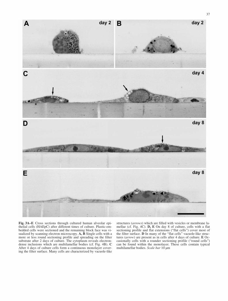
Fig. 3A–E Cross sections through cultured human alveolar epi-thelial cells (HAEpC) after different times of culture. Plastic-em-bedded cells were sectioned and the remaining block face was vi-sualized by scanning electron microscopy. A, B Single cells with amore or less round sectioning profile and spreading on the filtersubstrate after 2 days of culture. The cytoplasm reveals electron-dense inclusions which are multilamellar bodies (cf. Fig. 4B). CAfter 4 days of culture cells form a continuous monolayer cover-ing the filter surface. Many cells are characterized by vacuole-like
structures (arrows) which are filled with vesicles or membrane la-mellae (cf. Fig. 4C). D, E On day 8 of culture, cells with a flatsectioning profile and flat extensions (“flat cells”) cover most ofthe filter surface. D In many of the “flat cells” vacuole-like struc-tures (arrow) are present as in cells after 4 days of culture. E Oc-casionally cells with a rounder sectioning profile (“round cells”)can be found within the monolayer. These cells contain typicalmultilamellar bodies. Scale bar 10 µm

38

served. These cells showed no significant spreading onthe substrate, and some of them also laid on the surfaceof the flattened cells (Fig. 2D). Ultrathin sections revealed a morphology indicative of leukocytes (notshown). Their number seemed to decrease with time ofculture. On day 10 of culture none of these cells could beobserved.
On day 4 of the culture, gaps between the cells werealmost closed. At least 98% (n=15) of the filter surfacewas covered by cells (Fig. 2C, D). The cell morphologywas similar to that of the cells on day 2 with free edgesbearing multiple filopodia. Typical apical contact zones,which comprised apical tight junctions sometimes fol-lowed by adherent junctions, were much more prominentthan on day 2 because cells formed larger contact areas(Fig. 4A). The number of characteristic membrane-bound organelles containing membrane lamellae in theirlumina (see above) was lower than in cells on day 2. Inmost of the cells membrane lamellae seemed to be re-duced in size or condensed and were frequently associat-ed with vesicles, which gave the organelles a vacuole-like appearance (Figs. 3C, 4C).
After 8 and 10 days of the culture the cells formed aflat, almost closed monolayer on the filter surface (filterarea covered by cells ≥98%, n=16) (Figs. 2B, C, 3D, E).The average thickness of the epithelium was less com-pared to that of the epithelium on days 2 and 4 (cf.Fig. 3A–E). The remaining gaps between cells werespanned by numerous filopodia of the surrounding cells.The overall density of microvilli was reduced in compar-ison to cells on day 2 and 4 of culture and seemed to cor-relate with the thickness of the monolayer. The thinnerthe monolayer, the lower the density of microvilli. Inmost cells the flattened peripheral attenuations lackedmicrovilli. Cell shape remained variable across themonolayer with two distinct classes of cells distin-guished based on their cross-section profile and intracel-lular ultrastructure: (1) cells with a rounder profile(“round cells”) and numerous membrane-bound struc-tures containing lamellar membranes (Fig. 3E); and (2)
cells with a flattened and extended profile lacking lamel-lar structures; only membrane-bound structures withcondensed membranes were visible in some of thesecells. The majority of the filter surface was covered bythese “flat cells” (Fig. 3D, E).
In some of the “flat cells” numerous membrane in-vaginations occurred in restricted regions of the apicalplasma membrane. In the scanning electron microscope,they appear as concentric holes of a defined size (meanmaximum diameter=45 nm, SD=13, n=135; Fig. 2G, H).Their distribution correlates with the occurrence of ome-ga-shaped or flask-like membrane invaginations in sec-tions which are typical sectioning profiles of caveolae(Fig. 4D). Immunostaining showed that these structurescan be specifically labeled by antibodies against caveo-lin-1, the major structural protein of caveolae (Fig. 4E,F). The total number of caveolae increased from day 8 to10. Single caveolae were occasionally present in apicalmembranes of all cells including “round cells.”
Immunofluorescence microscopy
Immunofluorescence studies using a polyclonal anti-caveolin-1 antibody revealed the presence of caveolin-1in most of the cells after 3 days in culture (Fig. 5A, day3). Caveolin-1 labeling was seen as fluorescent dots inthe cytoplasm. The number of these dots and their sizeincreased with time of the culture (day 9, Fig. 5B). Im-munofluorescence studies on A549 cells gave a morediffuse pattern with a fluorescence signal that was weakcompared with that in HAEpC at later stages of the cul-ture (Fig. 5E). Moreover, no changes in labeling patternor intensity of the fluorescence for caveolin-1 were ob-served during the entire culture.
HAEpC and A549 were also tested for Sp-C in immu-nofluorescence studies. Sp-C labeling was found in near-ly all HAEpC on day 4 (Fig. 5C) with intense staining inthe perinuclear region or faint reticular staining through-out the cytoplasm indicating a labeling of the Golgi/ERcompartment. In the later stages (day 8), most of cellswere nearly negative for Sp-C or revealed single dots inthe cytoplasm. Only a few cells on day 8 still showedSp-C labeling in the area of the Golgi/ER in a patternsimilar to that of earlier stages (Fig. 5D). In A549, im-munofluorescence for Sp-C resulted in strong staining(Fig. 5F) comparable to the early stages of HAEpC. In-tense labeling for Sp-C in these cells was found aroundthe nucleus and in the cytoplasm, where it showed a reticular pattern suggesting a localization of Sp-C in association with the Golgi/ER complex. No change inSp-C levels or distribution in relation to time of cultureof A549 cells was observed, indicating a steady produc-tion of Sp-C. Immunofluorescence for the intracellularadhesion molecule ICAM-1 (Fig. 5G) in HAEpC demon-strated an increase in ICAM-1 levels with time of cul-ture. ICAM-1 was situated in small dots, which were either distributed all over the cell or only present the cellperiphery.
39
Fig. 4A–F Transmission electron microscopy of cultured humanalveolar epithelial cells (HAEpC) in cross section. A Two cells ofa monolayer after 4 days of culture which show an apical to baso-lateral polarity and typical epithelial cell contacts, i.e., tight junc-tions (arrows). The inset shows a magnification of the cell con-tacts. B Multilamellar bodies (mlb) of a cell on day 2 of culture(cf. Fig. 3A, B). The morphology of the multilamellar bodies iscomparable to that of multilamellar bodies in ATII cells of thelung (cf. Fig. 2A, B). C Multilamellar bodies (mlb) and vacuole-like (v) structures in a cell on day 4 of culture. The vacuole-likestructures contain small vesicles and few membrane lamellae. DFlask-like invaginations of the plasma membrane in a “flat cell”which are indicative of caveolae (day 8 of culture). Note that ve-sicular structures in deeper layers of the cortical cytoplasm (ar-rowheads) seem to be connected to the invaginations of the plas-ma membrane. E, F Immunogold labeling of flask-like membraneinvaginations with anti-caveolin-1 (aCav1). The gold particles areconcentrated around the sectioning profile of the invaginated plas-ma membrane (mt mitochondrion, mv microvilli, rer rough endo-plasmic reticulum). Scale bars 1 µm (A, B), 0.5 µm (C), 0.1 µm(D–F, inset)

The formation of tight junctions in HAEpC was previ-ously proven by the synthesis of the tight junctional pro-tein zo-1 forming a regular belt around the single cells(Elbert et al. 1999). Immunofluorescence using monoclo-
nal antibodies for occludin (Fig. 5H) and staining of actinfilaments by phalloidin-TRITC (not shown) resulted in avery similar labeling. These results indicate the formationof epithelial cell-cell contacts between the cultured cells.
40
Fig. 5A–H Immunofluores-cence of caveolin-1 and Sp-Cin HAEpC and A549 cells. Im-munoreactivity is displayed ingreen while nuclei appear red.A HAEpC on day 3 postseed-ing stained with anti-caveolin-1antibody reveal weak caveolin-1 levels in HAEpC in nearly allcells. Immunolabeling for ca-veolin-1 appears in little dots inthe cytoplasm. B In later stagesof the cell culture (day 9) cave-olin-1 labeling is situated indots forming larger aggregatesin the cytoplasm and the fluo-rescence for caveolin-1 is in-creased. E In comparison,A549 cells only display weakfluorescence for caveolin-1 andonly a few dots were found inthe cytoplasm. No change incaveolin-1 signal during culturetime is observed in A549. C Immunofluorescence inHAEpC for Sp-C on day 4postseeding reveals intensestaining in nearly all cells. Thelabeling for Sp-C is particularlyintense in the perinuclear re-gion, and extends into the peripheral cytoplasm, where itis reticular with intermingleddots. D On day 8 most of thecells are almost negative forSp-C or reveal only single dotsin the cytoplasm. Some cellsstill reveal an immunofluores-cence pattern comparable to theearly stages. F In A549 cellsSp-C labeling is intense and localized in the perinuclear re-gion, from where it sends retic-ular strands throughout the cy-toplasm similar to the labelingof HAEpC in early stages. Fur-ther, A549 show no changes inintensity or concerning the localization of Sp-C during cul-ture time. G Immunostainingfor ICAM-1 in HAEpC on day9. ICAM-1 was present in all ofthe cells but expressed to vari-ous extents in single cells. H HAEpC stained for occludinon day 9 using a mouse mono-clonal antibody. Labeling foroccludin demonstrated the for-mation of continuous circum-ferential intercellular contacts,which are typical of tight junc-tions. Scale bars A–H 50 µm

RT-PCR and Western blotting for caveolin-1
The caveolin-1 signal in HAEpC over time of culturewas quantified by RT-PCR and Western blotting. Caveo-lin-1 expression tested at the mRNA level by RT-PCRrevealed a product of 562 bp, which was consistent withexpected size. GAPDH was used as internal standard.For quantification of the caveolin-1 product, densitome-try analysis was performed. The ratio between caveolin-1 and GAPDH was estimated over time of culture. Thefreshly isolated cells showed a notable expression of ca-veolin-1 mRNA, which increased from day 3 up to day 8(Fig. 6A). The same pattern could be found by Westernblot analysis (Fig. 6B). Caveolin-1 (22 kDa) was detect-ed in freshly isolated cells and a clear increase in caveo-lin-1 synthesis from day 3 to day 8 was observed. Thepositive control, lysate of human lung, revealed a bandof the same size.
Flow cytometry
In comparison to other quantitative methods such as RT-PCR and Western blotting, flow cytometry offers thepossibility of evaluating the synthesis of marker proteinsat the level of a single cell level. This allows discrimina-tion of cell subpopulations according to cell size, granu-larity and marker proteins. For HAEpC, flow cytometrywas done with freshly isolated cells and cells culturedfor 2, 4 and 8 days after isolation. In the freshly isolatedcells, a weak signal for caveolin-1 was observed(Fig. 7A). The mean of the fluorescence channel FL-1 inthe gated population of freshly isolated cells on day 0was in the range of 50 units. However, in addition to thegated population, other cell populations on the basis of areduced morphological size (low forward scatter, FSC)and lack of caveolin-1 were detected. On day 2, the ca-veolin-1 synthesis within the gated HAEpC population
41
Fig. 6A, B Analysis of caveo-lin-1 in HAEpC during cultureby RT-PCR and immunoblot-ting. A shows RT-PCR for ca-veolin-1 (from left to right) infreshly isolated cells (lane 1),and in cells cultured for 3 days(lane 2), 5 days (lane 3) and8 days (lane 4). GAPDH wasused as internal standard. Thebands for caveolin-1 are locat-ed on the gel at a position con-sistent with the expected size of562 bp. Quantification was per-formed by densitometry analy-sis upon the above image bydetermining optical density.The time course reveals rela-tively high expression rates forcaveolin-1 in freshly isolatedcells followed by a reduced ca-veolin-1 expression on day 3.With time of culture a steadyincrease in caveolin-1 is no-ticed. B Western blotting forcaveolin-1 in cells after differ-ent times of culture. Humanlung lysate was used as control.A double band of 22 kDa is de-tected. For quantification theoptical density per unit areawas determined as shown bybar chart. The time course re-veals the presence of caveolin-1 in freshly isolated cells. Onday 3 the caveolin-1 signal isreduced followed by an in-crease on day 8

was increased significantly and the cells formed a homo-geneous caveolin-1-positive population. On day 4 andday 8 caveolin-1 levels within the gated cells had in-creased further. In addition, the entire population wasmore homogeneous with over 90% of the cultured cellsrepresented within the gated population and showed high
levels of caveolin-1. Other subpopulations of lower sizeand insignificant caveolin-1 signals disappeared, indicat-ing that they may constitute erythrocytes, leukocytes orother irrelevant cells. On day 10 no further increase incaveolin-1-specific fluorescence was detectable (notshown). In addition to the increase in caveolin-1 synthe-sis, an increase in cell size was also detected consistentwith flattening and cell spreading. Means of FL-1 weredetermined from three cell isolates (Fig. 7A).
As a control, FACS analysis was performed withA549 cells. These cells formed homogeneous cell popu-lations and showed high levels of Sp-C and low levels ofcaveolin-1, which corresponds to an ATII cell pattern.The intensity of the FL-1 signal for caveolin-1 or respec-tively Sp-C was not influenced by culture time in thiscell line (data not shown).
Sp-C, an ATII cell marker, was screened in HAEpCby flow cytometry over time of culture. A representativetime course for Sp-C FACS analysis is depicted inFig. 7B. In freshly isolated cells only a faint FL-1 signalfor Sp-C was present. On day 2, the signal for Sp-C in-creased and reached a maximum for the analyzed timecourse of the culture. On day 4 Sp-C levels decreased
42
Fig. 7A, B Flow cytometry of caveolin-1 and Sp-C synthesis inHAEpC over time of culture. A Time course of caveolin-1 immu-noreactivity depicted as dot blot (left). Freshly isolated cells reveala low signal for caveolin-1. Besides HAEpC also other cell popu-lations with lower cell size are detected. With time of culture, ca-veolin-1 signal increases (days 2, 4). On day 8 HAEpC (gated re-gion is indicated by a box) form a homogeneous highly caveolin-1-positive population. In addition the increase in the forward scat-ter signal suggests an increase in cell size with time of culture.Means for caveolin-1 signal in comparison to the isotypic controldetermined in three isolates are depicted as a bar chart on theright. B Time course for Sp-C (gray line) depicted in histograms(left) compared to the isotypic control (black line). In freshly iso-lated cells no Sp-C is detected. On day 2 the signal for Sp-C in-creases and reaches a maximum. On day 4 HAEC show a decreasein Sp-C expression. On day 8 no signal for Sp-C is detected. Themean of Fl-1 for Sp-C was determined in comparison to isotypiccontrol in three isolates and depicted as a bar chart on the right

slightly and disappeared on day 8. The means of Sp-Cwere determined in three isolates (Fig. 7B).
Discussion
The morphological investigation of HAEpC at differenttime points postseeding showed that cells form an epi-thelial monolayer with typical cell contacts and an apicalto basal polarity. The formation of an epithelial barrier isindicated by high TEER values and immunolocalizationof cell contact proteins zo-1, and occludin together withcortical actin (compare also Elbert et al. 1999), whichare critical components in the formation of tight junc-tions (Fanning et al. 1998). In addition to the develop-ment of an overall epithelial morphology, cells differen-tiate into two types. On day 2 postseeding the culture ismainly characterized by cells of variable shape but witha clearly elevated nucleus. These cells possess numerousorganelles containing membrane lamellae which reveal aclose similarity to the typical multilamellar bodies stor-ing surfactant components in cuboidal ATII cells presentin the donor tissue. On days 8 and 10 of culture two celltypes could be distinguished within the epithelial mono-layer: (1) “round cells” retaining a rounder cell shapeand (2) “flat cells” covering the majority of the filter sur-face. Both cell types revealed a morphological similaritywith the two alveolar cell types forming the epitheliumin the lung and therefore can be termed ATII- and ATI-like cells respectively. In particular, the ATII-likecell revealed a similar morphology to the predominantcell type on day 2 of culture, which most likely repre-sents the isolated ATII cells (see above). ATI-like cellsare flat and cover most of the filter surface, similar toATI cells in the alveolar epithelium. In addition, ATI-likecells developed plasma membrane invaginations whichpossessed typical features of caveolae, i.e., an omega-shaped or flask-like sectioning profile and a cross-reac-tion with antibodies against caveolin-1. Both aspects, theflat morphology and the occurrence of caveolae, are fea-tures of ATI cells of the alveolar epithelium. However,ATI-like cells are still considerably thicker than ATIcells and the number of caveolae is also comparativelylow in ATI-like cells. Moreover, in ATI-like cells vacu-ole-like structures were often observed which are remi-niscent of residual or incomplete multilamellar bodies ofthe ATII cells. The morphology of the ATI-like cellscould represent a snapshot of the hypothesized differen-tiation process from the ATII-cell type towards the ATIcell type (Uhal 1997; Fehrenbach 2001), where the ATI-like cells show characteristics of both ATII and ATI celltypes, i.e., a flat morphology with increasing number ofcaveolae (ATI cell) and residual multilamellar bodies(ATII cell). This notion is supported by the morphologi-cal observations of cells on day 4 of culture, which showa flatter morphology than cells on day 2 and an overallreduced number of intact multilamellar bodies. On day 4of culture the filter area is already entirely covered bycells which might mark the end of the growth phase and
the beginning of the differentiation process into differentcell types.
The morphological differentiation of the HAEpC isaccompanied by a change in the synthesis of marker pro-teins for the two cell types of the alveolar epithelium.Caveolin-1 has been proposed as a marker that is selec-tively increased in the ATI cell (Campbell et al. 1999;Newman et al. 1999). The results from RT-PCR suggestan increase in caveolin-1-specific mRNA during the cul-ture. This was also seen at the protein level for caveolin-1 in Western blotting. In parallel, an increase in caveo-lin-1 was also observed by immunofluorescence studiesand flow cytometry over the time course of the culture.Significant differences in caveolin-1 levels were onlyfound within freshly isolated cells investigated by differ-ent methods. In freshly isolated cells a relatively highsignal for caveolin-1 was observed by RT-PCR andWestern blotting, whereas in flow cytometry the signalfor caveolin-1 was weak. This discrepancy might be dueto contaminating subpopulations of cells, such as endo-thelial cells (rarely seen by electron microscopy) or fibroblasts, within the freshly isolated ATII cells, whichwere selectively excluded by FACS settings but includedwithin RT-PCR and Western blot analysis. However, allresults show an increase in caveolin-1 over time of culti-vation, which is, according to the flow cytometry data,concentrated in a growing population of cells. This resultis in good correlation with the morphology of the culti-vated cells which achieve ATI-like properties over timelike caveolae and an increase in surface area covered byan individual cell. An increase in the forward scatter(FSC) signal in FACS analysis indeed showed that high-ly caveolin-1-positive cells also increase in cell size ondays 4 and 8 of the culture.
Caveolae may play a fundamental role in macromole-cule absorption from the lung alveolar air space and inregulation or maintenance in the alveolar type I pheno-type. Recent data from caveolin-1 knockout mice (Drabet al. 2001; Razani et al. 2001) and more recently fromcaveolin-2 knockout mice (Razani et al. 2002) supportthe role of caveolin-1 serving as a key structural proteinfor caveolae formation. The knockout mice studies re-vealed that loss of caveolin-1 led to exercise intoleranceand severe morphological changes in the alveolar barrierincluding hyperproliferation, thickening of the alveolarsepta and ATII hyperplasia. The recent publication byRazani et al. (2002) shows that loss of caveolin-2 ap-pears to be responsible for the pulmonary defects detect-ed in the knockout mice.
The synthesis of the ATII cell marker Sp-C was re-stricted to the earlier stages of HAEpC cultures (days 2and 4) as shown by flow cytometry. In the later cultures(day 8) the Sp-C signal was reduced, which can be corre-lated with the morphological observation of a reducednumber of cells with intact multilamellar bodies. Al-though we expected high rates of Sp-C as ATII marker infreshly isolated cells, Sp-C signal in freshly isolated cellswas low, which might be due to the loss of the solubleSp-C during isolation and the mechanical stress by the
43

long isolation process. Indeed, Wirtz and Dobbs (1990)previously reported surfactant release after mechanicalstretching of ATII cells, which can be explained by the invivo function of surfactant to lower the surface tension.Low levels of Sp-C in freshly isolated cells might alsoresult from a downregulation of surfactant productionduring the isolation and its new initiation in the earlystages of the culture. Confocal microscopy revealed achange in the localization of Sp-C from the Golgi/ERaround the nucleus to distal subcellular compartmentswhere the proprotein is processed into the mature Sp-Cpeptide of 3.7 kDa as previously reported by Beers andLomax (1995) and Vorbroker et al. (1995). In addition tothe change in the localization of Sp-C, similar time patterns for Sp-C expression were described by Borok etal. (1998) at the mRNA level with Sp-C expression alsorestricted to days 4 and 6 in primary cultures of rat cells.Nevertheless, downregulation of the Sp-C production ondays 4 and 8 can be explained by the beginning of differ-entiation of ATII cells into ATI-like cells, which is indi-cated by the morphology and caveolin-1 expression asdescribed above. The differentiation from ATII to ATI-like cells is also supported by the increase in ICAM-1with time of culture as revealed by immunofluorescencemicroscopy. The transmembrane adhesion moleculeICAM-1 is constitutively expressed on the apical surfaceof ATI cells in vivo and in culture the shift from a domi-nant ATII phenotype to a dominant ATI phenotype corre-lates with an increase in ICAM-1 expression (Christensenet al. 1993; Rosseau et al. 2000).
In summary, our study demonstrated a differentiationprocess of the HAEpC with ongoing culture. The latecultures (days 8 and 10) of HAEpC develop morphologi-cal and molecular features which are of basic importancefor the alveolar epithelium in vivo. A good correlationbetween the morphological findings in the donor tissue,resembling the in vivo situation, and the differentiatedHAEpC was shown. These results suggest that HAEpCmight be suitable as an in vitro model to investigate cellbiological processes and transport mechanisms withpharmaceutical implications in human alveolar cells. Akey element relevant for pharmaceutical studies arecaveolae, which are supposed to be involved in molecu-lar transport across the epithelium. In this context, thehuman alveolar epithelial cell culture model describedhere may allow elucidation of caveolae-mediated trans-port in the lung, which might be used as a new approachin drug delivery via the pulmonary route.
Acknowledgements The Department of Thoracic and Cardiovas-cular Surgery, Saarland University, Homburg, Germany (Prof. Dr.H.-J. Schäfers) and the Clinic for Thoracic and CardiovascularSurgery in Völklingen, Germany (Dr. H. Isringhaus, Dr. H. Huwer),are thanked for the supply of human biopsies. We finally thankNorbert Pütz, Birgit Leis, Gabi Kiefer, Heike Stumpf and RolandFuchs for their excellent technical assistance.
References
Alcorn JL, Smith ME, Smith JF, Margraf LR, Mendelson CR(1997) Primary culture of human type II pneumocytes: mainte-nance of a differentiated phenotype and transfection with re-combinant adenovirus. Am J Respir Cell Mol Biol 17:672–682
Beers MF, Lomax C (1995) Synthesis and processing of hydro-phobic surfactant protein C by isolated rat type II cells. AmJ Physiol 269:L744–753
Beers MF, Kime CY, Dodia C, Fisher AB (1994) Localization,synthesis, and processing of surfactant protein SP-C in ratlung analyzed by epitope-specific antipeptide antibodies.J Biol Chem 269:20318–20328
Berod A, Hartman BK, Pujol JF (1981) Importance of fixation inimmunohistochemistry: use of formaldehyde solutions at vari-able pH for the localization of tyrosine hydroxylase. J Histo-chem Cytochem 29:844–850
Borok Z, Lubman RL, Spencer ID, Zhang XL, Zabski SM,King LS, Lee DM, Agre P, Crandall ED (1998) Keratinocytegrowth factor modulates alveolar epithelial cell phenotype invitro: expression of aquaporin 5. Am J Respir Cell Mol Biol18:554–561
Campbell L, Hollins AJ, Al Eid A, Newman GR, Ruhland von C,Gumbleton M (1999) Caveolin expression and caveolae bio-genesis during transdifferentiation in lung alveolar epithelialprimary cultures. Biochem Biophys Res Com 262:744–751
Cheek JM, Evans MJ, Crandall ED (1989) Type I cell-like mor-phology in tight alveolar epithelial monolayers. Exp Cell Res184:375–387
Christensen PJ, Kim S, Simon S, Toews G, Paine R III (1993) Differentiation related expression of ICAM-1 by rat alveolarepithelial cells. Am J Respir Cell Mol Biol 8:9–15
Crapo JD, Barry BE, Gehr P, Bachhofen M, Weibel ER (1982)Cell number and cell characteristics of normal human lung.Am Rev Respir Dis 125:332–337
Cunningham AC, Zhang JG, Moy JV, Ali S, Kirby JA (1997) Acomparison of the antigen-presenting capabilities of class IIMHC-expressing human lung epithelial and endothelial cells.Immunology 91:458–463
Danto SI, Zabski SM, Crandall ED (1992) Reactivity of alveolarcells in primary culture with type I monoclonal antibodies.Am J Cell Mol Biol 6:296–306
Danto SI, Shannon JM, Borok Z, Zabski SM, Crandall ED (1995)Reversible transdifferentiation of alveolar epithelial cells. AmJ Respir Cell Mol Biol 12:497–502
Dobbs LG, Williams MC, Gonzales R (1988) Monoclonal anti-bodies specific to apical surfaces of rat alveolar type I cellsbind to surfaces of cultured, but not freshly isolated, type IIcells. Biochim Biophys Acta 970:146–156
Dobbs LG, Pian MS, Maglio M, Dumars S, Allen L (1997) Main-tenance of the differentiated type II cell phenotype by culturewith an apical air surface. Am J Physiol 273:L347–354
Drab M, Verkade P, Elger M, Kasper M, Lohn M, Lauterbach B,Menne J, Lindschau C, Mende F, Luft FC, Schedl A, Haller H,Kurzchalia TV (2001) Loss of caveolae, vesicular dysfunctionand pulmonary defects in caveolin-1 gene-disrupted mice. Sci-ence 293:2449–2452
Elbert KJ, Schäfer UF, Schäfers HJ, Kim KJ, Lee VH, Lehr CM(1999) Monolayers of human alveolar epithelial cells in pri-mary culture for pulmonary drug absorption and transportstudies. Pharm Res 16:601–608
Fanning AS, Jameson BJ, Jesaitis LA, Anderson JM (1998) Thetight protein zo-1 establishes a link between the transmem-brane protein occluding and the actin cytoskeleton. J BiolChem 273:29745–29753
Fehrenbach H (2001) Alveolar epithelial type II cell: defender ofthe alveolus revisited. Respir Res 2:33–46
Giard DJ, Aaronson SA, Tordaro GJ, Arnstein P, Kersey JH, Dosik H, Parks WP (1973) In vitro cultivation of human tu-mors: establishment of cell lines derived from a series of solidtumors. J Natl Cancer Inst 51:1417–1423
44

Godfrey RAW (1997) Human airway epithelial tight junctions.Microsc Res Techn 38:488–499
Gumbleton M, Abulrob AG, Campbell L (2000) Caveolae: an alternative transport compartment. Pharm Res 17:1035–1048
Hurlstone AF, Reid G, Reeves JR, Fraser J, Srtathdee G, RahillyM, Parkinson EK, Black DM (1999) Analysis of the caveolin-1 gene at human chromosome 7q31.1 in primary tumours andtumour derived cell lines. Oncogene 18:1881–1890
Kalina M, Mason RJ, Shannon JM (1992) Surfactant protein C isexpressed in alveolar type II cells but not in Clara cells of ratlung. Am Respir J Cell Mol Biol 6:594–600
Kasper M, Reimann T, Hempel U, Wenzel KW, Bierhaus A,Schuh D, Dimmer V, Haroske G, Mueller M (1998) Loss ofcaveolin-1 expression in type-I pneumocytes as an indicator ofsubcellular alterations during fibrogenesis. Histochem CellBiol 109:41–48
Kreda SM, Gynn M, Fenstermacher DD, Boucher RC, Gabriel SE(2001) Expression and localization of epithelial aquaporins in the adult human lung. Am J Respir Cell Mol Biol 24:224–234
Luft JH (1961) Improvements in epoxy resin embedding methods.J Biophys Biochem Cytol 9:409–416
Mathias NR, Yamashita F, Lee VHL (1996) Respiratory epithelialcell culture models for evaluation of ion and drug transport.Adv Drug Deliv Rev 22:215–249
Newman GR, Campbell L, Ruhland von C, Jasani N, GumbletonM (1999) Caveolin and its subcellular immunolocalization inlung epithelium: implications for alveolar type I cell function.Cell Tissue Res 295:111–120
Phelps DS, Floros J (1991) Localization of pulmonary surfactantproteins using immunohistochemistry and tissue in situ hy-bridization. Exp Lung Res 17:985–995
Razani B, Engelman JA, Wang XB (2001) Caveolin null mice areviable but show evidence of hyperproliferative and vascularabnormalities. J Biol Chem 276:38121–38138
Razani B, Wang XB, Engelman JA, Battista M, Lagaud G,Zhang XL, Kneitz B, Hou H Jr, Christ GJ, Edelmann W, Lisanti MP (2002) Caveolin-2-deficient mice show evidenceof severe pulmonary dysfunction without disruption of caveo-lae. Mol Cell Biol 22:2329–2344
Rosseau S, Selhorst J, Wiechmann K, Leissner K, Maus U, Mayer K, Grimminger F, Seeger W, Lohmeyer J (2000) Mono-cyte migration through the alveolar epithelial barrier: adhesionmolecule mechanisms and impact of cytokines. J Immunol164:427–435
Roth J, Taatjes DJ, Tokuyasu KT (1990) Contrasting of LowicrylK4 M thin sections. Histochemistry 95:123ff
Uhal BD (1997) Cell cycle kinetics in the alveolar epithelium. AmJ Physiol 272:L1031–1045
Wirtz HR, Dobbs LG (1990) Calcium mobilization and exocytosisafter one mechanical stretch of lung epithelial cells. Science250:1266–1269
Vanderbilt JN, Dobbs LG (1998) Characterization of the gene andthe promoter for RTI40, a differentiation marker of type I al-veolar epithelial cells. Am J Respir Cell Mol Biol 19:662–671
Vorbroker DK, Voorhout WF, Weaver TE, Whitsett JA (1995)Posttranslational processing of surfactant protein C in rat typeII cells. Am J Physiol 259:L727–L733
45