Pancreatic Differentiation of Stem Cells Reveals ...
11
Pancreatic Differentiation of Stem Cells Reveals Pathogenesis of a Syndrome of Ketosis-Prone Diabetes Diane Yang, 1,2,3 Sanjeet Patel, 4 Wojciech J. Szlachcic, 5 Jolanta Chmielowiec, 1,2 Diane Scaduto, 6 Nagireddy Putluri, 1 Arun Sreekumar, 1 James Suliburk, 7 Michael Metzker, 8 Ashok Balasubramanyam, 9 and Malgorzata Borowiak 1,2,3,5 Diabetes 2021;70:2419–2429 | https://doi.org/10.2337/db20-1293 Genetic analysis of an adult patient with an unusual course of ketosis-prone diabetes (KPD) and lacking islet autoanti- bodies demonstrated a nucleotide variant in the 5 0 -untrans- lated region (UTR) of PDX1,a b-cell development gene. When differentiated to the pancreatic lineage, his induced pluripotent stem cells stalled at the definitive endoderm (DE) stage. Metabolomics analysis of the cells revealed that this was associated with leucine hypersensitivity dur- ing transition from the DE to the pancreatic progenitor (PP) stage, and RNA sequencing showed that defects in leu- cine-sensitive mTOR pathways contribute to the differenti- ation deficiency. CRISPR/Cas9 manipulation of the PDX1 variant demonstrated that it is necessary and sufficient to confer leucine sensitivity and the differentiation block, likely due to disruption of binding of the transcriptional reg- ulator NFY to the PDX1 5 0 -UTR, leading to decreased PDX1 expression at the early PP stage. Thus, the combination of an underlying defect in leucine catabolism characteristic of KPD with a functionally relevant heterozygous variant in a critical b-cell gene that confers increased leucine sensitiv- ity and inhibits endocrine cell differentiation resulted in the phenotype of late-onset b-cell failure in this patient. We define the molecular pathogenesis of a diabetes syndrome and demonstrate the power of multiomics analysis of patient-speci fic stem cells for clinical discovery. Ketosis-prone diabetes (KPD) comprises a group of atypi- cal diabetes syndromes characterized by presentation with diabetic ketoacidosis (DKA) (1), which implies severe b-cell dysfunction. These include a syndrome in which adult patients lacking evidence of islet autoimmunity experience prolonged periods of insulin-independent gly- cemic control following the index DKA before relapsing to insulin dependence (2,3). Our previous metabolomics and stable isotope kinetic investigations of patients with this form of KPD revealed underlying hypercatabolism of branched-chain amino acids (BCAAs), promoting ketogen- esis and slowed tricarboxylic acid cycling (4). Here, we performed genetic analysis coupled with detailed multio- mics investigations in induced pluripotent stem cells (iPSCs) derived from a KPD patient with an unusual clini- cal course and a potentially functional variant in the pan- creas and duodenum homeobox-1 (PDX1) gene. In the course of differentiation of this patient’s iPSCs through defined cellular stages of the pancreatic lineage, we identi- fied both the leucine hypercatabolism common in KPD patients and a specific molecular mechanism that together explain the pathogenesis. A Hispanic man was diagnosed with diabetes at 39 years of age and placed on metformin. Three years later 1 Center for Cell and Gene Therapy, Baylor College of Medicine, Texas Children’s Hospital and Houston Methodist Hospital, Houston, TX 2 Department of Molecular and Cellular Biology, Baylor College of Medicine, Houston, TX 3 McNair Medical Institute, Baylor College of Medicine, Houston, TX 4 Division of Cardiothoracic Surgery, Department of Surgery, Keck School of Medicine, University of Southern California, Los Angeles, CA 5 Adam Mickiewicz University, Poznan, Poland 6 HealthLabs.com, Houston, TX 7 Department of Surgery, Baylor College of Medicine, Houston, TX 8 RedVault Biosciences, Houston, TX 9 Division of Diabetes, Endocrinology and Metabolism, Baylor College of Medicine, Houston, TX Corresponding authors: Ashok Balasubramanyam, [email protected], and Malgorzata Borowiak, [email protected] Received 3 January 2021 and accepted 28 July 2021 D.Y. and S.P. are joint first authors. This article contains supplementary material online at https://doi.org/10.2337/ figshare.15070170. © 2021 by the American Diabetes Association. Readers may use this article as long as the work is properly cited, the use is educational and not for profit, and the work is not altered. More information is available at https:// www.diabetesjournals.org/content/license. GENETICS/GENOMES/PROTEOMICS/METABOLOMICS Diabetes Volume 70, October 2021 2419
Transcript of Pancreatic Differentiation of Stem Cells Reveals ...

Pancreatic Differentiation of Stem Cells RevealsPathogenesis of a Syndrome of Ketosis-Prone DiabetesDiane Yang,1,2,3 Sanjeet Patel,4 Wojciech J. Szlachcic,5 Jolanta Chmielowiec,1,2 Diane Scaduto,6
Nagireddy Putluri,1 Arun Sreekumar,1 James Suliburk,7 Michael Metzker,8 Ashok Balasubramanyam,9 andMalgorzata Borowiak1,2,3,5
Diabetes 2021;70:2419–2429 | https://doi.org/10.2337/db20-1293
Genetic analysis of an adult patient with an unusual courseof ketosis-prone diabetes (KPD) and lacking islet autoanti-bodies demonstrated a nucleotide variant in the 50-untrans-lated region (UTR) of PDX1, a b-cell development gene.When differentiated to the pancreatic lineage, his inducedpluripotent stem cells stalled at the definitive endoderm(DE) stage. Metabolomics analysis of the cells revealedthat this was associated with leucine hypersensitivity dur-ing transition from the DE to the pancreatic progenitor (PP)stage, and RNA sequencing showed that defects in leu-cine-sensitive mTOR pathways contribute to the differenti-ation deficiency. CRISPR/Cas9 manipulation of the PDX1variant demonstrated that it is necessary and sufficient toconfer leucine sensitivity and the differentiation block,likely due to disruption of binding of the transcriptional reg-ulator NFY to the PDX1 50-UTR, leading to decreased PDX1expression at the early PP stage. Thus, the combination ofan underlying defect in leucine catabolism characteristic ofKPD with a functionally relevant heterozygous variant in acritical b-cell gene that confers increased leucine sensitiv-ity and inhibits endocrine cell differentiation resulted in thephenotype of late-onset b-cell failure in this patient. Wedefine themolecular pathogenesis of a diabetes syndromeand demonstrate the power of multiomics analysis ofpatient-specific stemcells for clinical discovery.
Ketosis-prone diabetes (KPD) comprises a group of atypi-cal diabetes syndromes characterized by presentationwith diabetic ketoacidosis (DKA) (1), which implies severeb-cell dysfunction. These include a syndrome in whichadult patients lacking evidence of islet autoimmunityexperience prolonged periods of insulin-independent gly-cemic control following the index DKA before relapsing toinsulin dependence (2,3). Our previous metabolomics andstable isotope kinetic investigations of patients with thisform of KPD revealed underlying hypercatabolism ofbranched-chain amino acids (BCAAs), promoting ketogen-esis and slowed tricarboxylic acid cycling (4). Here, weperformed genetic analysis coupled with detailed multio-mics investigations in induced pluripotent stem cells(iPSCs) derived from a KPD patient with an unusual clini-cal course and a potentially functional variant in the pan-creas and duodenum homeobox-1 (PDX1) gene. In thecourse of differentiation of this patient’s iPSCs throughdefined cellular stages of the pancreatic lineage, we identi-fied both the leucine hypercatabolism common in KPDpatients and a specific molecular mechanism that togetherexplain the pathogenesis.
A Hispanic man was diagnosed with diabetes at 39years of age and placed on metformin. Three years later
1Center for Cell and Gene Therapy, Baylor College of Medicine, Texas Children’sHospital and Houston Methodist Hospital, Houston, TX2Department of Molecular and Cellular Biology, Baylor College of Medicine,Houston, TX3McNair Medical Institute, Baylor College of Medicine, Houston, TX4Division of Cardiothoracic Surgery, Department of Surgery, Keck School ofMedicine, University of Southern California, Los Angeles, CA5Adam Mickiewicz University, Poznan, Poland6HealthLabs.com, Houston, TX7Department of Surgery, Baylor College of Medicine, Houston, TX8RedVault Biosciences, Houston, TX9Division of Diabetes, Endocrinology and Metabolism, Baylor College ofMedicine, Houston, TX
Corresponding authors: Ashok Balasubramanyam, [email protected], andMalgorzata Borowiak, [email protected]
Received 3 January 2021 and accepted 28 July 2021
D.Y. and S.P. are joint first authors.
This article contains supplementary material online at https://doi.org/10.2337/figshare.15070170.
© 2021 by the American Diabetes Association. Readers may use this articleas long as the work is properly cited, the use is educational and not forprofit, and the work is not altered. More information is available at https://www.diabetesjournals.org/content/license.
GENETIC
S/G
ENOMES/P
ROTEOMIC
S/M
ETABOLOMIC
S
Diabetes Volume 70, October 2021 2419

he was admitted for an episode of DKA. The medical his-tory was otherwise significant only for occasional bouts ofdrinking. He was negative for islet cell autoantibodies toGAD65, islet antigen 2, and zinc transporter 8. The fast-ing serum C-peptide level was undetectable, with noresponse to glucagon upon repeated testing over severalyears. The patient has been on a basal-bolus insulin regi-men following the first episode of DKA, with poor glyce-mic control (HbA1c 10–11%) and multiple recurrences ofDKA.
The patient’s late onset of diabetes without evidence ofislet autoimmunity and initial treatment with metforminalone suggest that he retained significant insulin secretoryreserve following diagnosis (2–5). Three years later hedeveloped DKA, then never recovered b-cell function andhas remained permanently dependent on insulin therapy.He has been euthyroid, without clinical or biochemicalevidence of autoimmune disease or liver, kidney, or car-diac dysfunction. He has moderate diabetic retinopathy.His BMI has ranged from 23.5 to 25 kg/m2. The currenttreatment regimen includes insulin, canagliflozin, metfor-min, lisinopril, and atorvastatin.
RESEARCH DESIGN AND METHODS
Human Dermal Fibroblasts Reprogramming to iPSCsUsing a protocol approved by the Baylor College of Medi-cine Institutional Review Board and after obtaininginformed consent, we collected skin samples from partici-pants with punch biopsy. Dermal fibroblasts were iso-lated, expanded, and frozen in preparation for inductioninto iPSCs. Fibroblasts were reprogrammed into iPSCswith use of a nonintegration method based on modifiedmRNA with a StemRNA Reprogramming Kit (Stemgent;Cambridge, MA). Cell lines were evaluated with G-bandedkaryotyping analysis (Supplementary Fig. 1E). Cells werecharacterized based on expression of pluripotency genes(REX1, OCT3/4, SOX2, NANOG, and HTERT) evaluatedwith quantitative PCR (qPCR) and immunofluorescence(Supplementary Fig. 1C and data not shown). Lastly, iPSClines were differentiated with a spontaneous differentiationassay, and expression of markers of the three germ layers,GATA4, AFP, CTnT, FLK1, PAX6, TUBB3, BRA, and TUJ1,was assessed with qPCR.
Human iPSCs MaintenanceThe iPSCs were maintained on Matrigel (BD Biosciences;San Jose, CA) in Essential 8 (E8) media (STEMCell Tech-nologies; Vancouver, Canada). Cells were passaged every3–5 days at 80% confluency with TrypLE Express (Invitro-gen, Carlsbad, CA). After passage, cells were kept in 10mmol/L Y-27632 (Stemgent) for 24 h.
Pancreatic Human iPSC DifferentiationFor initiation of differentiation, the cells were dissociatedinto single cells and seeded at 150,000 cell/cm2 in E8media supplemented with 10 mmol/L Y27632 (Stemgent).
Differentiation began 2 days following seeding. The leu-cine concentrations used in the differentiation mediumwere chosen based on the literature and titration curves.The following media and growth factors/small moleculeswere used for pancreatic differentiation.
0.4 mmol/L Leucine RPMIDay 1, RPMI-1640 media 1 3 mmol/L CHIR99021 (Stem-gent) 1 100 ng/mL recombinant human (rh) Activin A(R&D Systems; Minneapolis, MN); days 2–3, 1100 ng/mL rh Activin A 1 0.2% FBS; days 4–5, 1 2% FBS 1 50ng/mL keratinocyte growth factor (KGF) (PeproTech;Rocky Hill, NJ); days 6–9, DMEM/B27 1 50 ng/mLKGF 1 2 mmol/L retinoic acid (MilliporeSigma; Burling-ton, MA) 1 0.25 mmol/L SANT-1 (MilliporeSigma) 1 100ng/mL rh Noggin (R&D Systems); and days 10–14,DMEM/B27 1 1 mmol/L PdBU1 1 mmol/L Alk5i 1 100ng/mL rh Noggin (R&D Systems).
1 mmol/L Leucine ProtocolDay 1, MCDB-131 media with 0.1% BSA 1 10 mmol/L glu-cose 1 100 ng/mL rh Activin A and 3 mmol/L CHIR99021;days 2–3, MCDB-131 media 1 0.1% BSA 1 10 mmol/L glu-cose 1 100 ng/mL rh Activin A; days 4–5, MCDB-131 10.1% BSA, 1 10 mmol/L glucose, 1 vitamin C (VitC) 1 50ng/mL KGF; days 6–9, MCDB-1311 2% BSA1 5.5 mmol/Lglucose 1 0.25 mmol/L VitC 1 1:200 Insulin-Transferrin-Selenium (ITS) (Invitrogen) 1 50 ng/mL KGF 1 2 mmol/Lretinoic acid 1 0.25 mmol/L SANT-1 1 100 ng/mL rh Nog-gin; days 10–12, MCDB-131 1 2% BSA 1 5.5 mmol/L glu-cose 1 0.25 mmol/L VitC 1 1:200 ITS 1 0.25 mmol/LSANT-1 1 100 ng/mL rh Noggin 1 1 mmol/L PdBU; days13–15, MCDB-131 1 2% BSA 1 5.5 mmol/L glucose 10.25 mmol/L VitC 1 1:200 ITS 1 100 ng/mL rh Noggin 11 mmol/L AlK5i.
RNA SequencingControl and patient iPSCs and cells differentiated to thedefinitive endoderm (DE) and pancreatic progenitor (PP)stages were harvested, and RNA extracted using TRIzol(Thermo Fisher Scientific) according to the manufacturer’sprotocol. The quality of extracted RNA samples was deter-mined with a 2100 Bioanalyzer and the Agilent RNA 6000Nano Kit (Agilent Technologies; Santa Clara, CA). Sampleswith RNA integrity numbers (RIN) >9 were used formRNA library preparation with the TruSeq strandedmRNA Library Prep Kit LT (Illumina, San Diego, CA). Theconcentration of the libraries was determined using Quan-titative PCR KAPA Library Quantification Kit for Illumina(Kapa Biosystems), and the sequencing runs were per-formed on a NextSeq 500 (Illumina).
CRISPR/Cas9-Mediated PDX1 Variant Correction inPatient iPSCsThe two guide RNAs targeting the PDX1 sequence (sequen-ces in Supplementary Table 4) were cloned into px335,which contains the sequence of a human codon-optimized
2420 KPD Defect Identified Using Patient Stem Cells Diabetes Volume 70, October 2021

SpCas9 nickase. The DNA constructs, including the twoguide RNAs in px335 and a 91 base pairs (bp) ssODN witha corrected PDX1 variant sequence and mutated PAMsequence, were delivered using 4D-Nucleofector (Lonza;Morristown, NJ) with a P3 primary cell kit and CA137 pro-gram. The targeted cells were plated in Geltrex-coated cellculture plates (Thermo Fisher Scientific) until coloniesformed. The colonies were individually picked and sepa-rated into 96-well plates to expand. For verification of gen-e-targeting success, DNA was extracted, and the PDX1variant site was amplified using PCR with the forward andreverse primer (Supplementary Table 4). The 867-bp PCRproduct was digested with BaeG1 and visualized on an aga-rose gel. The wild-type sequence fragments were 306, 230,181, 84, and 67 bp; the mutant fragments were 411, 306,84, and 67 bp.
Introduction of PDX1 Variant Into iCas9 HumanEmbryonic Stem CellsSingle guide (sg)RNAs and DNA donors were manufac-tured as ssDNA oligos (MilliporeSigma), and the sequen-ces are provided in Supplementary Table 4. To getfunctional sgRNAs, the second DNA strand was synthe-sized using high-fidelity PCR (Q5 Hot Start polymerase;New England Biolabs, Ipswich, MA) with the universalprimers T7_F and Tracr_R, and the resulting product wascleaned with a GeneJET PCR Purification Kit (ThermoFisher Scientific). In vitro transcription was carried outwith a MEGAshortscript T7 Transcription Kit and clea-ned using a MEGAclear Transcription Clean-Up Kit(Thermo Fisher Scientific). Human embryonic stem cell(hESC) Hues8-iCas9 cells (3) were cultured to 90% conflu-ency and treated with 2 mg/mL doxycycline 24 h beforetransfection to induce Cas9 expression. Cells were dissoci-ated into single cells with TrypLE Express (Thermo FisherScientific) and resuspended in 5 � 105 cells per 100 mLE8 medium (Thermo Fisher Scientific). iCas9 cells werereversely transfected using Lipofectamine Stem reagent(Thermo Fisher Scientific) with 1 mg each sgRNA (singleor multiplexed) and 200 pmol ssDNA donor. First, theLipofectamine-oligo complexes were plated on a single24-well plate well coated with Vitronectin (Thermo FisherScientific) and filled with E8 medium, and 100 mL cellsadded. The next day, cells were washed and medium waschanged. Cells were incubated with 10 mmol/L Y-27632for 48 h and with doxycycline for 24 h posttransfection.After 48–72 h, cells were passaged and collected for geno-typing. DNA was isolated with a Genomic Mini kit (A&ABiotechnology; Gdynia, Poland) and the PDX1 region wasamplified using PCR with GoTaq G2 Hot Start polymerase(Promega; Madison, WI) with 1% DMSO and F1R (PDX1specific) or F31R (SNP specific) primer pairs. Primersequences are provided in Supplementary Table 4. Reac-tion products were run on 1.5% agarose gel, and followingPCR clean-up, F1R products were sequenced using theSanger method. The sequencing results were deconvoluted
using Inference of CRISPR Edits (ICE) analysis (Synthego;Silicon Valley, CA) to estimate editing efficiency.
Gas Chromatography–Mass Spectrometry Array andMetabolomics AnalysisExtracted samples underwent high-performance liquidchromatography (HPLC) coupled to Agilent 6490 TripleQuadrupole mass spectrometry in the electrospray ioniza-tion positive mode. The HPLC column was an XBridgeAmide, 3.5 mm, 4.6 � 100 mm (Waters, Milford, MA).Mobile phases A and B were 0.1% formic acid in waterand acetonitrile, respectively, with gradient flow 0–3 min85% B, 3–12 min 30% B, 12–15 min 2% B, and 16 min95% B and re-equilibration through 23 min to the initialstarting condition of 85% B. The flow rate of the solventsfor the analysis was 0.3 mL/min. Samples were isolatedand data were processed using the quantitative mode ofthe MassHunter software (Agilent). Exported data werenormalized using internal standards; normalized datawere used for further analysis. To ensure quality data,quality controls (liver tissue extract) were used to monitorthe extraction efficiency, instrument variation, and repro-ducibility as previously described (6). Normalized abun-dances of metabolites underwent principal componentsanalysis (PCA) (RStudio, version 1.2.5033) with use of theprcomp package. The R packages were used to make fig-ures. For heat maps, the package pheatmap was used tocreate figures demonstrating normalized expression orabundances in RNA sequencing and metabolomics experi-ments, respectively. For the waterfall plot of the secondprincipal component (PC2) loadings, the package ggplotwas used on the values derived from the principle compo-nents analysis.
mTOR InhibitionFor mTOR inhibition, cells were treated at days 4–9 of dif-ferentiation with varying concentrations of broad PI3K/mTOR inhibitor PI-103 (AdooQ BioScience, Irvine, CA) orspecific mTOR inhibitor Torin2 (AdooQ BioScience) orwith DMSO as a control. PI-103 was added at 20 nmol/L(half-maximal effective concentration for mTORC1 inhibi-tion), 2 nmol/L, and 0.2 nmol/L concentrations and Torin2at 0.25, 0.025, and 0.0025 nmol/L.
siRNA KnockdownDharmacon ON-TARGETplus SMARTpool anti-NFYB orcontrol siRNAs (Horizon Discovery, Cambridge, U.K.) weredelivered to differentiating PP cells at 20 nmol/L concentra-tion using Lipofectamine RNAiMAX Reagent (ThermoFisher Scientific) according to the manufacturer’s instruc-tions. Cells were collected at 24–48 h posttransfection forqPCR analyses and at 72 h for flow cytometry.
Data and Resource AvailabilityAll data generated or analyzed during this study areincluded in this article (and in Supplementary Material).
diabetes.diabetesjournals.org Yang and Associates 2421

The RNA-sequencing data sets generated and analyzedduring the current study are available in the Gene Expres-sion Omnibus (GEO) repository under accession no.GSE165385.
RESULTS
The patient’s genomic DNA was sequenced against a panelof genes associated with monogenic diabetes, and the resultswere compared with ethnic-specific control DNA sequences.The patient had a single, rare, heterozygous nucleotide vari-ant (6), C>T located 18 bases upstream of the translationalstart site in the 50-untranslated region (UTR) of PDX1 (Fig.1A). This gene is critical for pancreas and b-cell develop-ment and maintenance and for insulin production (7,8).
To determine how the PDX1 variant might contribute tosevere b-cell dysfunction in this patient, we derived iPSCsfrom the patient’s skin fibroblasts, differentiated them inthe pancreatic lineage, and studied stage-specific functionaldefects in comparison with iPSCs from an unrelated healthyadult control subject without diabetes. We obtained info-rmed consent for skin biopsies under a Baylor College ofMedicine Institutional Review Board–approved protocol tostudy patient-specific iPSCs. We used a nonintegratingreprogramming approach to generate iPSCs from skin fibro-blasts (Supplementary Fig. 1A). Reprogramming efficiencywas similar in the cells of both participants (data not
shown). The PDX1 �18 C>T variant was confirmed in theiPSCs of the patient (Supplementary Fig. 1B).
iPSCs from the patient and control subject had normalkaryotypes and doubling times and expressed pluripo-tency markers such as OCT3/4, SOX2, REX1, NANOG,and HTERT (Supplementary Figs. 1C–E and 2A and datanot shown). Both iPSC lines were differentiated in stan-dard culture medium with 0.4 mmol/L leucine throughdefined stages of the pancreatic lineage, each verifiedusing well-characterized protein markers (Fig. 1B) (9,10).Both patient and control iPSCs transformed to DE at sim-ilar rates, as shown by the percentage of cells positive forthe DE-specific markers SOX17 and FOXA2 using flowcytometry (Fig. 1C and Supplementary Fig. 2B). However,at the PP stage, the percentage of PDX11 cells diverged,with the patient’s cells showing a diminished capacity todifferentiate beyond the DE stage (Fig. 1C and D andSupplementary Fig. 2C). We extended this analysis to sixadditional patient-specific iPSC clonal cell lines andobserved the same differentiation defect at the PP stage(Supplementary Fig. 3). Hence, we focused on the DE-to-PP transition for further analyses.
We previously showed that patients with autoantibody-negative KPD and initially preserved insulin secretoryreserve (denoted “A�b1” KPD) have a metabolomic signa-ture of altered plasma levels of BCAAs due to kinetic defectsin leucine/isoleucine catabolism (4). We hypothesized that
Figure 1—iPSCs derived from a KPD patient with a single nucleotide variant in the PDX1 gene 50-UTR have diminished potential to differ-entiate into pancreatic progenitors. A: Schematic depiction of the single nucleotide variant in the PDX1 50-UTR of the patient with KPD. B:Scheme of in vitro iPSC differentiation along the pancreatic lineage through DE, PP, EP, and b-like cell stages. C: Quantification of stage-specific marker protein expression during pancreatic in vitro differentiation for control and patient iPSCs. Each data point represents flowcytometry analysis of the selected protein. Data are presented as mean ± 95% CI. P values were determined by two-way ANOVA. n 5 7.D: Representative flow cytometry plot showing percentage of live cells expressing the PDX1 pancreatic cell marker in control subject (C)and patient (P) cells at the PP stage. n5 8.
2422 KPD Defect Identified Using Patient Stem Cells Diabetes Volume 70, October 2021
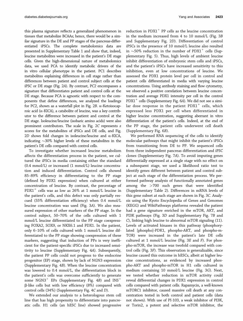
this plasma signature reflects a generalized phenomenon intissues that metabolize BCAAs; hence, there would be a sim-ilar signature in the DE and PP stages of the pancreas-differ-entiated iPSCs. The complete metabolomics data arepresented in Supplementary Table 1 and show that, indeed,leucine metabolites were increased in the patient’s DE stagecells. Given the high-dimensional nature of metabolomicsdata, we used PCA to identify metabolic drivers of thein vitro cellular phenotype in the patient. PC1 describesmetabolites explaining differences in cell stage rather thandifferences between patient and control subject cells at theiPSC or DE stage (Fig. 2A). By contrast, PC2 encompasses asignature that differentiates patient and control cells at theDE stage. Because PCA is agnostic with respect to the com-ponents that define differences, we analyzed the loadingsfor PC2, shown as a waterfall plot in Fig. 2B. a-Ketoisocap-roic acid (a-KICA), a catabolite of leucine, was a key contrib-utor to the difference between patient and control at theDE stage. Isoleucine/leucine (isobaric amino acids) were alsoprominent contributors. The scale in Fig. 2C represents zscores for the metabolites of iPSCs and DE cells, and Fig.2D shows fold changes in isoleucine/leucine and a-KICA,indicating �30% higher levels of these metabolites in thepatient’s DE cells compared with control cells.
To investigate whether increased leucine metabolismaffects the differentiation process in the patient, we cul-tured the iPSCs in media containing either the standard(0.4 mmol/L) or increased (1 mmol/L) leucine concentra-tion and induced differentiation. Control cells showed85–89% efficiency in differentiating to the PP stage(defined by PDX1 expression) when cultured at eitherconcentration of leucine. By contrast, the percentage ofPDX11 cells was as low as 26% at 1 mmol/L leucine inthe patient’s cells, and this defect was only partially res-cued (55% differentiation efficiency) when 0.4 mmol/Lleucine concentration was used (Fig. 3A). We also mea-sured expression of other markers of the PP stage. In thecontrol subject, 50–70% of the cells cultured with 1mmol/L leucine differentiated to the PP stage coexpress-ing FOXA2, SOX9, or NKX6.1 and PDX1. In the patient,only 6–10% of cells cultured with 1 mmol/L leucine dif-ferentiated to the PP stage showing coexpression of thesemarkers, suggesting that induction of PPs is very ineffi-cient for the patient-specific iPSCs due to increased sensi-tivity to leucine (Supplementary Fig. 4A). Subsequently,the patient PP cells could not progress to the endocrineprogenitor (EP) stage, shown by lack of NGN3 expression(Supplementary Fig. 4B). When the leucine concentrationwas lowered to 0.4 mmol/L, the differentiation block inthe patient’s cells was overcome sufficiently to generatesome NGN31 EPs (Supplementary Fig. 4B) and INS1
b-like cells but with low efficiency (8%) compared withcontrol cells (34%) (Supplementary Fig. 4C and D).
We extended our analysis to a heterologous stem cellline that has high propensity to differentiate into pancre-atic cells. H1 cells (an hESC line) showed progressive
reduction in PDX11 PP cells as the leucine concentrationin the medium increased from 4 to 10 mmol/L (Fig. 3Band Supplementary Fig. 2D). Differentiation of controliPSCs in the presence of 10 mmol/L leucine also resultedin �50% reduction in the number of PDX11 cells (Sup-plementary Fig. 5). Thus, high levels of ambient leucineinhibit differentiation of embryonic stem cells and iPSCs,and the patient’s iPSCs have increased sensitivity to thisinhibition, even at low concentrations of leucine. Weassessed the PDX1 protein level per cell in control andpatient cells differentiated in media with varying leucineconcentrations. Using antibody staining and flow cytometry,we observed a positive correlation between leucine concen-tration and average PDX1 intensity per cell in the controlPDX11 cells (Supplementary Fig. 6A). We did not see a simi-lar dose response in the patient PDX11 cells, whichexpressed less PDX1 per cell when differentiated inhigher leucine concentration, suggesting aberrant in vitrodifferentiation of the patient’s cells. Indeed, at the end ofthe PP stage, the patient cells underwent cell death(Supplementary Fig. 6B).
We performed RNA sequencing of the cells to identifymolecular pathways that might inhibit the patient’s iPSCsfrom transitioning from DE to PP. We sequenced cellsfrom three independent pancreas differentiation and iPSCclones (Supplementary Fig. 7A). To avoid imputing genesdifferentially expressed at a single stage with no effect ona subsequent stage, we used a likelihood ratio test toidentify genes different between patient and control sub-ject at each stage of the differentiation process. We per-formed pathway analysis of functionally annotated genesamong the >700 such genes that were identified(Supplementary Table 2). Differences in mRNA levels ofthis gene subset at each stage are shown in Fig. 3C. Analy-sis using the Kyoto Encyclopedia of Genes and Genomes(KEGG) and WikiPathways platforms revealed the patienthad a gene signature enriched in the mTOR, AKT, andPI3K pathways (Fig. 3D and Supplementary Fig. 7B andC), linking high leucine to abnormal mTOR signaling (11).Levels of activated kinases in this pathway (phosphory-lated [phospho]-PDK1, phospho-AKT, and phospho-m-TOR) were increased in the patient’s late DE cellscultured at 1 mmol/L leucine (Fig. 3E and F). For phos-pho-mTOR, the increase was twofold compared with con-trol cells (Fig. 3F). This observation is generalizable, sinceleucine caused this outcome in hESCs, albeit at higher leu-cine concentrations, as evidenced by increased phos-pho-AKT and phospho-mTOR in H1 cells cultured inmedium containing 10 mmol/L leucine (Fig. 3G). Next,we tested whether reduction in mTOR activity couldreveal differential changes in PDX1 expression in controlcells compared with patient cells. Rapamycin, a well-knownmTORC1 inhibitor, caused massive cell death at any con-centration tested in both control and patient cells (datanot shown). With use of PI-103, a weak inhibitor of PI3K,or Torin2, a potent and selective mTOR inhibitor, the
diabetes.diabetesjournals.org Yang and Associates 2423

control cells demonstrated a dose-dependent increase inPDX1 expression level per cell, whereas the patient cellsdemonstrated a small but significant decrease in Pdx1expression level per cell (Supplementary Fig. 8). Again,these data suggest that the patient’s cells at the PP stage
are functionally aberrant and do not represent true pancre-atic progenitor cells.
We next tested whether the 50-UTR variant in PDX1 isnecessary and sufficient to confer the unique sensitivity ofthe patient’s iPSC cells to leucine-induced differentiation
Figure 2—Leucine metabolism is altered in DE cells derived from patient iPSCs. A: PCA of control and patient cells at the iPSC and DEstages; dimension reduction using PCA of the metabolomics data shows that PC2 separates patient and control cells at the DE stage. n52. B: Rank ordering of the PC2 loadings shows prominent signals of a-KICA and iso/leucine. C: Row-normalized heat map of all metabo-lites confirms higher level of leucine and isoleucine in patient DE cells compared with control DE cells. D: Normalized iso/leucine anda-KICA levels in control and patient iPSCs and DE indicate higher content of these metabolites in patient DE. n5 2.
2424 KPD Defect Identified Using Patient Stem Cells Diabetes Volume 70, October 2021

inhibition. CRISPR/Cas9-mediated correction of the variantin the patient’s iPSCs (Fig. 4A and B) increased the expres-sion of PDX1, SOX9, and NKX6.1, key markers of the PPstage, when cultured in a medium containing 1 mmol/L leu-cine (Fig. 4C). After correction, quantification of the PDX11
cells showed a level of PDX1 expression in the patient’s cells
similar to that of the control cells (Fig. 4D). Furthermore,when corrected patient cells were differentiated through theendocrine lineage to b-like cells, we saw an efficiency ofinduction to CHGA1 and INS1 cells similar to that for con-trol iPSCs (Fig. 4E). The reverse experiment was performedto determine whether the variant-induced sensitivity to
Figure 3—(PDX1)c.�18 C>T confers increased susceptibility to leucine-mediated inhibition of differentiation to pancreatic progenitors. A:PDX1 expression induction in patient iPSCs is lower than that of control iPSCs when differentiated in media containing 1 mmol/L leucineand still lower when differentiated in media containing 0.4 mmol/L leucine. PDX1 expression is higher and unchanged at either leucineconcentration in control iPSCs. B: High leucine levels (4–10 mmol/L) impair PDX1 expression in H1 hESC-derived PP cells. C: Heat map ofmRNA levels of the top 100 of �700 genes statistically different between patient and control cells across three stages (iPSC, DE, and PP),with use of a likelihood ratio test. D: KEGG pathway analysis confirms the enrichment of the PI3K-AKT pathway in the patient’s cells. E:Phospho-PDK1, -AKT, and -mTOR levels are increased in the patient’s late DE cells compared with the control subject’s late DE cellswhen cultured with 1 mmol/L leucine in the medium. b-Actin was used as loading control. F: Quantification of immunoblots from E con-firms increased levels of phospho-PDK1, phospho-AKT, and phospho-mTOR in the patient’s late DE cells. n 5 4. G: High leucine levelscause increased phosphorylation of mTOR and AKT in H1 hESC-derived late DE cells. C, control subject; P, patient; p-, phosphorylated.
diabetes.diabetesjournals.org Yang and Associates 2425

Figure 4—Correction of the 50-UTR PDX1 variant in patient iPSCs rescues differentiation potential at the PP stage, and de novo knock-inof the 50-UTR variant in control iPSCs recapitulates the abnormal phenotype. A: Scheme of CRISPR/Cas9-mediated gene editing to cor-rect the nucleotide variant in the �18 position of the PDX1 50-UTR in the patient’s cells. B: Restriction enzyme digestion of PCR productsshows that the pattern of fragments cut by BaeG1 in the corrected PDX1 50-UTR sequence differs from that in the patient’s originalsequence. C: Correction of the PDX1 50-UTR variant restores efficient induction of PDX11, NKX6.11, and SOX91 PP cells from iPSCs.Representative flow cytometry plots show the percentage of PDX11/NKX6.11 and PDX11/SOX91 cells at the PP stage in the original (P)compared with the corrected (P-corrected) patient iPSCs. D: Quantification of PP cells confirms restoration of percent PDX11 cells inpatient-corrected (P-corr) cells to the level of control subject (C) cells. n5 6. E: Quantification of CHGA1 and INS1 cells shows similar effi-ciency of endocrine cell induction from patient-corrected cells compared with control cells. n 5 6. F: Scheme of strategy to introduce thepatient-specific variant (c.�18C.T) into the PDX1 50-UTR of HUES8 cells expressing inducible Cas9. G: ICE analysis of CRISPR editingevents with Sanger sequencing of genomic DNA shows 1% of targeted cells have homology-directed repair and the c.�18C>T substitu-tion. H: Introduction of the c.�18C>T PDX1 variant in wild-type HUES8 hESC leads to defective induction of PDX1 expression duringin vitro pancreas differentiation. Representative plot, n5 4. mt, mutant; P, patient; WT, wild type.
2426 KPD Defect Identified Using Patient Stem Cells Diabetes Volume 70, October 2021

leucine is specific to the patient’s cellular background or ageneralizable phenomenon. We used the iCas9 wild-typehESC line, HUES8, with an inducible Cas9 system underdoxycycline control (12) to introduce the �18 C>T variantin PDX1. We then performed single cell colony expansionwith genomic sequencing of PCR products to identify cellscontaining the PDX1 variant (Fig. 4F and G) in only one ofthe two alleles. Introducing this variant into a different cel-lular background produced the same differentiation defect;the number of PDX11 cells decreased to less than one-thirdof the number generated from PP-stage cells with wild-typePDX1 cultured with 1 mmol/L leucine (Fig. 4H). Thus, thePDX1 50-UTR variant that inhibits differentiation also con-fers leucine sensitivity, leading to a severe differentiationdefect in the transition from DE to PP.
Next, we investigated the mechanism whereby the �18bp 50-UTR variant impacts PDX1 expression during iPSC-directed differentiation. Using immunofluorescence, wenoted fewer PDX11 cells in differentiating patient cellsthan in differentiating control cells. We confirmed lowertotal PDX1 protein level in patient cells compared withcontrol subject cells by immunoblot analysis (Fig. 5A). Wethen investigated Pdx1 protein level per cell using anti-body staining and flow cytometry and determined thatthe patient’s PDX11 cells express less protein; thus, thereis less robust PDX1+ induction in the patient’s cells notonly at the population level but also at the individual celllevel (Fig. 5B). We then analyzed the wild-type DNAsequence around the 50-UTR region where the �18 bpvariant resides and found a binding motif for the tran-scriptional regulator protein NF-YA (which absolutelyrequires the 5-nt sequence CCAAT) and is part of the NF-Y trimeric protein complex (13,14) (Fig. 5C). The NF-Ycomplex behaves as a pioneer factor that regulates cellstate transitions during differentiation by creating acces-sible chromatin when a cell type–specific master regulatorlike PDX-1 is required for expression (15). Specifically, thetransactivation domain-containing NF-YA subunit bindsthe CCAAT sequence, whereas NF-YB and NF-YC bind thehistone fold, likely contributing the pioneer factor func-tion of the trimeric complex (16,17). Electromobility shiftassay revealed that NF-YA does indeed bind to the wild-type DNA sequence in vitro but does not bind to themutant DNA sequence (CTAAT) (Fig. 5D). We hypothe-sized that the NF-Y complex behaves as an activator ofthe Pdx1 promoter; hence, loss of NF-YB would decreaseNF-YA binding to the wild-type sequence beyond thetranscription start site and decrease wild-type PDX1 tran-scription. We therefore knocked down NF-YB in the con-trol cells and found that, indeed, PDX1 expressiondecreased, even with a limited (�20%) knockdown of NF-YB (Fig. 5E and F). In patient cells, the effect of theknockdown was allele dependent in the heterozygousstate; we saw no decrease in the expression of the mutantPDX1 allele but decreased expression of the wild-typePDX1 allele. We then performed flow cytometry and saw
the same effect of partial NF-YB knockdown on PDX1 pro-tein expression (Fig. 5G). Collectively, these experimentssuggest that transcriptional regulation of PDX1 is affectedin the patient’s cells due to the 50-UTR mutant. The patient-specific C>T alteration within the binding site for NF-Y dis-rupts NF-YA binding, displacing a transcriptional regulatorof PDX1 and decreasing PDX1 expression.
DISCUSSION
Altered BCAA metabolism plays an important role in thepathogenesis of T2D (11,18), and we have shown thataccelerated leucine catabolism, fueling ketone production,is a biochemical hallmark of KPD patients (4). The pre-sent data show that exposure to high levels of leucinecauses a PP differentiation defect in both this KPDpatient’s iPSCs and a heterologous ESC line. Leucinehypercatabolism associated with mTOR hyperactivationlikely sets the stage for a PP differentiation defect, sincemultiple translational programs are overactivated byhyperphosphorylation of mTOR (19), including some thatinhibit pancreatic endocrine cell development (20). TheBernal-Mizrachi group demonstrated a clear link betweenleucine concentration and the development of pancreaticprogenitors, likely mediated by the mTOR pathway(21,22). In KPD patients, this defective leucine metabo-lism and mTOR signaling appear to render PP cells sensi-tive to leucine-mediated inhibition and induce b-celldysfunction that is usually not severe enough to causeearly-onset, complete, irreversible b-cell failure. In thispatient, an additional “hit”—a variant in PDX1 thatdiminishes PDX1 expression at a critical stage of endo-crine cell differentiation and augments sensitivity to theinimical effects of leucine catabolism on PP differentia-tion—caused him to lapse over time to complete b-cellfailure and presentation with DKA.
Diabetic phenotypes associated with other known var-iants in PDX1 manifest as either late-onset mild mono-genic diabetes if heterozygous (the rare maturity-onsetdiabetes of the young 4 syndrome [7]) or neonatal diabe-tes if homozygous (23–25). The heterozygous 50-UTR var-iant in this patient had slower and less dramatic effectson b-cell differentiation than those that underlie PDX1-associated neonatal diabetes. Variants in b-cell functiongenes that are not highly deleterious (early stops, splicingdefects), or inherited in a heterozygous fashion withsome rescue by the normal allele, may permit relativelynormal function for many years, manifesting clinical dis-ease only later in life. In contrast, highly deleterious orhomozygous variants give rise to disease early in life. Dif-ferences in phenotypic severity observed in iPSC diseasemodels in vitro and disease progression in vivo have beennoted previously (26). The patient’s late onset of non-insulin-requiring diabetes with subsequent progression tob-cell failure resembles the chronology of heterozygousPdx11/� mice that manifest progressive b-cell loss with
diabetes.diabetesjournals.org Yang and Associates 2427

increased stress during adulthood, resulting in �50%reduced b-cell mass at 1 year of age (27–29).
The clinical significance of this variant in the proximalpromoter region of PDX1 is underscored by our findingthat its minor allele frequency was 3.8% among AfricanAmericans and 1.4% among Hispanics in a cohort of 81patients with KPD and absent b-cell function (frequencies$3� higher than in race/ethnicity-matched control sub-jects) (30). Identification of a potentially pathogenic var-
iant in a b-cell–regulated gene with detailed multiomicsinvestigation of patient-specific iPSCs differentiated intothe pancreatic endocrine lineage is a promising approachto identify the mechanism of b-cell failure in patientswith complex, atypical forms of diabetes (31).
Acknowledgments. The authors are grateful for Dr. Danwei Huangfu(Memorial Sloane-Kettering Cancer Center) for providing the HUES8-iCas9 cellline. The authors thank Jessica Teaw, Dr. Vivekananda Shetty, and Chandra
Figure 5—(PDX1)c.�18 C>T mutation alters NF-Y transcription factor binding to the PDX1 promoter. A: Global expression of PDX1 islower in patient PP cells than in control subject PP cells as assessed by Western blot. n5 2 for control subject (C), n5 3 for patient (P) bio-logical repeats; blots were performed in two technical repeats. B: Violin plots of PDX1 signal intensities in individual PDX11 PP cells ana-lyzed by flow cytometry. PDX1 expression per cell is significantly decreased in patient compared with control subject cells. Solid line,median; dotted lines, 1st and 3rd quartiles. n 5 10,041 for C and 5,679 for P, each with three biological repeats. C: Scheme of NF-Y tran-scription factor family members (NF-YA and NF-YB) binding motif (factorbook.org) discovered as part of the Encyclopedia of DNA Ele-ments (ENCODE) project, which suggests potential regulation of PDX1 transcription by NF-Y family members and altered regulation bythe c.�18 C>T mutant allele. D: Electrophoretic mobility shift assay shows binding of nuclear extract (NE) to the 50-UTR sequence of thePDX1 gene (lane 2). The specific binding protein in the NE is NF-YA, as shown by appearance of a supershift band upon treatment withNF-YA–specific antibody (ab) (lane 3). Neither DNA-NE binding nor the supershift occurs with the mutant 50-UTR DNA (lanes 9 and 10).The supershift band is not present with negative control anti-PDX1 (lanes 4 and 11) and anti–USF-1 (lanes 5 and 12) antibodies. “Cold”(lanes 6–7 and 13–14) indicates competition with unlabeled oligonucleotides. E: qPCR results showing silencing of the NFYB transcript inpatient PP cells (n 5 6) 48 h after anti-NFYB siRNA or control siRNA delivery. NRQ, normalized relative quantity. F: qPCR results showingdecrease in PDX1 wild-type (Wt) allele transcripts in control (n 5 3) and patient (n 5 6) PP cells 48 h after anti-NFYB siRNA or controlsiRNA delivery. The mutant allele (PDX1-mut) appears unaffected. G: Violin plots of PDX1 signal intensities in individual PDX11 PP cellsanalyzed by flow cytometry. PDX1 expression per cell is significantly decreased in siNFYB compared with siCTRL-treated cells, in bothcontrol subject and patient cells, 72 h post–siRNA delivery. n 5 3,875 and 2,502 for C siNFYB and siCTRL, respectively, and n 5 6,294and 2,837 for P siNFYB and siCTRL. Analyzed cells are from three biological repeats.
2428 KPD Defect Identified Using Patient Stem Cells Diabetes Volume 70, October 2021

Ambati (Baylor College of Medicine) for mass spectrometry support; AgnieszkaWojtkowiak-Szlachcic (Adam Mickiewicz University) for assistance with karyotyp-ing; Edyta Urbaniak (Adam Mickiewicz University) for technical support duringrevision; and Dr. Katherine Sippel (Baylor College of Medicine) for expert editing.Funding. This work was supported by funds from the Rutherford Chair inDiabetes Research (to A.B.) and the McNair Medical Institute Scholar’s Pro-gram, National Institutes of Health grant P30-DK079638, Polish National
Science Center (NCN) Polonez program 15/19/P/NZ3/03452, and EuropeanUnion Horizon 2020 and Marie Skłodowska-Curie Actions grant 665778 (all toM.B.). This project was carried out within the TEAM program of the Founda-
tion for Polish Science co-financed by the European Union under the Euro-pean Regional Development Fund (POIR.04.04.00-00-20C5/16). TheMetabolomics Core was supported by a Cancer Prevention and ResearchInstitute of Texas “Proteomic and Metabolomic Core Facility” Support Award
(RP170005), National Cancer Institute Cancer Center Support GrantP30CA125123, and intramural funds from the Dan L. Duncan Cancer Center.Duality of Interest. No potential conflicts of interest relevant to this
article were reported.Author Contributions. D.Y., W.J.S., J.C., and M.B. performed allexperiments related to the iPSCs. D.S. and M.M. performed the initial geneticstudies of KPD patients. N.P. and A.S. performed the metabolomics assays.
J.S. performed the skin biopsies. S.P. performed all bioinformatics and path-way analyses. A.B. diagnosed and classified the KPD patient and with M.B.developed the patient-specific iPSC study protocol. S.P., A.B., and M.B. per-
formed the primary analyses of the data. S.P., A.B., and M.B. drafted themanuscript, which was reviewed, edited, and approved by all the coauthors.A.B. and M.B. are the guarantors of this work and, as such, had full accessto all the data in the study and take responsibility for the integrity of the data
and the accuracy of the data analysis.
References
1. Balasubramanyam A, Nalini R, Hampe CS, Maldonado M. Syndromes ofketosis-prone diabetes mellitus. Endocr Rev 2008;29:292–3022. Maldonado M, Hampe CS, Gaur LK, et al. Ketosis-prone diabetes:
dissection of a heterogeneous syndrome using an immunogenetic and beta-cell functional classification, prospective analysis, and clinical outcomes. JClin Endocrinol Metab 2003;88:5090–50983. Mauvais-Jarvis F, Sobngwi E, Porcher R, et al. Ketosis-prone type 2
diabetes in patients of sub-Saharan African origin: clinical pathophysiologyand natural history of beta-cell dysfunction and insulin resistance. Diabetes2004;53:645–6534. Patel SG, Hsu JW, Jahoor F, et al. Pathogenesis of A�b1 ketosis-prone
diabetes. Diabetes 2013;62:912–9225. Nalini R, Ozer K, Maldonado M, et al. Presence or absence of a known
diabetic ketoacidosis precipitant defines distinct syndromes of "A-b1"ketosis-prone diabetes based on long-term b-cell function, human leukocyteantigen class II alleles, and sex predilection. Metabolism 2010;59:1448–14556. Haaland WC, Scaduto DI, Maldonado MR, et al. A�b� subtype of
ketosis-prone diabetes is not predominantly a monogenic diabetic syndrome.Diabetes Care 2009;32:873–8777. Holland AM, Hale MA, Kagami H, Hammer RE, MacDonald RJ. Experimental
control of pancreatic development and maintenance. Proc Natl Acad Sci U S A2002;99:12236–122418. Stoffers DA, Ferrer J, Clarke WL, Habener JF. Early-onset type-II diabetes
mellitus (MODY4) linked to IPF1. Nat Genet 1997;17:138–1399. Borowiak M, Melton DA. How to make beta cells? Curr Opin Cell Biol
2009;21:727–73210. Sneddon JB, Borowiak M, Melton DA. Self-renewal of embryonic-stem-
cell-derived progenitors by organ-matched mesenchyme. Nature 2012;491:765–768
11. Newgard CB, An J, Bain JR, et al. A branched-chain amino acid-relatedmetabolic signature that differentiates obese and lean humans and contributesto insulin resistance. Cell Metab 2009;9:311–32612. Gonz�alez F, Zhu Z, Shi ZD, et al. An iCRISPR platform for rapid,multiplexable, and inducible genome editing in human pluripotent stem cells.Cell Stem Cell 2014;15:215–22613. Dorn A, Bollekens J, Staub A, Benoist C, Mathis D. A multiplicity ofCCAAT box-binding proteins. Cell 1987;50:863–87214. Mantovani R. A survey of 178 NF-Y binding CCAAT boxes. Nucleic AcidsRes 1998;26:1135–114315. Oldfield AJ, Yang P, Conway AE, et al. Histone-fold domain protein NF-Ypromotes chromatin accessibility for cell type-specific master transcriptionfactors. Mol Cell 2014;55:708–72216. Nardini M, Gnesutta N, Donati G, et al. Sequence-specific transcriptionfactor NF-Y displays histone-like DNA binding and H2B-like ubiquitination. Cell2013;152:132–14317. Huber EM, Scharf DH, Hortschansky P, Groll M, Brakhage AA. DNA minorgroove sensing and widening by the CCAAT-binding complex. Structure2012;20:1757–176818. Hern�andez-Alvarez MI, D�ıaz-Ramos A, Berdasco M, et al. Early-onset andclassical forms of type 2 diabetes show impaired expression of genes involvedin muscle branched-chain amino acids metabolism. Sci Rep 2017;7:1385019. Son SM, Park SJ, Lee H, et al. Leucine signals to mTORC1 via itsmetabolite acetyl-coenzyme A. Cell Metab 2019;29:192–201.e720. Rachdi L, Aïello V, Duvilli�e B, Scharfmann R. L-leucine alters pancreaticb-cell differentiation and function via the mTor signaling pathway. Diabetes2012;61:409–41721 Alejandro EU, Bozadjieva N, Blandino-Rosano M, et al. Overexpression ofkinase-dead mTOR impairs glucose homeostasis by regulating insulinsecretion and not b-cell mass. Diabetes 2017;66:2150–216222. Elghazi L, Blandino-Rosano M, Alejandro E, Cras-M�eneur C, Bernal-Mizrachi E. Role of nutrients and mTOR signaling in the regulation ofpancreatic progenitors development. Mol Metab 2017;6:560–57323. Nicolino M, Claiborn KC, Sen�ee V, Boland A, Stoffers DA, Julier C. A novelhypomorphic PDX1 mutation responsible for permanent neonatal diabetes withsubclinical exocrine deficiency. Diabetes 2010;59:733–74024. De Franco E, Shaw-Smith C, Flanagan SE, et al. Biallelic PDX1 (insulinpromoter factor 1) mutations causing neonatal diabetes without exocrinepancreatic insufficiency. Diabet Med 2013;30:e197–e20025. Caetano LA, Santana LS, Costa-Riquetto AD, et al. PDX1 -MODY anddorsal pancreatic agenesis: new phenotype of a rare disease. Clin Genet2018;93:382–38626. Shi ZD, Lee K, Yang D, et al. Genome editing in hPSCs reveals GATA6haploinsufficiency and a genetic interaction with GATA4 in human pancreaticdevelopment. Cell Stem Cell 2017;20:675–688.e627. Ahlgren U, Jonsson J, Edlund H. The morphogenesis of the pancreaticmesenchyme is uncoupled from that of the pancreatic epithelium in IPF1/PDX1-deficient mice. Development 1996;122:1409–141628. Ahlgren U, Jonsson J, Jonsson L, Simu K, Edlund H. beta-cell-specificinactivation of the mouse Ipf1/Pdx1 gene results in loss of the beta-cellphenotype and maturity onset diabetes. Genes Dev 1998;12:1763–176829. Zhu Z, Li QV, Lee K, et al. Genome editing of lineage determinants inhuman pluripotent stem cells reveals mechanisms of pancreatic developmentand diabetes. Cell Stem Cell 2016;18:755–76830. Scaduto D, Ozer K, Nalini R, et al. Characterization of a novel 50-UTRputative CCAAT-box variant in the human PDX-1 gene promoter withina ketosis-prone diabetes (KPD) cohort (Abstract). Diabetes 2011;60(Suppl. 1)31. Cardenas-Diaz FL, Osorio-Quintero C, Diaz-Miranda MA, et al.Modeling monogenic diabetes using human ESCs reveals developmentaland metabolic deficiencies caused by mutations in HNF1A. Cell Stem Cell2019;25:273–289.e5
diabetes.diabetesjournals.org Yang and Associates 2429