Detection of Intracellular Proteins by Flow Cytometry
44
Detection of Intracellular Proteins by Flow Cytometry Sara Johansson, PhD BD European Scientific Support
Transcript of Detection of Intracellular Proteins by Flow Cytometry

Detection of Intracellular Proteinsby Flow Cytometry
Sara Johansson, PhDBD European Scientific Support
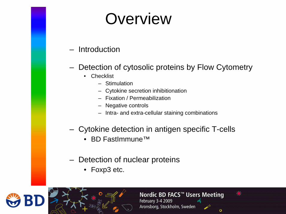
Overview
– Introduction
– Detection of cytosolic proteins by Flow Cytometry• Checklist
– Stimulation– Cytokine secretion inhibitionation– Fixation / Permeabilization– Negative controls– Intra- and extra-cellular staining combinations
– Cytokine detection in antigen specific T-cells• BD FastImmune™
– Detection of nuclear proteins• Foxp3 etc.
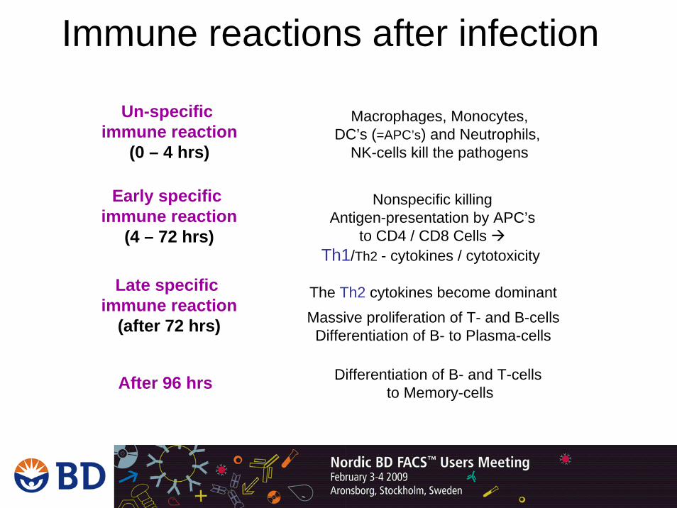
Immune reactions after infection
Un-specific immune reaction
(0 – 4 hrs)
Macrophages, Monocytes,DC’s (=APC’s) and Neutrophils,
NK-cells kill the pathogens
Early specific immune reaction
(4 – 72 hrs)
Nonspecific killingAntigen-presentation by APC’s
to CD4 / CD8 Cells Th1/Th2 - cytokines / cytotoxicity
Late specific immune reaction
(after 72 hrs)
The Th2 cytokines become dominantMassive proliferation of T- and B-cellsDifferentiation of B- to Plasma-cells
After 96 hrs Differentiation of B- and T-cells to Memory-cells

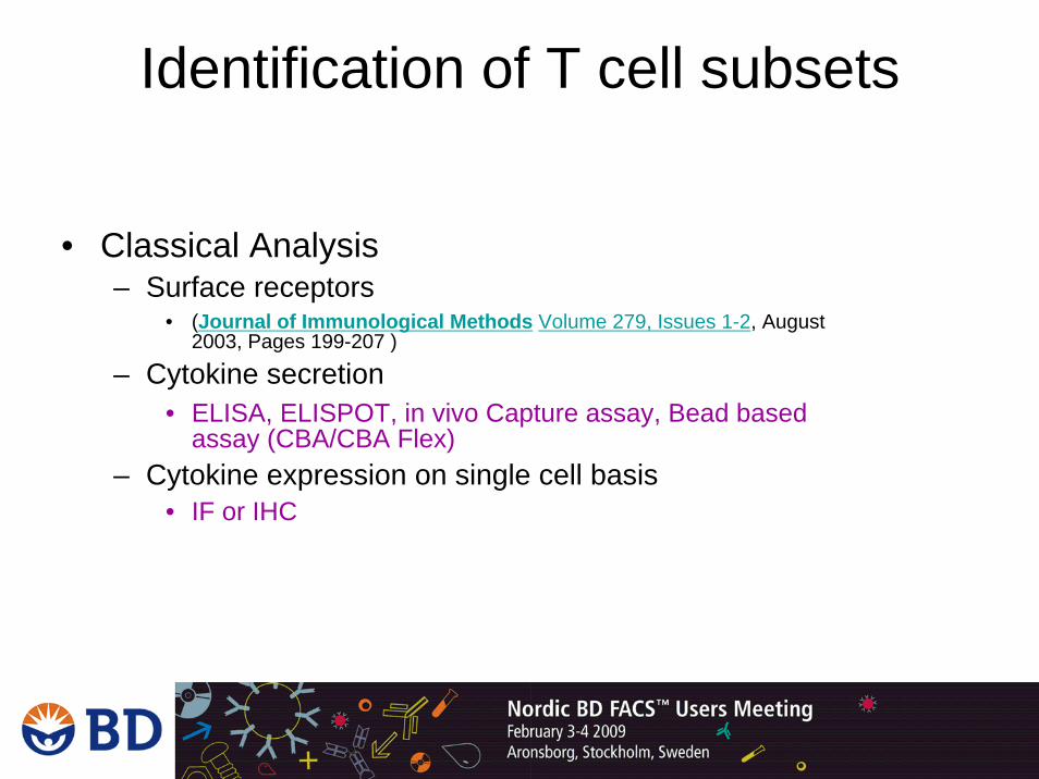
Identification of T cell subsets
• Classical Analysis – Surface receptors
• (Journal of Immunological Methods Volume 279, Issues 1-2, August 2003, Pages 199-207 )
– Cytokine secretion• ELISA, ELISPOT, in vivo Capture assay, Bead based
assay (CBA/CBA Flex)– Cytokine expression on single cell basis
• IF or IHC

Detection of Cytosolic Protein-Cytokines-
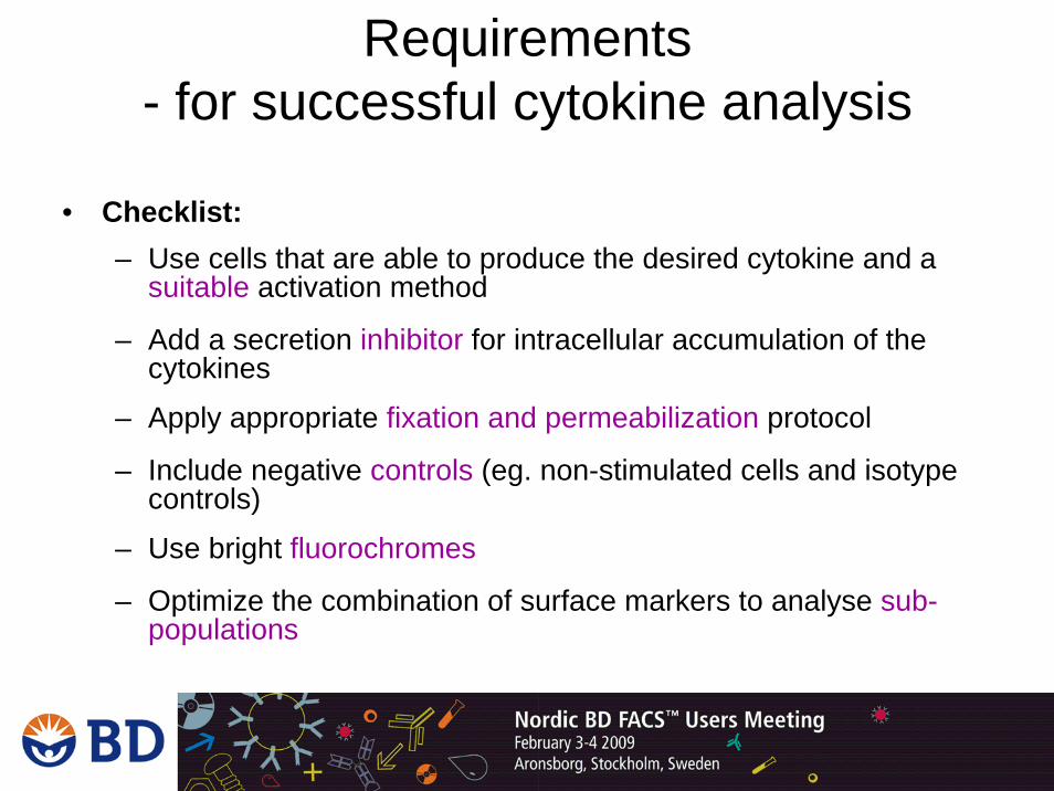
Requirements - for successful cytokine analysis
• Checklist:– Use cells that are able to produce the desired cytokine and a
suitable activation method
– Add a secretion inhibitor for intracellular accumulation of the cytokines
– Apply appropriate fixation and permeabilization protocol
– Include negative controls (eg. non-stimulated cells and isotype controls)
– Use bright fluorochromes
– Optimize the combination of surface markers to analyse sub-populations
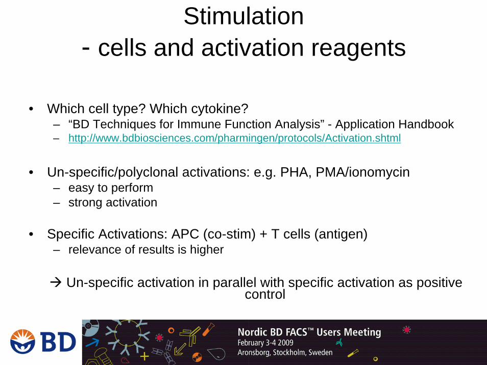
Stimulation- cells and activation reagents
• Which cell type? Which cytokine?– “BD Techniques for Immune Function Analysis” - Application Handbook– http://www.bdbiosciences.com/pharmingen/protocols/Activation.shtml
• Un-specific/polyclonal activations: e.g. PHA, PMA/ionomycin– easy to perform – strong activation
• Specific Activations: APC (co-stim) + T cells (antigen)– relevance of results is higher
Un-specific activation in parallel with specific activation as positive control
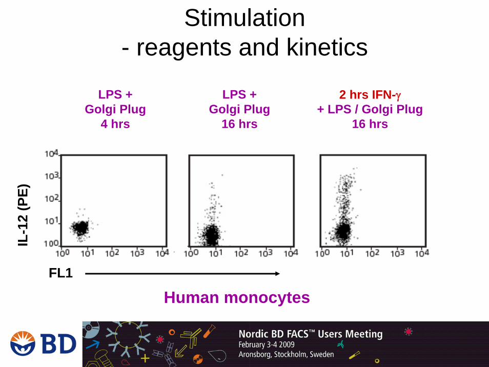
Stimulation- reagents and kinetics
2 hrs IFN-γ+ LPS / Golgi Plug
16 hrs
Human monocytes
IL-1
2 (P
E)
FL1
LPS +Golgi Plug
4 hrs
LPS +Golgi Plug
16 hrs

Stimulation- what BD offers
• Leukocyte activation cocktail– PMA + Ionomycin + GolgiPlug (Brefeldin A) (# 550583)
• Immobilized or soluble CD3 (NA/LE)– human = clones UCHT1 or HIT3a (# 555329 / 555336)– mouse = clone 145-2C11 (# 553057)
• Soluble CD28 (NA/LE)– human = clone 28.2 (# 555725)– mouse = clone 37.51 (# 553294)

Stimulation- what BD offers
• CD3 pre-coated plates– Human # 354725– Mouse # 354720– Control # 354730
• Cytokines used for cell activation– IFN-γ: 50 mg / ml (# 554617)– IL-2: 0.1 mg/ml (# 554603)– IL-4: 0.1 mg/ml (# 554605)– IL-6: 0.1 mg/ml (# 550071)
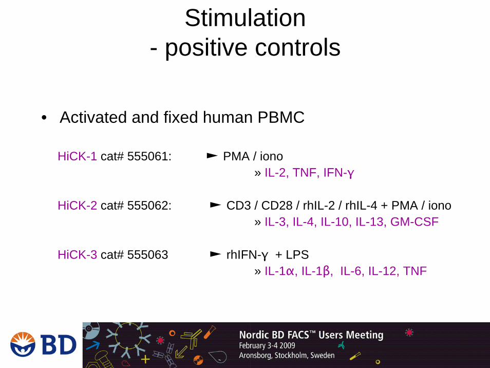
Stimulation - positive controls
• Activated and fixed human PBMC
HiCK-1 cat# 555061: ► PMA / iono » IL-2, TNF, IFN-γ
HiCK-2 cat# 555062: ► CD3 / CD28 / rhIL-2 / rhIL-4 + PMA / iono» IL-3, IL-4, IL-10, IL-13, GM-CSF
HiCK-3 cat# 555063 ► rhIFN-γ + LPS» IL-1α, IL-1β, IL-6, IL-12, TNF
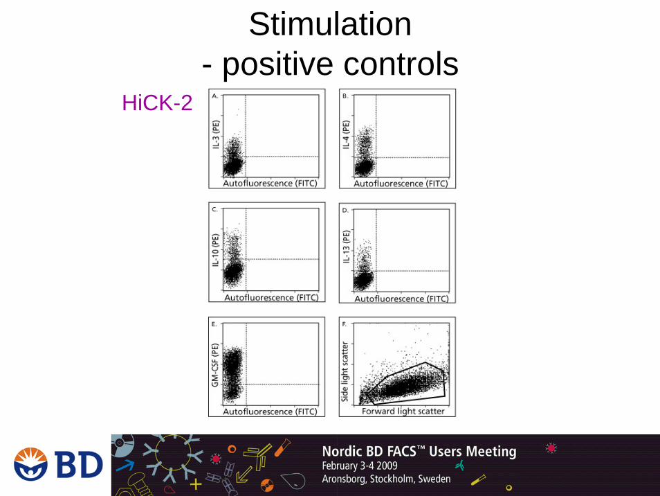
Stimulation- positive controls
HiCK-2

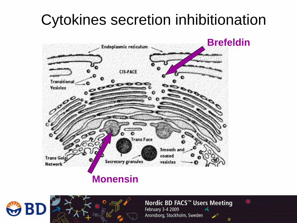
Cytokine secretion inhibition
• Cytokines are rapidly secreted upon expression which results inlow/not detectable intracellular concentrations
• For Flow cytometry analysis we block the secretion of the cytokines with:– Brefeldin A 1mg / ml in DMSO (BD GolgiPlug™ # 555029)
• 1 µg/million cells FINAL CONC.– Monensin 2mg / ml in EtOH (BD GolgiStop™ # 554724)
• 8 µg/million cells FINAL CONC.
• Secretion inhibitors are toxic. Exposure of cells should be as short as possible!
BD
Gol
giP
lug
Bre
feld
inA
BD
Gol
giS
top
Mon
ensi
n Superior Superior
Superior
Equal
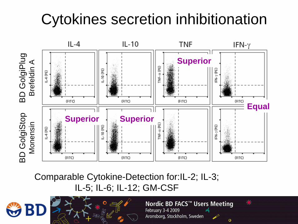
Comparable Cytokine-Detection for:IL-2; IL-3; IL-5; IL-6; IL-12; GM-CSF
Cytokines secretion inhibitionation
Cell fixation and permeabilization
• Fixation 3-4% Paraformaldehyde
• Permeabilization 0.05 – 0.2% Saponin (or other detergent) – Saponin is a reversible detergent– Required in all steps after permeabilization
• Washing steps• Antibody incubation

Cell fixation and permeabilization - what BD Offers
• BD Cytofix/Cytoperm™– Without protein inhibitionation # 554714– With Golgi Stop # 554715– With Golgi Plug # 555028
• BD Cytofix™ Buffer– Only fixation (# 554655)
• BD Perm/Wash™ Buffer– Only permeabilization (# 554723)
Negative controls - Isotype controls
• Compared to specific Ab’s, isotype controls have to be: – same species– same immunoglobulin type (IgG 1/2/3/4 or IgM) – same fluorochrome conjugate– purified from free fluorochromes– same concentration
• Compared to specific Ab’s, isotype controls can have: – same constant light chains (λ or κ) – same IgG2 type (IgG2a or IgG2b / ratIgG2c)

Negative controls- Fluorescence Minus One
• Un-stained cells or isotype controls are not always sufficient for determining positive vs. negative expression in multi-color experiments.
• An alternative is to stain cells with all reagents except the one of interest: FMO
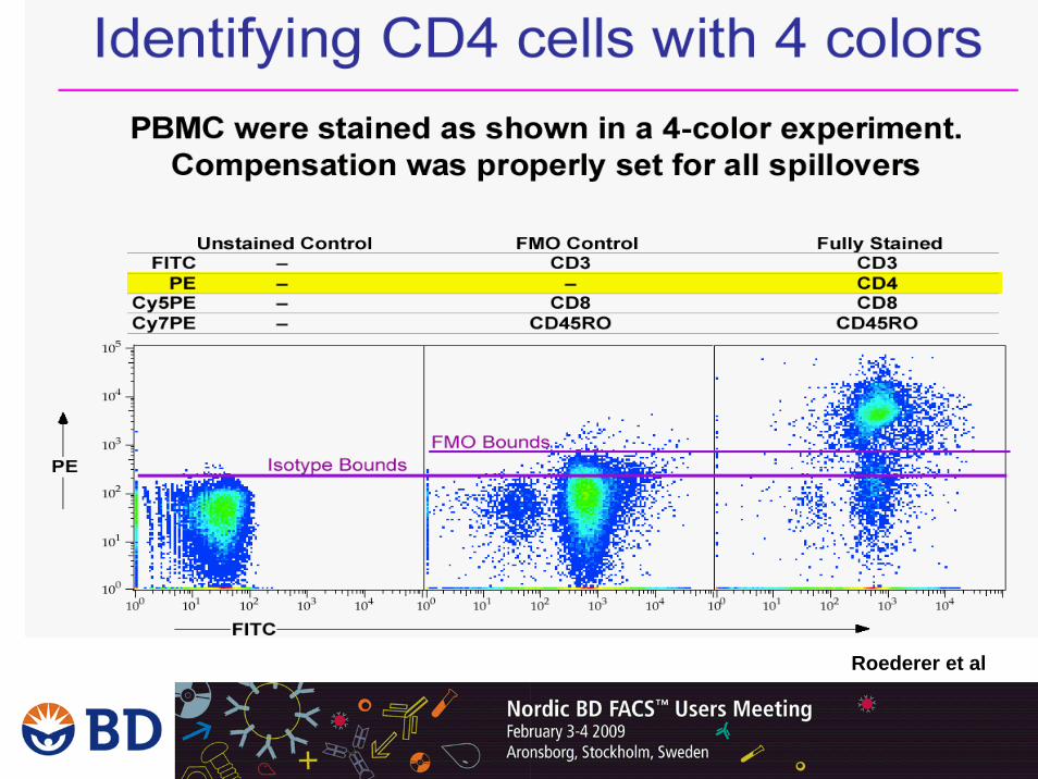
Fluorescence Minus One -FMO
Roederer et al

Fluorochrome selection - intra- and extra-cellular staining
• Fluorochromes:– Cytokines:
• PE, APC or Alexa Fluor 647 – Activation marker:
• APC, PE or Alexa Fluor 647• Note! inhibitionation also affects transport to surface
– Identification markers: • FITC, PerCP / PerCP-Cy5.5, Horizon V450, Pacific
Blue, APC-H7, APC-Cy7
• http://www.bdbiosciences.com/colors/
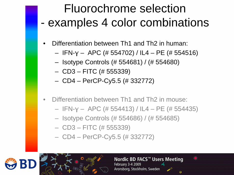
Fluorochrome selection - examples 4 color combinations• Differentiation between Th1 and Th2 in human:
– IFN-γ – APC (# 554702) / IL4 – PE (# 554516) – Isotype Controls (# 554681) / (# 554680)– CD3 – FITC (# 555339)– CD4 – PerCP-Cy5.5 (# 332772)
• Differentiation between Th1 and Th2 in mouse:– IFN-γ – APC (# 554413) / IL4 – PE (# 554435) – Isotype Controls (# 554686) / (# 554685)– CD3 – FITC (# 555339)– CD4 – PerCP-Cy5.5 (# 332772)
Cytokine Detection in Antigen Specific T cells
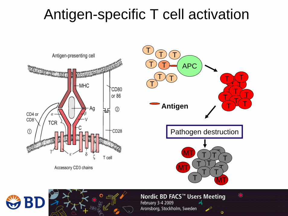
Antigen-specific T cell activation
T
Antigen
TT
T T
T
T
T
T APC
TT
T
TT
T T
Pathogen destruction
TTT TTT
MT
T
MT
MT
T T
TT
T
T

BD FastImmune™• Detection of antigen-specific immune reactions
in human T cell sub-populations
• Can be used for monitoring of immune status by detection of antigen-specific T cells – During disease – Testing of vaccine efficiency
• Optimized products and protocols for detection of activated T cell sub-populations of less than 0.1% of total T cells
• Test system for parallel detection of:– Cell surface markers (CD3; CD4; CD8)– Activation markers (CD69)– Immune reaction markers (Cytokines: IFN-γ,
etc.)
• Optimized for parallel intra- and extra-cellular Flow analysis
BD FastImmune™
• High concentrations of soluble Peptide-Mixes bind to TCR– Mimic specific APC – T cell interaction– Induce activation of T cells
• Cytokine production• Proliferation
• Problem - TCR contact without co-stimulation –> ANERGY
• Solution - soluble antigen (Peptide – Mixes) + co-stimulatory reagents
• Anti-CD28 • Anti-CD49d
• Positive control: Staphylococcus EnteroToxin B (SEB) 1ug/ml
BD FastImmune™- principle
• The multicolour combination:– Surface markers CD4 or CD8 in PerCP-Cy5.5 or
APC • Down-regulation of CD4 and CD8 in activated samples
– Activation marker CD69 in PE or APC• Expression inhibitioned by Brefeldin A in Activation
Cocktail
– IFN-γ / TNF / IL-2 in FITC• Some cytokines are highly expressed
BD FastImmune™

• Tested Peptide Mixes for antigen-specific activation of T Cells– CMV Peptide Mix (# 551969)– CMV-Lysate (ABI: 10-144000)– SEB positive Control (Sigma: S4881)
BD FastImmune™

1. Blood Draw 2. Incubation at 37 degree
Store blood at RT to avoid platelet activation Use within 8hrs after collection•100 ul/test•Only heparinized blood
Act
ivat
ed C
ells
Con
trol
cel
ls
BD FastImmune™- protocol
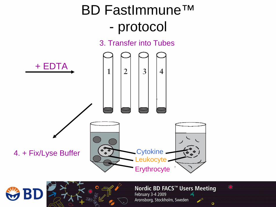
+ EDTA
3. Transfer into Tubes
CytokineLeukocyteErythrocyte
4. + Fix/Lyse Buffer
BD FastImmune™- protocol
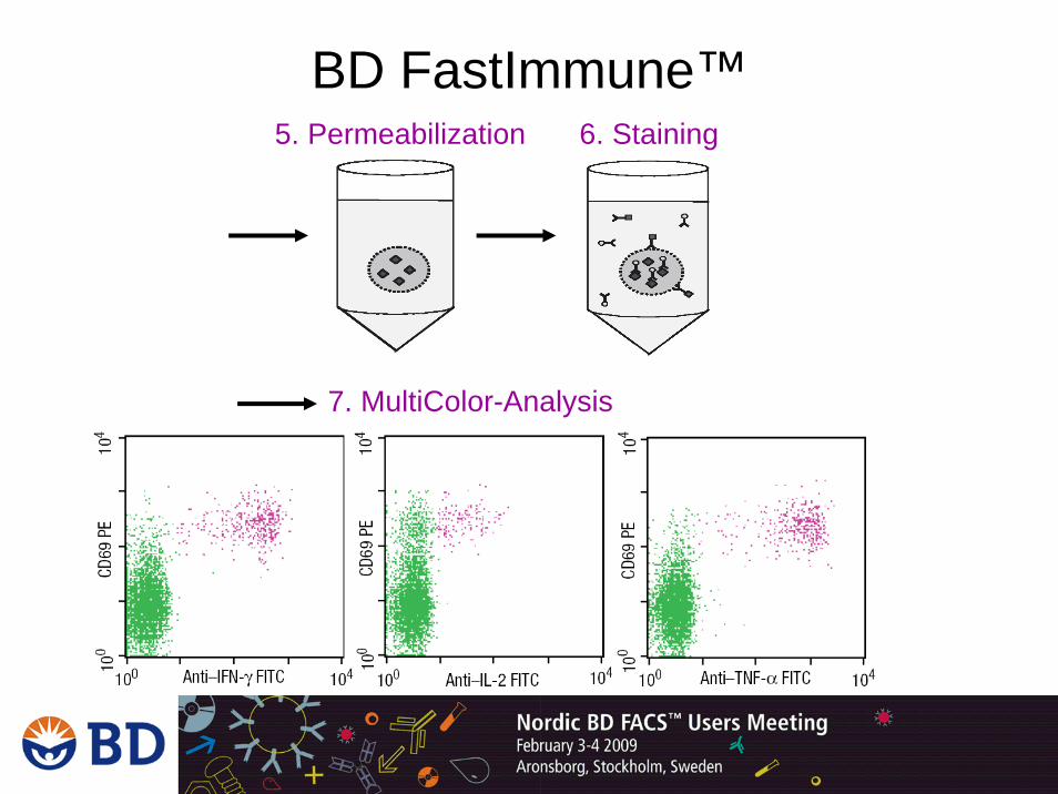
6. Staining5. Permeabilization
7. MultiColor-Analysis
BD FastImmune™
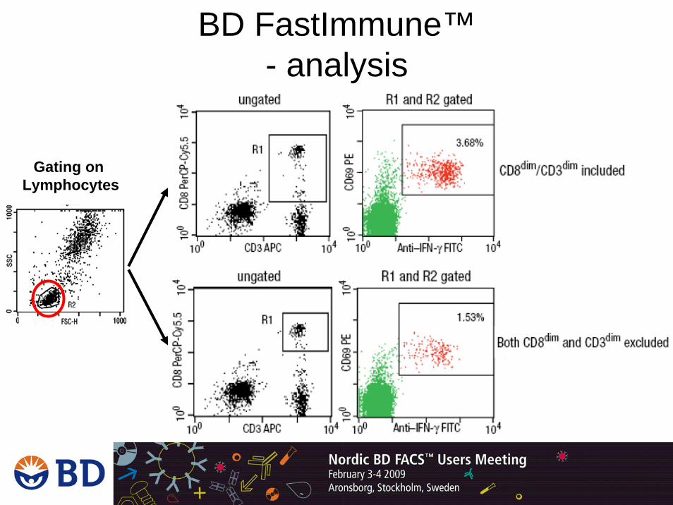
Gating on Lymphocytes
BD FastImmune™- analysis

• Special focus on antigen-specific T cell activation (CD4+ and CD8+) directly in human whole blood
• Anti CD28 / CD49d co-stimulatory reagent enables antigen specific activation in vitro
• Products are single reagents, sets and kits
• Can be combined with BD DimerX™ for detection of antigen specific CD8+ cells
BD FastImmune™- summary

Detection of Intranuclear Protein-FoxP3-
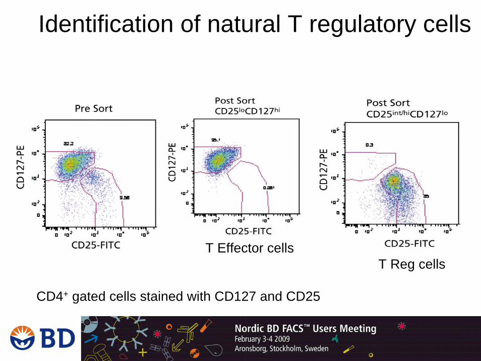
CD4+ gated cells stained with CD127 and CD25
T Effector cellsT Reg cells
Identification of natural T regulatory cells
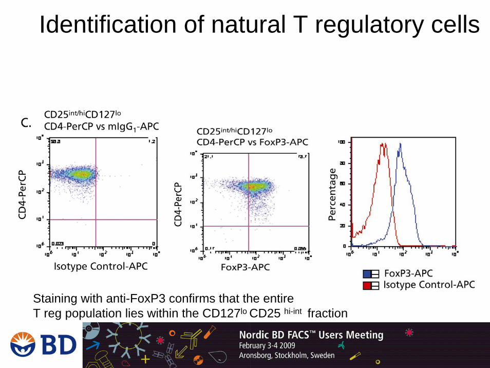
Staining with anti-FoxP3 confirms that the entireT reg population lies within the CD127lo CD25 hi-int fraction
Identification of natural T regulatory cells
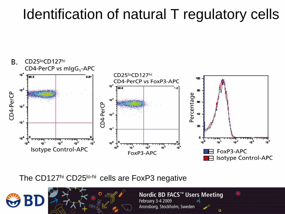
The CD127hi CD25lo-hi cells are FoxP3 negative
Identification of natural T regulatory cells
• Human Regulatory T Cell Cocktail– CD4-FITC, CD25-PE-Cy7, CD127-AF647 cat #
• Single colours FoxP3 antibodies:– Anti-FoxP3 clone 259D/C7 (PE, AF647 and AF488)
• Human FoxP3 Staining kits (different color combinations):– Anti-FoxP3 clone 259D/C7– Anti-CD4 clone RPA-T4– Anti-CD25 clone 2A3– Buffer Set
• Human FoxP3 Buffer Set
Identification of natural T regulatory cells- what BD offers
FoxP3 Staining- protocol
• Fix the cells in 2ml of Human FoxP3 Buffer A • Incubate 10 min at RT • Wash with 2ml of Staining Buffer (FBS)• Permeabilize the cells in 0.5ml of Human FoxP3 Buffer C• Incubate 30min at RT• Wash with 2ml of Staining Buffer (FBS)• Stain with anti-human FoxP3 in 100ul of Staining Buffer• Incubate 30min at RT• Wash and re-suspend in Staining Buffer (FBS) and re-suspend• Analysis
– Optional: add 300ul of 1% PFA and store at 4C, analyse within 24h
– Acquire at least 15,000 to 25,000 CD4 positive lymphocytes
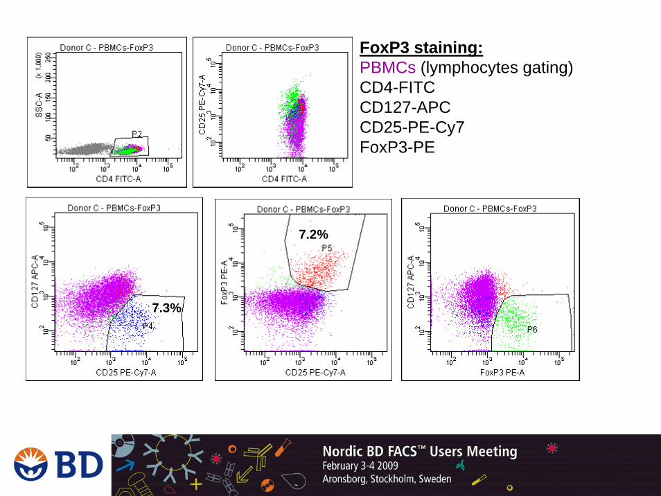
FoxP3 staining:PBMCs (lymphocytes gating)CD4-FITCCD127-APCCD25-PE-Cy7FoxP3-PE
7.2%
7.3%

FoxP3 staining:Whole Blood (lymphocytes gating)CD4-FITCCD127-APCCD25-PE-Cy7FoxP3-PE
7.0%
7.0%

BD European Scientific Support
Danmark (45) 80 882 193Suomi (358) 800 11 63 17Norge (47) 800 18530Sverige (46) 8 5069 2154