Staining Intracellular Antigens for Flow...
12
Flow Cytometry – BestProtocols® Page 1 of 12 Staining Intracellular Antigens for Flow Cytometry Research Use Only For additional questions, please contact Technical Support at +1-888-810-6168 (US) or +43 1 796 4040 120 (Europe/International), or send us an email at [email protected] ______________________________________________________________________________________ Revised 08-02-16 © Affymetrix, Inc. All rights reserved Tel: +1-888.999.1371 or +1-858.642.2058 • Fax: +1-858.642.2046 • ebioscience.affymetrix.com • [email protected] Protocol A: Two-step protocol: intracellular (cytoplasmic) proteins Protocol B: One-step protocol: intracellular (nuclear) proteins Protocol C: Two-step protocol for Fixation/Methanol Introduction A modification of the basic immunofluorescent staining and flow cytometric analysis protocol can be used for the simultaneous analysis of surface molecules and intracellular antigens at the single-cell level by flow cytometry. Typically, cells are fixed with formaldehyde to stabilize the cell membrane, and then permeabilized with detergent or alcohol to allow antibodies against intracellular antigens access to stain intracellularly. When staining proteins inside the cell, it is important to consider their location as this may dictate the protocol and buffer system that will perform optimally. For example, nuclear proteins and many secreted proteins work well with the Foxp3/Transcription Factor Staining Buffer Set (cat. no. 00-5523), while secreted proteins such as cytokines and chemokines work well with the Intracellular Fixation & Permeabilization Buffer Set (cat. no. 88-8824). Lastly, there are several phosphorylated signaling proteins that may not work in the two previously-mentioned buffer systems but will work with the Fixation/Methanol Protocol. Antibody performance in different buffer systems and protocols should be determined empirically. Please contact Technical Support ([email protected]) for more information. General Notes 1. For optimal performance of fluorochrome-conjugated antibodies, store vials at 4°C. Protect from light. Do not freeze. 2. Prior to use, quickly spin the antibody vial to recover the maximum volume. Vortexing the antibody vial is not recommended. 3. Except where noted in the protocol, all staining should be done on ice or at 4°C with minimal exposure to light. 4. The fixation and permeabilization steps that are required for the detection of intracellular antigens may alter the light scatter properties of cells and may increase non-specific background staining. Including extra protein such as BSA or fetal calf serum (FCS) in the staining buffer may help reduce non-specific background. The use of Fixable Viability Dyes (FVD) is recommended to help eliminate dead cells during the analysis.
Transcript of Staining Intracellular Antigens for Flow...
Flow Cytometry – BestProtocols® Page 1 of 12
Staining Intracellular Antigens for Flow Cytometry Research Use Only
For additional questions, please contact Technical Support at +1-888-810-6168 (US) or +43 1 796 4040 120 (Europe/International),
or send us an email at [email protected]
______________________________________________________________________________________ Revised 08-02-16
© Affymetrix, Inc. All rights reserved Tel: +1-888.999.1371 or +1-858.642.2058 • Fax: +1-858.642.2046 • ebioscience.affymetrix.com • [email protected]
Protocol A: Two-step protocol: intracellular (cytoplasmic) proteins Protocol B: One-step protocol: intracellular (nuclear) proteins Protocol C: Two-step protocol for Fixation/Methanol
Introduction A modification of the basic immunofluorescent staining and flow cytometric analysis protocol can be used for the simultaneous analysis of surface molecules and intracellular antigens at the single-cell level by flow cytometry. Typically, cells are fixed with formaldehyde to stabilize the cell membrane, and then permeabilized with detergent or alcohol to allow antibodies against intracellular antigens access to stain intracellularly. When staining proteins inside the cell, it is important to consider their location as this may dictate the protocol and buffer system that will perform optimally. For example, nuclear proteins and many secreted proteins work well with the Foxp3/Transcription Factor Staining Buffer Set (cat. no. 00-5523), while secreted proteins such as cytokines and chemokines work well with the Intracellular Fixation & Permeabilization Buffer Set (cat. no. 88-8824). Lastly, there are several phosphorylated signaling proteins that may not work in the two previously-mentioned buffer systems but will work with the Fixation/Methanol Protocol. Antibody performance in different buffer systems and protocols should be determined empirically. Please contact Technical Support ([email protected]) for more information.
General Notes
1. For optimal performance of fluorochrome-conjugated antibodies, store vials at 4°C. Protect from light. Do not freeze.
2. Prior to use, quickly spin the antibody vial to recover the maximum volume. Vortexing the antibody vial is not recommended.
3. Except where noted in the protocol, all staining should be done on ice or at 4°C with minimal exposure to light.
4. The fixation and permeabilization steps that are required for the detection of intracellular antigens may alter the light scatter properties of cells and may increase non-specific background staining. Including extra protein such as BSA or fetal calf serum (FCS) in the staining buffer may help reduce non-specific background. The use of Fixable Viability Dyes (FVD) is recommended to help eliminate dead cells during the analysis.

Flow Cytometry – BestProtocols® Page 2 of 12
Staining Intracellular Antigens for Flow Cytometry Research Use Only
For additional questions, please contact Technical Support at +1-888-810-6168 (US) or +43 1 796 4040 120 (Europe/International),
or send us an email at [email protected]
______________________________________________________________________________________ Revised 08-02-16
© Affymetrix, Inc. All rights reserved Tel: +1-888.999.1371 or +1-858.642.2058 • Fax: +1-858.642.2046 • ebioscience.affymetrix.com • [email protected]
Protocol A: Two-step protocol: intracellular (cytoplasmic) proteins The following protocol allows the simultaneous analysis of cell surface molecules and intracellular antigens at the single-cell level. In this protocol, fixation is followed by permeabilization resulting in the creation of pores in the cell membrane that require the continuous presence of the permeabilization buffer during all subsequent steps. This allows antibodies to have access to the cytoplasm of the cell. Thus, all intracellular staining must be done in the presence of the permeabilization buffer. This protocol is recommended for the detection of cytoplasmic proteins, cytokines, or other secreted proteins in individual cells following activation in vitro or in vivo. For cytokine detection, the appropriate stimulation conditions and kinetics of cytokine production will vary depending on the cell type and the particular cytokine being assayed. For example, to stimulate T cells to produce IFN-γ, TNF-α, IL-2, and IL-4, a combination of PMA (a phorbol ester, a protein kinase C activator) and Ionomycin (a calcium ionophore) or anti-CD3 antibodies can be used. To induce IL-6, IL-10, or TNF-α production by monocytes, stimulation with lipopolysaccharide (LPS) can be used. For in vitro stimulation of cells, it is necessary to block secretion of cytokines with protein transport inhibitors, such as Monensin or Brefeldin A Solution, during the final hours of the stimulation protocol. It is advised that investigators evaluate the use and efficacy of different protein transport inhibitors in their specific assay system. For the detection of nuclear proteins such as transcription factors, please see Protocol B: One-step protocol: intracellular (nuclear) proteins below. For detection of some phosphorylated signaling molecules such as MAPK and STAT proteins, it may be preferential to use Protocol C: Two-step protocol: Fixation/Methanol, below.
Materials
12x75 mm round bottom test tubes [Optional] Fixable Viability Dyes (FVD) eFluor® 455UV (cat. no. 65-0868), eFluor® 450
(cat. no. 65-0863), eFluor® 506 (cat.no 65-0866), eFluor® 660 (cat. no. 65-0864), or eFluor® 780 (cat. no. 65-0865)
Directly conjugated antibodies Intracellular Fixation & Permeabilization Buffer Set (cat. no. 88-8824) Flow Cytometry Staining Buffer (cat. no. 00-4222) Cell Stimulation Cocktail (plus protein transport inhibitors) (500X) (cat. no. 00-4975) or
Protein Transport Inhibitor Cocktail (500X) (cat. no. 00-4980) or Brefeldin A Solution (cat. no. 00-4506) or Monensin Solution (cat. no. 00-4505)
Buffer and solution preparation
Prepare a 1X working solution of Permeabilization Buffer by mixing 1 part 10X concentrate with 9 parts distilled water. Each sample will require 8.5 mL of 1X Permeabilization Buffer.

Flow Cytometry – BestProtocols® Page 3 of 12
Staining Intracellular Antigens for Flow Cytometry Research Use Only
For additional questions, please contact Technical Support at +1-888-810-6168 (US) or +43 1 796 4040 120 (Europe/International),
or send us an email at [email protected]
______________________________________________________________________________________ Revised 08-02-16
© Affymetrix, Inc. All rights reserved Tel: +1-888.999.1371 or +1-858.642.2058 • Fax: +1-858.642.2046 • ebioscience.affymetrix.com • [email protected]
Flow Cytometry – BestProtocols® Page 4 of 12
Staining Intracellular Antigens for Flow Cytometry Research Use Only
For additional questions, please contact Technical Support at +1-888-810-6168 (US) or +43 1 796 4040 120 (Europe/International),
or send us an email at [email protected]
______________________________________________________________________________________ Revised 08-02-16
© Affymetrix, Inc. All rights reserved Tel: +1-888.999.1371 or +1-858.642.2058 • Fax: +1-858.642.2046 • ebioscience.affymetrix.com • [email protected]
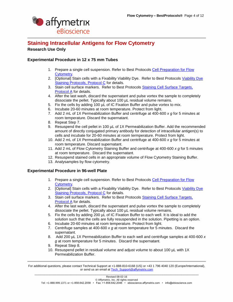
Experimental Procedure in 12 x 75 mm Tubes
1. Prepare a single cell suspension. Refer to Best Protocols Cell Preparation for Flow
Cytometry. 2. [Optional] Stain cells with a Fixability Viability Dye. Refer to Best Protocols Viability Dye
Staining Protocols, Protocol C for details. 3. Stain cell surface markers. Refer to Best Protocols Staining Cell Surface Targets,
Protocol A for details. 4. After the last wash, discard the supernatant and pulse vortex the sample to completely
dissociate the pellet. Typically about 100 µL residual volume remains. 5. Fix the cells by adding 100 µL of IC Fixation Buffer and pulse vortex to mix. 6. Incubate 20-60 minutes at room temperature. Protect from light. 7. Add 2 mL of 1X Permeabilization Buffer and centrifuge at 400-600 x g for 5 minutes at
room temperature. Discard the supernatant. 8. Repeat Step 7. 9. Resuspend the cell pellet in 100 µL of 1X Permeabilization Buffer. Add the recommended
amount of directly conjugated primary antibody for detection of intracellular antigen(s) to cells and incubate for 20-60 minutes at room termperature. Protect from light.
10. Add 2 mL of 1X Permeabilization Buffer and centrifuge at 400-600 x g for 5 minutes at room temperature. Discard supernatant.
11. Add 2 mL of Flow Cytometry Staining Buffer and centrifuge at 400-600 x g for 5 minutes at room temperature. Discard the supernatant.
12. Resuspend stained cells in an appropriate volume of Flow Cytometry Staining Buffer. 13. Analysamples by flow cytometry.
Experimental Procedure in 96-well Plate
1. Prepare a single cell suspension. Refer to Best Protocols Cell Preparation for Flow Cytometry.
2. [Optional] Stain cells with a Fixability Viability Dye. Refer to Best Protocols Viability Dye Staining Protocols, Protocol C for details.
3. Stain cell surface markers. Refer to Best Protocols Staining Cell Surface Targets, Protocol A for details.
4. After the last wash, discard the supernatant and pulse vortex the sample to completely dissociate the pellet. Typically about 100 µL residual volume remains.
5. Fix the cells by adding 200 µL of IC Fixation Buffer to each well. It is ideal to add the solution such that the cells are fully resuspended in the solution. Pipetting is an option.
6. Incubate 20-60 minutes at room temperature. Protect from light. 7. Centrifuge samples at 400-600 x g at room temperature for 5 minutes. Discard the
supernatant. 8. Add 200 µL 1X Permeabilization Buffer to each well and centrifuge samples at 400-600 x
g at room temperature for 5 minutes. Discard the supernatant. 9. Repeat Step 8. 10. Resuspend pellet in residual volume and adjust volume to about 100 µL with 1X
Permeabilization Buffer.
Flow Cytometry – BestProtocols® Page 5 of 12
Staining Intracellular Antigens for Flow Cytometry Research Use Only
For additional questions, please contact Technical Support at +1-888-810-6168 (US) or +43 1 796 4040 120 (Europe/International),
or send us an email at [email protected]
______________________________________________________________________________________ Revised 08-02-16
© Affymetrix, Inc. All rights reserved Tel: +1-888.999.1371 or +1-858.642.2058 • Fax: +1-858.642.2046 • ebioscience.affymetrix.com • [email protected]
11. [Optional] Block with 2% normal mouse/rat serum by adding 2 µL directly to the cells. Incubate at room temperature for 15 minutes.
12. Without washing, add the recommended amount of directly conjugated antibody for detection of intracellular antigen(s) to cells and incubate for at least 30 minutes at room temperature. Protect from light.
13. Add 200 µL of 1X Permeabilization Buffer to each well and centrifuge samples at 400-600 x g at room temperature for 5 minutes. Discard the supernatant.
14. Repeat Step 13. 15. Resuspend stained cells in an appropriate volume of Flow Cytometry Staining Buffer. 16. Analy by flow cytometry.

Flow Cytometry – BestProtocols® Page 6 of 12
Staining Intracellular Antigens for Flow Cytometry Research Use Only
For additional questions, please contact Technical Support at +1-888-810-6168 (US) or +43 1 796 4040 120 (Europe/International),
or send us an email at [email protected]
______________________________________________________________________________________ Revised 08-02-16
© Affymetrix, Inc. All rights reserved Tel: +1-888.999.1371 or +1-858.642.2058 • Fax: +1-858.642.2046 • ebioscience.affymetrix.com • [email protected]
Protocol B: One-step protocol: intracellular (nuclear) proteins The following protocol allows the simultaneous analysis of cell surface molecules and intracellular antigens, including nuclear antigens, at the single-cell level. This protocol combines fixation and permeabilization into a single step. It is recommended for the detection of nuclear antigens such as transcription factors but is also useful for the detection of many cytokines. For compatibility of the Foxp3/Transcription Factor Staining Buffer Set (cat. no. 00-5523) with cytokine antibodies, please refer to http://www.ebioscience.com/resources/application/flow-cytometry/antibody-fixation-considerations.htm for antibody clone performance.
Materials
12 x 75 mm round bottom test tubes or 96-well V- or U-bottom microtiter plate [Optional] Fixable Viability Dyes (FVD) eFluor® 455UV, 450, 506, 520, 660 and 780 (cat.
nos. 65-0868, 65-0863, 65-0866, 65-0867, 65-0864, 65-0865, respectively) [Optional] Normal Mouse Serum (cat. no. 24-5544) [Optional] Normal Rat Serum (cat. no. 24-5555) Directly conjugated antibodies Foxp3/Transcription Factor Staining Buffer Set (cat. no. 00-5523) Flow Cytometry Staining Buffer (cat. no. 00-4222)
Buffers and solution preparation
Prepare fresh Foxp3 Fixation/Permeabilization working solution by mixing 1 part of Foxp3 Fixation/Permeabilization Concentrate with 3 parts of Foxp3 Fixation/Permeabilization Diluent. One mL of the working solution is required for each sample, if staining in tubes.
Prepare a 1X working solution of Permeabilization Buffer by mixing 1 part of 10X Permeabilization Buffer with 9 parts of distilled water. 8.5 mL of the working solution is required for each sample, if staining in tubes.
Experimental Procedure in 12 x 75 mm Tubes
1. Prepare a single-cell suspension. Refer to ‘Cell Preparation Protocols for Flow
Cytometry’. 2. [Optional] Stain cells with a Fixable Viability Dye. Refer to ‘Viabilty Dye Staining, Protocol
C’ for more details. 3. Stain cell surface markers. Refer to ‘Staining Cell Surface Targets, Protocol A’ for
details. 4. After the final wash, discard the supernatant and pulse vortex the sample to completely
dissociate the pellet. Typically about 100 µL of residual volume remains. 5. Add 1 mL of Foxp3 Fixation/Permeabilization working solution to each tube and pulse
vortex. 6. Incubate for 30-60 minutes at 2-8°C or room temperature. Protect from light. (Mouse
samples can be incubated for up to 18 hours at 2-8°C in the dark). 7. Add 2 mL of 1X Permeabilization Buffer to each tube and centrifuge samples at 400-600
x g for 5 minutes at room temperature. Discard the supernatant.

Flow Cytometry – BestProtocols® Page 7 of 12
Staining Intracellular Antigens for Flow Cytometry Research Use Only
For additional questions, please contact Technical Support at +1-888-810-6168 (US) or +43 1 796 4040 120 (Europe/International),
or send us an email at [email protected]
______________________________________________________________________________________ Revised 08-02-16
© Affymetrix, Inc. All rights reserved Tel: +1-888.999.1371 or +1-858.642.2058 • Fax: +1-858.642.2046 • ebioscience.affymetrix.com • [email protected]
8. [Optional] Repeat Step 7. 9. Resuspend pellet in residual volume of 1X Permeabilization Buffer. This is typically 100
µL after decanting. 10. [Optional] Block with 2% normal mouse/rat serum by adding 2 µL directly to the cells.
Incubate for 15 minutes at room temperature. 11. Without washing, add the recommended amount of directly conjugated antibody for
detection of intracellular antigen(s) to cells and incubate for at least 30 minutes at room temperature. Protect from light.
12. Add 2 mL of 1X Permeabilization Buffer to each tube and centrifuge samples at 400-600 x g for 5 minutes at room temperature. Discard the supernatant.
13. Repeat Step 12. 14. Resuspend stained cells in an appropriate volume of Flow Cytometry Staining Buffer. 15. Analyze samples by flow cytometer.
Experimental Procedure in 96-well Plate
1. Prepare a single-cell suspension. Refer to ‘Cell Preparation Protocols for Flow Cytometry’.
2. [Optional] Stain cells with a Fixable Viability Dye. Refer to ‘Viabilty Dye Staining, Protocol C’ for more details.
3. Stain cell surface markers. Refer to ‘Staining Cell Surface Targets, Protocol A’ for details.
4. After the final wash, discard the supernatant and pulse vortex the sample to completely dissociate the pellet.
5. Add 200 µL of Foxp3 Fixation/Permeabilization working solution to each well. It is ideal to add the solution such that the cells are fully resuspended in the solution. Pipetting is an option.
6. Incubate for 30-60 minutes at 2-8°C or room temperature. Protect from light. (Mouse samples can be incubated for up to 18 hours at 2-8°C in the dark).
7. Centrifuge samples at 400-600 x g for 5 minutes at room temperature. Discard the supernatant.
8. Add 200 µL 1X Permeabilization Buffer to each well and centrifuge samples at 400-600 x g for 5 minutes at room temperature. Discard the supernatant.
9. Repeat Step 8. 10. Resuspend pellet in residual volume and adjust volume to about 100 µL with 1X
Permeabilization Buffer. 11. [Optional] Block with 2% normal mouse/rat serum by adding 2 µL directly to the cells.
Incubate for 15 minutes at room temperature. 12. Without washing, add the recommended amount of directly conjugated antibody for
detection of intracellular antigen(s) to cells and incubate for at least 30 minutes at room temperature. Protect from light.
13. Add 200 µL of 1X Permeabilization Buffer to each well and centrifuge samples at 400-600 x g for 5 minutes at room temperature. Discard the supernatant.
14. Repeat Step 13. 15. Resuspend stained cells in an appropriate volume of Flow Cytometry Staining Buffer.

Flow Cytometry – BestProtocols® Page 8 of 12
Staining Intracellular Antigens for Flow Cytometry Research Use Only
For additional questions, please contact Technical Support at +1-888-810-6168 (US) or +43 1 796 4040 120 (Europe/International),
or send us an email at [email protected]
______________________________________________________________________________________ Revised 08-02-16
© Affymetrix, Inc. All rights reserved Tel: +1-888.999.1371 or +1-858.642.2058 • Fax: +1-858.642.2046 • ebioscience.affymetrix.com • [email protected]
16. Analyze by flow cytometer.

Flow Cytometry – BestProtocols® Page 9 of 12
Staining Intracellular Antigens for Flow Cytometry Research Use Only
For additional questions, please contact Technical Support at +1-888-810-6168 (US) or +43 1 796 4040 120 (Europe/International),
or send us an email at [email protected]
______________________________________________________________________________________ Revised 08-02-16
© Affymetrix, Inc. All rights reserved Tel: +1-888.999.1371 or +1-858.642.2058 • Fax: +1-858.642.2046 • ebioscience.affymetrix.com • [email protected]
Protocol C: Two-step Protocol for Fixation/Methanol The following protocol allows for the simultaneous analysis of cell surface molecules and some intracellular phosphorylated signaling proteins. In this protocol, fixation is followed by treatment of cells with methanol. For phospho-protein detection, the appropriate stimulation conditions and kinetics of phosphorylation will vary depending on the cell type and the particular signaling event being assayed. For example, to induce phospho-STAT1 (Y701) phosphorylation, macrophages can be activated with IFNγ, while phospho-ERK1/2 (T202/Y204) is induced in T cells in response to PMA (a phorbol ester and protein kinase C activator) or anti-CD3 crosslinking.
General Notes
1. Fluorochrome-conjugated antibodies can be used to stain surface proteins for the purpose of immunophenotyping cells that will be further analyzed for phosphorylated proteins; however, additional considerations for staining are warranted.
Antibody staining for surface markers on live cells has been shown to alter expression of signaling proteins due to possible stimulation/suppression of signaling events. Because of this, surface staining is not recommended prior to cell stimulation. Instead, stain surface proteins at the same step as the intracellular protein staining. Please note that some proteins will also have intracellular pools, in addition to surface localization, which should be considered. Antibody clones to surface proteins that will recognize fixed cells/epitopes will need to be evaluated and used. Refer to “Antibody Clone Performance Following Fixation/Permeabilization” in Technical Support Resources.
If surface staining is required before fixation (due to epitope destruction caused by fixation), cells may be stained with fluorochrome-conjugated antibodies before the Fixation/Methanol steps only if the conjugated fluorochromes are resistant to methanol exposure (refer to table below).
MeOH Resistant Fluorochromes MeOH Sensitive Fluorochromes
Alexa Fluor 488 PE
eFluor 660 PE-tandems
Alexa Fluor 647 PerCP
eFluor 450 PerCP-tandems
FITC APC
APC-tandems
2. For adherent cells, we recommend fixing the cells in the plates/well. After fixation, scrape cells or treat with EDTA solution to harvest and continue with protocol. Trypsin can be used if surface staining is not needed or if the surface staining protein is resistant to tryspin digestion.

Flow Cytometry – BestProtocols® Page 10 of 12
Staining Intracellular Antigens for Flow Cytometry Research Use Only
For additional questions, please contact Technical Support at +1-888-810-6168 (US) or +43 1 796 4040 120 (Europe/International),
or send us an email at [email protected]
______________________________________________________________________________________ Revised 08-02-16
© Affymetrix, Inc. All rights reserved Tel: +1-888.999.1371 or +1-858.642.2058 • Fax: +1-858.642.2046 • ebioscience.affymetrix.com • [email protected]
Materials
12 x 75 mm round bottom test tubes or 96-well V- or U- bottom microtiter plates Tissue culture media of choice Cell stimulants of choice Primary antibodies (directly conjugated) Flow Cytometry Staining Buffer (cat. no. 00-4222) IC Fixation Buffer (cat. no. 00-8222) 90-100% methanol (HPLC grade) [Optional] Fc Block: Anti-Mouse CD16/CD32 Purified (cat. no. 14-0161) or Human Fc
Receptor Binding Inhibitor Purified (cat. no. 14-9161)
Experimental Procedure
1. Prepare cells of interest for stimulation in appropriate media. 2. Count cells and resuspend in appropriate media at 1-5 x 10⁶ cells/mL.
3. Stimulate cells at 37C with appropriate treatment for desired time point(s). Remember
to incubate untreated cells at 37C as a negative control. 4. [Optional] If surface staining is needed prior to fixation (in Step 5), stain cell surface
antigen(s) as described in ‘Staining Cell Surface Targets for Flow Cytometry Protocols’ with antibodies conjugated to methanol-resistant fluorochromes.
5. At the end of the stimulation period, fix cells to stop stimulation by adding an equal volume of IC Fixation Buffer directly to cells and vortex to mix.
6. Incubate samples for 10-60 minutes at room temperature. Protect from light. 7. Centrifuge samples at 400-600 x g for 5 minutes at room temperature. Discard the
supernatant. 8. Resuspend the cell pellet in residual volume and add 1 mL of ice-cold 90-100%
methanol. Vortex to mix and incubate for at least 30 minutes at 2-8C.
NOTE: Once in methanol, cells can be stored at <-20C for up to 4 weeks.
9. Wash cells with an excess volume of Flow Cytometry Staining Buffer. 10. Centrifuge cells at 400-600 x g for 5 minutes at room temperature. Discard the
supernatant. 11. Resuspend cells at 1x10⁷ cells/mL in Flow Cytometry Staining Buffer and aliquot 1x10⁶
cells (100 µL) into separate flow tubes. 12. [Optional] Cells can be blocked for nonspecific Fc-mediated binding using Anti-Mouse
CD16/CD32 Purified or Human Fc Receptor Binding Inhibitor Purified before staining. 13. Add the recommended amount of directly conjugated antibody to each tube and incubate
for 30-60 minutes at room temperature. Protect from light.

Flow Cytometry – BestProtocols® Page 11 of 12
Staining Intracellular Antigens for Flow Cytometry Research Use Only
For additional questions, please contact Technical Support at +1-888-810-6168 (US) or +43 1 796 4040 120 (Europe/International),
or send us an email at [email protected]
______________________________________________________________________________________ Revised 08-02-16
© Affymetrix, Inc. All rights reserved Tel: +1-888.999.1371 or +1-858.642.2058 • Fax: +1-858.642.2046 • ebioscience.affymetrix.com • [email protected]
NOTE: If needed, surface staining and intracellular phospho-staining can be performed simultaneously. As not all antibody clones will bind to a fixed epitope, refer “Antibody Clone Performance Following Fixation/Permeabilization” table online.
14. Add 2 mL of Flow Cytometry Staining Buffer and centrifuge at 400-6—x g for 4-5 minutes at room temperature. Discard supernatant.
15. Repeat Step 14. 16. Resuspend stained cells in an appropriate volume of Flow Cytometry Staining Buffer. 17. Analyze samples by flow cytometer.
Experimental Procedure in 96-well Plate
1. Prepare cells of interest for stimulation in appropriate media. 2. Count cells and resuspend in appropriate media at 1-5 x 10⁶ cells/mL. 3. Add 100 µL media containing appropriate stimulants to wells.
4. Add 100 µL cells to wells and incubate at 37C for desired time point(s). Remember to
incubate untreated cells at 37C as a negative control. 5. [Optional] If surface staining is needed prior to fixation (in Step 6), stain cell surface
antigen(s) as described in ‘Staining Cell Surface Targets for Flow Cytometry Protocols’ with antibodies conjugated to methanol-resistant fluorochromes.
6. At the end of the stimulation period, fix cells to stop stimulation by adding 200 µL of IC Fixation Buffer directly to wells.
7. Incubate plate 10-60 minutes at room tempature in the dark. Protect from light. 8. Centrifuge plate at 600 x g for 4-5 minutes at room temperature. Discard the supernatant. 9. Resuspend the cell pellets in residual volume and add 100 µL ice-cold 90-100%
methanol to wells. Vortex to mix and incubate plate for at least 30 minutes at 2-8C or on ice.
NOTE: Once in methanol, cells can be stored at <-20C for up to 4 weeks.
10. Add 200 µL Flow Cytometry Staining Buffer and centrifuge cells at 600 x g for 4-5 minutes at room temperature. Discard the supernatant.
11. Repeat Step 10. 12. [Optional] Cells can be blocked for nonspecific Fc-mediated binding using Anti-Mouse
CD16/CD32 Purified or Human Fc Receptor Binding Inhibitor Purified before staining. 13. Add the recommended amount of directly conjugated antibody to each well and incubate
for 30-60 minutes at room temperature. Protect from light. NOTE: If needed, surface staining and intracellular phospho-staining can be performed simultaneously. As not all antibody clones will bind to a fixed epitope, refer “Antibody Clone Performance Following Fixation/Permeabilization” table online.
14. Add 200 µL Flow Cytometry Staining Buffer and centrifuge at 600 x g for 4-5 minutes. Discard the supernatant.
15. Repeat Step 14.

Flow Cytometry – BestProtocols® Page 12 of 12
Staining Intracellular Antigens for Flow Cytometry Research Use Only
For additional questions, please contact Technical Support at +1-888-810-6168 (US) or +43 1 796 4040 120 (Europe/International),
or send us an email at [email protected]
______________________________________________________________________________________ Revised 08-02-16
© Affymetrix, Inc. All rights reserved Tel: +1-888.999.1371 or +1-858.642.2058 • Fax: +1-858.642.2046 • ebioscience.affymetrix.com • [email protected]
16. Resuspend stained cells in an appropriate volume of Flow Cytometry Staining Buffer. 17. Analyze samples by flow cytometer.