CYTOTOXICITY AND APOPTOSIS - Shodhgangashodhganga.inflibnet.ac.in/bitstream/10603/6583/9/09_chapter...
29
39 3 CHAPTER THREE CYTOTOXICITY AND APOPTOSIS ACTIVITIES OF WITHANIA SOMNIFERA AND TINOSPORA CORDIFOLIA EXTRACTS
Transcript of CYTOTOXICITY AND APOPTOSIS - Shodhgangashodhganga.inflibnet.ac.in/bitstream/10603/6583/9/09_chapter...
39��
3 �
�
�
�
�
�
�
�
�
�
�
�
�
�
�
�
�
�
�
�
CHAPTER THREE CYTOTOXICITY AND APOPTOSIS
ACTIVITIES OF WITHANIA SOMNIFERA AND TINOSPORA CORDIFOLIA EXTRACTS
40��
3.1 INTRODUCTION
Various studies on breast cancer published from India reflect the disease profile and
treatment characteristics unique to the urban rich and the middle class patients (Kuraparthy et
al., 2007). Recent reports indicate that breast cancer is emerging as prevalent cancer amongst
women, surpassing cervical cancer in India (Kuraparthy et al., 2007). Chemotherapy is a
major modality of breast cancer treatment. Chemotherapy aims at treatment of cancer by
destroying the rapidly proliferating cancer cells. It is also called a systemic treatment as the
drug enters through the bloodstream, travels throughout the body and kills cancer cells at their
sites (Pluen et al., 2001). Intensive chemotherapy regimens using cytotoxic agents do
effectively kill certain malignancies, especially of hematopoietic origin and some solid
cancers. However, malignant tumors are often resistant to chemotherapy and even develop
acquired chemoresistance or show multi-drug resistance as a consequence of the previous
treatment (Gottesman, 2002). Among the cellular mechanisms proposed to mediate multidrug
resistance, over expression of a family of plasma membrane efflux transporters, ATP-binding
cassette (ABC) transporters, has received extensive investigation. It is observed that the over
expression of these ABC transporters, predominantly by ABCB1 (MDR1), ABCC1 (MRP1)
and ABCG2 (BCRP) (Shukla et al., 2008) in cancer cells limits the intracellular accumulation
of anticancer drugs for efficient activity through the active extrusion of the cytotoxic drugs.
Classical multidrug resistance to plant based hydrophobic compounds has been shown to be
due to the elevated expression of cell-membrane transporters, which result in an increased
efflux of the cytotoxic drugs from the cancer cells, thus lowering their intracellular
concentrations (Nobili et al., 2011).
Plants are being used as indigenous cure in folklore or traditional system of medicine for
treatment of various kinds of illness including cancer (Pandey et al., 2011). Recently, a
greater emphasis has been given towards the research on complementary and alternative
medicine that deals with cancer management (Sawadogo et al., 2012). In traditional medicine,
plants are being used for healing purposes and are effective as they contain biologically active
principles which are non toxic (Duffy et al., 2012). With an understanding of cell biology,
mechanism based bioassays have become increasingly important and bio-activity guided
phytochemical investigation has resulted in the isolation and characterization of several new
molecules possessing interesting medicinal properties (Pan et al., 2012). Ayurveda, a
41��
traditional sect of Indian system of medicine mainly based on plant drugs had been successful
since very early times for the prevention or suppressing ailments (Liu, 2011). Plants represent
the principal therapy in traditional medicine since time immemorial (Ogilvie, 2003). Early
documentation about the use of medicinal plants has been mentioned in Discorides and
Ayurveda (Spitzer, 2011). Epidemiological studies suggest that consumption of diets
containing fruits and vegetables which are the major sources of phytochemicals and
micronutrients reduce the risk of developing cancer (Davis and Milner, 2010). Certain
products from plants are known to induce apoptosis in neoplastic cells but not in normal cells
(Sharif et al., 2012).
Apoptosis is a highly organized cell death process characterized by loss of plasma
membrane phospholipid asymmetry, enzymatic cleavage of the DNA into oligonucleosomal
fragments, and segmentation of the cells into membrane-bound apoptotic bodies (Cotter,
2009). Apoptosis has been recognized to play an important role in the maintenance of tissue
homeostasis by the selective elimination of excessive cells (Schmitt, 2003). Induction of
apoptosis of cancer cells is recognized as a valuable tool for cancer treatment. The agents that
are capable of inducing selective apoptosis of cancer cells are receiving considerable attention
in the development of cancer chemotherapeutic drugs for cancer.
The current study investigated, two indigenous medicinal plants, Withania somnifera of
the family Solanaceae and Tinospora cordifolia of the family Menispermaceae. The selection
of plants were based on valuable information obtained from Ayurveda on anticancer
properties, detailed ethno-botanical review, results obtained from the preliminary anticancer
screening and contain potential anticancer agents like sesquiterpene lactones. The
pharmacological activities of the selected plants, Withania somnifera and Tinospora
cordifolia have been investigated by various in vitro and in vivo experiments. However,
detailed investigations on anticancer properties of the two selected medicinal plants are
unknown. In this direction, we have studied the detailed anticancer activities of Withania
sonifera and Tinospora cordifolia in human breast cancer cells using a cell proliferation
assay, fluorescent microscopy based assays of apoptosis, DNA fragmentation assay and flow
cytometry based cell cycle analysis. Agents that are proficient to induce apoptosis in cancer
cells without harming normal cells have considerable attention on development of novel
anticancer drugs (Cotter, 2009). Therefore, the current study also investigated the cancer cell

42��
specific activity of Withania sonifera and Tinospora cordifolia in comparison with human
normal epithelial cells.
3.2 MATERIALS AND METHODS
3.2.1 Identification, collection and preparation of crude extracts of selected medicinal plants
The stem part of Tinospora cordifolia (Wild.) Hook. F. & Thomas of the family
Menispermaceae and the roots of Withania somnifera Dunal of the family Solanaceae were
collected from the Udupi district, Karnataka (India) in the month of January 2009 and were
authenticated by Dr. Gopala Krishna Bhatt, Taxonomist, Professor and Head, Department of
the botany, Poorna Prajna College, Udupi, Karnataka. The authenticated specimen was kept in
the Herbarium of Manipal College of pharmaceutical sciences, Manipal. Shade dried and
coarsely powdered plant (2 kg) was extracted in a soxhlet apparatus for 72 hours using
absolute ethanol as solvent. The crude ethanolic extracts were concentrated in a rotary
evaporator under reduced pressure and stored in a desiccator and dried completely. Similarly,
for the preparation of aqueous extracts of selected medicinal plants, shade dried and coarsely
powdered plant materials were extracted with double distilled water in a glass beaker by
repeated warming at 50-60°C for 72 hours. Each extract was then filtered through whattman
filter paper. Crude aqueous extracts were concentrated in a rotary evaporator under reduced
pressure and stored in a desiccator and dried completely. The crude ethanolic and aqueous
extracts were further dried by lyophilization. For the biological studies, crude ethanolic
extracts were dissolved in DMSO (Calbiochem) at 20 mg/ml and aqueous extracts were
dissolved in sterile MilliQ water at 10 mg/ml.
3.2.2 Drugs and chemicals
Doxorubicin hydrochloride solution, Dulbecco’s modified eagle medium (DMEM),
Trypsin-EDTA, Hank’s Balanced Salt Solution (HBSS), Thiazolyl Blue Tetrazolium Bromide
(MTT), Acridine orange, Ethidium bromide, Hoechst 33342 (Bisbenzimide
Trihydrochloride), Propidium iodide, RNase A, Agarose, HEPES buffer, Proteinase K,
Nonidet P40, Tris-HCl, were purchased Sigma Chemical Co. (St. Louis, MO, USA). Fetal
bovine serum (FBS) was purchased from Gibco, Invitrogen, USA. Dimethyl Sulfoxide
43��
(DMSO) was purchased from Calbiochem. All other fine chemicals were obtained from
Qualigens fine chemicals (Mumbai, India).
3.2.3 Cell culture
MCF7 (human breast carcinoma, ER+, tumorigenic and non-invasive), MDA MB 231
(human breast carcinoma, ER-, tumorigenic and invasive) HeLa (Human cervical carcinoma,
tumorigenic and invasive) and HaCaT (Human keratinocyte, immortalized) were cultured in
DMEM with high glucose supplemented with 10% FBS, 100 units/ml penicillin and 100
μg/ml streptomycin, in a humidified 5% CO2 incubator at 37°C. Cells were cultured in
healthy condition and exponentially growing cells were taken for the experiments.
3.2.4 Cytotoxicity assay (Methyl tetrazolium-MTT assay)
3.2.4.1 Determination of optimal cell number for assay
In order to determine optimal cell number required for the assay, serial dilutions of the
MCF7, MDA MB 231 and HaCaT cells (2,000, 4,000, 6,000, 8,000, 12,000, 14,000, 16,000
and 18,000 cells/100 �l) were made in cell culture media and seeded in 96 well microtiter
tissue culture plates. Cells were cultured, in a humidified 5% CO2 incubator at 37°C for 24
hours. At the end of the incubation period, 20 �l of MTT solution (Stock concentration, 5.00
mg/ml in HBSS) was added to each well and incubated for 4 hours under the same conditions.
Thereafter, medium containing MTT was gently replaced by 200 �l DMSO to dissolve
formazan crystals and the absorbance values were measured by a microtiter plate reader
(Biotek ELx800 - MS) at 540 nm with a reference wavelength of 630 nm. A graph was
plotted with the number of cells in X-axis and absorbance at 570/630 nm in Y-axis. Optimal
cell densities of cell lines corresponding to absorbance values of 0.9 to 1.0 in the assay were
selected for each of the cell lines such as MCF7, MDA MB 231 HeLa, and HaCaT to
facilitate measurement of both stimulation and inhibition of cell proliferation within the linear
range.
3.2.4.2 Evaluation of cytotoxicity
Briefly, specified cell types were trypsinized and resuspended in the culture medium to
get a defined cell number for MCF7 (14,000/100 �l), MDA MB 231 (16,000/100 �l), HeLa
(6,000/100 �l) and HaCaT (12,000/100 �l) in a 96-well microtiter tissue culture plate and
44��
cultured in a humidified 5% CO2 incubator at 37°C for 24 hours. Defined concentrations of
the extracts in culture media were freshly prepared by serial dilution to get final concentration
of 12.5, 25, 50, 100 and 200 μg/ml (for Tinospora cordifolia ethanolic extract); 10, 20, 40, 60
and 100 μg/ml (for Withania somnifera ethanolic extract); 25, 50, 100 and 200 μg/ml (for
Tinospora cordifolia and Withania somnifera aqueous extracts). Serial dilution was carried
out in cell culture media in such a way that the final concentration of DMSO in the well did
not exceed 0.50% (v/v). After 24 hours of incubation, cells were treated with above
mentioned concentrations of Tinospora cordifolia and Withania somnifera extracts in
triplicates for 48 hours. Doxorubicin hydrochloride, an anticancer drug was used as a positive
control. Equal volume of DMSO was used as a vehicle control. At the end of treatment, 20 μl
of MTT (Stock was made in HBSS at 5.00 mg/ml) reagent was added to each well and
incubated for further 4 hours. Thereafter, the culture medium was removed and formazan
crystals were dissolved in 200μl of DMSO. The plates were read in a 96 well microplate
reader (Biotek-ELx-800) at a wavelength of 570 nm with a reference wavelength of 630nm.
Percentage cell viability (Y-axis) was calculated from O.D values and plotted against
concentration in μg/ml (X-axis). The absorbance values of both test and the control (untreated
cells) were used for calculating the percentage cell viability (% Cell viability = O.D of
Test/O.D of Control x 100). Cell viability in untreated control was normalized to 100%. The
dose response curve was plotted with concentration of the drug in the ordinate and percentage
cell viability in the abscissa. IC50 values of the extracts were obtained from the graph as the
concentration which decreases percentage cell viability to 50. IC50 values for Tinospora
cordifolia ethanolic and aqueous extracts; Withania somnifera ethanolic and aqueous extracts
and doxorubicin hydrochloride were determined by nonlinear regression (curve fit) analysis
by plotting log (inhibitors) vs. normalized response using software, Graph pad prism 5.
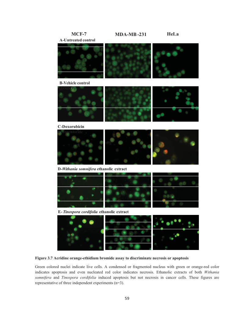
3.2.5 Acridine orange-ethidium bromide assay
MCF7, MDA MB 231, HeLa and HaCaT cells were seeded in a 6-well tissue culture plate
at an optimal cell number and cultured for 24 hours. Cells at the exponential growth phase
were treated with Tinospora cordifolia ethanolic extract, Withania somnifera ethanolic extract
and doxorubicin (positive control) at the IC50 values (mentioned in Table 3.1) for 48 hours.
Untreated cells and DMSO treated cells were considered as untreated control and solvent
control respectively. At the end of the treatment, adherent cells were harvested by
45��
trypsinization and pooled with suspended dead cells. Thereafter, cells were centrifuged at
2000 rpm for 5 minutes at RT, washed with HBSS twice. Cells were then stained with a
mixture of acridine orange and ethidium bromide (2 μg/ml) for 10 minutes in a 37°C CO2
incubator. At the end of staining, cells were washed with ice-cold HBSS twice and the pellet
was re-suspended in 100 μl of HBSS. Thereafter, cells were mounted on a glass slide with
cover slip and observed in an inverted fluorescent microscope equipped with 450-490 nm
excitation source and 520/570 nm filters of emission wavelengths. Morphological
characteristics of both necrosis and apoptosis were observed in both 20X and 40X
magnifications. Number of live cells (even nucleated green colored), early apoptotic cells
(green colored condensed or fragmented nuclei), late apoptotic cells (orange-red colored
condensed or fragmented nuclei) and necrotic cells (even nucleated orange-red colored) were
determined.
3.2.6 Hoechst 33342 assay
MCF7, MDA MB 231, HeLa and HaCaT cells were seeded in a 6-well tissue culture plate
at an optimal cell number and cultured for 24 hours. Cells at the exponential growth phase
were treated with Tinospora cordifolia ethanolic extract, Withania somnifera ethanolic extract
and doxorubicin (positive control) at the IC50 values (mentioned in Table 3.1) for 48 hours.
Untreated cells and DMSO treated cells were considered as untreated control and solvent
control respectively. At the end of treatment, adherent cells were harvested by trypsinization
and pooled with suspended dead cells. Thereafter, cells were centrifuged at 2000 rpm for 5
minutes at RT, washed with HBSS twice. Thereafter, the cell was stained with Hoechst 33342
(2 μg/ml) 10 minutes at 37°C. At the end of staining, cells were washed twice with ice-cold
HBSS and the pellet was resuspended in a minimum volume of HBSS. Cells were mounted
on a glass slide with a coverslip and observed under inverted fluorescent microscope equipped
with 350 nm excitation source and emission filter of 450 nm wavelength. Morphological
characteristics of apoptosis such as, condensed nuclei, fragmented nuclei and membrane
blebbing were observed in both 20X and 40X magnifications.
3.2.7 DNA fragmentation assay
MCF7, MDA MB 231, HeLa and HaCaT cells were seeded in a 6-well tissue culture plate
at an optimal cell number and cultured for 24 hours. Cells at the exponential growth phase

46��
were treated with Tinospora cordifolia ethanolic extract, Withania somnifera ethanolic extract
and doxorubicin (positive control) at the IC50 values (mentioned in Table 3.1) for 48 hours.
Untreated cells and DMSO treated cells were considered as untreated control and solvent
control respectively. At the end of treatment, adherent cells were harvested by trypsinization
and pooled with suspended dead cells. At the end of treatment, adherent cells were lysed with
300 �l of lysis buffer cocktail (1.00 mL stock which contains 10% NP-40 [100 μl] + 200 mM
EDTA [40 μl ] + 0.2 M Tris-HCl [50 μl; pH 7.5] + 0.50 mg proteinase K + 810 μl miliQ
water). The lysate was then pooled with floating dead cells. The cell lysate was incubated at
560C for 1 hour and further incubated with RNAse (100 μg/ml) at 560 C for 1 hour. At the end
of the process, samples were diluted with 30% glycerol and ran in agarose gel (1.50%
Agrarose in 1X TBE buffer). Electrophoresis was carried by using Bio-rad electrophoresis
unit out at 60 V, 400mA for 90 min using 1X TBE buffer.
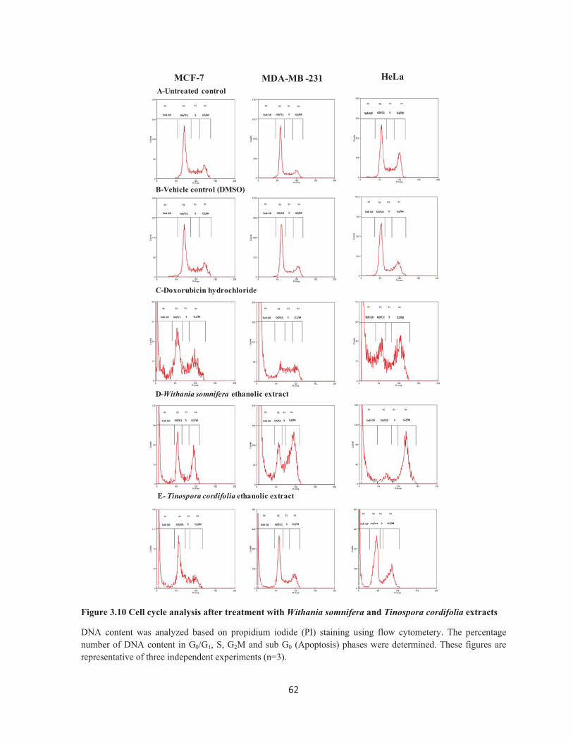
3.2.8 Cell cycle analysis by flow cytometry
Cell cycle analysis was performed by propidium iodide based measurements of the DNA
content of the cells by flow cytometry. Briefly, MCF7, MDA MB 231, HeLa and HaCaT cells
were seeded in a 6-well tissue culture plate at an optimal cell number and cultured for 24
hours. Cells at the exponential growth phase were treated with Tinospora cordifolia ethanolic
extract, Withania somnifera ethanolic extract and doxorubicin (positive control) at the IC50
values (mentioned in Table 3. 1) for 48 hours. Untreated cells and DMSO treated cells were
considered as untreated control and solvent control respectively. At the end of treatment,
adherent cells were harvested by trypsinization and pooled with suspended dead cells.
Thereafter, cells were centrifuged at 2000 rpm for 5 minutes at RT, washed with HBSS twice
and thereafter fixed with 70% ice cold ethanol and stored at -20°C. The cells were then
centrifuged at 3000 rpm for 5 minutes at RT and removed ethanol completely. Cells were
further washed with HBSS twice and re-suspended in 400 �l of HBSS. Thereafter, RNase A
(100 �g/ml) was added and incubated for 3 hours in water bath set at 560C. Propidium iodide
(20 �g/ml) was then added and incubated at RT for 15 minutes and analyzed by flow
cytometry (FACSCalibur, Becton Dickinson, USA). Propidium iodide was excited at 488nm
solid state laser and emission wavelength was detected at 585/42 nm (FL-2). The percentage
number of DNA in G0/G1, S phase, G2/M phase and sub-G0 phase in untreated cells and
treated cells were analyzed by software Summit v4.3 (Beckman Coulter, USA). Effects of

47��
Tinospora cordifolia ethanolic extract, Withania somnifera ethanolic extract and doxorubicin
in each and every phase of the cell cycle were determined.
3.3 RESULTS
3.3.1 Tinospora cordifolia and Withania somnifera extracts inhibits breast cancer cell
proliferation
Cytotoxic and growth inhibitory effects of the selected plant extracts on human cancer
cells were studied by MTT assay. Optimal cell densities corresponding to absorbance values
of 0.9 to 1.0 in MTT assay was selected for each of the cell lines to facilitate measurement of
both stimulation and inhibition of cell proliferation within the linear range (Fig. 3.1). The
optimal cell number to be seeded for a cytotoxicity assay in MCF7, MDA MB 231, HeLa, and
HaCaT determined from the plot and was found to be 14,000, 16,000, 6,000 and 12,000
cells/100 �l respectively. In order to evaluate the cytotoxic effects of the ethanolic and
aqueous extracts of Tinospora cordifolia and Withania somnifera, MCF7, MDA MB 231,
HeLa and HaCaT cells were treated with specified concentrations of the extracts for 48 hours.
Doxorubicin was used as a positive control. The ethanolic extracts of Tinospora cordifolia
and Withania somnifera showed cytotoxic and dose dependent inhibitory effects on cell
proliferation of human breast cancer cells (MCF7 and MDA MB 231) and cervical cancer
cells (HeLa). IC50 values of Tinospora cordifolia ethanolic extract in MCF7, MDA MB 231
and HeLa cells were found to be 84.40 ± 2.68 μg/ml, 66.39 ± 3.08 μg/ml and 155.30 ± 6.48
μg/ml, respectively (Fig. 3.2). Similarly, IC50 values of Withania somnifera ethanolic extract
in MCF7, MDA MB 231 and HeLa cells were found to be 22.33 ± 1.45 μg/ml, 31.99 ± 1.64
μg/ml and 30.12 ± 1.68 μg/ml, respectively (Fig. 3.4). Aqueous extracts of Tinospora
cordifolia and Withania somnifera did not show any concentration dependent cytotoxicity or
growth inhibitory activity in any of the cell lines (IC50 >300.0 μg/ml) (Fig. 3.3 and Fig. 3.5).
Thus, among the crude extracts screened for cytotoxicity and inhibition of cancer cell
proliferation, both Tinospora cordifolia and Withania somnifera ethanolic extracts
demonstrated dose dependent anticancer activity against human breast cancer cells (MCF7,
MDA MB 231) and cervical cancer cells (HeLa). The positive control, doxorubicin imparted
cytotoxic and dose dependent inhibition of cell proliferation and the IC50 values of
48��
doxorubicin on MCF7, MDA MB 231 and HeLa cells was found to be 1.25 ± 0.05 �M, 0.70 ±
0.03 �M and 2.12 ± 0.14 �M, respectively (Fig. 3.6).
The next question was whether Tinospora cordifolia and Withania somnifera extract-
mediated suppression of cell viability and growth was selectively to cancer cells, and not to
normal cells. This would be a highly desirable trait for a potential therapeutic anti-cancer
agent. This question was addressed by determining the cytotoxic and growth inhibitory effects
of bio-active Tinospora cordifolia and Withania somnifera ethanolic extracts on human
normal epithelial cells (HaCaT, an immortalized keratinocyte cell line). Results indicated that
ethanolic extract of Tinospora cordifolia possess cytotoxic and cell growth inhibitory effects
with an IC50 value of 194.10 ± 12.09 μg/ml (Fig. 3.2). Similarly, the ethanolic extract of
Withania somnifera possesses cytotoxic and cell growth inhibitory effects with an IC50 value
of 75.40 ± 4.33 μg/ml (Fig. 3.4). However, cell viability was not significantly affected by
Withania somnifera and Tinospora cordifolia ethanolic extract treatment at the concentrations
that were cytotoxic to MCF7, MDA MB 231 and HeLa cells. Moreover, an increased IC50
value for Withania somnifera and Tinospora cordifolia ethanolic extracts against HaCaT cells
as compared to MCF7, MDA MB 231 and HeLa cells indicate less cytotoxic effects. The well
known anticancer drug, doxorubicin has a dose dependent cytotoxicity in HaCaT with an IC50
value of 0.42 ± 0.01 �M (Fig. 3.6). Table 3.1 show a comparison profile of IC50 values for
Withania somnifera, Tinospora cordifolia extracts and the standard, doxorubicin in human
breast cancer cells (MCF7, MDA MB 435), human cervical cancer cells (HeLa) and human
normal epithelial cells (HaCaT). These data suggested that Withania somnifera and Tinospora
cordifolia ethanolic extracts do impart cancer cell-specific cytotoxic effects on human breast
and cervical cancer cells. The data represent ± SD of three independent experiments (n=3).
3.3.2 Withania somnifera and Tinospora cordifolia ethanolic extracts induce apoptosis
in human breast cancer cells
3.3.2.1 Acridine orange-ethidium bromide assay
In order to distinguish, if the cytotoxic effects of ethanolic extracts of Withania somnifera
and Tinospora cordifolia was due to apoptosis or necrosis, human breast cancer cells (MCF7,
MDA MB 231) and cervical cancer cells (HeLa) were treated with ethanolic extracts of
Withania somnifera and Tinospora cordifolia at their IC50 concentration as shown in Table

49��
3.1 for 48 hours. Doxorubicin hydrochloride and DMSO were used in the study design as
positive control and vehicle control respectively. At the end of the treatment, cells were
harvested and stained with a mixture of acridine orange and ethidium bromide and observed
under a fluorescent light microscope. Acridine orange can permeate cells and stain the nuclei
green (indicating live cells), while ethidium bromide which stains the nuclei orange-red, can
be taken up only by cells that have lost cytoplasmic membrane integrity, and hence indicate
dead cells. In addition, while uniformly stained green nuclei indicate live cells; green colored
condensed/fragmented nuclei indicate early apoptosis. Similarly, uniformly stained orange-
red colored cells indicate necrosis, while orange-red colored condensed/fragmented nuclei
indicate late apoptosis. As shown in the Fig. 3.7; untreated cells and vehicle control illustrate
uniform green colored nuclei indicating live cells, while cells treated with ethanolic extracts
of Withania somnifera and Tinospora cordifolia revealed condensed and fragmented green or
orange-red nuclei indicating apoptosis. The apoptotic phenotype was quite comparable with
anticancer drug, doxorubicin. These data suggest that ethanolic extracts of Withania
somnifera and Tinospora cordifolia induced apoptosis but not necrosis in cancer cells. All the
experiments were performed three times independently (n=3).
3.3.2.2 Hoechst 33342 assay
In order to confirm if the cytotoxic effects of ethanolic extracts of Withania somnifera and
Tinospora cordifolia was due to induction of apoptosis, human breast cancer cells (MCF7,
MDA MB 231) and cervical cancer cells (HeLa) were treated with extracts and the positive
control, doxorubicin at their IC50 concentration as shown in Table 3.1 for 48 hours. DMSO
was used as vehicle control. At the end of the treatment, cells were harvested and stained with
DNA binding fluorescent dye Hoechst 33342 to spot the number of apoptotic cells by
fluorescent microscopy. Hall mark properties of apoptosis such as condensed nuclei, DNA
fragmentation and shrinkage was observed in MCF7, MDA MB231 and HeLa cells upon
treatment with the ethanolic extracts of Withania somnifera, Tinospora cordifolia and
doxorubicin (Fig. 3.8). Apoptotic phenotype of extract treated cells was compared with the
doxorubicin treated cells. These results confirm that ethanolic extracts of Withania somnifera
and Tinospora cordifolia induced apoptosis in human breast cancer cells (MCF7, MDA MB
231) and cervical cancer cells (HeLa). All the experiments were performed three times
independently (n=3).

50��
3.3.2.3 DNA fragmentation assay
Fluorescent microscopy based nuclear staining studies indicate that Withania somnifera
and Tinospora cordifolia ethanolic extracts induced apoptosis and not necrosis in human
cancer cells. DNA fragmentation is a hall mark property of apoptosis. In order to validate the
apoptosis, human breast cancer cells (MCF7, MDA MB 231) and cervical cancer cells (HeLa)
were treated with ethanolic extracts of Withania somnifera and Tinospora cordifolia and
anticancer drug, doxorubicin hydrochloride at their IC50 concentration as shown in Table 3.1
for 48 hours. Doxorubicin hydrochloride and DMSO were used as positive control and
vehicle control respectively. At the end of the treatment, cells were harvested; genomic DNA
was isolated and subjected to agarose gel electrophoresis to detect DNA ladder formation, a
hall mark property of apoptosis. Results showed that formation of DNA fragments in MCF7,
MDA MB 231 and HeLa cells upon treatment with ethanolic extracts of Withania somnifera
and Tinospora cordifolia. DNA ladder phenotype was prominent in cells treated with
doxorubicin and was quite comparable with ethanolic extracts of Withania somnifera and
Tinospora cordifolia. The genomic DNA was intact without any ladder pattern in untreated
control and vehicle control groups (Fig. 3.9). These results corroborated that ethanolic
extracts of Withania somnifera and Tinospora cordifolia induced apoptosis in human breast
cancer cells (MCF7, MDA MB 231) and cervical cancer cells (HeLa). All the experiments
were performed three times independently (n=3).
3.3.3 Cell cycle specific inhibitory activity of ethanolic extracts of Withania somnifera
and Tinospora cordifolia on human breast cancer cells
In order to evaluate the cell cycle specific pharmacological effects of ethanolic extracts of
Withania somnifera and Tinospora cordifolia, human breast cancer cells (MCF7, MDA MB
231) and cervical cancer cells (HeLa) were cultured in the presence or absence of extracts at
their IC50 concentration for 48 hours. Cells were also treated with doxorubicin and vehicle
control (DMSO). At the end of the treatment period, cells were harvested, fixed with ice-cold
70% ethanol and stained with propidium iodide which binds to both DNA and RNA.
However, RNA was removed by treatment with RNAse and DNA content was measured by
flow cytometry as described in materials and methods. Percentage DNA content in G1/G0
phase, Synthetic (S) phase, G2/M phase and Sub-G0 phase were determined. As depicted in

51��
Fig. 3.10, untreated and vehicle control treated cells showed a normal cell cycle progression.
However, upon treatment with the ethanolic extracts of Withania somnifera and Tinospora
cordifolia cause a significant inhibition of cell cycle progression in human cancer cells. An
increased Sub-G0 phase indicates DNA fragmentation and apoptosis. Similarly, an elevated
G1/G0 phase and G2/M phase indicate cell cycle arrest and blockade of mitosis or cell division
respectively. A decrease in S-phase indicates inhibition of DNA replication. Withania
somnifera extract treatment resulted in the clear increase of G2/M phase and Sub-G0 phase as
compared with the untreated control. Similarly, Tinospora cordifolia extract treatment caused
an increase of Sub-G0 phase. Cells treated with doxorubicin showed an arrest of cells in G2/M
phase and elevated Sub G0 phase. The cell cycle specific effects of ethanolic extracts of
Withania somnifera, Tinospora cordifolia and doxorubicin on G1/G0 phase, Synthetic (S)
phase, G2/M phase and Sub-G0 phase were compared in Table 3.2. These data suggested that
treatment of human cancer cells with the extract of Withania somnifera results in apoptosis,
inhibition of DNA replication and mitosis. Similarly, Tinospora cordifolia extract caused
induction of apoptosis and inhibition of DNA replication of human cancer cells.
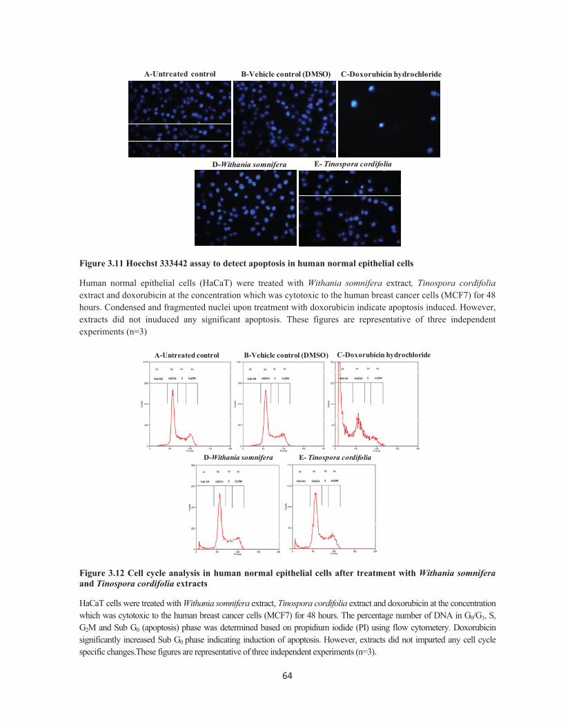
3.3.4 Apoptotic effects of ethanolic extracts of Withania somnifera and Tinospora
cordifolia in human normal epithelial cells
Comparison of cytotoxic and growth inhibitory activities of Withania somnifera and
Tinospora cordifolia ethanolic extracts on human cancer cells (MCF7, MDA MB 231 and
HeLa) and human normal epithelial cells (HaCaT) by MTT assay revealed that extracts
imparted a cancer cell-specific cytotoxicity. In order to understand whether Withania
somnifera and Tinospora cordifolia ethanolic extracts induce any apoptosis in normal
epithelial cells at the concentrations that were cytotoxic to human breast cancer cells; HaCaT
cells were treated with ethanolic extract of Withania somnifera, Tinospora cordifolia and
doxorubicin at 22 μg/ml, 84 μg/ml, 1.25 �M respectively (IC50 values of the respective drugs
in breast cancer cells, MCF7). At the end of the treatment, cells were harvested and
fluorescent microscopy based Hoechst 33342 assay was performed to detect apoptosis.
Results suggested that ethanolic extract of Withania somnifera, Tinospora cordifolia did not
show any significant nuclear condensation or DNA fragmentation in HaCaT cells. However,
doxorubicin treated cells demonstrated hall mark properties of apoptosis (Fig. 3.11). All the
experiments were performed three times independently (n=3).

52��
Similarly, in order to evaluate changes in cell cycle and apoptosis, HaCaT cells were
treated with ethanolic extract of Withania somnifera, Tinospora cordifolia and doxorubicin at
22 μg/ml, 84 μg/mL, 1.25 �M respectively (IC50 values of the respective drugs in MCF7). At
the end of the treatment period, cells were harvested, fixed with ice-cold 70% ethanol and
stained with propidium iodide and DNA content was measured by flow cytometry. Percentage
DNA content in G1/G0 phase, Synthetic (S) phase, G2/M phase and Sub-G0 phase were
determined. As depicted in the Fig. 3.12; untreated and vehicle control treated cells showed a
normal cell cycle progression. Upon treatment with ethanolic extract of Withania somnifera,
Tinospora cordifolia there was no any significant change in Sub-G0 phase, G1/G0 phase,
Synthetic (S) phase and G2/M phase. However, upon treatment with doxorubicin, there was a
significant increase in Sub-G0 phase, indicating apoptosis. A comparative analysis of
percentage DNA content in Sub-G0, G1/G0, Synthetic (S) and G2/M phase after treatment with
ethanolic extract of Withania somnifera, Tinospora cordifolia and doxorubicin in MCF7 and
HaCaT cells is shown in Table 3.3. These results demonstrated that ethanolic extracts of
Withania somnifera and Tinospora cordifolia possess cancer cell specific cytotoxicity and
apoptosis. This is a highly attractive trait for cancer chemotherapeutic agents.
3.4 DISCUSSION
The current study investigated pharmacological activities of two selected indigenous
medicinal plant extracts in human breast cancer (MCF7, MDA MB 231) and cervical cancer
cells (HeLa). A methodical evaluation of detailed cytotoxicity and cell proliferation effects in
human cancer cells reveals that ethanolic extracts of Withania somnifera and Tinospora
cordifolia harbors dose-dependent cytotoxicity and cell growth inhibitory activity. As per
National Cancer institute (NCI, USA) anti-cancer screening in panel of human cell lines, an
IC50 value of less than 100 μg/ml for medicinal plant extracts were considered as potential
anticancer agents and recommend for further isolation and characterization of bio-active
molecules. The data suggest that Withania somnifera ethanolic extract imparted very potent
cytotoxic and growth inhibitory activities in selected human cancer cells with IC50 values less
than 40 μg/ml. Similarly, Tinospora cordifolia ethanolic extract also exhibited potent
cytotoxic and growth inhibitory activities with IC50 values less than 90 μg/ml. Therefore,
ethanolic extracts of both Withania somnifera and Tinospora cordifolia may be considered for
further isolation and characterization of bio-active molecules.

53��
The current study also investigated that whether ethanolic extracts of Withania somnifera
and Tinospora cordifolia induces apoptosis in human breast cancer (MCF7, MDA MB 231)
and cervical cancer cells (HeLa). Acridine orange/ethidium bromide assay by fluorescent
microscopy revealed that ethanolic extracts of Withania somnifera and Tinospora cordifolia
induced apoptosis but not the necrosis in human cancer cells. Hoechst 33342 assay and DNA
fragmentation assay revealed the hall mark properties of apoptosis such as membrane
blebbing, nuclear condensation and DNA fragmentation in both human breast and cervical
cancer cells upon treatment with ethanolic extracts of Withania somnifera and Tinospora
cordifolia. These findings corroborated that the ethanolic extracts of Withania somnifera and
Tinospora cordifolia induced apoptosis in human breast and cervical cancer cells.
Cancer chemotherapeutic drugs are also classified based on their specificity on mammalian
cell cycle as cell cycle-specific and cell cycle non-specific drugs. Cell cycle specific drugs has
drawn considerable attention as they act on specific cancer cell cycle checkpoints (Go/G1
phase, S phase, G2/M phase) and inhibit cancer cell proliferation (Go/G1 phase arrest), or DNA
replication (Diminished S phase) or mitosis (Go/G1 phase arrest). We have also investigated
the cell cycle specific pharmacological effects of Withania somnifera and Tinospora
cordifolia ethanolic extracts in human breast cancer (MCF7, MDA MB 231) and cervical
cancer cells (HeLa) by propidium iodide (PI) based cell cycle analysis using flow cytometry.
Propidium iodide binds to both DNA and RNA, DNA content in different phases of cell cycle
(Go/G1 phase, S phase, G2/M phase and sub-G0) were measured based on fluorescent intensity
of propidium iodide. Elevated sub-G0 phase indicate DNA fragmentation or apoptosis. The
data suggest that treatment of human breast cancer and cervical cancer cells with the Withania
somnifera ethanolic extract resulted in significant arrest of cells at G2/M phase (G2/M arrest)
and prevent mitosis to occur. It also significantly increased Sub-G0 phase indicating induction
of apoptosis and diminishes S phase indicating inhibition of DNA replication. On the other
hand, treatment of human breast cancer and cervical cancer cells with the Tinospora
cordifolia ethanolic extract significantly elevates Sub-G0 phase indicating induction of
apoptosis and diminishes S phase indicating inhibition of DNA replication. These results
revealed the cancer cell cycle-specific activities of ethanolic extracts of Withania somnifera
and Tinospora cordifolia in human breast cancer and cervical cancer cells.

54��
The agents that are capable of inducing selective apoptosis of cancer cells have received
considerable interest in the development of novel cancer chemotherapeutic drugs. Hence, the
current study also addressed the question of whether Tinospora cordifolia and Withania
somnifera extract- mediated suppression of cell viability and growth was selectively to cancer
cells, which is a highly desirable trait for potential cancer preventive and therapeutic agents.
Cytotoxic and growth inhibitory study of Tinospora cordifolia and Withania somnifera
ethanolic extracts in human normal epithelial cells (HaCaT, an immortalized keratinocyte cell
line) indicated that ethanolic extract of Withania somnifera and Tinospora cordifolia possess
considerably less cytotoxic and cell growth inhibitory activity. Similarly, the potential of
Withania somnifera and Tinospora cordifolia ethanolic extracts to induce apoptosis
selectively in cancer cells was investigated by treating human normal epithelial cells with the
extracts at the concentrations which were cytotoxic to the human breast cancer cells.
Induction of apoptosis and effects on various phases of cancer cell cycle (Go/G1 phase, S
phase, G2/M phase and sub-G0) were measured by classical Hoechst 33342 assay and cell
cycle analysis using flow cytometry respectively. More interestingly, the results revealed that
ethanolic extracts of Withania somnifera and Tinospora cordifolia does not induce any
apoptosis in human normal epithelial cells. These results suggest that ethanolic extracts of
Withania somnifera and Tinospora cordifolia possess cancer cell specific cytotoxicity and
apoptosis which is a highly attractive trait for cancer chemotherapeutic agents.

55��
�
Figure 3.1 Determination of optimal cell number to be seeded for MTT assay
Optimal cell number to be seeded was determined in MCF7, MDA MB 231, HeLa and HaCaT cells. Each point represents the mean ± S.D triplicate samples in a representative experiment.
Figure 3.2 Cytotoxicity of Tinospora cordifolia ethanolic extract
Cytotoxic effects in human breast cancer cells (MCF7 and MDA MB 231), human cervical cancer cells (HeLa) and human normal epithelial cells (HaCaT) after treatment with Tinospora cordifolia ethanolic extract. Each point represents the mean ± S.D of three independent experiments performed in triplicate (n=3).
0 5000 10000 15000 200000.0
0.5
1.0
1.5 MCF7
MDA-MB-231
HaCaT
HeLa
Cell number
O D
at 5
40/6
30 n
m
0 50 100 150 2000
20
40
60
80
100
120 HeLa
MCF7
MDA-MB-231
HaCaT
Concentation �g/mL
% C
ell v
iabi
lity
56��
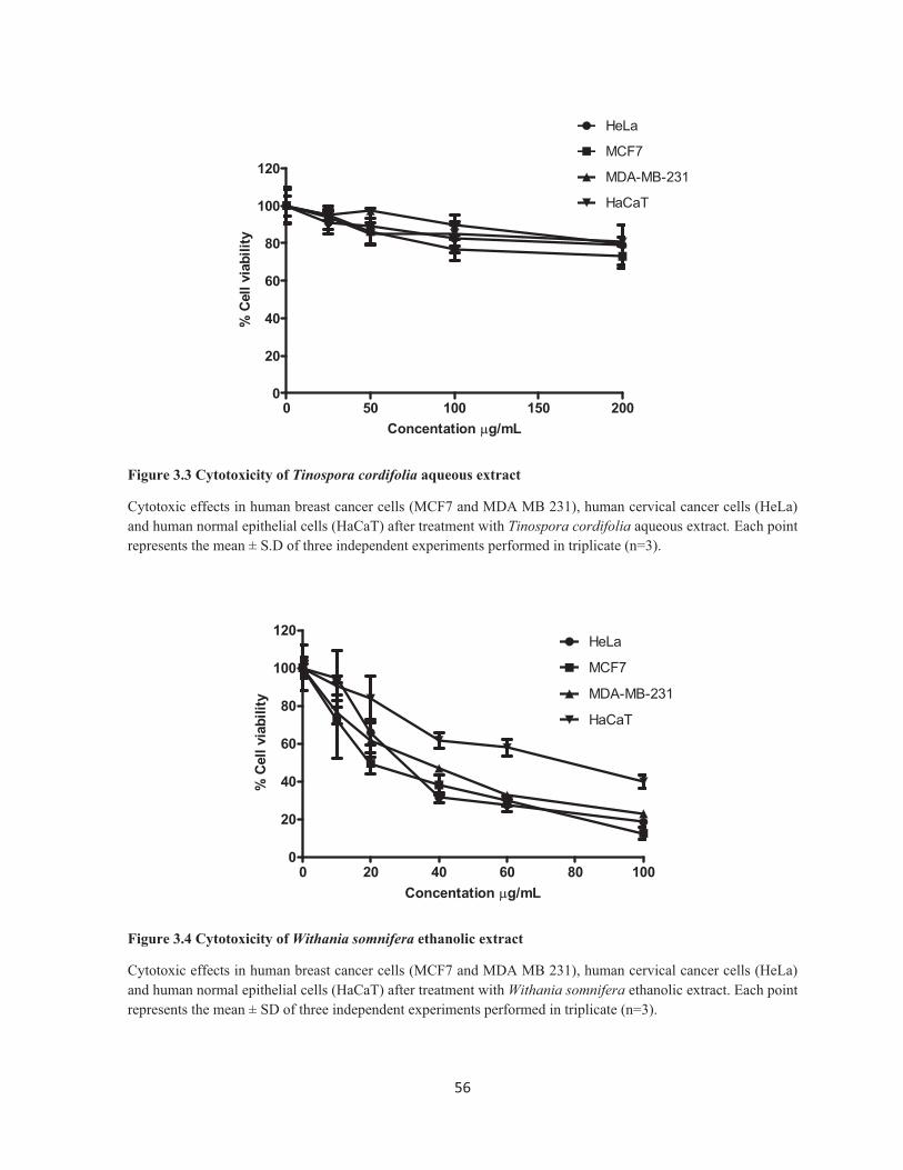
Figure 3.3 Cytotoxicity of Tinospora cordifolia aqueous extract
Cytotoxic effects in human breast cancer cells (MCF7 and MDA MB 231), human cervical cancer cells (HeLa) and human normal epithelial cells (HaCaT) after treatment with Tinospora cordifolia aqueous extract. Each point represents the mean ± S.D of three independent experiments performed in triplicate (n=3).
Figure 3.4 Cytotoxicity of Withania somnifera ethanolic extract
Cytotoxic effects in human breast cancer cells (MCF7 and MDA MB 231), human cervical cancer cells (HeLa) and human normal epithelial cells (HaCaT) after treatment with Withania somnifera ethanolic extract. Each point represents the mean ± SD of three independent experiments performed in triplicate (n=3).
0 50 100 150 2000
20
40
60
80
100
120
HeLa
MCF7
MDA-MB-231
HaCaT
Concentation �g/mL
% C
ell v
iabi
lity
0 20 40 60 80 1000
20
40
60
80
100
120HeLa
MCF7
MDA-MB-231
HaCaT
Concentation �g/mL
% C
ell v
iabi
lity

57��
Figure 3.5 Cytotoxicity of Withania somnifera aqueous extract
Cytotoxic effects in human breast cancer cells (MCF7 and MDA MB 231), human cervical cancer cells (HeLa) and human normal epithelial cells (HaCaT) after treatment with Withania somnifera aqueous extract. Each point represents the mean ± SD of three independent experiments performed in triplicate (n=3).
Figure 3.6 Cytotoxicity of Doxorubicin
Cytotoxic effects in human breast cancer cells (MCF7 and MDA MB 231), cervical cancer cells (HeLa) and human normal epithelial cells (HaCaT) after treatment with doxorubicin. Each point represents the mean ± SD of three independent experiments performed in triplicate (n=3).
0 50 100 150 2000
20
40
60
80
100
120
HeLa
MCF7
MDA-MB-231
HaCaT
Concentation �g/mL
% C
ell v
iabi
lity
0 1 2 3 40
20
40
60
80
100
120HeLa
MCF7
MDA-MB-231
HaCaT
Concentration �M/mL
% C
ell v
iabi
lity
58��
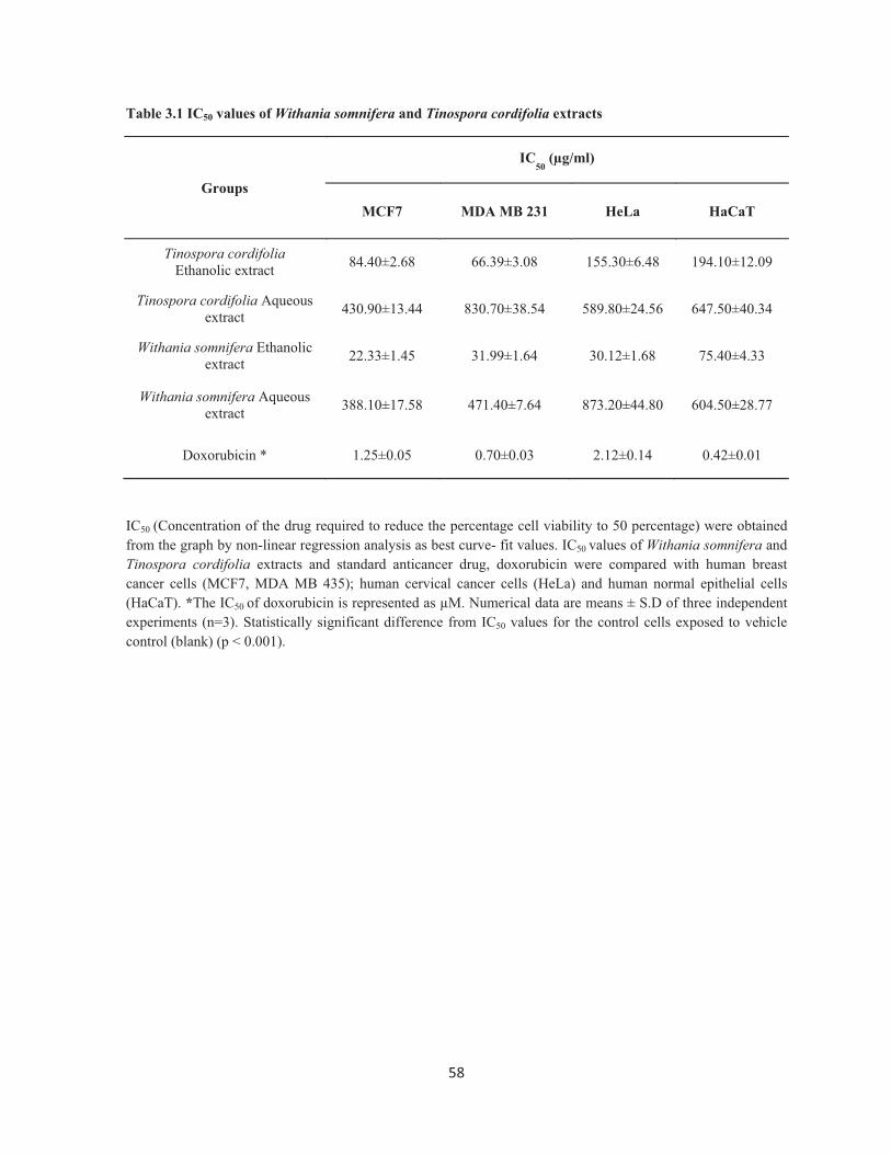
Table 3.1 IC50 values of Withania somnifera and Tinospora cordifolia extracts
Groups
IC50
(μg/ml)
MCF7 MDA MB 231 HeLa HaCaT
Tinospora cordifolia Ethanolic extract 84.40±2.68 66.39±3.08 155.30±6.48 194.10±12.09
Tinospora cordifolia Aqueous extract 430.90±13.44 830.70±38.54 589.80±24.56 647.50±40.34
Withania somnifera Ethanolic extract 22.33±1.45 31.99±1.64 30.12±1.68 75.40±4.33
Withania somnifera Aqueous extract 388.10±17.58 471.40±7.64 873.20±44.80 604.50±28.77
Doxorubicin * 1.25±0.05 0.70±0.03 2.12±0.14 0.42±0.01
IC50 (Concentration of the drug required to reduce the percentage cell viability to 50 percentage) were obtained from the graph by non-linear regression analysis as best curve- fit values. IC50 values of Withania somnifera and Tinospora cordifolia extracts and standard anticancer drug, doxorubicin were compared with human breast cancer cells (MCF7, MDA MB 435); human cervical cancer cells (HeLa) and human normal epithelial cells (HaCaT). *The IC50 of doxorubicin is represented as μM. Numerical data are means ± S.D of three independent experiments (n=3). Statistically significant difference from IC50 values for the control cells exposed to vehicle control (blank) (p < 0.001).
59��
Figure 3.7 Acridine orange-ethidium bromide assay to discriminate necrosis or apoptosis
Green colored nuclei indicate live cells. A condensed or fragmented nucleus with green or orange-red color indicates apoptosis and even nucleated red color indicates necrosis. Ethanolic extracts of both Withania somnifera and Tinospora cordifolia induced apoptosis but not necrosis in cancer cells. These figures are representative of three independent experiments (n=3).
A-Untreated control
B-Vehicle control (DMSO)
C-Doxorubicin hydrochloride
D-Withania somnifera ethanolic extract
E- Tinospora cordifolia ethanolic extract
HeLaMCF-7 MDA-MB -231
60��
Figure 3.8 Hoechst 33342 assay for the detection of apoptosis
Condensed and fragmented nuclei upon treatment with Withania somnifera and Tinospora cordifolia extracts indicated hall mark properties of apoptosis in breast cancer cells. These figures are representative of three independent experiments (n=3).
A-Untreated control
B-Vehicle control (DMSO)
C-Doxorubicin hydrochloride
D-Withania somnifera ethanolic extract
E- Tinospora cordifolia ethanolic extract
HeLaMCF-7 MDA-MB -231

61��
Figure 3.9 DNA fragmentation assay for the detection of apoptosis
DNA fragmentation assay in human breast cancer cells (MCF7, MDA MB 231) and cervical cancer cells (HeLa) indicated that Withania somnifera and Tinospora cordifolia induced DNA ladder formation, a hallmark property of apoptosis. A: Doxorubicin B: Withania somnifera ethanolic extract, C: Tinospora cordifolia ethanolic extract, D: Untreated control, E: Vehicle control (DMSO), M: DNA ladder (1kb). These figures are representative of three independent experiments (n=3).
M A B C D E
MCF-7
M A B C D EHeLa
MB CAD E
MDA-MB-231
62��
Figure 3.10 Cell cycle analysis after treatment with Withania somnifera and Tinospora cordifolia extracts
DNA content was analyzed based on propidium iodide (PI) staining using flow cytometery. The percentage number of DNA content in G0/G1, S, G2M and sub G0 (Apoptosis) phases were determined. These figures are representative of three independent experiments (n=3).
A-Untreated control
B-Vehicle control (DMSO)
C-Doxorubicin hydrochloride
D-Withania somnifera ethanolic extract
E- Tinospora cordifolia ethanolic extract
HeLaMCF-7 MDA-MB -231

63��
Table 3.2 Cell cycle specific effects of Withania somnifera and Tinospora cordifolia extracts in cancer cells
Groups
MCF7 MDA-MB-231
Sub G0 phase
Go/G1 phase
S phase G2/M phase
Sub G0 phase
Go/G1 phase
S phase G2/M phase
Untreated control 0.70±0.04 60.76±4.34 16.92±1.72 22.64±1.80 1.06±0.08 64.75±5.60 16.00±0.96 19.50±1.50
Vehicle control 1.90±0.10 57.38±5.38 16.76±2.13 25.20±2.45 0.62±0.10 62.72±4.80 14.37±0.77 23.57±2.56
Doxorubicin 30.46±2.07 34.90±3.24 10.73±0.98 25.14±2.96 62.14±4.80 14.17±1.23 11.96±1.23 12.83±0.76
Withania somnifera ethanolic extract
33.65±1.61 30.30±4.25 7.66±0.12 29.20±3.23 48.28±3.78 15.06±1.73 5.14±0.05 32.06±3.45
Tinospora cordifolia ethanolic extract
33.54±1.60 43.14±3.30 8.67±0.85 15.77±1.45 23.86±2.78 43.40±2.80 11.43±0.85 22.62±1.56
�
Groups
HeLa
Sub G0 phase
Go/G1 phase
S phase G2/M phase
Untreated control 0.86±0.03 50.14±3.75 17.37±0.95 32.91±1.96
Vehicle control (DMSO) 0.82±0.05 59.02±3.95 12.00±0.75 29.28±2.33
Doxorubicin 58.07±6.65 15.45±1/12 4.89±0.22 22.32±2.10
Withania somnifera ethanolic extract 37.53±3.25 11.09±0.95 4.14±0.18 47.83±3.78
Tinospora cordifolia ethanolic extract 18.64±1.56 49.96±1.20 4.06±0.25 31.40±2.66
Cell cycle analysis in cancer cells after treatment with Withania somnifera extract, Tinospora cordifolia extract and doxorubicin. DNA content was determined in G0/G1, S, G2M and sub G0 (Apoptosis) phase based on propidium iodide (PI) staining using flow cytometry. An increased Sub G0 phase indicating that extracts induced apoptosis in cancer cells. An increased G2M phase indicates cell cycle arrest and prevents mitosis. Numerical data are means ± S.D of three independent experiments (n=3). Statistically significant difference from the values for the control cells exposed to vehicle control (blank). p<0.001, determined by two way ANOVA using Bonferroni post test.
64��
Figure 3.11 Hoechst 333442 assay to detect apoptosis in human normal epithelial cells
Human normal epithelial cells (HaCaT) were treated with Withania somnifera extract, Tinospora cordifolia extract and doxorubicin at the concentration which was cytotoxic to the human breast cancer cells (MCF7) for 48 hours. Condensed and fragmented nuclei upon treatment with doxorubicin indicate apoptosis induced. However, extracts did not inuduced any significant apoptosis. These figures are representative of three independent experiments (n=3)
Figure 3.12 Cell cycle analysis in human normal epithelial cells after treatment with Withania somnifera and Tinospora cordifolia extracts
HaCaT cells were treated with Withania somnifera extract, Tinospora cordifolia extract and doxorubicin at the concentration which was cytotoxic to the human breast cancer cells (MCF7) for 48 hours. The percentage number of DNA in G0/G1, S, G2M and Sub G0 (apoptosis) phase was determined based on propidium iodide (PI) using flow cytometery. Doxorubicin significantly increased Sub G0 phase indicating induction of apoptosis. However, extracts did not imparted any cell cycle specific changes.These figures are representative of three independent experiments (n=3).
A-Untreated control B-Vehicle control (DMSO) C-Doxorubicin hydrochloride
D-Withania somnifera E- Tinospora cordifolia
A-Untreated control B-Vehicle control (DMSO) C-Doxorubicin hydrochloride
D-Withania somnifera E- Tinospora cordifolia

65��
Table 3.3 Cell cycle specific effects of Withania somnifera and Tinospora cordifolia extracts in normal epithelial cells
Groups
MCF7 HaCaT
Sub G0 phase
Go/G1 phase
S phase G2/M phase
Sub G0 phase
Go/G1 phase
S phase G2/M phase
Untreated control 0.70±0.04 60.76±4.34 16.92±1.72 22.64±1.80 2.25±0.07 58.84±3.57 18.40±1.22 22.00±0.95
Vehicle control (DMSO)
1.90±0.10 57.38±5.38 16.76±2.13 25.20±2.45 2.35±0.05 59.11±3.22 17.63±0.96 22.53±1.40
Doxorubicin 30.46±2.07 34.90±3.24 10.73±0.98 25.14±2.96 63.19±3.54 24.12±1.45 6.31±0.20 7.35±0.63
Withania somnifera ethanolic extract
33.65±1.61 30.30±4.25 7.66±0.12 29.20±3.23 6.24±0.72 58.76±2.90 14.43±0.56 21.47±0.70
Tinospora cordifolia ethanolic extract
33.54±1.60 43.14±3.30 8.67±0.85 15.77±1.45 6.56±0.65 58.63±3.11 15.07±0.88 21.24±0.88
Cell cycle analysis in human breast cancer cells (MCF7) and normal epithelial cells (HaCaT) after treatment with Withania somnifera extract, Tinospora cordifolia extract and doxorubicin. HaCaT cells were treated at the concentrations which was cytotoxic to human breast cancer cells (MCF7). DNA content was determined in G0/G1, S, G2M and sub G0 (apoptosis) phase. An increased Sub G0 phase indicated that extracts induces apoptosis specifically in cancer cells. However, currently used anticancer drug, doxorubicin induced apoptosis in both cancer and normal cells. All values are mean ± S.D of three independent experiments, p<0.001, determined by two way ANOVA using Bonferroni post test.

66��
3.5 REFERENCES
Cotter, T. G. 2009. Apoptosis and cancer: the genesis of a research field. Nature Reviews Cancer, 9,
501-507.
Davis, C. D. & Milner, J. A. 2010. Diet, Physical Activity, and Cancer Prevention. Nutrition Guide for
Physicians, 379-393.
Duffy, R., Wade, C. & Chang, R. 2012. Discovery of anticancer drugs from antimalarial natural
products: a MEDLINE literature review. Drug Discovery Today, Epub ahead of print.
Gottesman, M. M. 2002. Mechanisms of cancer drug resistance. Annual Review of Medicine, 53, 615-
627.
Jain, R. K. 2001. Delivery of molecular and cellular medicine to solid tumors1. Advanced Drug
Delivery Reviews, 46, 149-168.
Kuraparthy, S., Reddy, K. M., Yadagiri, L. A., Yutla, M., Venkata, P. B., Kadainti, S. & Reddy, R. P.
V. 2007. Epidemiology and patterns of care for invasive breast carcinoma at a community
hospital in Southern India. World J Surg Oncol, 5, 56.
Liu, W. J. H. 2011. Traditional Herbal Medicine Research Methods: Identification, Analysis,
Bioassay, and Pharmaceutical and Clinical Studies. Wiley.
Nobili, S., Landini, I., Mazzei, T. & Mini, E. 2011. Overcoming tumor multidrug resistance using
drugs able to evade P-glycoprotein or to exploit its expression. Medicinal Research Reviews,
Epub ahead of print.
Ogilvie, B. W. 2003. The many books of nature: Renaissance naturalists and information overload.
Journal of the History of Ideas, 64, 29-40.
Pan, L., Chai, H. & Kinghorn, A. 2012. Discovery of new anticancer agents from higher plants.
Frontiers in Bioscience (Scholar edition), 4, 142.
Pandey, M., Debnath, M., Gupta, S. & Chikara, S. K. 2011. Phytomedicine: An ancient approach
turning into future potential source of therapeutics. Journal of Pharmacognosy and
Phytotherapy, 3, 27-37.
Pluen, A., Boucher, Y., Ramanujan, S., Mckee, T. D., Gohongi, T., Di Tomaso, E., Brown, E. B.,
Izumi, Y., Campbell, R. B. & Berk, D. A. 2001. Role of tumor–host interactions in interstitial
diffusion of macromolecules: cranial vs. subcutaneous tumors. Proceedings of the National
Academy of Sciences, 98, 4628.
Sawadogo, W. R., Schumacher, M., Teiten, M. H., Dicato, M. & Diederich, M. 2012. Traditional
pharmacopoeia, plants and derived compounds for cancer therapy. Biochemical
Pharmacology, Epub ahead of print.

67��
Schmitt, C. A. 2003. Senescence, apoptosis and therapy-cutting the lifelines of cancer. Nature Reviews
Cancer, 3, 286-295.
Sharif, T., Alhosin, M., Auger, C., Minker, C., Kim, J. H., Etienne-Selloum, N., Bories, P.,
Gronemeyer, H., Lobstein, A. & Bronner, C. 2012. Aronia melanocarpa Juice Induces a
Redox-Sensitive p73-Related Caspase 3-Dependent Apoptosis in Human Leukemia Cells.
PloS One, 7, e32526.
Shukla, S., Wu, C. P. & Ambudkar, S. V. 2008. Development of inhibitors of ATP-binding cassette
drug transporters-present status and challenges. Expert Opinion on Drug Metabolism &
Toxicology, 4, 205-223.
Spitzer, O. 2011. Principles of Herbal Medicine. Clinical Naturopathic Medicine, Book chapter.