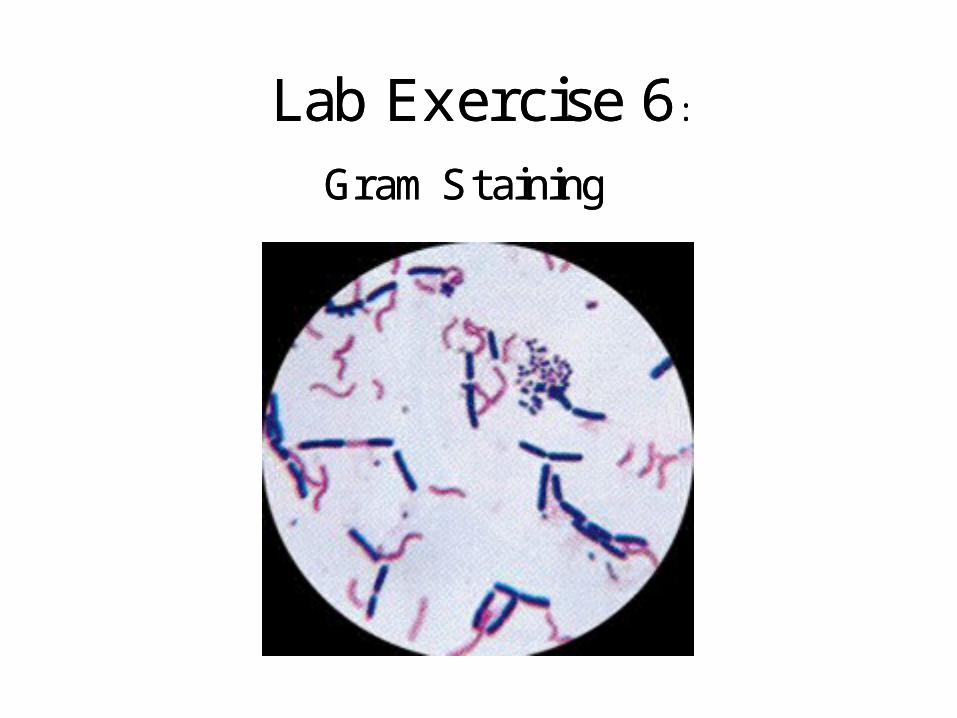
Crystal Gram’s (Primary stain) Gram’s (Primary stain) Gram’s Crystal violet Gram’s iodine...
12
Lab Exercise 6: Gram Staining Lab Exercise 6: Gram Staining
-
Upload
sophie-summers -
Category
Documents
-
view
230 -
download
1
Transcript of Crystal Gram’s (Primary stain) Gram’s (Primary stain) Gram’s Crystal violet Gram’s iodine...
Lab Exercise 6:
Gram Staining
Lab Exercise 6:
Gram Staining
Crystal Gram’s
(Primary stain)Gram’s
(Primary stain)Gram’s
Crystal violet
Gram’s iodine
SafraninAlcohol
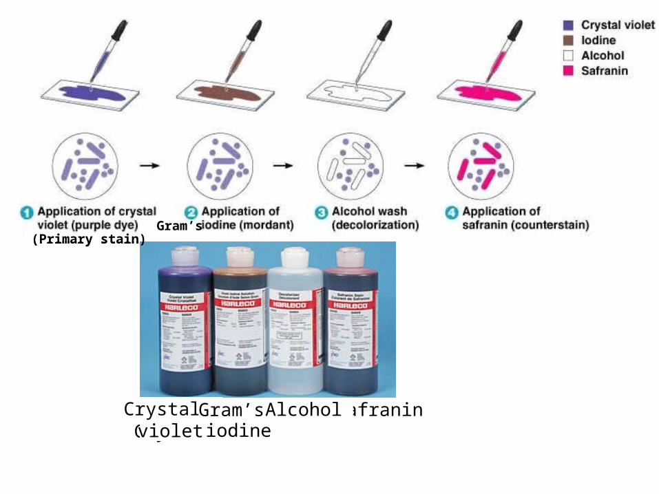
1. What is the name of this Gram stain solution?
2. How long was the stain lef t on the slide before being rinsed off ?
3. What is the name of the Gram stain solution?
4. What is the name of this Gram stain solution?
How long was this stain lef t on the slide before being rinsed off ?
1. What is the name of this Gram stain solution?
2. How long was the stain lef t on the slide before being rinsed off ?
3. What is the name of the Gram stain solution?
4. What is the name of this Gram stain solution?
How long was this stain lef t on the slide before being rinsed off ?

I dentif y the Gram-positive cells and the Gram-negative cells in these photographs.I dentif y the Gram-positive cells and the Gram-negative cells in these photographs.
1 2
3
4
5
Answers: 1. Gram-negative spirilli 2. Gram-positive bacilli 3. Gram-positive cocci
4. Gram-negative bacilli 5. Gram-positive cocci
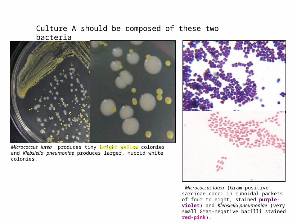
Culture A should be composed of these two bacteria
Micrococcus lutea produces tiny bright yellowbright yellow colonies and Klebsiella pneumoniae produces larger, mucoid white colonies.
Micrococcus lutea (Gram-positive sarcinae cocci in cuboidal packets of four to eight, stained purple-violet) and Klebsiella pneumoniae (very small Gram-negative bacilli stained red-pink).
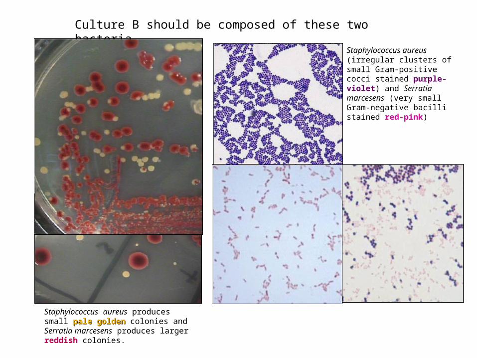
Culture B should be composed of these two bacteria
Staphylococcus aureus produces small pale goldenpale golden colonies and Serratia marcesens produces larger reddish colonies.
Staphylococcus aureus (irregular clusters of small Gram-positive cocci stained purple-violet) and Serratia marcesens (very small Gram-negative bacilli stained red-pink)

Culture C should be composed of these two bacteria
Escherichia coli produces small irregular translucent (T) white colonies and Bacillus megaterium produces larger, more rounded opaque (O) white colonies. These are sometimes difficult to distinguish in room light.
TO
Bacillus megaterium (large, thick Gram-positive bacilli, often with endospores, stained purple-violet) and Escherichia coli (small Gram-negative bacilli stained red-pink).
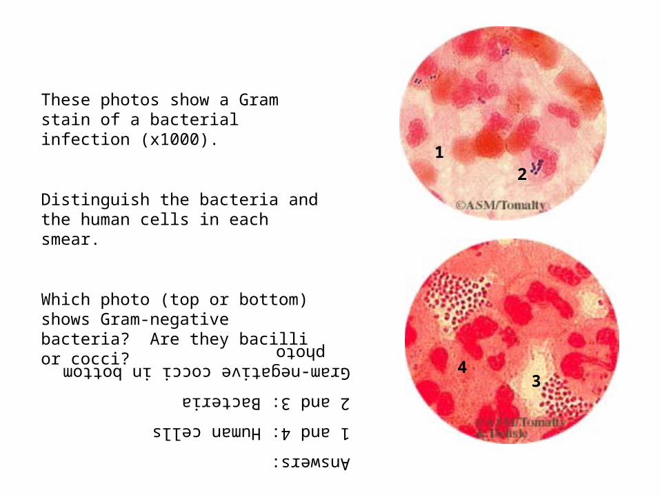
These photos show a Gram stain of a bacterial infection (x1000).
Distinguish the bacteria and the human cells in each smear.
Which photo (top or bottom) shows Gram-negative bacteria? Are they bacilli or cocci?
21
34
Answers:
1 and 4: Human cells
2 and 3: Bacteria
Gram-negative cocci in bottom photo
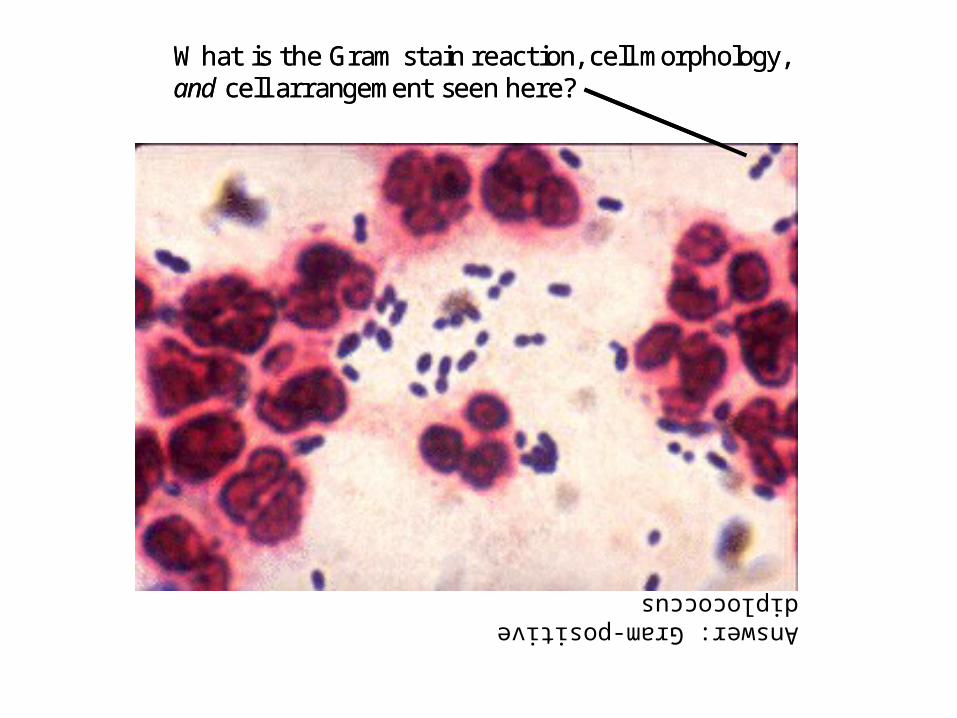
What is the Gram stain reaction, cell morphology, and cell arrangement seen here?What is the Gram stain reaction, cell morphology, and cell arrangement seen here?
Answer: Gram-positive diplococcus

What is the Gram stain reaction, cell morphology, and cell arrangement seen here?What is the Gram stain reaction, cell morphology, and cell arrangement seen here?
Answer: Gram-positive streptococcus

What is the Gram stain reaction, cell morphology, and cell arrangement seen here?What is the Gram stain reaction, cell morphology, and cell arrangement seen here?
Answer: Gram-negative bacilli
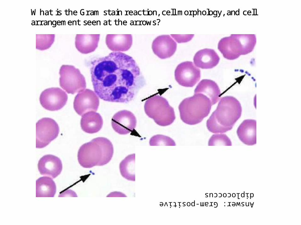
What is the Gram stain reaction, cell morphology, and cell arrangement seen at the arrows?What is the Gram stain reaction, cell morphology, and cell arrangement seen at the arrows?
Answer: Gram-positive diplococcus