Gram’s staining
15
GRAM’S STAINING Presented by – Gurjeet Kaur
-
Upload
gurjeet-gill -
Category
Science
-
view
39 -
download
0
Transcript of Gram’s staining

GRAM’S STAINING Presented by – Gurjeet Kaur
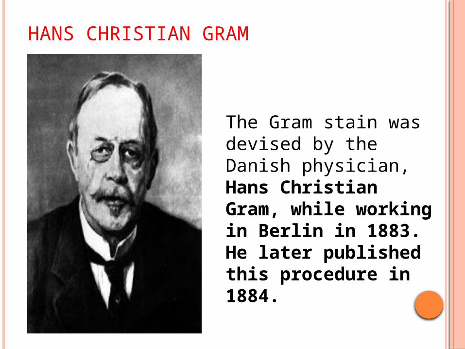
HANS CHRISTIAN GRAM
The Gram stain was devised by the Danish physician, Hans Christian Gram, while working in Berlin in 1883. He later published this procedure in 1884.

GRAM’S STAIN
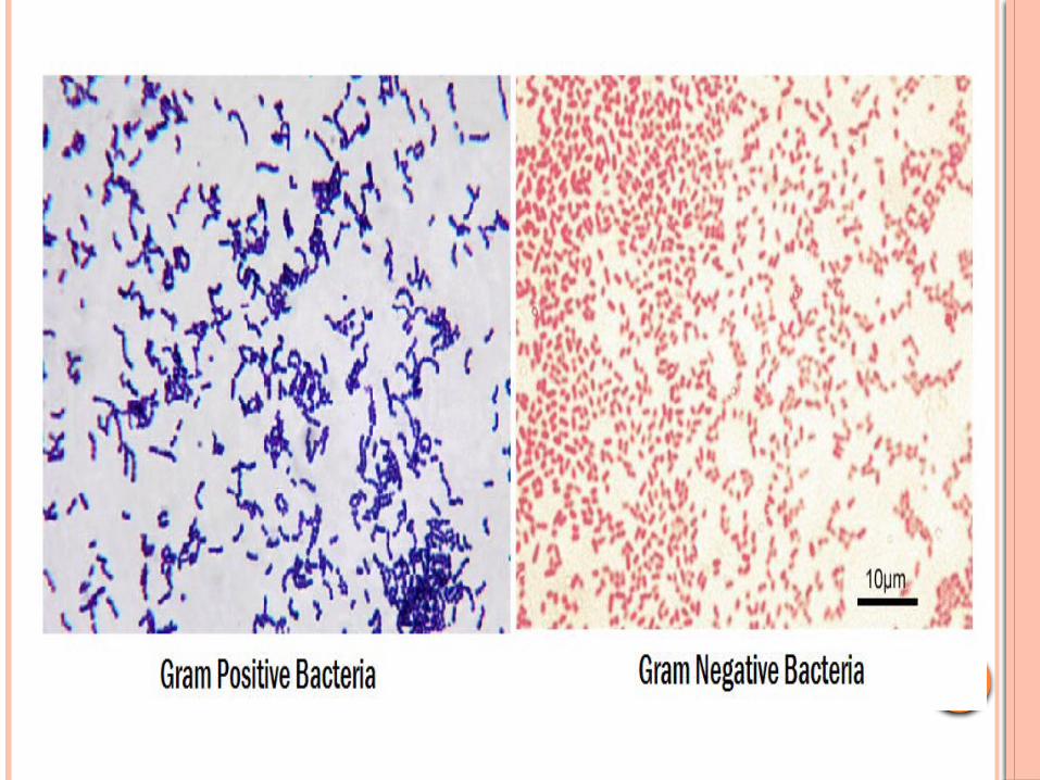
Gram staining (or Gram's method) is a method of differentiating bacterial species into two large groups
Gram Positive Gram Negative
Gram staining differentiates bacteria by the chemical and physical properties of their cell walls.

GRAM POSITIVE BACTERIA
• Gram positive bacteria have a thick cell wall of peptidoglycan.
• Peptidoglycan is a polymer consisting of sugar amino acids that form a mesh like outside the plasma membrane of bacteria forming cell wall.
• In Gram positive bacteria, between the cell wall and cell membrane, there is a "membrane teichoic acid".

GRAM NEGATIVE BACTERIA
Gram negative bacteria have an outer membrane of phospholipids and bacterial Lipopolysaccharides outside of their thin peptidoglycan layer. The space between the outer membrane and the peptidoglycan layer is called the periplasmic space.


PRINCIPLE OF GRAM’S STAINING The structure of the organism ‘s cell wall
determines whether the organism is gram positive or negative.
When stained with a primary stain and fixed by a mordant, some bacteria are able to retain the primary stain by resisting declorization while other get decolorized by decolorizer.
Those bacteria which retain the primary stain are called Gram positive.
Those bacteria which get decolorized and then get counterstained are called Gram negative.

1. Crystal violet - all bacteria take crystal violet- so all appears violet.
2. Iodine – Crystal Violet-iodine(CV-I) complex is formed.
3. Acetone- bacteria with high lipid content loose CV-I complex(appear colourless) but bacteria with less lipid content retains CV-I complex ( appear violet).
4. Safranine/ basic fuchsin – only colourless bacteria takes – appear pink.


PROCEDURE1. Make a smear & dry thoroughly in cool air. Fix
the dried film by passing it briefly through a bunsen flame.
2. Flood the slide with crystal violate sol. for upto 1 min. Wash off briefly with tap water & drain.
3.Flood the slide with gram’s iodine sol. & allow to act as a mordant for about 1 min. Wash off with tap water & drain.

4.Decolourise the smear with acetone for 10-30 sec. taking care not to overdecolourise & immediately wash off with water.
5.Flood the slide with safranin sol. & counterstain for about 30 sec, wash off with tap water, drain & blot dry with filter paper & examine under oil immersion objective.


EXAMPLES

Thank You