Cronicon - ECronicon Open Access | Scientific Publications ... · ing drill holes in the femur...
9
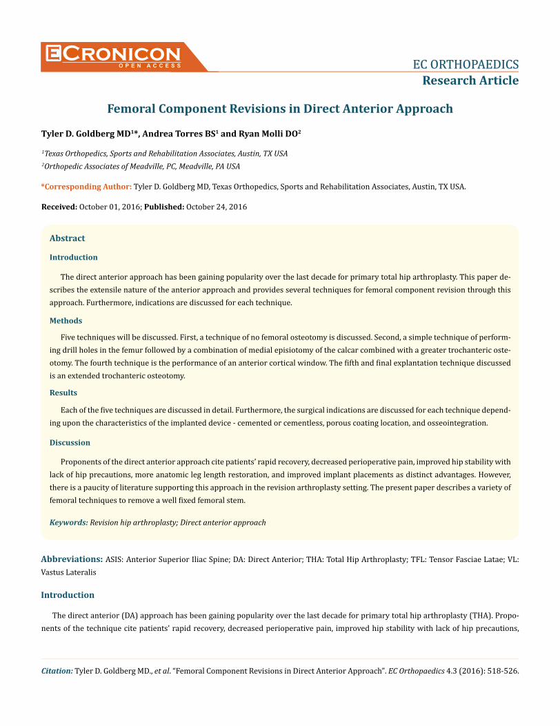
Cronicon OPEN ACCESS EC ORTHOPAEDICS Research Article Femoral Component Revisions in Direct Anterior Approach Tyler D. Goldberg MD 1 *, Andrea Torres BS 1 and Ryan Molli DO 2 1 Texas Orthopedics, Sports and Rehabilitation Associates, Austin, TX USA 2 Orthopedic Associates of Meadville, PC, Meadville, PA USA *Corresponding Author: Tyler D. Goldberg MD, Texas Orthopedics, Sports and Rehabilitation Associates, Austin, TX USA. Citation: Tyler D. Goldberg MD., et al. “Femoral Component Revisions in Direct Anterior Approach”. EC Orthopaedics 4.3 (2016): 518-526. Received: October 01, 2016; Published: October 24, 2016 Abstract Introduction The direct anterior approach has been gaining popularity over the last decade for primary total hip arthroplasty. This paper de- scribes the extensile nature of the anterior approach and provides several techniques for femoral component revision through this approach. Furthermore, indications are discussed for each technique. Methods Five techniques will be discussed. First, a technique of no femoral osteotomy is discussed. Second, a simple technique of perform- ing drill holes in the femur followed by a combination of medial episiotomy of the calcar combined with a greater trochanteric oste- otomy. The fourth technique is the performance of an anterior cortical window. The fifth and final explantation technique discussed is an extended trochanteric osteotomy. Results Each of the five techniques are discussed in detail. Furthermore, the surgical indications are discussed for each technique depend- ing upon the characteristics of the implanted device - cemented or cementless, porous coating location, and osseointegration. Discussion Proponents of the direct anterior approach cite patients’ rapid recovery, decreased perioperative pain, improved hip stability with lack of hip precautions, more anatomic leg length restoration, and improved implant placements as distinct advantages. However, there is a paucity of literature supporting this approach in the revision arthroplasty setting. The present paper describes a variety of femoral techniques to remove a well fixed femoral stem. Keywords: Revision hip arthroplasty; Direct anterior approach Abbreviations: ASIS: Anterior Superior Iliac Spine; DA: Direct Anterior; THA: Total Hip Arthroplasty; TFL: Tensor Fasciae Latae; VL: Vastus Lateralis Introduction The direct anterior (DA) approach has been gaining popularity over the last decade for primary total hip arthroplasty (THA). Propo- nents of the technique cite patients’ rapid recovery, decreased perioperative pain, improved hip stability with lack of hip precautions,
Transcript of Cronicon - ECronicon Open Access | Scientific Publications ... · ing drill holes in the femur...
CroniconO P E N A C C E S S EC ORTHOPAEDICS
Research Article
Femoral Component Revisions in Direct Anterior Approach
Tyler D. Goldberg MD1*, Andrea Torres BS1 and Ryan Molli DO2
1Texas Orthopedics, Sports and Rehabilitation Associates, Austin, TX USA2Orthopedic Associates of Meadville, PC, Meadville, PA USA
*Corresponding Author: Tyler D. Goldberg MD, Texas Orthopedics, Sports and Rehabilitation Associates, Austin, TX USA.
Citation: Tyler D. Goldberg MD., et al. “Femoral Component Revisions in Direct Anterior Approach”. EC Orthopaedics 4.3 (2016): 518-526.
Received: October 01, 2016; Published: October 24, 2016
Abstract
Introduction
The direct anterior approach has been gaining popularity over the last decade for primary total hip arthroplasty. This paper de-scribes the extensile nature of the anterior approach and provides several techniques for femoral component revision through this approach. Furthermore, indications are discussed for each technique.
Methods
Five techniques will be discussed. First, a technique of no femoral osteotomy is discussed. Second, a simple technique of perform-ing drill holes in the femur followed by a combination of medial episiotomy of the calcar combined with a greater trochanteric oste-otomy. The fourth technique is the performance of an anterior cortical window. The fifth and final explantation technique discussed is an extended trochanteric osteotomy.
Results
Each of the five techniques are discussed in detail. Furthermore, the surgical indications are discussed for each technique depend-ing upon the characteristics of the implanted device - cemented or cementless, porous coating location, and osseointegration.
Discussion
Proponents of the direct anterior approach cite patients’ rapid recovery, decreased perioperative pain, improved hip stability with lack of hip precautions, more anatomic leg length restoration, and improved implant placements as distinct advantages. However, there is a paucity of literature supporting this approach in the revision arthroplasty setting. The present paper describes a variety of femoral techniques to remove a well fixed femoral stem.
Keywords: Revision hip arthroplasty; Direct anterior approach
Abbreviations: ASIS: Anterior Superior Iliac Spine; DA: Direct Anterior; THA: Total Hip Arthroplasty; TFL: Tensor Fasciae Latae; VL: Vastus Lateralis
Introduction
The direct anterior (DA) approach has been gaining popularity over the last decade for primary total hip arthroplasty (THA). Propo-nents of the technique cite patients’ rapid recovery, decreased perioperative pain, improved hip stability with lack of hip precautions,

519
Femoral Component Revisions in Direct Anterior Approach
Citation: Tyler D. Goldberg MD., et al. “Femoral Component Revisions in Direct Anterior Approach”. EC Orthopaedics 4.3 (2016): 518-526.
more anatomic leg length restoration, and improved implant placements. Critics cite the complex learning curve required, narrow patient indications, and lack of femoral extensile exposure especially in revision settings as distinct disadvantages.
Kurtz., et al. have shown that the demand for primary THAs is projected to increase by 174% over the next several decades, with the prevalence of revision THAs rising by 137% in the same time [1]. Revision THA techniques such as extensile femoral osteotomies, once considered possible only through a posterior approach, are now routinely performed anteriorly as surgeons gain familiarity with the DA approach and expand the indications.
Currently, however, there is a paucity of literature with explanations of the techniques involved in anterior revisions of the femoral component. This paper describes the extensile nature of the anterior approach and includes multiple specific techniques for femoral component revisions. The authors’ indications for each technique are also discussed.
Indications and Contraindications
As with any surgical technique, certain clinical scenarios and patients are easier to address due to several different factors, including patient demographics, anatomic characteristics, reason for revision, stability of the current implants, extent of bone loss, implant fixation, previous approach, existing hardware, etc.
While there are no absolute indications or contraindications for anterior approach revision surgery, there are several factors that influ-ence a surgeon’s choice. As surgeons become more comfortable with the approach, they are able to expand their indications for simple THA revisions, and, subsequently, more complex cases.
Specialized instrumentation and retractors, including a femoral extension table, femoral elevators, and various osteotomes, facilitate the approach and make it more effective, though they are not necessary.
Direct Anterior Approach
The DA approach is a true intermuscular and internervous approach. Many authors have described variations of the approach in de-tail [2-4]. Briefly, the skin incision for the approach is located approximately 2 cm distal and 3 cm posterior to the anterior superior iliac spine (ASIS) over the body of the tensor fasciae latae (TFL) muscle belly. Superficially, the interval lies between the sartorius (femoral nerve) and TFL (superior gluteal nerve). Dissection is typically through the fascia of the TFL, retracting it laterally. The deep dissection occurs between the rectus femoris (femoral nerve) and gluteus medius (superior gluteal nerve). Below the floor of the rectus fascia lies the ascending branch of the lateral femoral circumflex artery and accompanying veins. These vessels must be ligated and controlled for safe exposure and minimal blood loss.
Extensile exposure, regardless of which osteotomy technique utilized, is the same when performing femoral revisions. First, the skin incision is extended posteriorly to the mid-coronal plane and extended distally as far as necessary for exposure. The TFL tendon can be retracted laterally to achieve access to the vastus ridge, vastus lateralis (VL), and lateral femur. Alternatively, a separate incision through the iliotibial band tendon distally affords access to the lateral aspect of the vastus lateralis.
To gain access to the femur the VL can be elevated off of its intermuscular septum from posterior to anterior or the VL can be split in line with its fibers. The perforating vessels that supply the VL must be ligated/cauterized to minimize blood loss. For maximal exposure and subsequent repair of the dissection, it is often helpful to leave a cuff of the VL tendon origin attached to the vastus ridge for reattach-ment upon completion of the procedure.
With femoral revisions, it is often necessary to have a direct approach down the femoral medullary canal in order to safely prepare the femur and implant long revision stems (often diaphyseal engaging). This can be challenging through the DA approach due to intact

Citation: Tyler D. Goldberg MD., et al. “Femoral Component Revisions in Direct Anterior Approach”. EC Orthopaedics 4.3 (2016): 518-526.
Femoral Component Revisions in Direct Anterior Approach
520
muscular attachments to the top of the femur (gluteus medius/minimus, short external rotators). To allow for better femoral exposure and excursion, the head of the table can be placed in a slight Trendelenburg position to effectively hyperextend the hip and DA specific femoral retractors facilitate the approach.
Techniques for Femoral Component Removal
Technique 1: No osteotomy
Some implants do not require an osteotomy of the femur in order to be retrieved. The proximal femur is exposed through external rotation, extension, and adduction, bringing the neck of the implant into view. This allows an extraction device to dock with the implant for extraction. Utilization of this technique often requires the rupture of “spot welds” on the implant using a flexible osteotome on the anterior and posterior sides of the implant. Pneumatic, flexible osteotomes (AcuDriver, Exactech, Inc) have been especially useful with this technique (Figure 1). Small curved rigid or flexible osteotomes can free the medial and lateral sides of the implant. It is also critical to make sure the lateral shoulder of the implant is clearly visualized and exposed to avoid unnecessary damage to the greater trochanter and its muscular attachments. This is often easiest with a small high speed acorn tip burr.
Figure 1: Pneumatic osteotomes can be used to aid in removing stem.
This technique maintains an intact femur, though it carries the potential for fracture (Figure 2). The surgeon must make sure that the entire femur is intact after stem removal as fractures can occur as far distally as the suprametaphyseal region. We routinely use this technique for femoral components that are well fixed distally but loose proximally, easily identified by proximal radiolucencies on plain radiographs, or cemented implants that have loosened from their mantle. This technique may be augmented with the addition longitu-dinal drill holes referenced below. It is helpful to consider a time limit before progressing to a more aggressive approach due the time consuming nature of this technique.
Technique 2: Femoral Shaft Drill Holes
For a more diaphyseal engaging stem or a stem that has been freed of osseous ingrowth and fixation proximally, there are often inac-cessible areas of fixation distally where femoral hoop stress will “hold on” to the implant. In these instances, one can identify the area of

Citation: Tyler D. Goldberg MD., et al. “Femoral Component Revisions in Direct Anterior Approach”. EC Orthopaedics 4.3 (2016): 518-526.
Femoral Component Revisions in Direct Anterior Approach
521
Figure 2: Radiograph of distal fracture of femur after removal of well fixed stem.
the femur that appears to be capturing the implant (with the help of fluoroscopy) and expose it through a separate, laterally based incision that is centered over the iliotibial band and splits the VL fibers in line with the skin incision. Anterior and posterior Bennett-type retrac-tors are then placed and several drill holes are made over the area of suspected maximal hoop stresses and connected with an osteotome or small bunion-style osteotomy saw to minimize bone removal (Figure 3, 4). It is critically important to place prophylactic cables below the osteotomy prior to creating the osteotomy to prevent distal fracture propagation and above the osteotomy after stem removal to pre-vent proximal fracture propagation.
Figure 3: Intraoperative photos showing the placement of femoral drill holes (a-c), connected with a saw (d).

Citation: Tyler D. Goldberg MD., et al. “Femoral Component Revisions in Direct Anterior Approach”. EC Orthopaedics 4.3 (2016): 518-526.
Femoral Component Revisions in Direct Anterior Approach
522
Figure 4: Femoral drill holes on bone model.
Technique 3 : Episiotomy +/- Greater Trochanter osteotomy
This technique is performed in two distinct steps. The first step is a medial calcar longitudinal osteotomy performed with a straight, narrow osteotome or small bunion type saw blade (Figure 5). This allows the proximal femur to “open” and release proximal hoop stresses, and thus allow more access to the anterior/posterior sides. The amount of displacement is typically 2-3 mm only and care must be taken to not completely fracture the femur. If the implant still remains well fixed, it is likely integrated by the lateral side. In this case, an osteotomy of the greater trochanter is then necessary. A curved osteotome or narrow reciprocating saw is used. One can perform a single plane or biplanar (chevron style) osteotomy. The osteotomy begins just lateral to the shoulder of the implant and the cut proceeds distally, aiming just distal to the vastus ridge (Figure 6). It is important to make the osteotomized bone as large as possible to increase the surface area for healing. The biplanar or chevron osteotomy also provides more inherent rotational stability and anatomical positioning during the healing phase.
Figure 5: Episiotomy on bone model, with red line indicating cut line and black line the placement of the AP screw.

523
Femoral Component Revisions in Direct Anterior Approach
Citation: Tyler D. Goldberg MD., et al. “Femoral Component Revisions in Direct Anterior Approach”. EC Orthopaedics 4.3 (2016): 518-526.
Figure 6: Greater trochanter osteotomy on bone model, with red line indicating cut line.
Once the implant is removed, fixation of the osteotomies requires only a small wire or cable fixing the medial episiotomy. Alternatively, a small 3.5 mm cortical screw can be placed across the medial calcar to fixate the episiotomy. The surgeon should not attempt to fix the greater trochanter fragment as it will often reduce anatomically once the hip length and offset are restored. This is due to the intact muscular sleeve or “deltoid of the hip” to the osteotomized fragment. This sleeve is made up of the gluteus medius, gluteus minimus, piriformis, obturator externus, and vastus lateralis. Attempts to fixate this piece will often destabilize it causing trochanteric escape and eventual non-union.
This technique is relatively easy to perform, potentially improves exposure, and reliably heals. We generally use this technique for proximally-coated, well-fixed, non-cemented stems. Fully-coated stems are also retrieved with this technique provided they are shorter in length.
Technique 4 : Anterior Cortical Window
This technique allows a broader intra-cortical view of the proximal implant by removing the anterior cortex of the femur. This al-lows excellent exposure of the anterior, medial, and lateral sides of the implant. With the hip in a reduced position, the IT band is incised distally and retracted laterally, thus exposing the proximal origin of the VL. The VL is next released from its proximal origin beginning laterally just distal to the gluteus minimus insertion. This is extended approximately 5 cm distally and then reflected medially in a sub-periosteal fashion. Next, a saw is used to incise the lateral cortex the length of the exposure and finished with a medial cut (Figure 7). A drill hole is placed in the corner of the 2 bony cuts. A protective cerclage cable is placed just below the osteotomy to prevent distal fracture propagation. The fragment is hinged medially allowing exposure of the underlying implant. The exposure of the lateral side of the implant requires dislocation, external rotation, and extension. Once the implant is retrieved, the cortical fragment is fixated with 1 - 2 simple wires/cables around the proximal femur. Typically, the cable is placed directly over the fragment so that it lies proximal to the lesser trochanter medially and distal to the vastus ridge laterally. The disadvantage of this method is the inability to directly expose the lateral side of the implant. Typically, this technique is used for stems that are both fully and proximally-coated as well as cemented stems with a poor cement mantle.

524
Femoral Component Revisions in Direct Anterior Approach
Citation: Tyler D. Goldberg MD., et al. “Femoral Component Revisions in Direct Anterior Approach”. EC Orthopaedics 4.3 (2016): 518-526.
Figure 7: Anterior cortical window on bone model, with blue line indicating cut line.
Technique 5 : Extended Trochanteric Osteotomy (ETO)
To perform an ETO, the proximal femur is exposed in a similar fashion to technique 3. However, the amount of VL reflected off the fe-mur is significantly more – upwards of 10-12 cm. Once the anterior cortex of the femur is exposed, the osteotomy is performed in an oppo-site fashion. The longitudinal cut is performed anteriorly for a distance of 8 - 12 cm as required by the underlying implant. Distally the cut is turned laterally finishing at the mid-coronal line (Figure 8). Once again, a drill hole is placed in the apex and protective cable is placed just distal to the osteotomy to prevent any propagation. Next osteotomes are used to hinge the fragment from medial to lateral cracking the femur laterally/posteriorly. This completely exposes the lateral, anterior, and posterior sides of the implant. Internal rotation allows greater visualization of the anterior/posterior sides of the implant. Once the implant is removed and new implant placed, the osteotomy is fixed with cerclage cables (Figure 9). The advantage of the technique includes full, broad exposure of the implant. The disadvantage is the time required and amount of dissection. We typically use the ETO for indications similar to posterior approach ETO’s - well-fixed, cemented stems, and fully porous-coated stems.
Figure 8: Extended trochanteric osteotomy on bone model, with red line indicating cut line.

Citation: Tyler D. Goldberg MD., et al. “Femoral Component Revisions in Direct Anterior Approach”. EC Orthopaedics 4.3 (2016): 518-526.
Figure 9: Radiographs taken six weeks postoperatively (a) after extended trochanteric osteotomy (ETO) performed. Radiographs taken three years postoperatively demonstrate excellent consolidation of bone (b).
Discussion
Bibliography
Femoral Component Revisions in Direct Anterior Approach
525
It has been shown that the incidence of primary as well as revision THA will steadily increase over the next several decades [2,5-7]. As this occurs and patients become more demanding of less invasive techniques, less pain and quicker functional recovery, surgeons must adapt to these demands by becoming more familiar and comfortable with surgical approaches/techniques that respect this.
A standard posterior or direct lateral approach may be best for revision cases for fresh postoperative infections, metal-on-metal pseu-dotumors, massive soft tissue necrosis. The majority of both simple and complex revision THA cases, however, can be safely and effec-tively addressed through the DA approach.
The DA approach is not yet a well-accepted approach for revision THA, despite its increase in popularity for primary cases. It is our belief that as more surgeons begin to utilize the DA approach for primary THAs they will shift their perspective likely begin to incorporate the DA approach for revision surgeries. This transition often occurs slowly as the surgeon progresses through simpler cases such as head and liner exchanges, followed by simple acetabular and femoral revisions, and finally more complex femoral revisions requiring truly extensile techniques.
The current skepticism with revision DA approach for revisions is based on the idea that it is not extensile. As we have explained, the approach can be extremely extensile while maintaining the many advantages of anatomically respecting intermuscular and internervous planes and allowing patients a quicker functional recovery. The challenge for most surgeons lies in shifting their mentality from using a posterior approach for revisions to using the DA approach and overcoming the learning curve. As surgeons more readily use the DA ap-proach, there will likely be a natural transition to using the approach for revision cases.
1. Kurtz S., et al. “Projections of Primary and Revision Hip and Knee Arthroplasty in the United States from 2005 to 2030”. Journal of Bone & Joint Surgery America 89.4 (2007): 780-785.
2. Post ZD., et al. “Direct Anterior Approach for Total Hip Arthroplasty: Indications, Technique, and Results”. Journal of the American Academy of Orthopaedic Surgeons 22.9 (2014): 595-603.

Citation: Tyler D. Goldberg MD., et al. “Femoral Component Revisions in Direct Anterior Approach”. EC Orthopaedics 4.3 (2016): 518-526.
Femoral Component Revisions in Direct Anterior Approach
526
3. Hallert O., et al. “The direct anterior approach: initial experience of a minimally invasive technique for total hip arthroplasty”. Journal of Orthopaedic Surgery and Research 7 (2012): 17.
4. Howell J., et al. “Minimally invasive hip replacement: rationale, applied anatomy, and instrumentation”. Orthopedic Clinics of North America 35.2 (2004): 107-18.
5. Bozic KJ., et al. “The Epidemiology of Revision Total Hip Arthroplasty in the United States”. Journal of Bone & Joint Surgery America 91.1 (2009): 128-133.
6. Kavanagh BF., et al. “Revision Total Hip Arthroplasty”. Journal of Bone & Joint Surgery America 67.4 (1985): 517-526.
7. Mahomed NN., et al. “Rates and outcomes of primary and revision total hip replacement in the United States medicare population”. Journal of Bone & Joint Surgery America 85.1 (2003): 27-32.
Volume 4 Issue 3 October 2016© All rights reserved by Tyler D. Goldberg MD., et al.