Conundrum: Measuring Myopia Progression
60
+ Controversies and Conundrums in Myopia Management By Mark Koszek Professional Education Officer (PEO) EyeQ Optometrists B.Optom, M.Optom, Grad.Cert Oc.Ther Board Member CCLSA NSW Division
Transcript of Conundrum: Measuring Myopia Progression
+
Controversies and Conundrums in Myopia Management
By Mark KoszekProfessional Education Officer (PEO)
EyeQ Optometrists
B.Optom, M.Optom, Grad.Cert Oc.Ther
Board Member CCLSA NSW Division
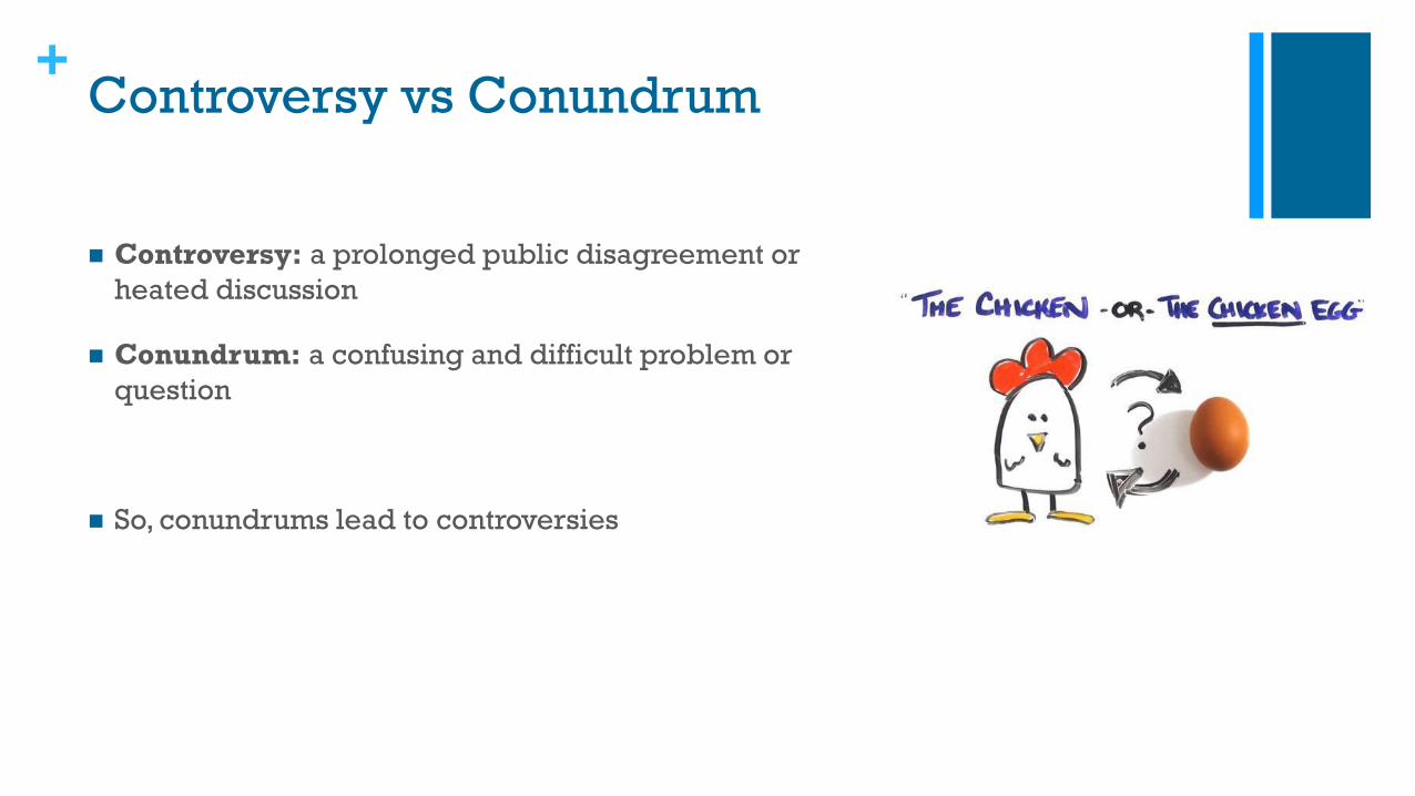
+Controversy vs Conundrum
◼ Controversy: a prolonged public disagreement or
heated discussion
◼ Conundrum: a confusing and difficult problem or
question
◼ So, conundrums lead to controversies
+Conundrum: Measuring Myopia Progression
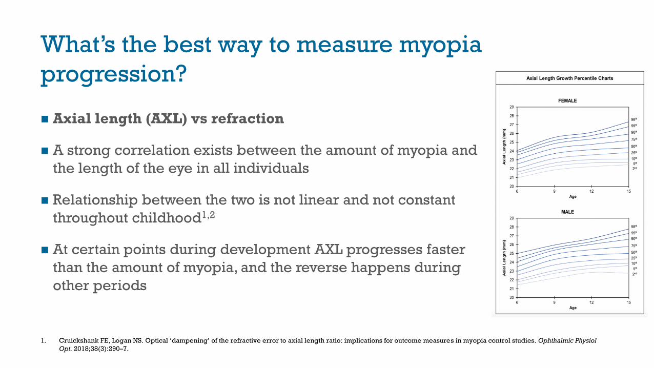
What’s the best way to measure myopia
progression?
◼ Axial length (AXL) vs refraction
◼ A strong correlation exists between the amount of myopia and
the length of the eye in all individuals
◼ Relationship between the two is not linear and not constant
throughout childhood1,2
◼ At certain points during development AXL progresses faster
than the amount of myopia, and the reverse happens during
other periods
1. Cruickshank FE, Logan NS. Optical ‘dampening’ of the refractive error to axial length ratio: implications for outcome measures in myopia control studies. Ophthalmic Physiol
Opt. 2018;38(3):290–7.
+CLEERE Study
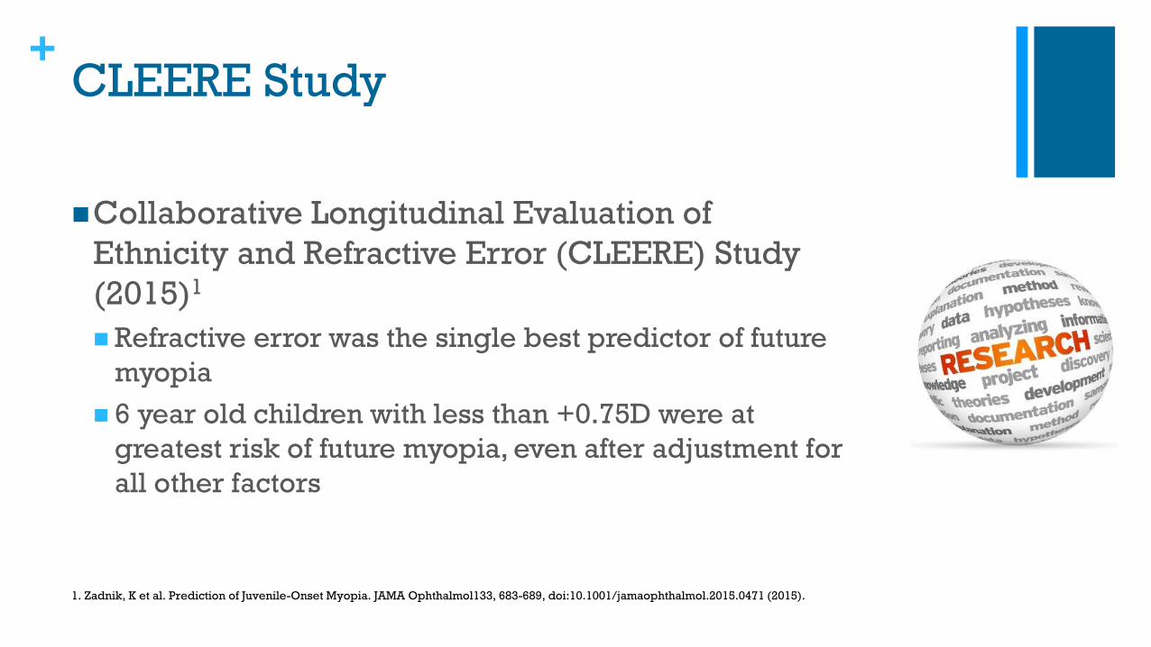
◼Collaborative Longitudinal Evaluation of
Ethnicity and Refractive Error (CLEERE) Study
(2015)1
◼ Refractive error was the single best predictor of future
myopia
◼ 6 year old children with less than +0.75D were at
greatest risk of future myopia, even after adjustment for
all other factors
1. Zadnik, K et al. Prediction of Juvenile-Onset Myopia. JAMA Ophthalmol133, 683-689, doi:10.1001/jamaophthalmol.2015.0471 (2015).
+Axial Length and Myopia
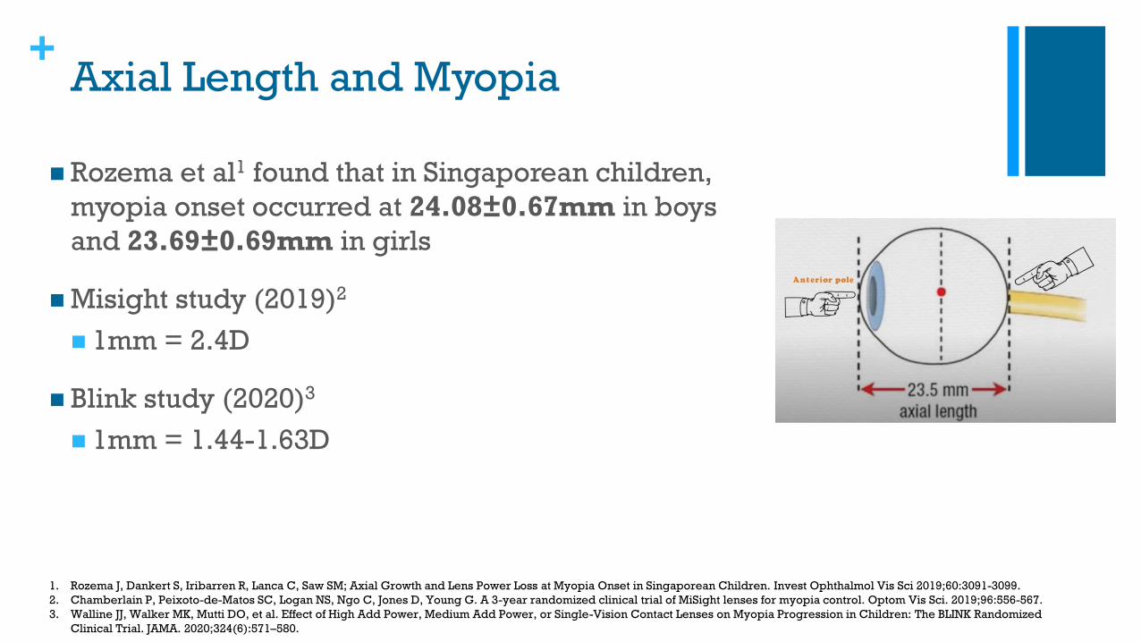
◼ Rozema et al1 found that in Singaporean children,
myopia onset occurred at 24.08±0.67mm in boys
and 23.69±0.69mm in girls
◼ Misight study (2019)2
◼ 1mm = 2.4D
◼ Blink study (2020)3
◼ 1mm = 1.44-1.63D
1. Rozema J, Dankert S, Iribarren R, Lanca C, Saw SM; Axial Growth and Lens Power Loss at Myopia Onset in Singaporean Children. Invest Ophthalmol Vis Sci 2019;60:3091-3099.
2. Chamberlain P, Peixoto-de-Matos SC, Logan NS, Ngo C, Jones D, Young G. A 3-year randomized clinical trial of MiSight lenses for myopia control. Optom Vis Sci. 2019;96:556-567.
3. Walline JJ, Walker MK, Mutti DO, et al. Effect of High Add Power, Medium Add Power, or Single-Vision Contact Lenses on Myopia Progression in Children: The BLINK Randomized
Clinical Trial. JAMA. 2020;324(6):571–580.
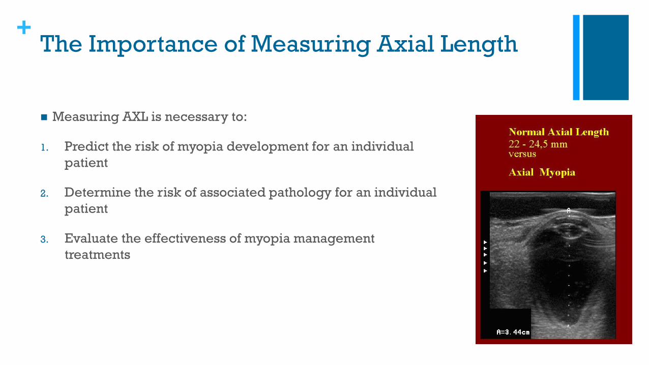
+The Importance of Measuring Axial Length
◼ Measuring AXL is necessary to:
1. Predict the risk of myopia development for an individual
patient
2. Determine the risk of associated pathology for an individual
patient
3. Evaluate the effectiveness of myopia management
treatments
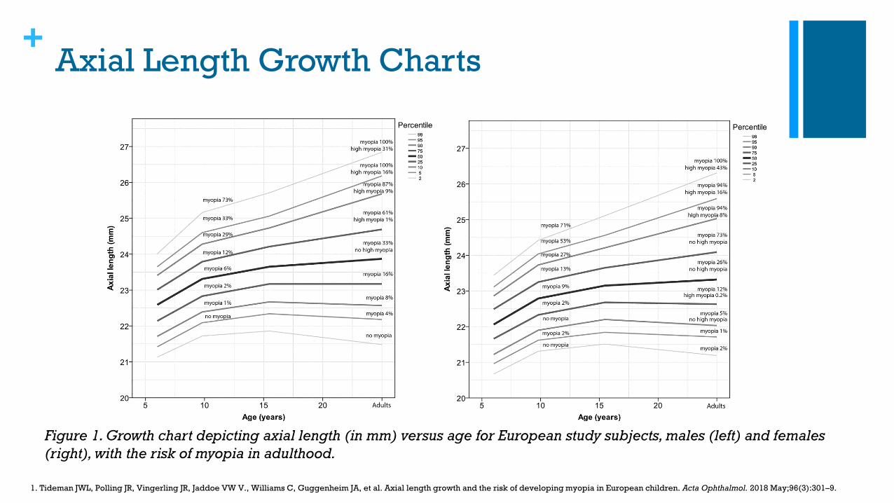
+Axial Length Growth Charts
Figure 1. Growth chart depicting axial length (in mm) versus age for European study subjects, males (left) and females
(right), with the risk of myopia in adulthood.
1. Tideman JWL, Polling JR, Vingerling JR, Jaddoe VW V., Williams C, Guggenheim JA, et al. Axial length growth and the risk of developing myopia in European children. Acta Ophthalmol. 2018 May;96(3):301–9.
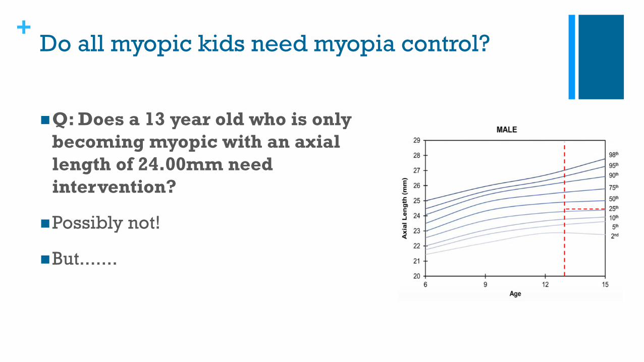
+Do all myopic kids need myopia control?
◼Q: Does a 13 year old who is only
becoming myopic with an axial
length of 24.00mm need
intervention?
◼Possibly not!
◼But.......
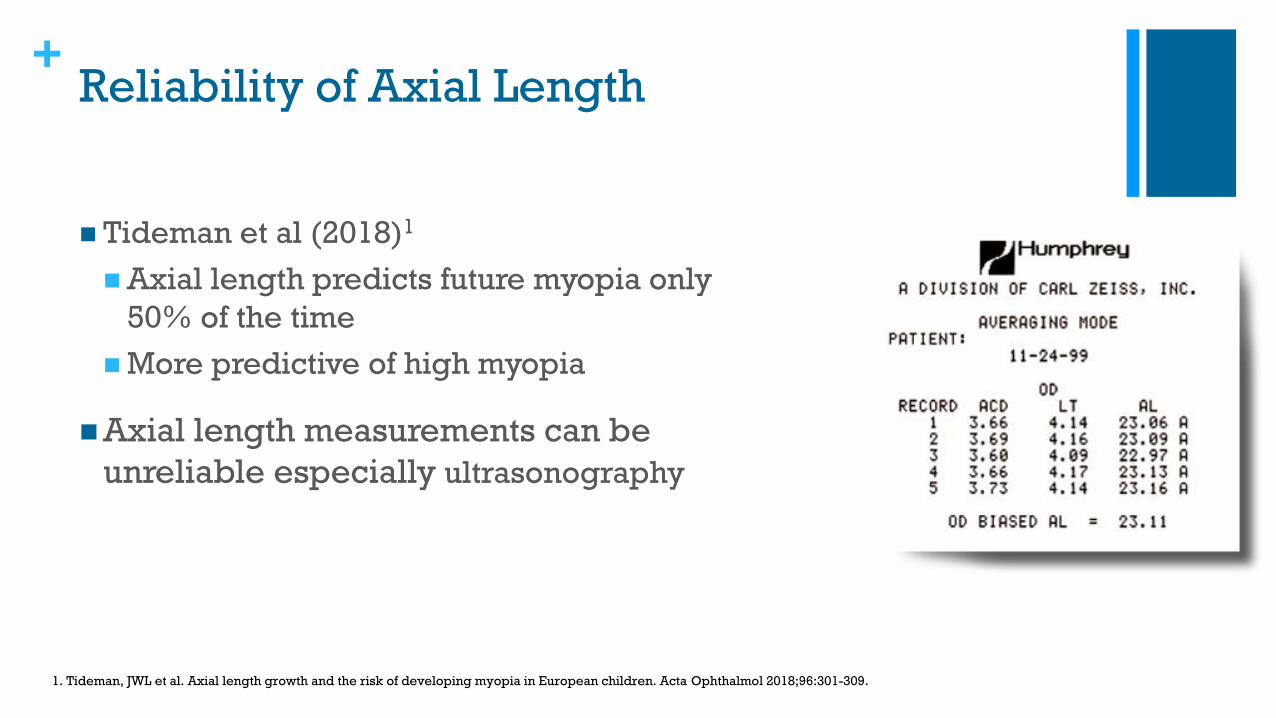
+Reliability of Axial Length
◼ Tideman et al (2018)1
◼ Axial length predicts future myopia only
50% of the time
◼ More predictive of high myopia
◼Axial length measurements can be
unreliable especially ultrasonography
1. Tideman, JWL et al. Axial length growth and the risk of developing myopia in European children. Acta Ophthalmol 2018;96:301-309.
+Gold Standard in Myopia Care
◼Cycloplegic refraction
◼Axial length
◼Utilise the growth charts
+
Controversy: Which treatment is best?
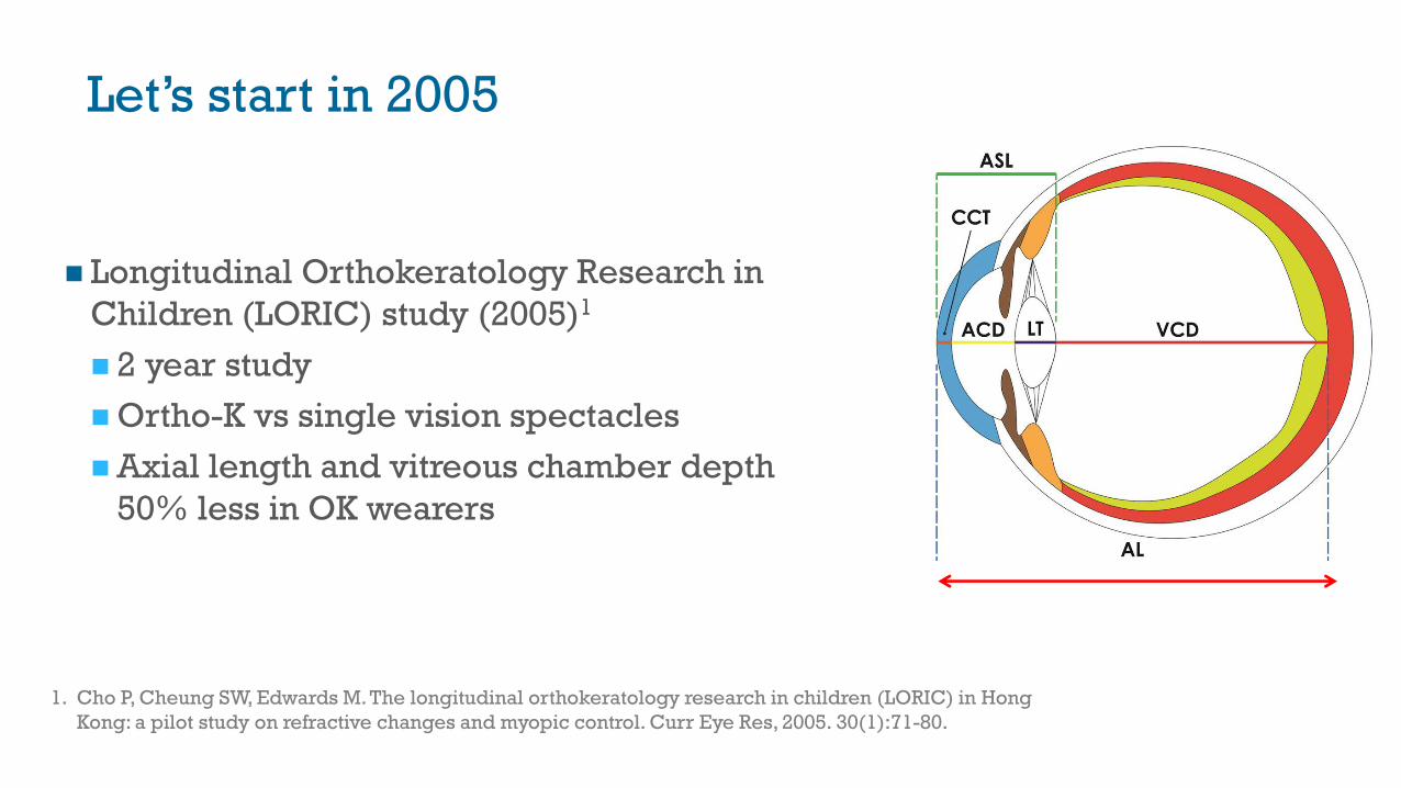
Let’s start in 2005
◼ Longitudinal Orthokeratology Research in
Children (LORIC) study (2005)1
◼ 2 year study
◼ Ortho-K vs single vision spectacles
◼ Axial length and vitreous chamber depth
50% less in OK wearers
1. Cho P, Cheung SW, Edwards M. The longitudinal orthokeratology research in children (LORIC) in Hong
Kong: a pilot study on refractive changes and myopic control. Curr Eye Res, 2005. 30(1):71-80.
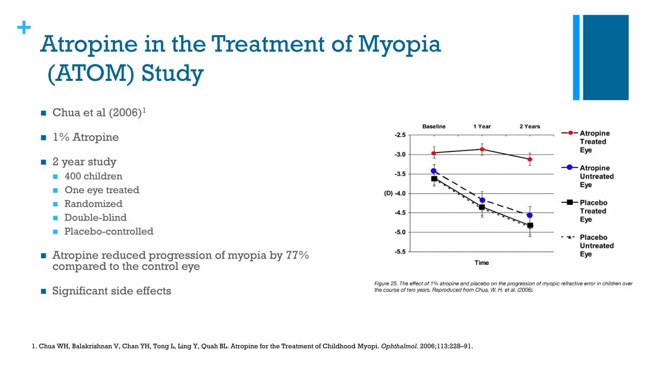
+Atropine in the Treatment of Myopia
(ATOM) Study
◼ Chua et al (2006)1
◼ 1% Atropine
◼ 2 year study
◼ 400 children
◼ One eye treated
◼ Randomized
◼ Double-blind
◼ Placebo-controlled
◼ Atropine reduced progression of myopia by 77% compared to the control eye
◼ Significant side effects
1. Chua WH, Balakrishnan V, Chan YH, Tong L, Ling Y, Quah BL. Atropine for the Treatment of Childhood Myopi. Ophthalmol. 2006;113:228–91.
+
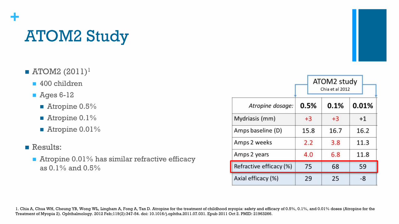
ATOM2 Study
◼ ATOM2 (2011)1
◼ 400 children
◼ Ages 6-12
◼ Atropine 0.5%
◼ Atropine 0.1%
◼ Atropine 0.01%
◼ Results:
◼ Atropine 0.01% has similar refractive efficacy
as 0.1% and 0.5%
1. Chia A, Chua WH, Cheung YB, Wong WL, Lingham A, Fong A, Tan D. Atropine for the treatment of childhood myopia: safety and efficacy of 0.5%, 0.1%, and 0.01% doses (Atropine for the
Treatment of Myopia 2). Ophthalmology. 2012 Feb;119(2):347-54. doi: 10.1016/j.ophtha.2011.07.031. Epub 2011 Oct 2. PMID: 21963266.
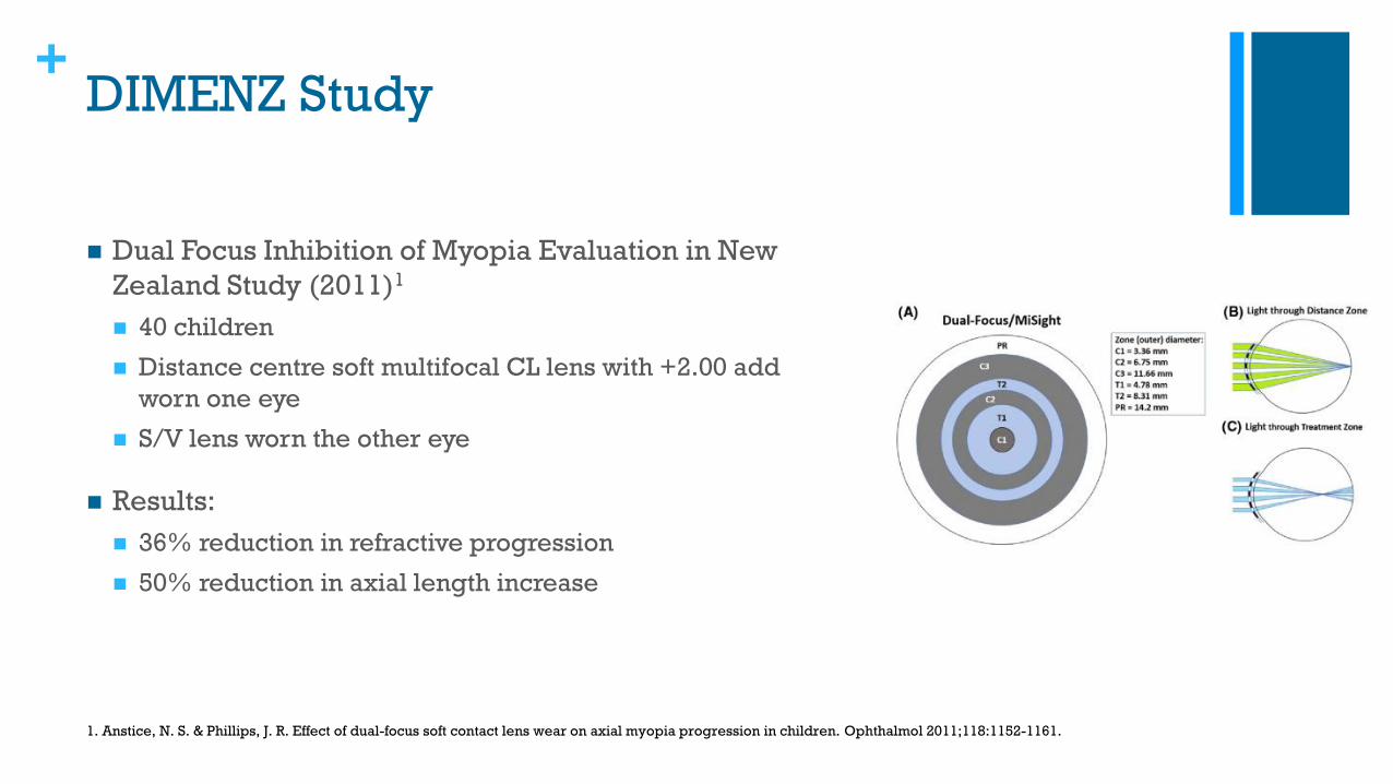
+DIMENZ Study
◼ Dual Focus Inhibition of Myopia Evaluation in New
Zealand Study (2011)1
◼ 40 children
◼ Distance centre soft multifocal CL lens with +2.00 add
worn one eye
◼ S/V lens worn the other eye
◼ Results:
◼ 36% reduction in refractive progression
◼ 50% reduction in axial length increase
1. Anstice, N. S. & Phillips, J. R. Effect of dual-focus soft contact lens wear on axial myopia progression in children. Ophthalmol 2011;118:1152-1161.
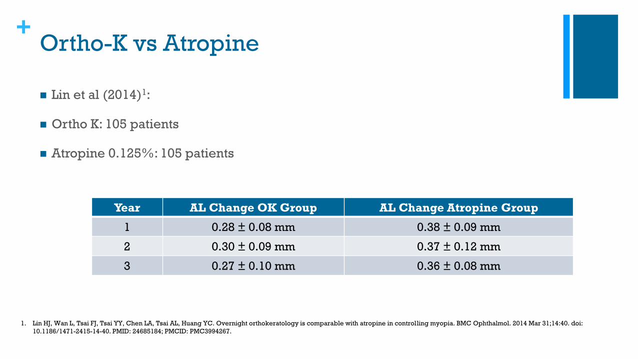
+Ortho-K vs Atropine
◼ Lin et al (2014)1:
◼ Ortho K: 105 patients
◼ Atropine 0.125%: 105 patients
1. Lin HJ, Wan L, Tsai FJ, Tsai YY, Chen LA, Tsai AL, Huang YC. Overnight orthokeratology is comparable with atropine in controlling myopia. BMC Ophthalmol. 2014 Mar 31;14:40. doi:
10.1186/1471-2415-14-40. PMID: 24685184; PMCID: PMC3994267.
Year AL Change OK Group AL Change Atropine Group
1 0.28 ± 0.08 mm 0.38 ± 0.09 mm
2 0.30 ± 0.09 mm 0.37 ± 0.12 mm
3 0.27 ± 0.10 mm 0.36 ± 0.08 mm
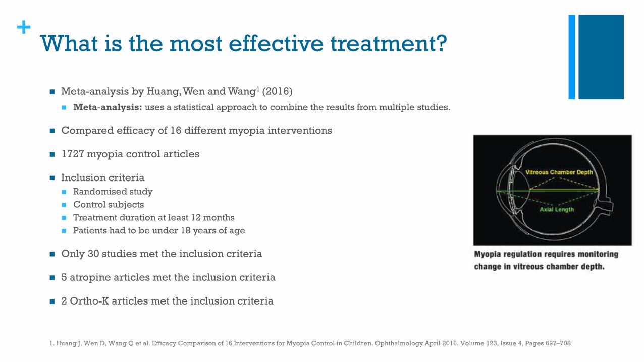
+What is the most effective treatment?
◼ Meta-analysis by Huang, Wen and Wang1 (2016)
◼ Meta-analysis: uses a statistical approach to combine the results from multiple studies.
◼ Compared efficacy of 16 different myopia interventions
◼ 1727 myopia control articles
◼ Inclusion criteria
◼ Randomised study
◼ Control subjects
◼ Treatment duration at least 12 months
◼ Patients had to be under 18 years of age
◼ Only 30 studies met the inclusion criteria
◼ 5 atropine articles met the inclusion criteria
◼ 2 Ortho-K articles met the inclusion criteria
1. Huang J, Wen D, Wang Q et al. Efficacy Comparison of 16 Interventions for Myopia Control in Children. Ophthalmology April 2016. Volume 123, Issue 4, Pages 697–708
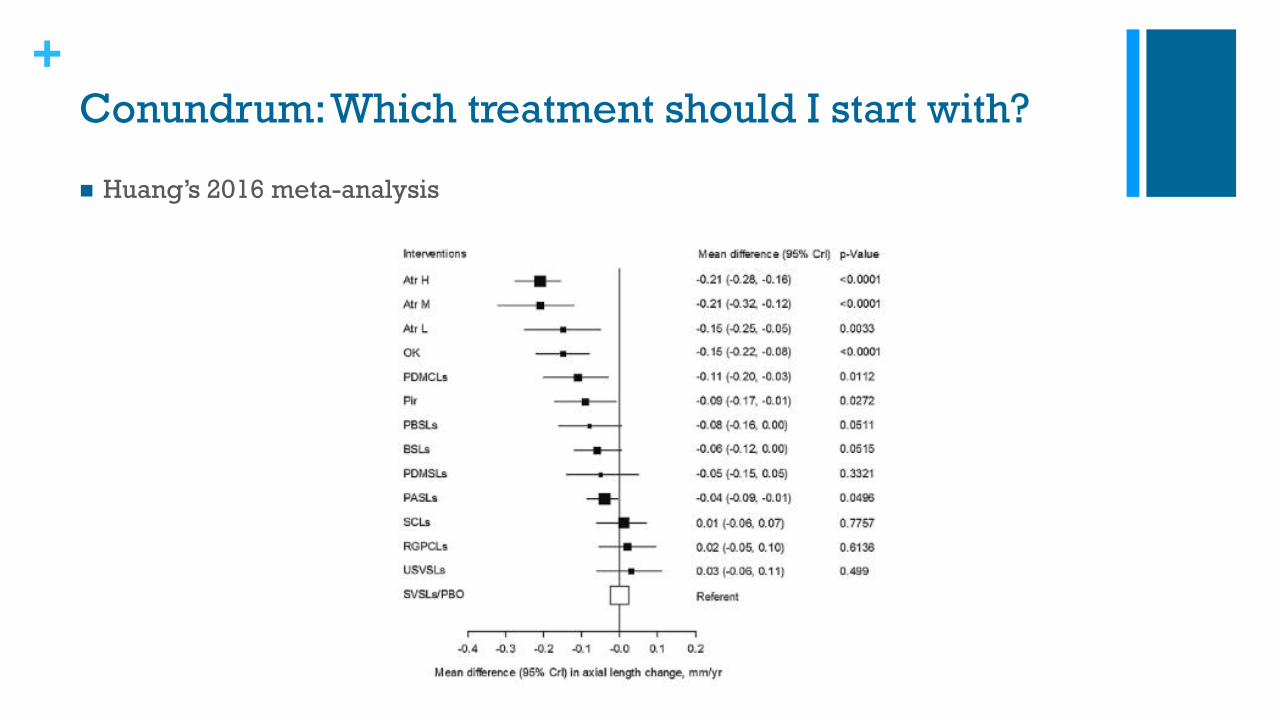
+Conundrum: Which treatment should I start with?
◼ Huang’s 2016 meta-analysis
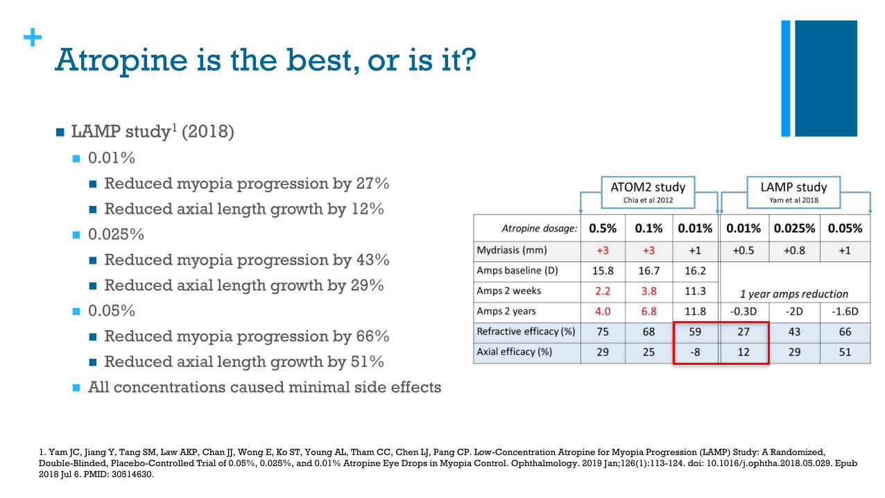
+Atropine is the best, or is it?
◼ LAMP study1 (2018)
◼ 0.01%
◼ Reduced myopia progression by 27%
◼ Reduced axial length growth by 12%
◼ 0.025%
◼ Reduced myopia progression by 43%
◼ Reduced axial length growth by 29%
◼ 0.05%
◼ Reduced myopia progression by 66%
◼ Reduced axial length growth by 51%
◼ All concentrations caused minimal side effects
1. Yam JC, Jiang Y, Tang SM, Law AKP, Chan JJ, Wong E, Ko ST, Young AL, Tham CC, Chen LJ, Pang CP. Low-Concentration Atropine for Myopia Progression (LAMP) Study: A Randomized,
Double-Blinded, Placebo-Controlled Trial of 0.05%, 0.025%, and 0.01% Atropine Eye Drops in Myopia Control. Ophthalmology. 2019 Jan;126(1):113-124. doi: 10.1016/j.ophtha.2018.05.029. Epub
2018 Jul 6. PMID: 30514630.
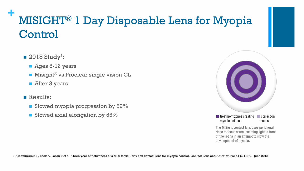
+MISIGHT® 1 Day Disposable Lens for Myopia
Control
◼ 2018 Study1:
◼ Ages 8-12 years
◼ Misight® vs Proclear single vision CL
◼ After 3 years
◼ Results:
◼ Slowed myopia progression by 59%
◼ Slowed axial elongation by 56%
1. Chamberlain P, Back A, Lazon P et al. Three year effectiveness of a dual focus 1 day soft contact lens for myopia control. Contact Lens and Anterior Eye 41:S71-S72 · June 2018
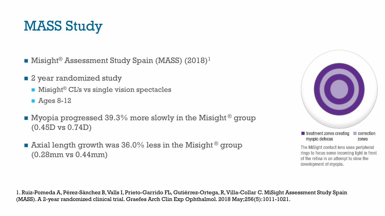
MASS Study
◼ Misight® Assessment Study Spain (MASS) (2018)1
◼ 2 year randomized study
◼ Misight® CL’s vs single vision spectacles
◼ Ages 8-12
◼ Myopia progressed 39.3% more slowly in the Misight ® group
(0.45D vs 0.74D)
◼ Axial length growth was 36.0% less in the Misight ® group
(0.28mm vs 0.44mm)
1. Ruiz-Pomeda A, Pérez-Sánchez B, Valls I, Prieto-Garrido FL, Gutiérrez-Ortega, R, Villa-Collar C. MiSight Assessment Study Spain
(MASS). A 2-year randomized clinical trial. Graefes Arch Clin Exp Ophthalmol. 2018 May;256(5):1011-1021.
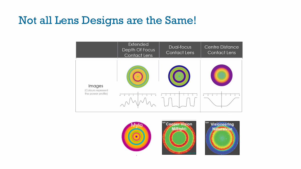
Not all Lens Designs are the Same!
Mylo

+Defocus Incorporated Multiple Segment
(DIMS) Spectacle Lenses
◼ Lam et al (2020)1
◼ 160 children
◼ DIMS vs single vision spectacles
◼ DIMS lenses slowed myopia progression 52%
◼ DIMS group 62% less axial elongation
1. Lam CSY, Tang WC, Tse DY, et al. Defocus Incorporated Multiple Segments (DIMS) spectacle lenses slow myopia progression: a 2-year randomised clinical trial. British Journal of
Ophthalmology 2020;104:363-368.
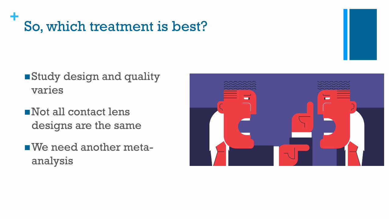
+So, which treatment is best?
◼Study design and quality
varies
◼Not all contact lens
designs are the same
◼We need another meta-
analysis
+Conundrum: Should I prescribe
multiple treatments?
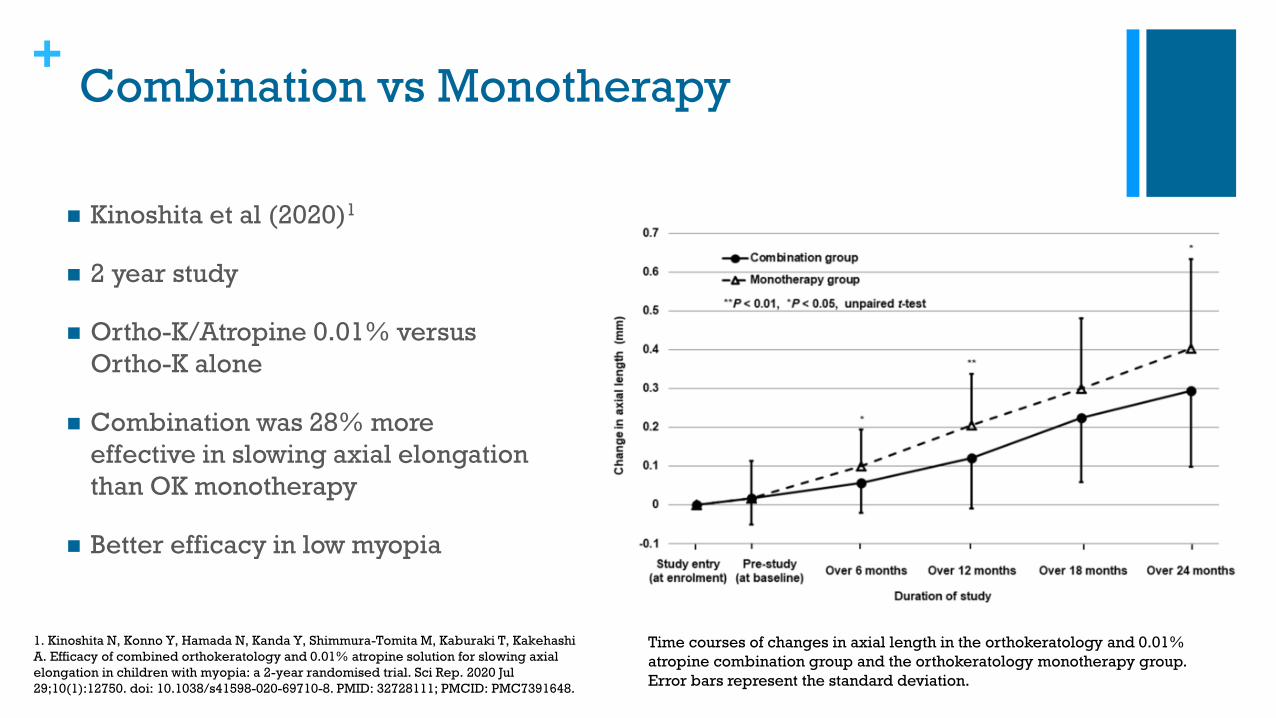
+Combination vs Monotherapy
◼ Kinoshita et al (2020)1
◼ 2 year study
◼ Ortho-K/Atropine 0.01% versus
Ortho-K alone
◼ Combination was 28% more
effective in slowing axial elongation
than OK monotherapy
◼ Better efficacy in low myopia
Time courses of changes in axial length in the orthokeratology and 0.01%
atropine combination group and the orthokeratology monotherapy group.
Error bars represent the standard deviation.
1. Kinoshita N, Konno Y, Hamada N, Kanda Y, Shimmura-Tomita M, Kaburaki T, Kakehashi
A. Efficacy of combined orthokeratology and 0.01% atropine solution for slowing axial
elongation in children with myopia: a 2-year randomised trial. Sci Rep. 2020 Jul
29;10(1):12750. doi: 10.1038/s41598-020-69710-8. PMID: 32728111; PMCID: PMC7391648.
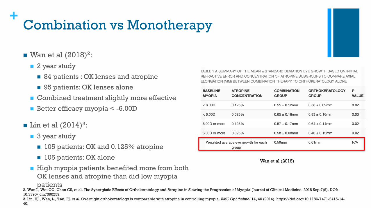
+Combination vs Monotherapy
◼ Wan et al (2018)2:
◼ 2 year study
◼ 84 patients : OK lenses and atropine
◼ 95 patients: OK lenses alone
◼ Combined treatment slightly more effective
◼ Better efficacy myopia < -6.00D
◼ Lin et al (2014)3:
◼ 3 year study
◼ 105 patients: OK and 0.125% atropine
◼ 105 patients: OK alone
◼ High myopia patients benefited more from both
OK lenses and atropine than did low myopia
patients2. Wan L, Wei CC, Chen CS, et al. The Synergistic Effects of Orthokeratology and Atropine in Slowing the Progression of Myopia. Journal of Clinical Medicine. 2018 Sep;7(9). DOI:
10.3390/jcm7090259.
3. Lin, HJ., Wan, L., Tsai, FJ. et al. Overnight orthokeratology is comparable with atropine in controlling myopia. BMC Ophthalmol 14, 40 (2014). https://doi.org/10.1186/1471-2415-14-
40.
Wan et al (2018)
+Conundrum: The Cause of Myopia
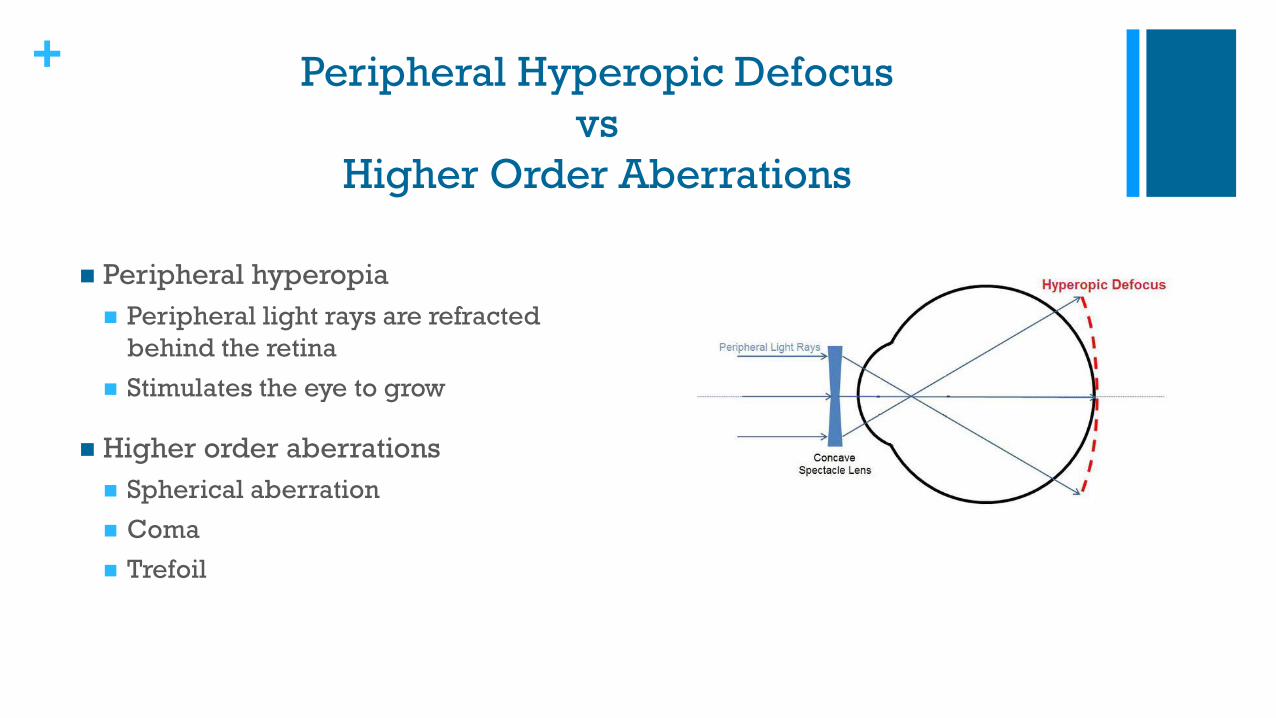
+ Peripheral Hyperopic Defocus
vs
Higher Order Aberrations
◼ Peripheral hyperopia
◼ Peripheral light rays are refracted
behind the retina
◼ Stimulates the eye to grow
◼ Higher order aberrations
◼ Spherical aberration
◼ Coma
◼ Trefoil
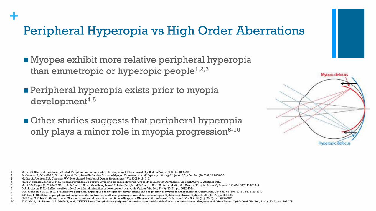
+Peripheral Hyperopia vs High Order Aberrations
◼ Myopes exhibit more relative peripheral hyperopia
than emmetropic or hyperopic people1,2,3
◼ Peripheral hyperopia exists prior to myopia
development4,5
◼ Other studies suggests that peripheral hyperopia
only plays a minor role in myopia progression6-10
1. Mutti DO, Sholtz RI, Friedman NE, et al. Peripheral refraction and ocular shape in children. Invest Ophthalmol Vis Sci 2000;41:1022–30.
2. Seidemann A, Schaeffel F, Guirao A, et al. Peripheral Refractive Errors in Myopic, Emmetropic, and Hyperopic Young Subjects. J Opt Soc Am (A) 2002;19:2363–73.
3. Mathur A, Atchison DA, Charman WN. Myopia and Peripheral Ocular Aberrations. J Vis 2009;9:15. 1–2.
4. Mutti D, Sinnott L, Jones L, et al. Relative Peripheral Refractive Error and the Risk of Juvenile-Onset Myopia. Invest Ophthalmol Vis Sci 2008;49: E-Abstract 5426.
5. Mutti DO, Hayes JR, Mitchell GL, et al. Refractive Error, Axial Length, and Relative Peripheral Refractive Error Before and after the Onset of Myopia. Invest Ophthalmol Vis Sci 2007;48:2510–9.
6. D.A. Atchison, R. RosénThe possible role of peripheral refraction in development of myopia Optom. Vis. Sci., 93 (9) (2016), pp. 1042-1044.
7. D.A. Atchison, S.M. Li, H. Li, et al.Relative peripheral hyperopia does not predict development and progression of myopia in children Invest. Ophthalmol. Vis. Sci., 56 (10) (2015), pp. 6162-6170.
8. T.T. Lee, P. ChoRelative peripheral refraction in children: twelve-month changes in eyes with different ametropias Ophthalmic Physiol. Optic., 33 (3) (2013), pp. 283-293.
9. C.C. Sng, X.Y. Lin, G. Gazzard, et al.Change in peripheral refraction over time in Singapore Chinese children Invest. Ophthalmol. Vis. Sci., 52 (11) (2011), pp. 7880-7887.
10. D.O. Mutti, L.T. Sinnott, G.L. Mitchell, et al., CLEERE Study GroupRelative peripheral refractive error and the risk of onset and progression of myopia in children Invest. Ophthalmol. Vis. Sci., 52 (1) (2011), pp. 199-205.
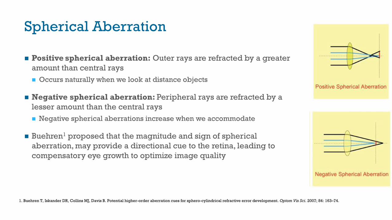
Spherical Aberration
◼ Positive spherical aberration: Outer rays are refracted by a greater
amount than central rays
◼ Occurs naturally when we look at distance objects
◼ Negative spherical aberration: Peripheral rays are refracted by a
lesser amount than the central rays
◼ Negative spherical aberrations increase when we accommodate
◼ Buehren1 proposed that the magnitude and sign of spherical
aberration, may provide a directional cue to the retina, leading to
compensatory eye growth to optimize image quality
1. Buehren T, Iskander DR, Collins MJ, Davis B. Potential higher-order aberration cues for sphero-cylindrical refractive error development. Optom Vis Sci. 2007; 84: 163–74.
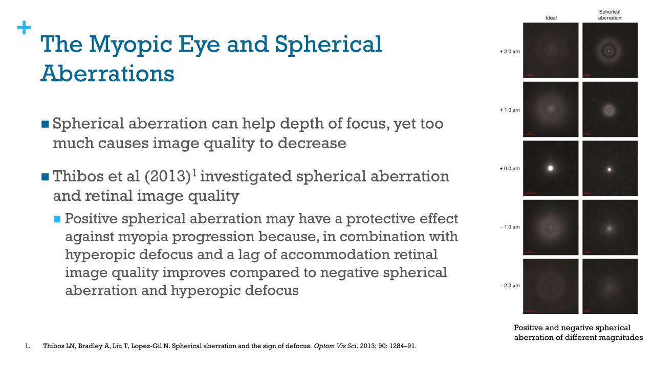
+The Myopic Eye and Spherical
Aberrations
◼ Spherical aberration can help depth of focus, yet too
much causes image quality to decrease
◼ Thibos et al (2013)1 investigated spherical aberration
and retinal image quality
◼ Positive spherical aberration may have a protective effect
against myopia progression because, in combination with
hyperopic defocus and a lag of accommodation retinal
image quality improves compared to negative spherical
aberration and hyperopic defocus
1. Thibos LN, Bradley A, Liu T, Lopez-Gil N. Spherical aberration and the sign of defocus. Optom Vis Sci. 2013; 90: 1284–91.
Positive and negative spherical
aberration of different magnitudes
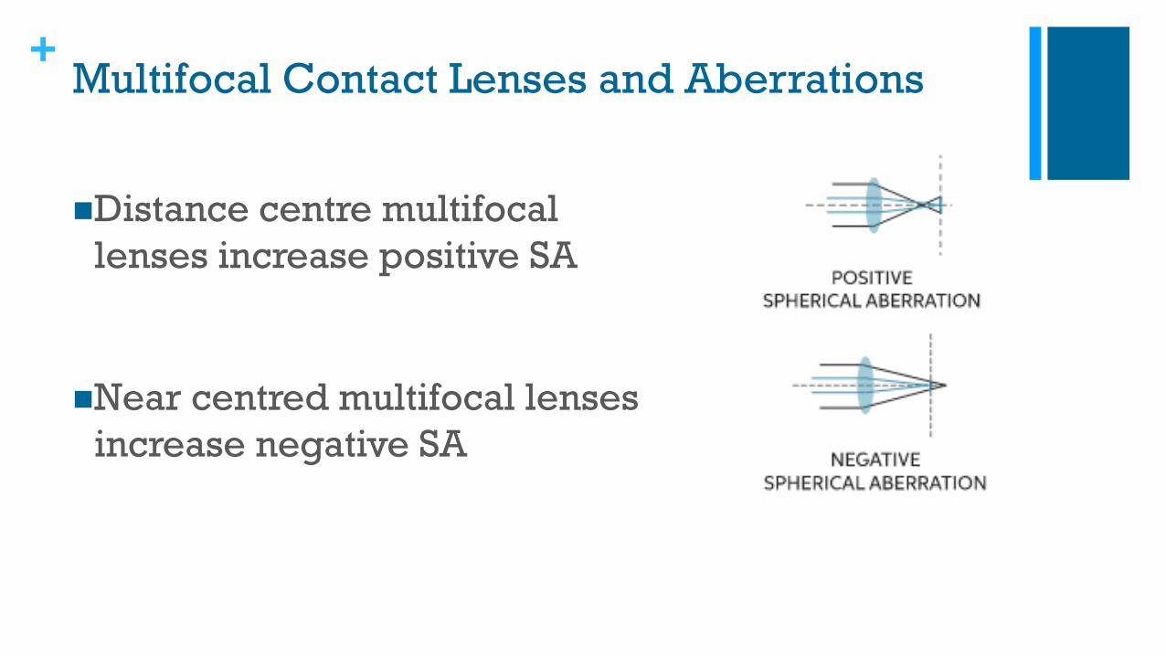
+Multifocal Contact Lenses and Aberrations
◼Distance centre multifocal
lenses increase positive SA
◼Near centred multifocal lenses
increase negative SA

+Positive Spherical Aberration Contact Lenses
◼ Positive spherical aberration contact lens study (2016)1
◼ 127 children
◼ Ages 8-11
◼ Soft CL with positive spherical aberration versus soft spherical CL’s
◼ Results:
◼ 38.6% less axial elongation after 12 months
◼ Only 0.14D reduction in refractive change after 12 months
◼ Why the poor refractive response?
◼ Follow-Up Study (2019)1
◼ Positive SA lenses cause a decrease in accommodation which was correlated with greater myopia progression
◼ The authors propose that a strong accommodative response slows myopia progression
1. 1. Cheng X, Xu J, Chehab K, Exford J, Brennan N. Soft Contact Lenses with Positive Spherical Aberration for Myopia Control. Optom Vis Sci. 2016 Apr;93(4):353-66. doi:
10.1097/OPX.0000000000000773. PMID: 26704144.
2. Cheng, X., Xu, J., & Brennan, N. A. (2019). Accommodation and its role in myopia progression and control with soft contact lenses. Ophthalmic and Physiological Optics, 39(3), 162-
171.
+Ortho-K and Accommodation
◼ Ortho-K has been shown to improve accommodation
◼ Study (2014)3:
◼ 49 children
◼ Ages: 7-14
◼ Wore ortho-K lenses for 2 years
◼ 2 groups
◼ ‘Below average’ accommodation
◼ ‘Above average’ accommodation
◼ Results:
◼ Children with ‘below average’: Amplitude of accommodation improved by 4D after OK wear
◼ ‘Below average’ group also had a better myopia control effect
1. Zhu M, Feng H, Zhu J, Qu X. The impact of amplitude of accommodation on controlling the development of myopia in orthokeratology. Chinese J Ophthalmol. 2014;50:14-9.
+Conundrum: How does increased near
work cause myopia?
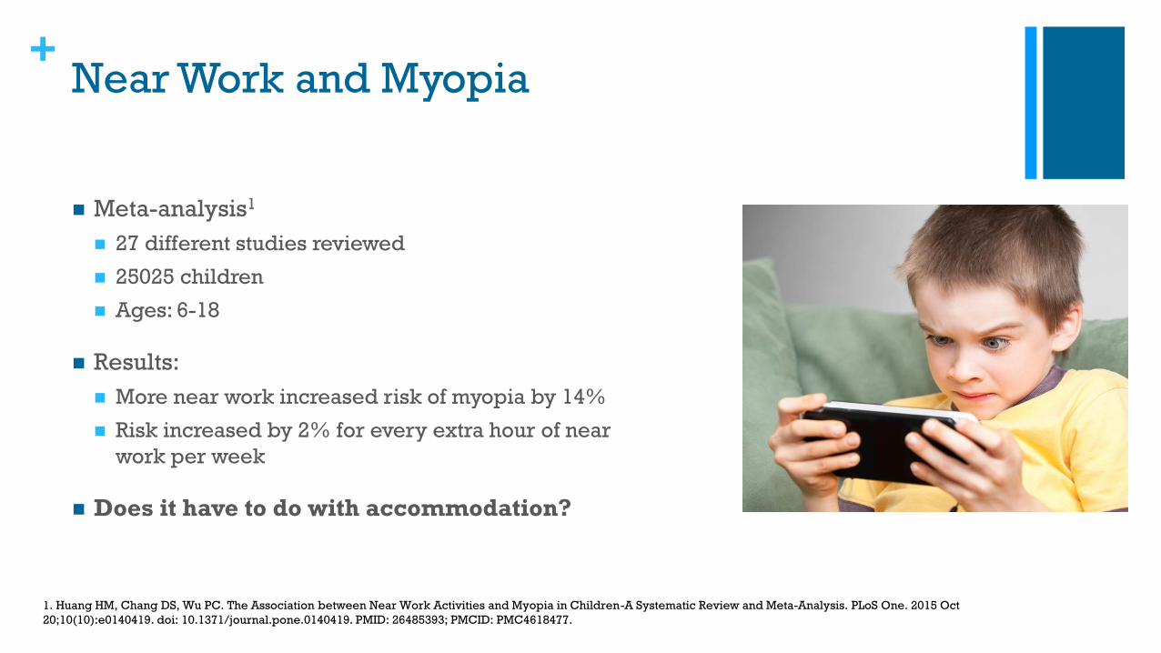
+Near Work and Myopia
◼ Meta-analysis1
◼ 27 different studies reviewed
◼ 25025 children
◼ Ages: 6-18
◼ Results:
◼ More near work increased risk of myopia by 14%
◼ Risk increased by 2% for every extra hour of near
work per week
◼ Does it have to do with accommodation?
1. Huang HM, Chang DS, Wu PC. The Association between Near Work Activities and Myopia in Children-A Systematic Review and Meta-Analysis. PLoS One. 2015 Oct
20;10(10):e0140419. doi: 10.1371/journal.pone.0140419. PMID: 26485393; PMCID: PMC4618477.
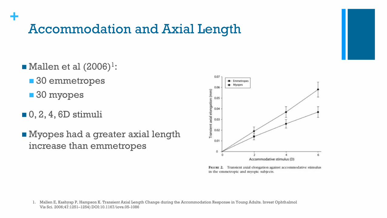
+Accommodation and Axial Length
◼ Mallen et al (2006)1:
◼ 30 emmetropes
◼ 30 myopes
◼ 0, 2, 4, 6D stimuli
◼ Myopes had a greater axial length
increase than emmetropes
1. Mallen E, Kashyap P, Hampson K. Transient Axial Length Change during the Accommodation Response in Young Adults. Invest Ophthalmol
Vis Sci. 2006;47:1251–1254) DOI:10.1167/iovs.05-1086
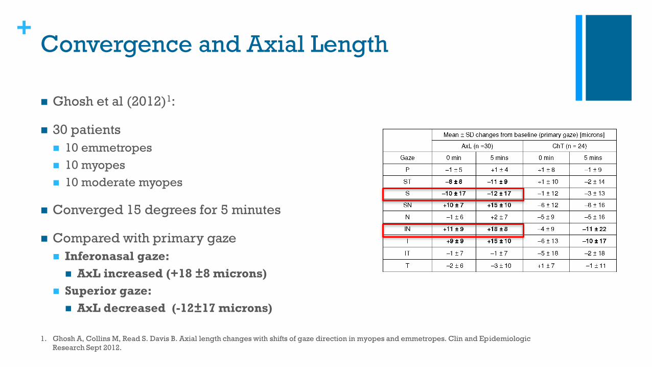
+Convergence and Axial Length
◼ Ghosh et al (2012)1:
◼ 30 patients
◼ 10 emmetropes
◼ 10 myopes
◼ 10 moderate myopes
◼ Converged 15 degrees for 5 minutes
◼ Compared with primary gaze
◼ Inferonasal gaze:
◼ AxL increased (+18 ±8 microns)
◼ Superior gaze:
◼ AxL decreased (-12±17 microns)
1. Ghosh A, Collins M, Read S. Davis B. Axial length changes with shifts of gaze direction in myopes and emmetropes. Clin and Epidemiologic
Research Sept 2012.
+Convergence and Axial Length Changes
High vs Low Myopes
◼ Ghosh et al (2012)1:
◼ Axial elongation: Mean change from baseline at
5 minutes
◼ Inferonasal gaze:
◼ Moderate myopes: +25 ± 6 μm
◼ Low myopes: +12 ± 4 μm
◼ Emmetropes: +13 ± 6 μm
1. Ghosh A, Collins M, Read S. Davis B. Axial length changes with shifts of gaze direction in myopes and emmetropes. Clin and Epidemiologic
Research Sept 2012.
+Axial Length Changes with Large Degrees of
Convergence
◼ Bayramlar et al (1999)1:
◼ 124 young males
◼ 20 cm target distance
◼ Accommodative convergence rather than
accommodation alone increased AxL
◼ Are myopic scleras more susceptible to
stretching?
1. Bayramlar H Cekic O Hepsen IF. Does convergence, not accommodation, cause axial-length elongation at near? A biometric study in teens. Ophthalmic
Res . 1999;31:304–308.
+Scleral Stiffness and Myopia
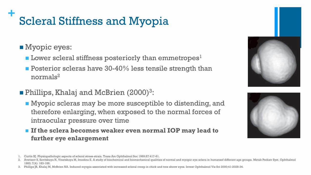
◼ Myopic eyes:
◼ Lower scleral stiffness posteriorly than emmetropes1
◼ Posterior scleras have 30-40% less tensile strength than
normals2
◼ Phillips, Khalaj and McBrien (2000)3:
◼ Myopic scleras may be more susceptible to distending, and
therefore enlarging, when exposed to the normal forces of
intraocular pressure over time
◼ If the sclera becomes weaker even normal IOP may lead to
further eye enlargement
1. Curtin BJ. Physiopathologic aspects of scleral stress-strain. Trans Am Ophthalmol Soc 1969;67:417-61.
2. Avetisov S, Savitskaya N, Vinetskaya M, Imodina E. A study of biochemical and biomechanical qualities of normal and myopic eye sclera in humansof different age groups. Metab Pediatr Syst. Ophthalmol
1983; 7(4): 183-188.
3. Phillips JR, Khalaj M, McBrien NA. Induced myopia associated with increased scleral creep in chick and tree shrew eyes. Invest Ophthalmol Vis Sci 2000;41:2028-34.

+Conundrum: Does increased IOP cause
myopia?
+Inflationary Model
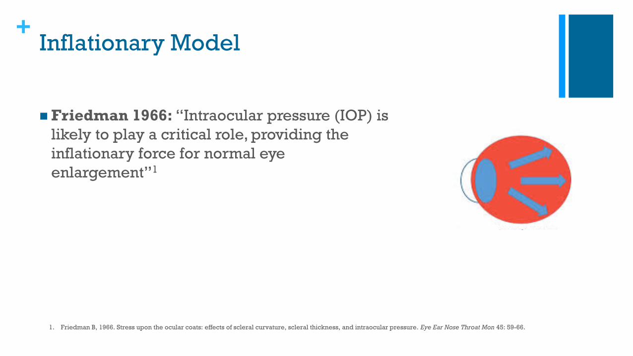
◼ Friedman 1966: “Intraocular pressure (IOP) is
likely to play a critical role, providing the
inflationary force for normal eye
enlargement”1
1. Friedman B, 1966. Stress upon the ocular coats: effects of scleral curvature, scleral thickness, and intraocular pressure. Eye Ear Nose Throat Mon 45: 59-66.
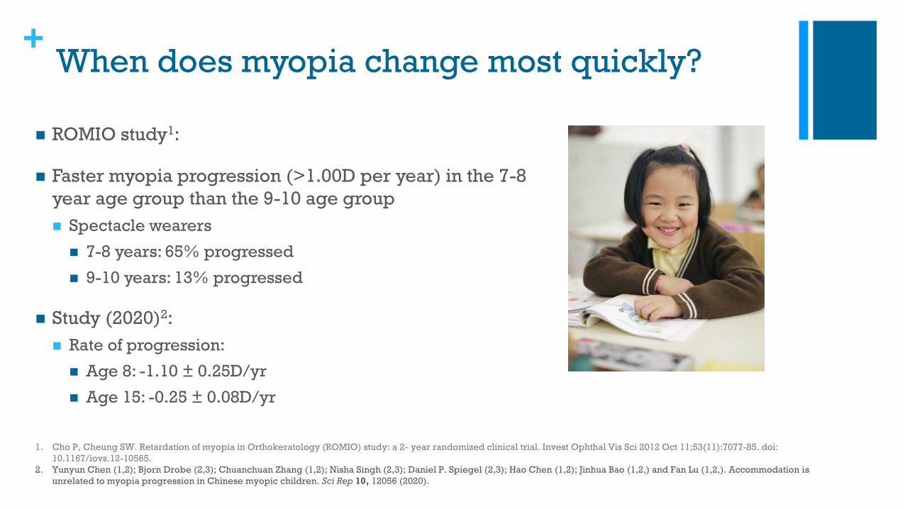
+When does myopia change most quickly?
◼ ROMIO study1:
◼ Faster myopia progression (>1.00D per year) in the 7-8
year age group than the 9-10 age group
◼ Spectacle wearers
◼ 7-8 years: 65% progressed
◼ 9-10 years: 13% progressed
◼ Study (2020)2:
◼ Rate of progression:
◼ Age 8: -1.10 ± 0.25D/yr
◼ Age 15: -0.25 ± 0.08D/yr
1. Cho P, Cheung SW. Retardation of myopia in Orthokeratology (ROMIO) study: a 2- year randomized clinical trial. Invest Ophthal Vis Sci 2012 Oct 11;53(11):7077-85. doi:
10.1167/iovs.12-10565.
2. Yunyun Chen (1,2); Bjorn Drobe (2,3); Chuanchuan Zhang (1,2); Nisha Singh (2,3); Daniel P. Spiegel (2,3); Hao Chen (1,2); Jinhua Bao (1,2,) and Fan Lu (1,2,). Accommodation is
unrelated to myopia progression in Chinese myopic children. Sci Rep 10, 12056 (2020).
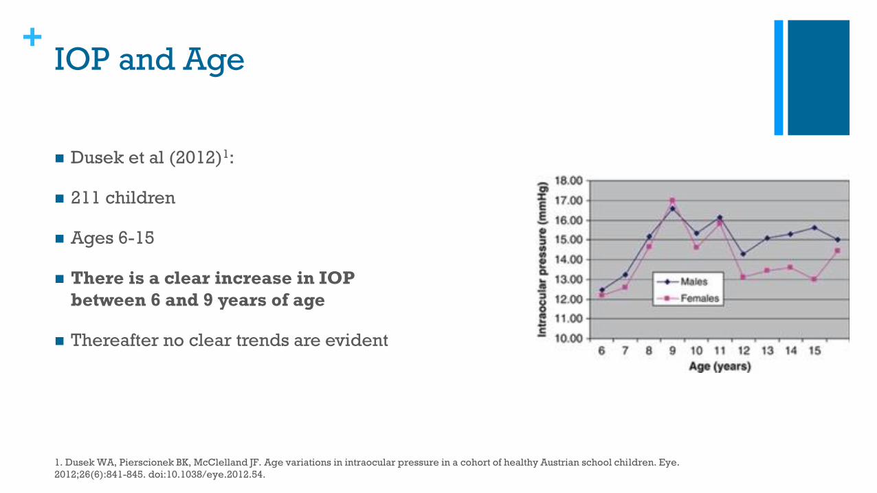
+IOP and Age
◼ Dusek et al (2012)1:
◼ 211 children
◼ Ages 6-15
◼ There is a clear increase in IOP
between 6 and 9 years of age
◼ Thereafter no clear trends are evident
1. Dusek WA, Pierscionek BK, McClelland JF. Age variations in intraocular pressure in a cohort of healthy Austrian school children. Eye.
2012;26(6):841-845. doi:10.1038/eye.2012.54.
+Near Work and IOP
◼ Ha et al (2018)1:
◼ 39 subjects
◼ <40 years
◼ Looked at smartphone for 5, 15, and 25 minutes
◼ Daylight:
◼ Baseline IOP: 13.7 ± 1.8 mmHg
◼ After 5 mins: 14.1 ± 1.8 mmHg
◼ After 15 mins: 15.5 ± 1.7 mmHg
◼ After 25 mins: 15.3 ± 1.8 mmHg
◼ Low Light:
◼ Baseline IOP: 13.9 ± 1.9 mmHg
◼ After 5 mins: 15.6 ± 1.8 mmHg
◼ After 15 mins: 17.3 ± 1.9 mmHg
◼ After 25 mins: 17.0 ± 1.7 mmHg 1. Ha A, Kim YK, Park YJ, Jeoung JW, Park KH. Intraocular pressure change during reading or writing on
smartphone. PLoS One. 2018;13(10):e0206061. Published 2018 Oct 25. doi:10.1371/journal.pone.0206061

+Accommodation and IOP
◼ Yan et al (2014)1:
◼ 86 subjects
◼ 46 progressing myopes
◼ 40 emmetropes
◼ No accommodation stimulus
◼ No difference in IOP between progressing myopes and
emmetropes
◼ Accommodation stimulus (0D to 6.00D)
◼ Progressing myopes: IOP increased (+1.02±2.07 mm Hg)
◼ Emmetropes: Unchanged (−0.76±3.22 mm Hg)
1. Yan L, Huibin L, Xuemin L. Accommodation-induced intraocular pressure changes in progressing myopes and emmetropes. Eye
(Lond). 2014;28(11):1334-40.

+IOP and Myopia: ‘Evidence Against’
◼ Lee et al (2004)1: 636 children
◼ Ages: 9-11
◼ No association between IOP, refractive error or axial length
◼ Chang et al (2009)2:
◼ 63 children
◼ No significant correlation between IOP and AL
1. Lee AJ, Saw S-M, Gazzard G, Cheng A, Tan DTH. Intraocular pressure associations with refractive error and axial length in children. The British
Journal of Ophthalmology. 2004;88(1):5-7.
2. Chang PY, Chang SW, Wang JY. Assessment of corneal biomechanical properties and intraocular pressure with the Ocular Response Analyzer
in childhood myopia. Br J Ophthalmol 2010;94:877e881. doi:10.1136/bjo.2009.158568
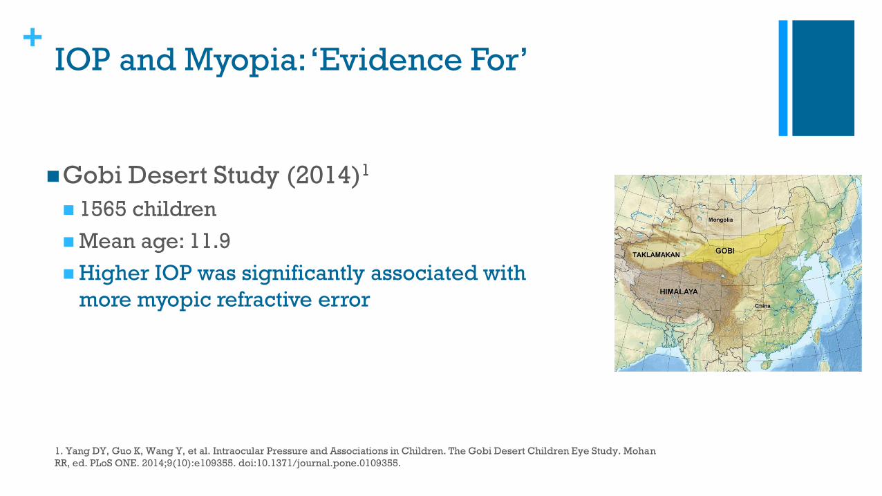
+IOP and Myopia: ‘Evidence For’
◼Gobi Desert Study (2014)1
◼ 1565 children
◼ Mean age: 11.9
◼ Higher IOP was significantly associated with
more myopic refractive error
1. Yang DY, Guo K, Wang Y, et al. Intraocular Pressure and Associations in Children. The Gobi Desert Children Eye Study. Mohan
RR, ed. PLoS ONE. 2014;9(10):e109355. doi:10.1371/journal.pone.0109355.
+IOP and Myopia: ‘Evidence For’
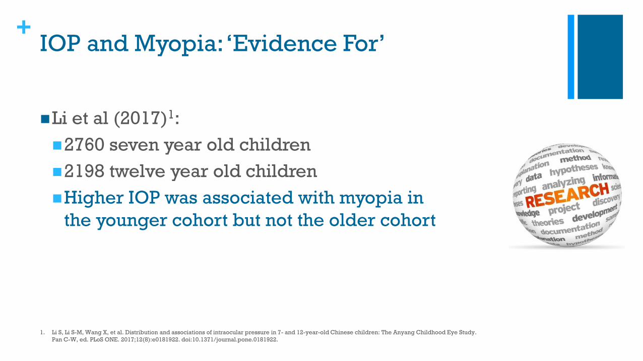
◼Li et al (2017)1:
◼2760 seven year old children
◼2198 twelve year old children
◼Higher IOP was associated with myopia in
the younger cohort but not the older cohort
1. Li S, Li S-M, Wang X, et al. Distribution and associations of intraocular pressure in 7- and 12-year-old Chinese children: The Anyang Childhood Eye Study.
Pan C-W, ed. PLoS ONE. 2017;12(8):e0181922. doi:10.1371/journal.pone.0181922.

+IOP and Axial Length
◼ Li et al (2018)2: 1558 children
◼ 12 year old children
◼ IOP had essentially no relationship with myopia
progression
◼ Why is the evidence so variable?
◼ Trans-laminar pressure difference (TLPD)
1. Li S, Iribarren R, Li H, et al Intraocular pressure and myopia progression in Chinese children: the Anyang Childhood Eye Study
British Journal of Ophthalmology Published Online First: 01 June 2018. doi: 10.1136/bjophthalmol-2017-311831
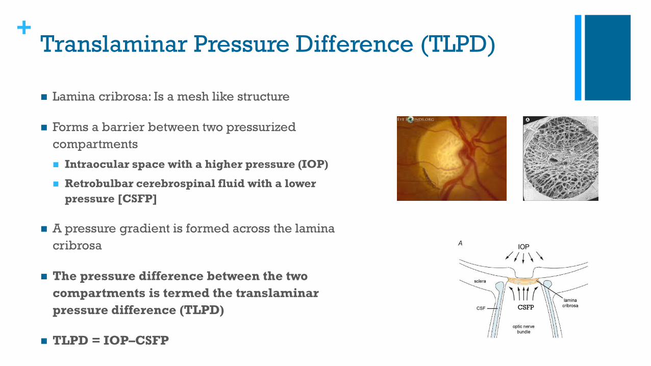
+Translaminar Pressure Difference (TLPD)
◼ Lamina cribrosa: Is a mesh like structure
◼ Forms a barrier between two pressurized
compartments
◼ Intraocular space with a higher pressure (IOP)
◼ Retrobulbar cerebrospinal fluid with a lower
pressure [CSFP]
◼ A pressure gradient is formed across the lamina
cribrosa
◼ The pressure difference between the two
compartments is termed the translaminar
pressure difference (TLPD)
◼ TLPD = IOP–CSFP
CSFP
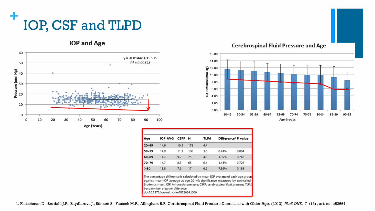
+IOP, CSF and TLPD
1. Fleischman D., Berdahl J.P., Zaydlarova J., Stinnett S., Fautsch M.P., Allingham R.R. Cerebrospinal Fluid Pressure Decreases with Older Age. (2012) PLoS ONE, 7 (12) , art. no. e52664.
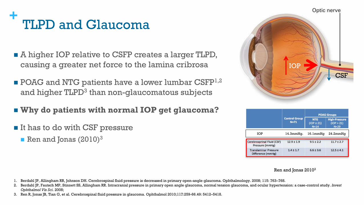
+TLPD and Glaucoma
◼ A higher IOP relative to CSFP creates a larger TLPD,
causing a greater net force to the lamina cribrosa
◼ POAG and NTG patients have a lower lumbar CSFP1,2
and higher TLPD3 than non-glaucomatous subjects
◼ Why do patients with normal IOP get glaucoma?
◼ It has to do with CSF pressure
◼ Ren and Jonas (2010)3
1. Berdahl JP, Allingham RR, Johnson DH. Cerebrospinal fluid pressure is decreased in primary open-angle glaucoma. Ophthalmology. 2008; 115: 763–768.
2. Berdahl JP, Fautsch MP, Stinnett SS, Allingham RR. Intracranial pressure in primary open angle glaucoma, normal tension glaucoma, and ocular hypertension: a case-control study. Invest
Ophthalmol Vis Sci. 2008;
3. Ren R, Jonas JB, Tian G, et al. Cerebrospinal fluid pressure in glaucoma. Ophthalmol 2010;117:259-66.49: 5412–5418.
Ren and Jonas 20103
IOP
CSF
IOP 14.3mmHg. 16.1mmHg 24.2mmHg
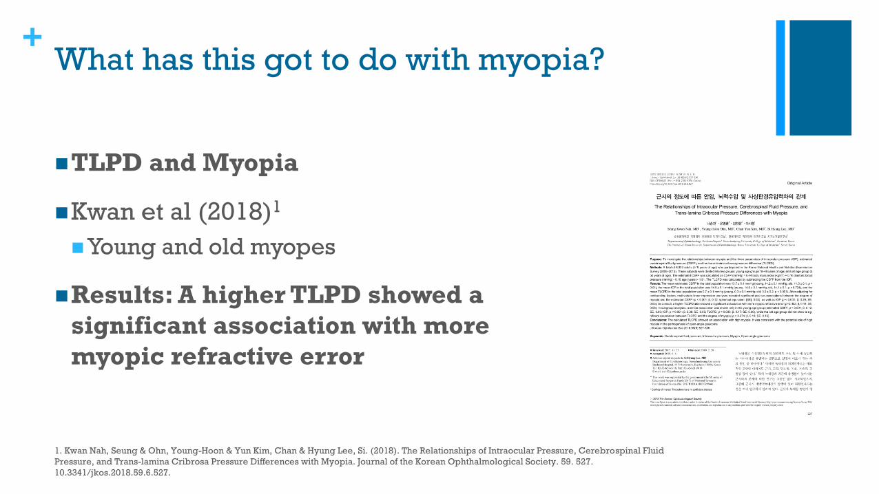
+What has this got to do with myopia?
◼TLPD and Myopia
◼Kwan et al (2018)1
◼Young and old myopes
◼Results: A higher TLPD showed a
significant association with more
myopic refractive error
1. Kwan Nah, Seung & Ohn, Young-Hoon & Yun Kim, Chan & Hyung Lee, Si. (2018). The Relationships of Intraocular Pressure, Cerebrospinal Fluid
Pressure, and Trans-lamina Cribrosa Pressure Differences with Myopia. Journal of the Korean Ophthalmological Society. 59. 527.
10.3341/jkos.2018.59.6.527.
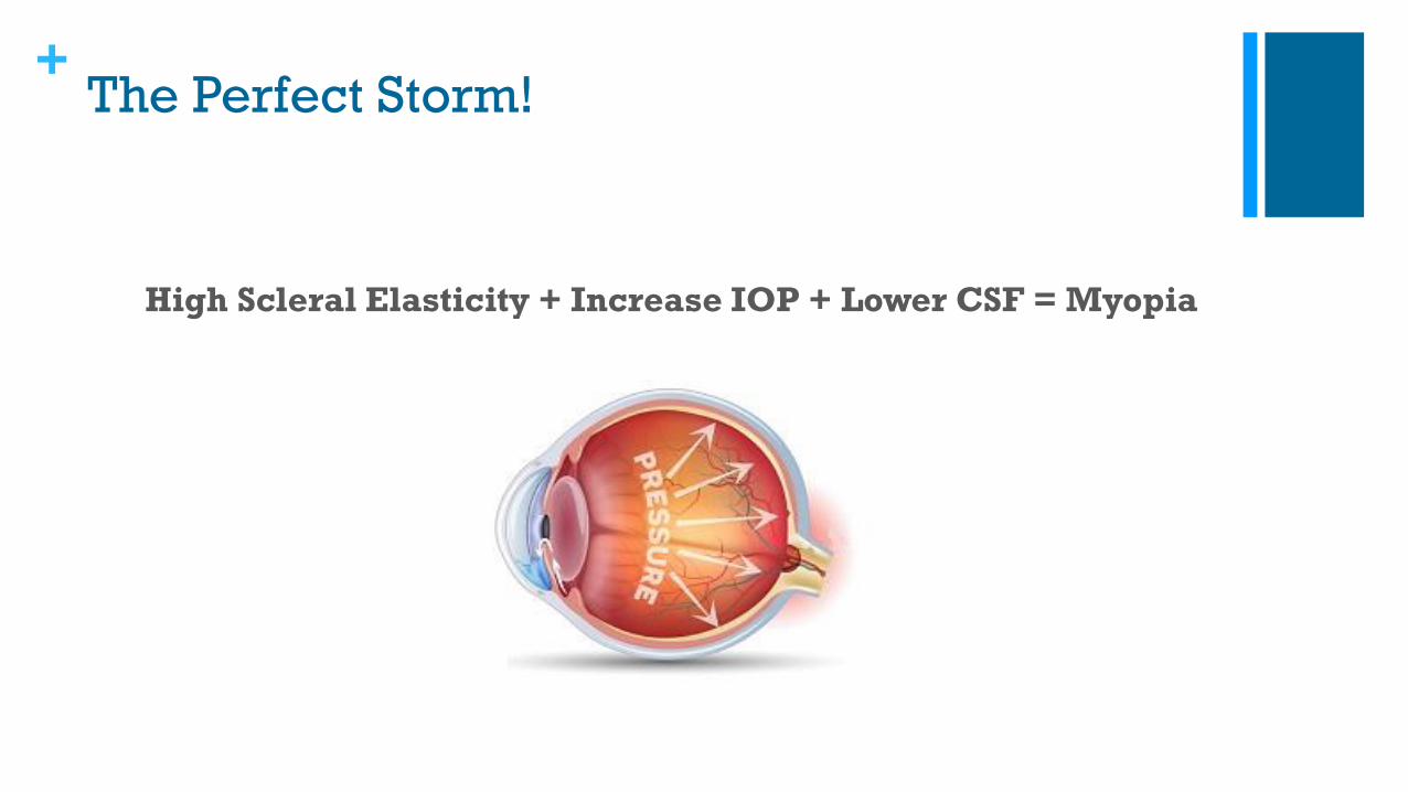
+The Perfect Storm!
High Scleral Elasticity + Increase IOP + Lower CSF = Myopia
+Take Home Points
◼ Measure axial length
◼ Consult the eye growth charts
◼ Counsel patients about their near behaviors:
◼ Regular breaks
◼ Look up into the distance
◼ Working distance no closer than 40cm
◼ Reading in good light
◼ Multiple therapies for high risk patients

+
Thank You!