Congenital Toxoplasmosis: Continued Parasite Proliferation ... · within the fetal brain even when...
5
MAJOR ARTICLE Congenital Toxoplasmosis: Continued Parasite Proliferation in the Fetal Brain Despite Maternal Immunological Control in Other Tissues David J. P. Ferguson, 1 Colene Bowker, 2 Katie J. M. Jeffery, 3 Paul Chamberlain, 4 and Waney Squier 5 1 Nuffield Department of Clinical Laboratory Science, 2 Department of Cellular Pathology, 3 Department of Microbiology, 4 Nuffield Department of Obstetrics and Gynaecology, and 5 Department of Neuropathology, Oxford University Hospital, United Kingdom Background. Congenital toxoplasmosis is a serious condition but little is known of the natural history of parasite development and associated fetal tissue destruction. Methods. Two cases identified by ultrasound underwent induced abortion at 21 and 30 weeks’ gestation. At autopsy, the placenta and fetal organs were examined by histology and immunocytochemistry employing anti- Toxoplasma stage-specific antibodies to confirm diagnosis and also provide information on the stage of parasite development. Results. In both cases, maternal serology prior to termination showed both specific immunoglobulin M (IgM) and immunoglobulin G (IgG), whereas retrospective analysis of an earlier sample (12–14 weeks’ gestation) showed only IgM reactivity consistent with infection occurring in the first trimester. The finding of a number of tissue cysts but few or no tachyzoites within the placenta and fetal adrenal and heart is characteristic of a chronic infection. However, in contrast, there were still areas of the fetal brain with large numbers of actively dividing, tissue-destructive tachyzoites. Conclusions. These observations show that continued parasite proliferation and tissue destruction can occur within the fetal brain even when there is a marked maternal immune response including maternal IgG. This finding strongly suggests that there may be benefits from treating cases of recently acquired congenital infection to destroy any remaining proliferating parasites located in immunologically protected sites such as the fetal brain. Keywords. congenital toxoplasmosis; Toxoplasma gondii; pathology; immunocytochemistry; parasite proliferation. Toxoplasma gondii is a protozoan parasite with a world- wide distribution. It is a coccidian parasite with the cat as its definitive host; however, any warm-blooded animal, including humans, may act as intermediate hosts [1]. In intermediate hosts, after an initial acute sys- temic phase with proliferating parasites (tachyzoites), it reverts to a chronic infection with parasites (brady- zoites) limited to tissue cysts in the brain or musculature resulting in lifelong persistent infection. Although infection can cause devastating disease in immunocom- promised individuals, it causes no, or mild and tran- sient, symptoms in immunocompetent individuals. However, if a woman (or other female mammal) becomes infected for the first time during pregnancy, proliferating parasites, circulating in the blood, may cross the placenta during the acute phase and infect the developing fetus [2, 3]. The results of this prenatal infec- tion vary widely, ranging from intrauterine fetal death to birth of children who may present with severe symp- toms or remain asymptomatic [2, 4]. The chances of congenital transmission and severity of the symptoms can vary depending on when in gestation infection occurs [5]. Because Toxoplasma has a tropism for the central nervous system, severe symptoms are caused by the parasite replicating in the brain or eye. Different countries have divergent policies on sero- logical screening for evidence of primary maternal Received 2 July 2012; accepted 2 October 2012; electronically published 16 October 2012. Correspondence: David J. P. Ferguson, PhD, DSc, Nuffield Department of Clini- cal Laboratory Science, John Radcliffe Hospital, Oxford OX3 9DU, United Kingdom ([email protected]). Clinical Infectious Diseases 2013;56(2):204–8 © The Author 2012. Published by Oxford University Press on behalf of the Infectious Diseases Society of America. All rights reserved. For Permissions, please e-mail: [email protected]. DOI: 10.1093/cid/cis882 204 • CID 2013:56 (15 January) • Ferguson et al
Transcript of Congenital Toxoplasmosis: Continued Parasite Proliferation ... · within the fetal brain even when...

M A J O R A R T I C L E
Congenital Toxoplasmosis: Continued ParasiteProliferation in the Fetal Brain Despite MaternalImmunological Control in Other Tissues
David J. P. Ferguson,1 Colene Bowker,2 Katie J. M. Jeffery,3 Paul Chamberlain,4 and Waney Squier5
1Nuffield Department of Clinical Laboratory Science, 2Department of Cellular Pathology, 3Department of Microbiology, 4Nuffield Department ofObstetrics and Gynaecology, and 5Department of Neuropathology, Oxford University Hospital, United Kingdom
Background. Congenital toxoplasmosis is a serious condition but little is known of the natural history ofparasite development and associated fetal tissue destruction.
Methods. Two cases identified by ultrasound underwent induced abortion at 21 and 30 weeks’ gestation. Atautopsy, the placenta and fetal organs were examined by histology and immunocytochemistry employing anti-Toxoplasma stage-specific antibodies to confirm diagnosis and also provide information on the stage of parasitedevelopment.
Results. In both cases, maternal serology prior to termination showed both specific immunoglobulin M (IgM)and immunoglobulin G (IgG), whereas retrospective analysis of an earlier sample (12–14 weeks’ gestation)showed only IgM reactivity consistent with infection occurring in the first trimester. The finding of a number oftissue cysts but few or no tachyzoites within the placenta and fetal adrenal and heart is characteristic of a chronicinfection. However, in contrast, there were still areas of the fetal brain with large numbers of actively dividing,tissue-destructive tachyzoites.
Conclusions. These observations show that continued parasite proliferation and tissue destruction can occurwithin the fetal brain even when there is a marked maternal immune response including maternal IgG. Thisfinding strongly suggests that there may be benefits from treating cases of recently acquired congenital infection todestroy any remaining proliferating parasites located in immunologically protected sites such as the fetal brain.
Keywords. congenital toxoplasmosis; Toxoplasma gondii; pathology; immunocytochemistry; parasite proliferation.
Toxoplasma gondii is a protozoan parasite with a world-wide distribution. It is a coccidian parasite with the catas its definitive host; however, any warm-bloodedanimal, including humans, may act as intermediatehosts [1]. In intermediate hosts, after an initial acute sys-temic phase with proliferating parasites (tachyzoites), itreverts to a chronic infection with parasites (brady-zoites) limited to tissue cysts in the brain or musculatureresulting in lifelong persistent infection. Although
infection can cause devastating disease in immunocom-promised individuals, it causes no, or mild and tran-sient, symptoms in immunocompetent individuals.However, if a woman (or other female mammal)becomes infected for the first time during pregnancy,proliferating parasites, circulating in the blood, maycross the placenta during the acute phase and infect thedeveloping fetus [2, 3]. The results of this prenatal infec-tion vary widely, ranging from intrauterine fetal deathto birth of children who may present with severe symp-toms or remain asymptomatic [2, 4]. The chances ofcongenital transmission and severity of the symptomscan vary depending on when in gestation infectionoccurs [5]. Because Toxoplasma has a tropism for thecentral nervous system, severe symptoms are caused bythe parasite replicating in the brain or eye.
Different countries have divergent policies on sero-logical screening for evidence of primary maternal
Received 2 July 2012; accepted 2 October 2012; electronically published 16October 2012.
Correspondence: David J. P. Ferguson, PhD, DSc, Nuffield Department of Clini-cal Laboratory Science, John Radcliffe Hospital, Oxford OX3 9DU, United Kingdom([email protected]).
Clinical Infectious Diseases 2013;56(2):204–8© The Author 2012. Published by Oxford University Press on behalf of the InfectiousDiseases Society of America. All rights reserved. For Permissions, please e-mail:[email protected]: 10.1093/cid/cis882
204 • CID 2013:56 (15 January) • Ferguson et al
sadej
Typewritten Text
Clinical Infectious Diseases
sadej
Typewritten Text
Subscription Information for:
sadej
Typewritten Text
sadej
Typewritten Text
sadej
Typewritten Text

infection during pregnancy and also on the benefits of treatingmothers in whom infection has been identified. If seroconver-sion is detected during pregnancy, then it is possible that con-genital infection may occur. In countries where there is noscreening, the first evidence of congenital infection can comefrom abnormalities observed during prenatal ultrasound ex-aminations or only at the time of birth, when the newborn isalready severely affected. In addition, and especially when in-fection occurs at late gestational age, infection of the fetusmight be asymptomatic. The 2 cases reported in this studywere identified by prenatal ultrasound examination and subse-quently the pregnancy was terminated. In each case, the pla-centa and fetus were examined by histology andimmunocytochemistry to identify not only the presence ofT. gondii, but also the developmental stages present in relationto continuing host tissue destruction. The developmental stagewas identified by the expression of stage-specific proteins, inthis case antibody to a surface protein (SAG1) only expressedby the actively proliferating tachyzoites [6] and a cytoplasmicprotein (BAG1) only expressed by the chronic phase brady-zoites located within tissue cysts [7]. These observationsfurther our understanding of the natural history of T. gondiiinfection in pregnancy.
CASE REPORTS
Case 1The mother was a 28-year-old in her second pregnancy. Herfirst pregnancy resulted in a healthy live-born baby at term. Aroutine anomaly scan at 20 weeks had been reported asnormal. Repeat ultrasonography at 30 weeks’ gestation re-vealed severe bilateral ventriculomegaly with almost total de-struction of the cerebral cortical tissue posteriorly. A finethread-like echo pattern in the posterior horns of the lateralventricles was thought to represent intracranial bleeding. Thepregnancy was terminated.
Mother’s serology at termination was positive for bothToxoplasma immunoglobulin M (IgM; Siemens ADVIACentaur Toxoplasma IgM assay, index 25.9) and immunoglob-ulin G (IgG; Siemens ADVIA Centaur Toxoplasma IgGassay). When a serum sample taken at 12 weeks was retrospec-tively analyzed, it was positive for Toxoplasma IgM (index19.7) but negative for IgG.
At autopsy, the placenta and fetal organs, including thebrain, were processed for histopathological examination.
Case 2The mother was a 28-year-old in her first pregnancy. Routineanomaly scanning at 21 weeks’ gestation revealed mild bilat-eral ventriculomegaly (anterior and posterior horns of thelateral ventricles: 13 mm), echogenic bowel with ascites, and a
head circumference of less than the 3rd centile for gestation.The pregnancy was terminated.
Mother’s serology at termination was positive for bothToxoplasma IgM (Siemens ADVIA Centaur Toxoplasma IgMassay, index >40) and IgG (Siemens ADVIA Centaur Toxo-plasma IgG assay). When a serum sample taken at 14 weeks’gestation was retrospectively analyzed, it was positive for IgM(index >40) but negative for IgG.
At autopsy, the placenta and fetal organs, including thebrain, were processed for histopathological examination.
MATERIALS AND METHODS
HistologyTissues were fixed in formaldehyde and processed for wax his-tology. Sections were stained with hematoxylin and eosinprior to examination.
ImmunohistochemistrySections were de-waxed and treated by pressure cookingbefore immunostaining. The sections were double labeled withToxoplasma stage-specific antibodies (tachyzoite-specificrabbit anti-SAG1 raised to the recombinant protein [6] andbradyzoite-specific mouse anti-BAG1 raised to the recombi-nant protein [7]) and visualized using goat antirabbit immu-noglobulin conjugated to fluorescein isothiocyanate and goatantimouse immunoglobulin conjugated to Texas red andcounterstained with 4/,6-diamidino-2-phenylindole.
RESULTS
Case 1Histological examination confirmed the diagnosis of T. gondiiby the identification of a number of tissue cysts within theplacenta and umbilical cord (Figure 1A) plus a few cysts in theadrenal gland and heart. Double-labeled immunocytochemis-try confirmed these structures as Toxoplasma tissue cysts dueto the presence of SAG1–/BAG1+ bradyzoites (Figure 1A and1B). In addition, a few SAG1+/Bag1– tachyzoites, intermediateSAG1+/BAG1+, and also SAG1–/BAG1– parasites were ob-served within the umbilical cord (not shown). It is probablethat these represent dead (lysed) parasites.
When the brain was examined, there appeared to be areaswith apparently normal architecture and areas with markednecrosis. When sections from the “normal” area were exam-ined the architecture appeared intact but with some inflamma-tory cell infiltration. In addition, a few tissue cysts wereidentified confirmed by immunocytochemistry as containingSAG1–/BAG1+ bradyzoites (Figure 1D). However, no SAG1+/BAG1– proliferating tachyzoites were observed. In contrast, inother areas of the brain, particularly in those regions adjacent
Parasite Proliferation in Congenital Toxoplasmosis • CID 2013:56 (15 January) • 205

to necrotic foci, very large numbers of proliferating tachyzoiteswere observed (Figure 1C and 1E). No SAG1–/BAG1+ brady-zoites containing tissue cysts were observed in these lesions(Figure 1C).
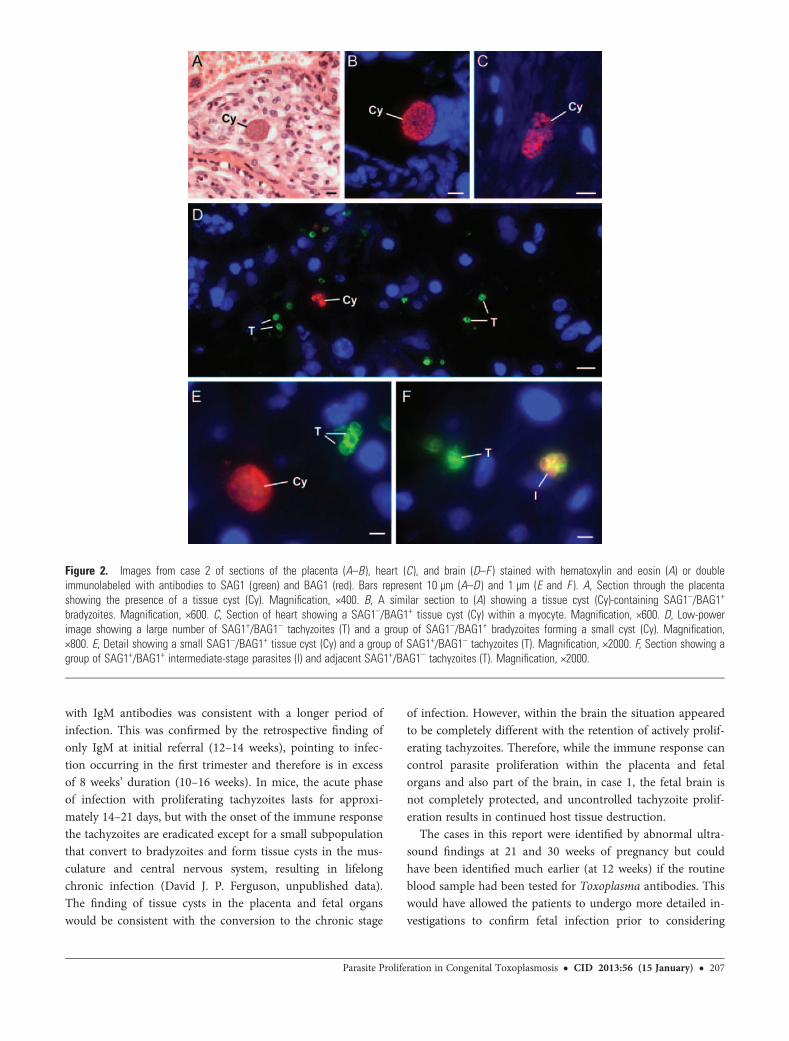
Case 2By histology, low numbers of tissue cysts were identifiedwithin the placenta (Figure 2A) but parasites were not identi-fied in fetal organs except the brain. Immunocytochemistryconfirmed the identity of the tissue cysts as containingSAG1–/BAG1+ (Figure 2B). In addition, a few small tissuecysts were identified within myocytes of the heart (Figure 2C).However, no SAG1+/BAG1– proliferating tachyzoites were ob-served in the placenta or fetal tissues.
In contrast, in sections from various regions of the brain,areas with numerous individual and small groups of proliferat-ing SAG1+/BAG1– tachyzoites were observed (Figure 2D). Inaddition, a few parasites were SAG1+/BAG1+ representing in-termediate stages (Figure 2F) and a few small SAG1–/BAG1+
tissue cysts were also present (Figure 2E).
DISCUSSION
The autopsy examination of the placentas and fetal tissue con-firmed the diagnosis of toxoplasmosis. Review of the serologi-cal investigations showed positive results for both IgM andIgG at time of termination of pregnancy (21 and 30 weeks,respectively). The presence of specific IgG antibodies together
Figure 1. Images from case 1 of the umbilical cord (A), heart (B ), and brain (C–E ) stained with hematoxylin and eosin (A) or double immunolabeledwith antibodies to SAG1 and BAG1 and visualized with fluorescein isothiocyanate (green) and Texas red (red), respectively (B–E ). Bars represent 10 µm(A–D ) and 1 µm (E ). A, Section through the umbilical cord showing the presence of 2 tissue cysts (Cy). Magnification, ×200; inset shows immunostainedsection of a tissue cyst containing SAG1–/BAG1+ bradyzoites. Magnification, ×800. B, Section of a SAG1–/BAG1+ tissue cyst (Cy) in the heart. Magnifi-cation, ×800. C, Low-power image through a damaged area showing the very large numbers of proliferating SAG1+/BAG1– tachyzoites (T). Magnification,×600. D, Section of a SAG1–/BAG1+ tissue cyst (Cy) surrounded by normal neural tissue. Magnification, ×800. E, Detail showing dividing tachyzoites (T).Magnification, ×3000.
206 • CID 2013:56 (15 January) • Ferguson et al
with IgM antibodies was consistent with a longer period ofinfection. This was confirmed by the retrospective finding ofonly IgM at initial referral (12–14 weeks), pointing to infec-tion occurring in the first trimester and therefore is in excessof 8 weeks’ duration (10–16 weeks). In mice, the acute phaseof infection with proliferating tachyzoites lasts for approxi-mately 14–21 days, but with the onset of the immune responsethe tachyzoites are eradicated except for a small subpopulationthat convert to bradyzoites and form tissue cysts in the mus-culature and central nervous system, resulting in lifelongchronic infection (David J. P. Ferguson, unpublished data).The finding of tissue cysts in the placenta and fetal organswould be consistent with the conversion to the chronic stage
of infection. However, within the brain the situation appearedto be completely different with the retention of actively prolif-erating tachyzoites. Therefore, while the immune response cancontrol parasite proliferation within the placenta and fetalorgans and also part of the brain, in case 1, the fetal brain isnot completely protected, and uncontrolled tachyzoite prolif-eration results in continued host tissue destruction.
The cases in this report were identified by abnormal ultra-sound findings at 21 and 30 weeks of pregnancy but couldhave been identified much earlier (at 12 weeks) if the routineblood sample had been tested for Toxoplasma antibodies. Thiswould have allowed the patients to undergo more detailed in-vestigations to confirm fetal infection prior to considering
Figure 2. Images from case 2 of sections of the placenta (A–B ), heart (C ), and brain (D–F ) stained with hematoxylin and eosin (A) or doubleimmunolabeled with antibodies to SAG1 (green) and BAG1 (red). Bars represent 10 µm (A–D ) and 1 µm (E and F ). A, Section through the placentashowing the presence of a tissue cyst (Cy). Magnification, ×400. B, A similar section to (A) showing a tissue cyst (Cy)-containing SAG1–/BAG1+
bradyzoites. Magnification, ×600. C, Section of heart showing a SAG1–/BAG1+ tissue cyst (Cy) within a myocyte. Magnification, ×600. D, Low-powerimage showing a large number of SAG1+/BAG1– tachyzoites (T) and a group of SAG1–/BAG1+ bradyzoites forming a small cyst (Cy). Magnification,×800. E, Detail showing a small SAG1–/BAG1+ tissue cyst (Cy) and a group of SAG1+/BAG1– tachyzoites (T). Magnification, ×2000. F, Section showing agroup of SAG1+/BAG1+ intermediate-stage parasites (I) and adjacent SAG1+/BAG1– tachyzoites (T). Magnification, ×2000.
Parasite Proliferation in Congenital Toxoplasmosis • CID 2013:56 (15 January) • 207

whether to undergo termination or anti-Toxoplasma treat-ment. An ongoing controversy exists over whether to treatwomen with anti-Toxoplasma drugs if seroconversion occursduring pregnancy [8]. There is circumstantial evidence for theefficacy of treatment if initiated early [8–11], consistent withworse outcomes reported in those countries without screeningand therefore no prenatal treatment [12]. The present reportshows that there can be continuing parasite proliferationwithin the brain of the fetus for extended periods. Both fetalbrains described showed immune privilege, and it could beargued that treatment would have a beneficial affect by de-stroying any remaining proliferating parasites within thebrain.
In conclusion, in both cases the placenta and fetal organsshowed features consistent with conversion to the non-tissue-destructive chronic phase with few or no proliferatingparasites. However the brains of both fetuses showed evidenceof numerous proliferating acute-stage tachyzoites resulting incontinued tissue destruction.
Note
Potential conflicts of interest. All authors: No reported conflicts.All authors have submitted the ICMJE Form for Disclosure of Potential
Conflicts of Interest. Conflicts that the editors consider relevant to thecontent of the manuscript have been disclosed.
References
1. Ferguson DJP. Toxoplasma gondii: 1908–2008, homage to Nicolle,Manceaux and Splendore. Mem Inst Oswaldo Cruz 2009; 104:133–48.
2. Remington JS, McLeod R, Thulliez P, Desmonts G. Toxoplasmosis. In:Remington JS, Klein J, Wilson CB, Baker CJ, eds. Infectious disease ofthe fetus and newborn infant. Philadelphia: Elsevier Saunders,2006:947–1092.
3. Robert-Gangneux F, Murat JB, Fricker-Hidalgo H, Brenier-PinchartMP, Gangneux JP, Pelloux H. The placenta: a main role in congenitaltoxoplasmosis? Trends Parasitol 2011; 27:530–6.
4. Montoya JG, Remington JS. Management of Toxoplasma gondii infec-tion during pregnancy. Clin Infect Dis 2008; 47:554–66.
5. Dunn D, Wallon M, Peyron F, Petersen E, Peckham C, Gilbert R.Mother-to-child transmission of toxoplasmosis: risk estimates for clin-ical counselling. Lancet 1999; 353:1829–33.
6. Harning D, Spenter J, Metsis A, Vuust J, Petersen E. RecombinantToxoplasma gondii surface antigen 1 (P30) expressed in Escherichiacoli is recognized by human Toxoplasma-specific immunoglobulin M(IgM) and IgG antibodies. Clin Diagn Lab Immunol 1996; 3:355–7.
7. Bohne W, Gross U, Ferguson DJP, Heesemann J. Cloning and charac-terization of a bradyzoite-specifically expressed gene (hsp30/bag1) ofToxoplasma gondii, related to genes encoding small heat-shock pro-teins of plants. Mol Microbiol 1995; 16:1221–30.
8. Cortina-Borja M, Tan HK, Wallon M, et al., European MulticentreStudy on Congenital Toxoplasmosis (EMSCOT). Prenatal treatmentfor serious neurological sequelae of congenital toxoplasmosis: an ob-servational prospective cohort study. PLoS Med 2010; 7:e1000351.
9. Gras L, Wallon M, Pollak A, et al., European Multicenter Study onCongenital Toxoplasmosis. Association between prenatal treatmentand clinical manifestations of congenital toxoplasmosis in infancy: acohort study in 13 European centres. Acta Paediatr 2005; 94:1721–31.
10. Hotop A, Hlobil H, Gross U. Efficacy of rapid treatment initiation fol-lowing primary Toxoplasma gondii infection during pregnancy. ClinInfect Dis 2012; 54:1545–52.
11. McLeod R, Kieffer F, Sautter M, Hosten T, Pelloux H. Why prevent,diagnose and treat congenital toxoplasmosis? Mem Inst Oswaldo Cruz2009; 104:320–44.
12. McLeod R, Boyer KM, Lee D, et al., the Toxoplasmosis Study Group.Prematurity and severity are associated with Toxoplasma gondii alleles(NCCCTS, 1981–2009). Clin Infect Dis 2012; 54:1595–605.
208 • CID 2013:56 (15 January) • Ferguson et al