Compartmentalized Culture of Perivascular Stroma and ... · Compartmentalized Culture of...
12
Reproductive Tissue Engineering Compartmentalized Culture of Perivascular Stroma and Endothelial Cells in a Microfluidic Model of the Human Endometrium JUAN S. GNECCO, 1,2 VIRGINIA PENSABENE , 3,4 DAVID J. LI, 5 TIANBING DING, 1 ELLIOT E. HUI, 5 KAYLON L. BRUNER-TRAN, 1 and KEVIN G. OSTEEN 1,2,6 1 Women’s Reproductive Health Research Center, Vanderbilt University Medical Center, Nashville, TN, USA; 2 Department of Pathology, Immunology and Microbiology, Vanderbilt University Medical Center, Nashville, TN, USA; 3 School of Electronic and Electrical Engineering, University of Leeds, Woodhouse Lane, Leeds LS2 9JT, UK; 4 School of Medicine, Leeds Institute of Biomedical and Clinical Sciences, University of Leeds, Leeds, UK; 5 Department of Biomedical Engineering, University of California, Irvine, CA, USA; and 6 Veteran Affairs Tennessee Valley Healthcare System, Nashville, TN, USA (Received 6 October 2016; accepted 11 January 2017; published online 20 January 2017) Associate Editor Christiani Amorim oversaw the review of this article. Abstract—The endometrium is the inner lining of the uterus. Following specific cyclic hormonal stimulation, endometrial stromal fibroblasts (stroma) and vascular endothelial cells exhibit morphological and biochemical changes to support embryo implantation and regulate vascular function, respec- tively. Herein, we integrated a resin-based porous membrane in a dual chamber microfluidic device in polydimethylsiloxane that allows long term in vitro co-culture of human endometrial stromal and endothelial cells. This transparent, 2-lm porous membrane separates the two chambers, allows for the diffusion of small molecules and enables high resolution bright field and fluorescent imaging. Within our primary human co-culture model of stromal and endothelial cells, we simulated the temporal hormone changes occurring during an idealized 28- day menstrual cycle. We observed the successful differentiation of stroma into functional decidual cells, determined by mor- phology as well as biochemically as measured by increased production of prolactin. By controlling the microfluidic prop- erties of the device, we additionally found that shear stress forces promoted cytoskeleton alignment and tight junction formation in the endothelial layer. Finally, we demonstrated that the endometrial perivascular stroma model was sustain- able for up to 4 weeks, remained sensitive to steroids and is suitable for quantitative biochemical analysis. Future utiliza- tion of this device will allow the direct evaluation of paracrine and endocrine crosstalk between these two cell types as well as studies of immunological events associated with normal vs. disease-related endometrial microenvironments. Keywords—Endometrium, Stroma, Organs-on-a-chip, Mi- crofluidic, Porous membrane. INTRODUCTION The female reproductive tract is composed of interactive organs, including uterus and ovaries that are physiologically regulated by endocrine signals at a spatial–temporal level. 14 The endometrium lines the inner cavity of the uterus and is the primary maternal tissue responsible for establishing embryo implantation and the successful maintenance of pregnancy. 2 Somatic cell numbers and immune cell ratios within the en- dometrium vary significantly across each phase of the menstrual cycle. However, histologic analysis of the cycling endometrium consistently reveals luminal and glandular epithelial cells supported by specialized reticular stromal fibroblasts (stroma), a vascular sys- tem and a dynamic flux of immune cells 39 (Fig. 1). Endometrial tissue homeostasis, cellular prolifera- tion, metabolism and reproductive function are medi- ated by the crosstalk between these cell types through paracrine and endocrine pathways. 3,9,11,31–33 The cyclical changes of ovarian sex steroids production, oestrogen and progesterone, dictate timing and func- tional capabilities of the endometrium to support em- bryo implantation. These steroids account for the distinct phases of the menstrual cycle by driving cell- specific morphological and biochemical changes (as depicted in Fig. 1d). In the endometrium of humans and some other primates, 20,37,42 during the secretory phase of a non- pregnant menstrual cycle, increasing levels of ovarian progesterone trigger a partial, spontaneous decidual- ization process within the stroma. Address correspondence to Virginia Pensabene, School of Elec- tronic and Electrical Engineering, University of Leeds, Woodhouse Lane, Leeds LS2 9JT, UK. Electronic mail: [email protected] Annals of Biomedical Engineering, Vol. 45, No. 7, July 2017 (ȑ 2017) pp. 1758–1769 DOI: 10.1007/s10439-017-1797-5 0090-6964/17/0700-1758/0 ȑ 2017 The Author(s). This article is published with open access at Springerlink.com 1758
Transcript of Compartmentalized Culture of Perivascular Stroma and ... · Compartmentalized Culture of...
Reproductive Tissue Engineering
Compartmentalized Culture of Perivascular Stroma and Endothelial Cells
in a Microfluidic Model of the Human Endometrium
JUAN S. GNECCO,1,2 VIRGINIA PENSABENE ,3,4 DAVID J. LI,5 TIANBING DING,1 ELLIOT E. HUI,5
KAYLON L. BRUNER-TRAN,1 and KEVIN G. OSTEEN1,2,6
1Women’s Reproductive Health Research Center, Vanderbilt University Medical Center, Nashville, TN, USA; 2Department ofPathology, Immunology and Microbiology, Vanderbilt University Medical Center, Nashville, TN, USA; 3School of Electronicand Electrical Engineering, University of Leeds, Woodhouse Lane, Leeds LS2 9JT, UK; 4School of Medicine, Leeds Institute ofBiomedical and Clinical Sciences, University of Leeds, Leeds, UK; 5Department of Biomedical Engineering, University of
California, Irvine, CA, USA; and 6Veteran Affairs Tennessee Valley Healthcare System, Nashville, TN, USA
(Received 6 October 2016; accepted 11 January 2017; published online 20 January 2017)
Associate Editor Christiani Amorim oversaw the review of this article.
Abstract—The endometrium is the inner lining of the uterus.Following specific cyclic hormonal stimulation, endometrialstromal fibroblasts (stroma) and vascular endothelial cellsexhibit morphological and biochemical changes to supportembryo implantation and regulate vascular function, respec-tively.Herein, we integrated a resin-based porousmembrane ina dual chamber microfluidic device in polydimethylsiloxanethat allows long term in vitro co-culture of human endometrialstromal and endothelial cells. This transparent, 2-lm porousmembrane separates the two chambers, allows for the diffusionof small molecules and enables high resolution bright field andfluorescent imaging. Within our primary human co-culturemodel of stromal and endothelial cells, we simulated thetemporal hormone changes occurring during an idealized 28-daymenstrual cycle.We observed the successful differentiationof stroma into functional decidual cells, determined by mor-phology as well as biochemically as measured by increasedproduction of prolactin. By controlling the microfluidic prop-erties of the device, we additionally found that shear stressforces promoted cytoskeleton alignment and tight junctionformation in the endothelial layer. Finally, we demonstratedthat the endometrial perivascular stroma model was sustain-able for up to 4 weeks, remained sensitive to steroids and issuitable for quantitative biochemical analysis. Future utiliza-tion of this device will allow the direct evaluation of paracrineand endocrine crosstalk between these two cell types as well asstudies of immunological events associated with normal vs.disease-related endometrial microenvironments.
Keywords—Endometrium, Stroma, Organs-on-a-chip, Mi-
crofluidic, Porous membrane.
INTRODUCTION
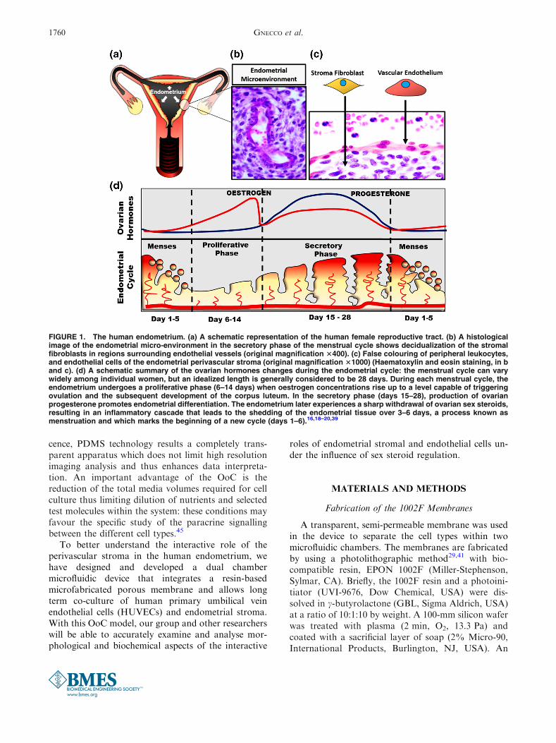
The female reproductive tract is composed ofinteractive organs, including uterus and ovaries thatare physiologically regulated by endocrine signals at aspatial–temporal level.14 The endometrium lines theinner cavity of the uterus and is the primary maternaltissue responsible for establishing embryo implantationand the successful maintenance of pregnancy.2 Somaticcell numbers and immune cell ratios within the en-dometrium vary significantly across each phase of themenstrual cycle. However, histologic analysis of thecycling endometrium consistently reveals luminal andglandular epithelial cells supported by specializedreticular stromal fibroblasts (stroma), a vascular sys-tem and a dynamic flux of immune cells39 (Fig. 1).
Endometrial tissue homeostasis, cellular prolifera-tion, metabolism and reproductive function are medi-ated by the crosstalk between these cell types throughparacrine and endocrine pathways.3,9,11,31–33 Thecyclical changes of ovarian sex steroids production,oestrogen and progesterone, dictate timing and func-tional capabilities of the endometrium to support em-bryo implantation. These steroids account for thedistinct phases of the menstrual cycle by driving cell-specific morphological and biochemical changes (asdepicted in Fig. 1d).
In the endometrium of humans and some otherprimates,20,37,42 during the secretory phase of a non-pregnant menstrual cycle, increasing levels of ovarianprogesterone trigger a partial, spontaneous decidual-ization process within the stroma.
Address correspondence to Virginia Pensabene, School of Elec-
tronic and Electrical Engineering, University of Leeds, Woodhouse
Lane, Leeds LS2 9JT,UK. Electronic mail: [email protected]
Annals of Biomedical Engineering, Vol. 45, No. 7, July 2017 (� 2017) pp. 1758–1769
DOI: 10.1007/s10439-017-1797-5
0090-6964/17/0700-1758/0 � 2017 The Author(s). This article is published with open access at Springerlink.com
1758
In all women, the morphological and biochemicaldifferentiation of stroma into specialized decidual cellsis clinically recognized as being critical for the suc-cessful establishment and maintenance of pregnancy toterm. Decidualized stroma exhibit an epithelial-likecuboidal morphology and develop the biochemicalcapacity to release several pro-gestational moleculesincluding prolactin and insulin-like growth factorbinding protein-1. Histological evidence of secretoryphase decidualization has been observed to originate inthose regions directly surrounding the vasculature(Fig. 1b). While this basic, visual observation is wellaccepted in the field, the degree to which paracrinesignalling mechanisms between vascular cells andadjacent specialized fibroblasts drives early decidual-ization at this specific site has not yet been determined.At present, a lack of appropriate in vitro models ofhuman endometrial cells that allow analysis of poten-tial interactions between key cell types necessary tosupport the successful establishment and maintenanceof pregnancy hinders progress in understanding thiscritically important aspect of reproductive tract func-tion.2,12
Many researchers, including our group, havedemonstrated an important role for stromal-epithelialcross talk in normal endometrial function while dys-regulated cell–cell communication is associated withnumerous disease processes11,24,31,40; however, theinteraction between stroma and their adjacent vascularendothelium has received less investigative attention.1
While steroid hormone receptors are largely concen-trated in endometrial stromal and epithelial cells(Fig. 1d), it is equally true that the proliferative en-dometrium becomes increasingly thicker due toendometrial vascularization, as spiral arteries growwithin the stroma in response to increasing levels ofoestrogen. In regards to this biological relationship,Albrecht et al.1 co-cultured human endometrial so-matic cells (i.e. epithelial and stromal cells) withmyometrial microvascular endothelial cells using atranswell assay and observed an increase in endothelialtube formation and vascular endothelial growth factorproduction under the influence of oestrogen.1 Togetherwith established histological observations, Albrecht’sin vitro study strongly suggests that interactionsbetween endothelial and stromal cells occur via activeparacrine communication.
In relation to the culture system described herein,the possibility to precisely recreate and visualize mor-phological and functional changes in a microfluidicmodel will be critical to better understand not onlyoestrogen action, but also progesterone-directed com-munication between these specific cell types during thesecond half of the human menstrual cycle. In additionto the cyclic oestrogen and progesterone mediated
events described above, it is also important to note thatbiologically significant changes also occur in relationto the immunomodulatory function of endothelial cellsat the end of the secretory phase of the menstrualcycle.21–23,46
A more physiologic model system of the perivas-cular stroma should provide not only a better basicknowledge of normal steroid-mediated endometrialfunction but also holds promise to reveal how alteredpatterns of cell–cell communication promotes thepathogenesis of diseases, including breakthroughbleeding, infertility, menorrhagia, endometriosis,pregnancy disorders and endometrial cancer.20,21
Unfortunately, most in vitro models fail to mimic thein vivo physiological conditions that the endotheliumexperiences, including bidirectional paracrine crosstalkbetween cells and hemodynamic forces. Moreover,current in vitro models do not allow for high resolutionreal-time examination of functionally significant mor-phological changes. Current transwell assays largelyenable modelling of specific cell barriers in humanorgans, such as epithelium4 or endothelium,36 withminimal capability to mimic and control the hemody-namic flow conditions observed in the vasculature.Several in vivo and in vitro studies have defined the roleof hemodynamic forces in the regulation of vascularfunction35,47: once exposed to shear stress from thecontinuous blood circulation, endothelial cells undergocytoskeletal remodelling (from cobblestone shaped instatic conditions to elongated cell body in the directionof flow in dynamic conditions)10 and become func-tionally different compared to cells cultured in staticconditions.32,33
Multi-compartmental 3D microfluidic cell culturedevices, so called ‘‘Organs-on-a-Chip’’ (OoC) havebeen introduced to address limitations of in vitromodelling.6,8,9,13,15,25,26,28,30 These models representrobust compartmentalized, heterogeneous cell culturesystems to simulate the physiology and anatomy ofhuman organs and thus should enhance our under-standing of in vivo mechanisms that are otherwisedifficult to study. Via microfluidic compartmentaliza-tion, the role of specific cell types can be identified aswell as the cell-specific effects of biomechanical forces(e.g. shear stress) and chemical (steroid stimulation)cues that are externally introduced and controlled inthe system. Key analytical functions of OoC includereal-time imaging of a cell culture, maintenance of longterm cultures (a minimum of 4 weeks), non-invasiveselective staining and the analysis of secretion andmetabolism of the individual cell types by samplingspent media from each compartment. Additionally,while commonly used transwell inserts rely on polye-ster and polycarbonate membranes that are opaque inbright field or may contribute to high auto fluores-
A Microfluidic Model of the Endometrial Perivascular Stroma 1759
cence, PDMS technology results a completely trans-parent apparatus which does not limit high resolutionimaging analysis and thus enhances data interpreta-tion. An important advantage of the OoC is thereduction of the total media volumes required for cellculture thus limiting dilution of nutrients and selectedtest molecules within the system: these conditions mayfavour the specific study of the paracrine signallingbetween the different cell types.45
To better understand the interactive role of theperivascular stroma in the human endometrium, wehave designed and developed a dual chambermicrofluidic device that integrates a resin-basedmicrofabricated porous membrane and allows longterm co-culture of human primary umbilical veinendothelial cells (HUVECs) and endometrial stroma.With this OoC model, our group and other researcherswill be able to accurately examine and analyse mor-phological and biochemical aspects of the interactive
roles of endometrial stromal and endothelial cells un-der the influence of sex steroid regulation.
MATERIALS AND METHODS
Fabrication of the 1002F Membranes
A transparent, semi-permeable membrane was usedin the device to separate the cell types within twomicrofluidic chambers. The membranes are fabricatedby using a photolithographic method29,41 with bio-compatible resin, EPON 1002F (Miller-Stephenson,Sylmar, CA). Briefly, the 1002F resin and a photoini-tiator (UVI-9676, Dow Chemical, USA) were dis-solved in c-butyrolactone (GBL, Sigma Aldrich, USA)at a ratio of 10:1:10 by weight. A 100-mm silicon waferwas treated with plasma (2 min, O2, 13.3 Pa) andcoated with a sacrificial layer of soap (2% Micro-90,International Products, Burlington, NJ, USA). An
FIGURE 1. The human endometrium. (a) A schematic representation of the human female reproductive tract. (b) A histologicalimage of the endometrial micro-environment in the secretory phase of the menstrual cycle shows decidualization of the stromalfibroblasts in regions surrounding endothelial vessels (original magnification 3400). (c) False colouring of peripheral leukocytes,and endothelial cells of the endometrial perivascular stroma (original magnification 31000) (Haematoxylin and eosin staining, in band c). (d) A schematic summary of the ovarian hormones changes during the endometrial cycle: the menstrual cycle can varywidely among individual women, but an idealized length is generally considered to be 28 days. During each menstrual cycle, theendometrium undergoes a proliferative phase (6–14 days) when oestrogen concentrations rise up to a level capable of triggeringovulation and the subsequent development of the corpus luteum. In the secretory phase (days 15–28), production of ovarianprogesterone promotes endometrial differentiation. The endometrium later experiences a sharp withdrawal of ovarian sex steroids,resulting in an inflammatory cascade that leads to the shedding of the endometrial tissue over 3–6 days, a process known asmenstruation and which marks the beginning of a new cycle (days 1–6).16,18–20,39
GNECCO et al.1760
8-lm layer of 1002F (diluted 1:1 in GBL) was de-posited by spin coating and then soft baked. The wa-fers were then exposed through a 125-mm chromium–quartz photomask (10 mW/cm2, 14 s). After a post-exposure bake, the patterned resist was developed inpropylene glycol methyl ether acetate. Each membraneis patterned with a 6.5 9 6.5 mm2 array of 2-lm poresenclosed within a 15-mm diameter circle.
PDMS Layer Design and Fabrication
The device was assembled using two 4.75 mm by6.2 mm microfluidic chambers separated by a semi-permeable 1002F membrane. The complete device wasfabricated by soft lithography17 in polydimethylsilox-ane (PDMS, Sylgard� 184, Dow Corning, MI, USA)(Fig. 2) as summarized in Fig. 2.
Assembly of the Two-Chambers Platform
The 1002F membranes were released from the sili-con master by dissolving the sacrificial Micro-90 soaplayer. The membranes were carefully removed andinspected for quality prior to bonding. After release,the membranes were kept in sterile H2O to removeresidual soap and to prevent drying. The top PDMSlayer and the 1002F membrane were then oxygen-
plasma treated (600 mTorr, 100 W, 45 s) and bondedtogether. Since both PDMS and 1002F are opticallytransparent, alignment of various components wascompleted under a stereomicroscope. The second layerwas then bonded with the same method on the 1002Fmembrane orthogonally to the top PDMS layer.
Oxygen-plasma treatment renders the exposed sur-faces hydrophilic. Hence, the assembled devices wereimmediately filled with sterile DI H2O and stored at4�C until used. For static experiments, 500 lL cloningcylinders (Fisher Scientific, Pittsburgh, PA, USA) werebonded with liquid PDMS to the inlet/outlet regions ofeach channel to form small reservoirs for the cellmedia.
Acquisition of Human Tissues
The Vanderbilt University Institutional ReviewBoard approved the procedures for tissue acquisition,performed only after patients gave informed consent.Primary human umbilical vein endothelial cells (HU-VECs) were isolated from umbilical cord,5 obtainedfrom de-identified term placenta collected frompatients who underwent elective caesarean sectionbetween 37 and 39 weeks of gestation. For this phaseof establishment, optimization and characterization ofthe OoC, HUVECS were selected since they represent
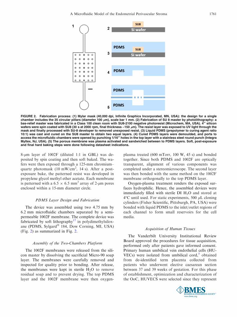
FIGURE 2. Fabrication process: (1) Mylar mask (40,000 dpi, Infinite Graphics Incorporated, MN, USA): the design for a singlechamber includes the 35 circular pillars (diameter 100 lm), scale bar 1 mm. (2) Fabrication of SU 8 master by photolithography: abas-relief master was fabricated in a Class 100 clean room with SU8-2100 negative photoresist (Microchem, MA, USA). 4¢¢ siliconwafers were spin coated with SU8 (30 s at 2000 rpm, final thickness ~150 lm). The resist layer was exposed to UV light through themask and finally processed with SU-8 developer to removed unexposed resist. (3) Liquid PDMS (prepolymer to curing agent ratio10:1) was cast and cured on the SU8 master to obtain two equal layers. (4) Cured PDMS layers were demoulded, and ports toaccess the microfluidic chambers were opened by punching 1/16¢¢ holes in the top layer with a stainless steel round punch (IntegraMyltex, NJ, USA). (5) The porous membrane was plasma activated and sandwiched between to PDMS layers. Soft, post-exposureand final hard baking steps were done following datasheet indications.
A Microfluidic Model of the Endometrial Perivascular Stroma 1761
the most common model based on human derivedprimary endothelial cells.34 After isolation, we consis-tently observed ‡95% purity of endothelial cells, vali-dated morphologically and by immunofluorescentstaining for CD31 (DAKO, USA) before loading in thedevices. Cells were cultured in EBM-2 medium sup-plemented with EGMTM-2 Single Quot� growth fac-tors (Lonza, USA), maintained at 37�C in a saturatedhumidity atmosphere containing 95% air/5% CO2,and they were sub-cultured before reaching 60–70%confluence (approximately every 2 days) up to passage5.
For acquisition of endometrial stroma, surgicallyexcised uterine tissues were collected from consenteddonors (ages 18–45) exhibiting predictable menstrualcycles and undergoing a hysterectomy for benignleiomyoma not associated with any additional inflam-matory ovarian or endometrial disease. Endometrialstroma were isolated by enzymatic digestion and filterseparation.40 As with our endothelial cell preparation,the purity of the stroma isolation was above 95% andwas quantified by morphological assessment and pos-itive staining for vimentin as previously described.40
Stroma were maintained in phenol red-free DMEM/F-12 with 10% charcoal-stripped calf serum, 1 nM 17-b oestradiol (Sigma Aldrich, USA) and 19 antibiotic–antimycotic solution (stromal complete growth med-ium). As required for experimental objectives, somestroma cultures received treatments with 0.5 mMmedroxyprogesterone acetate (MPA, Sigma Aldrich,USA) and/or 8-bromoadenosine-3¢,5¢-cyclicmonophosphate (cAMP, 0.5 mM, Sigma Aldrich,USA) to induce a decidualization response over aperiod of 14 days.
Cell Culture and Maintenance in Device
Isolated stromal and endometrial cells were initiallyexpanded within 75 mm flasks before transfer to ourmicrofluidic devices. Upon achieving approximately80% confluent monolayers, both endothelial andstromal cells were separately trypsinized, pelleted bycentrifugation (220 rcf), resuspended in full medium(1 9 106 cells/mL) and subsequently loaded into eachchamber using a 1 mL syringe. To enhance cell adhe-sion inside the device we utilized a thin coating of 1:50dilution of Matrigel (BD Bioscience, USA) on bothchambers to provide an extracellular matrix substrate.The cells were allowed to adhere for a minimum of30 min inside the incubator. 300 lL of EBM-2 mediumor complete growth medium, were added to theendothelial top chamber or stromal bottom chamber,respectively. Spent media from the reservoirs werecollected and replaced daily with fresh mediathroughout all static experiments.
For dynamic flow experiments, the endothelialchamber was perfused by using a syringe pump (Pi-coPlus, Harvard Apparatus, Cambridge, USA) and byconnecting Tygon� tubing (Cole-Parmer) directly intothe inlet port of the device. Shear stress conditionswere induced when the cells reached 60% confluence.The final value of 1 lL/min was defined with thisprotocol as minimal value for the cells to form a tightand reoriented endothelium. The stroma were main-tained under the same static culture conditions throughall experiments. Wall shear stress in the center of thechamber (approximately 6 9 1023 dyn s cm22) wascalculated as in Sung et al.44 treating the chamber as arelatively flat channel (with width � height). Bubbleformation was limited by loading the microfluidicchambers with DI water immediately after plasmatreatment and by equalizing the temperature of themedia in the syringe and inside the device beforestarting the perfusion.
Fluorescence Imaging
Mouse monoclonal anti human CD-31 primaryantibody (DAKO, USA) was used at a 1:50 dilutionand goat anti-mouse IgG Cy3 conjugate (JacksonImmuno Research, USA) was used as the secondaryantibody (1:200). Tight junction expression of zonaoccludens-1 (ZO-1) was measured with a mouse anti-human FITC-conjugated antibody (1:50, Invitrogen,USA). A rabbit antibody against human vimentin(1:200, Abcam, USA) was used with a FITC conju-gated goat anti-rabbit antibody as a secondary anti-body (1:100, Jackson ImmunoResearch, USA).Standard fixation and immunofluorescence stainingprotocols were performed as described in the productdatasheet. The cell nuclei were stained with 4¢6-di-amidino-2-phenylindole (DAPI, Sigma Aldrich) for1 min and then washed with 1X PBS.
Membrane Permeability Assay
A 3 mL syringe was filled with a 2.5 mg/mL solu-tion of FITC-dextran (150 kDa MW, Sigma Aldrich,USA) and mounted on a syringe pump. The syringewas connected to the top chamber with microboretubing, while 200 lL of 1X PBS were added to theoutlet reservoir and to the inlet and outlet of the bot-tom chamber. Perfusion was run at 2.5 lL/min.100 lL samples were collected from each of thereservoirs and replaced with 100 lL of PBS at 1, 2 and3 h intervals. Fluorescence intensity in the collectedeffluent was measured using a fluorescence microplatereader (GloMax� Multimode Readers, Promega) at470 nm excitation and 520–550 nm emission range(details in Supplemental Material).
GNECCO et al.1762
Effluent Collection and Protein Measurement
The devices were cultured initially for 14 days ineither complete growth medium, or complete EBM-2medium supplemented with oestradiol (1 nM) untilboth cell monolayers reached 80% confluence. Thecomplete growth medium for the stromal chamber wasthen supplemented with MPA (0.5 nM) and media wascollected and changed daily (300 lL) for an additional14 days from both inlet and outlet. Collected effluentswere then analysed by measuring prolactin production,a marker of decidualization, using an enzyme-linkedimmunosorbent assay (ELISA Duoset, R&D systems).The ELISA was performed according to the manu-facturer’s instructions using 50 lL samples. The platewas read using an absorbance microplate reader(GloMax� Multimode Readers, Promega). The resultsare representative of three different experiments. Dueto the patient-related variability among experiments,we normalized prolactin concentration to the minimalmean prolactin production at day 2. Statistical analysiswas performed by a 2 way ANOVA using a Bonferroni
correction. Statistical significance was calculated asp< 0.05.
RESULTS
Fabrication of a Dual-Chamber Microfluidic Devicewith a High-Resolution Porous Membrane
As noted above, the ability of researchers to under-stand the intercellular paracrine interactions that regu-late endometrial function within the perivascularmicroenvironment is limited by a lack of an appropriatemodel to accurately recreate the perfusion characteristicsand physical compartmentalization at this tissue site. Toovercome these constraints, we designed a microfluidicdevice and integrated a transparent semipermeablemembrane (Fig. 3) to model the endometrial vascularinterface between endothelial and endometrial stromalcells that mediate critical reproductive processes.
The device consists of 2 orthogonal microfluidicchambers, each one providing 29.45 mm2 of cell
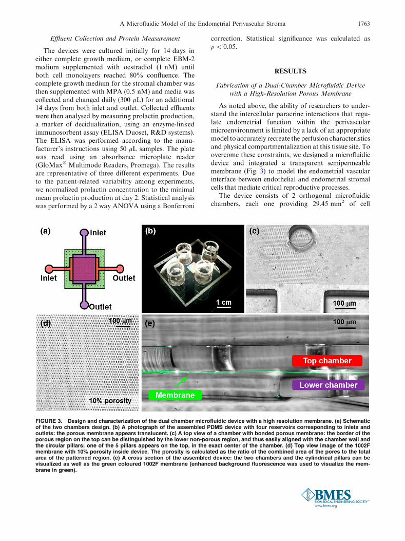
FIGURE 3. Design and characterization of the dual chamber microfluidic device with a high resolution membrane. (a) Schematicof the two chambers design. (b) A photograph of the assembled PDMS device with four reservoirs corresponding to inlets andoutlets: the porous membrane appears translucent. (c) A top view of a chamber with bonded porous membrane: the border of theporous region on the top can be distinguished by the lower non-porous region, and thus easily aligned with the chamber wall andthe circular pillars; one of the 5 pillars appears on the top, in the exact center of the chamber. (d) Top view image of the 1002Fmembrane with 10% porosity inside device. The porosity is calculated as the ratio of the combined area of the pores to the totalarea of the patterned region. (e) A cross section of the assembled device: the two chambers and the cylindrical pillars can bevisualized as well as the green coloured 1002F membrane (enhanced background fluorescence was used to visualize the mem-brane in green).
A Microfluidic Model of the Endometrial Perivascular Stroma 1763
growth area and a volume of 4.7 lL. The chambers aredivided by the 1002F resin-based membrane, andsealed by plasma bonding. The 1002F membrane isbiocompatible, has a 6-lm thickness (DekTak3 surfaceprofilometer) and 2-lm circular pores that allow sol-uble factor communication and cellular contactbetween cells cultured on the two sides.29 The pat-terned membrane fully spans the chamber to provide acompartmentalized region, suitable for cell co-cultureassays (Fig. 3). Due to the aspect ratio of the fluidicchamber, the flexibility of the membrane and the use ofautomated perfusion of the endothelial chamber, weincorporated a series of pillars within the chambersthat provide structural support to the membrane.While it is important to note that these pillars mayalter dynamic flow profile inside the chamber, we al-ways observed homogeneous cell distribution duringloading and uniform proliferation and polarizationthroughout the chamber (see next sections). Further-more, the density and size of membrane pores does notaffect the cell loading and the flow dynamic inside thedevice. Repetitive loading of cells in the two chambersdid not show leakage of cells through the pores (sincethe 2-lm diameter is smaller than the size of the indi-vidual cell).
The 1002F membrane is transparent and the porousregion can be distinguished from non-porous one(Fig. 3c). This simplifies layer alignment during deviceassembly and aids in determining the cell density ratiosfollowing device seeding. Being both PDMS and 1002Fmembrane transparent, we are able to characterize realtime cell maintenance inside the device as described indetail below and perform low backgroundimmunofluorescent imaging.
Characterization of the Microfluidic Model of theEndometrial Perivascular Stroma
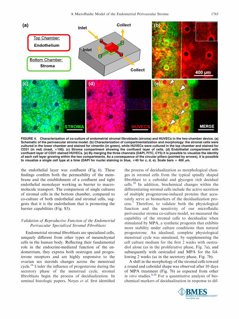
Little is known about the role of the vascularendothelium in regulating endometrial reproductiveprocesses due to a lack of adequate models thatphysiologically mimic the in vivo characteristics ofhuman tissue. To evaluate the cellular growth insidethe device, we seeded primary human endothelial cellsand endometrial stromal cells in the top and bottomchambers of the device, respectively. Both cell typeswere loaded simultaneously (1 9 106 cells/mL), lead-ing to a final cell seeding density of 5000 cells/mm2.Static culture conditions inside the OoC were main-tained for up to 28 days, corresponding to the lengthof an idealized menstrual cycle.
In order to confirm the compartmentalization of thetwo cell types, the vascular monolayer was selectivelystained for the endothelial cell marker CD31, while thestromal compartment was stained for vimentin
(Figs. 4b and 4c). We observed a clear compartmen-talization of the two cell types throughout the device.Each cell type was observed to adopt characteristicmorphologies similar to those seen in cells cultured ontraditional polystyrene cell culture dishes, with distinctcobblestone morphology exhibited by the endothelialmonolayer and a confluent layer of the stromafibroblasts that initially exhibited a striated morphol-ogy prior to decidualization (Fig. S2). Furthermore,there were no distinguishable morphological differ-ences in the endothelial layer between porous and non-porous regions of the membrane and was confirmed byCD31 immunofluorescent staining (Fig. S1a). Overtime, endothelial cells were able to colonize andestablish a confluent layer at the top and at the bottomof the chamber (Fig. S2d).
Validation of the Endothelial Monolayer Shear StressPolarization, Actin Cytoskeleton Realignment
and Barrier Function
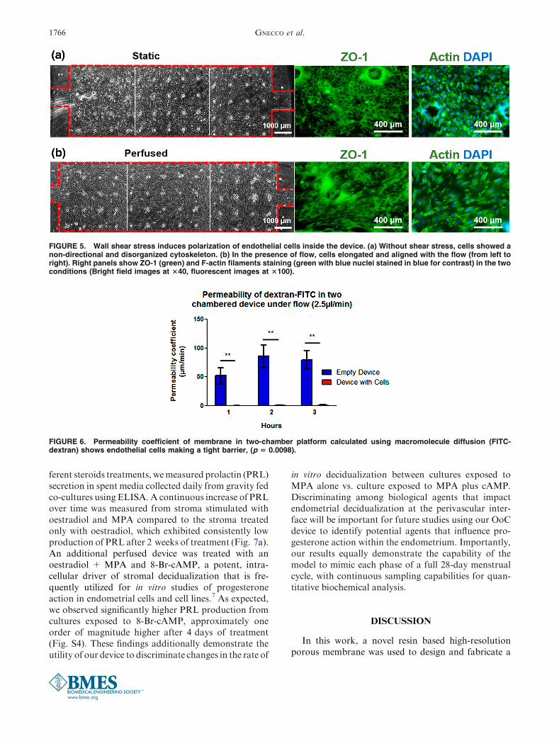
Vascular blood flow is an essential characteristic ofendothelial cell biology and thus in order to appro-priately mimic the fluid dynamic condition of theperivascular endometrium within our OoC, only theendothelial cells were dynamically perfused, while thestroma cells were cultured under static conditions. Weutilized morphological polarization as a marker ofculture conditions exhibiting physiologic shear stress.The endothelial cells were allowed to proliferate toconfluence in full medium in static conditions (Fig. 5a)and subsequently exposed to 1 lL/min flow for aminimum of 4 days (Fig. 5b). The stroma were kept instatic conditions since this cell type is not affected bydirect shear stress in physiological conditions withinthe endometrium.
Tight junction formation, observed by staining ZO-1, was positively identified in both conditions, but onlythe shear stressed monolayers formed clear parallelmargins around the cell body. These cells showedpolarization and changes in cell shape from polygonalto ellipsoidal (Fig. 5b), as result of alterations in theF-actin filaments and reorientation of stress fibers inthe cytoskeletal structure. As a proof of principle, longterm culture of a confluent endothelial layer wasmaintained under continuous perfusion for more than24 days (Supplemental Material). The dual-chamberdevice also permits assaying the functional integrity ofthe endothelial monolayer in forming a barrier to dif-fusive transport. We evaluated the permeability of theendothelial monolayer by measuring the transfer offluorescently labelled dextran (150 kD MW) betweenthe two chambers of the device. Compared to anunpopulated device, there was a significant reductionin the permeability coefficient of the membrane once
GNECCO et al.1764
the endothelial layer was confluent (Fig. 6). Thesefindings confirm both the permeability of the mem-brane and the establishment of a confluent and tightendothelial monolayer working as barrier to macro-molecule transport. The comparison of single culturesof stromal cells in the bottom chamber, compared toco-culture of both endothelial and stromal cells, sug-gests that it is the endothelium that is promoting thebarrier capabilities (Fig. S3).
Validation of Reproductive Function of the EndometrialPerivascular Specialized Stromal Fibroblasts
Endometrial stromal fibroblasts are specialized cells,uniquely different from other types of mesenchymalcells in the human body. Reflecting their fundamentalrole in the endocrine-mediated function of the en-dometrium, they express both oestrogen and proges-terone receptors and are highly responsive to theovarian sex steroids changes across the menstrualcycle.16 Under the influence of progesterone during thesecretory phase of the menstrual cycle, stromalfibroblasts begin the process of decidualization. Inseminal histologic papers, Noyes et al. first identified
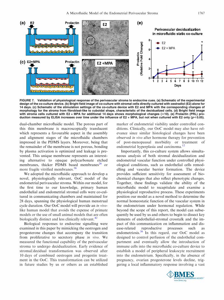
the process of decidualization as morphological chan-ges in stromal cells from the typical spindly shapedfibroblast to a cuboidal and glycogen rich decidualcells.39 In addition, biochemical changes within thedifferentiating stromal cells include the active secretionof multiple progesterone-induced proteins that accu-rately serve as biomarkers of the decidualization pro-cess.7 Therefore, to validate both the physiologicalfunction and the sensitivity of our microfluidicperivascular stroma co-culture model, we measured thecapability of the stromal cells to decidualize whenstimulated by MPA, a synthetic progestin that exhibitsmore stability under culture conditions than naturalprogesterone. An idealized, complete physiologicalmenstrual cycle was simulated, by supplementing thecell culture medium for the first 2 weeks with oestra-diol alone (as in the proliferative phase, Fig. 7a), andsubsequently with oestradiol and MPA for the fol-lowing 2 weeks (as in the secretory phase, Fig. 7b).
A shift in the morphology of the stromal cells towarda round and cuboidal shape was observed after 10 daysof MPA treatment (Fig. 7b) as expected from otherin vitro studies.4,20 For a quantitative analysis of bio-chemical markers of decidualization in response to dif-
FIGURE 4. Characterization of co-culture of endometrial stromal fibroblasts (stroma) and HUVECs in the two-chamber device. (a)Schematic of the perivascular stroma model. (b) Characterization of compartmentalization and morphology: the stromal cells werecultured in the lower chamber and stained for vimentin (in green), while HUVECs were cultured in the top chamber and stained forCD31 (in red) (inset, 3100). (c) Stroma compartment showing the confluent layer of cells. (d) Endothelial compartment withconfluent layer of CD31 stained HUVECs. (e) By merging the three channels (DAPI, FITC, CY5) it is possible to visualize the identityof each cell layer growing within the two compartments. As a consequence of the circular pillars (pointed by arrows), it is possibleto visualize a single cell type at a time (DAPI for nuclei staining in blue, 340 for c, d, e). Scale bars 5 400 lm.
A Microfluidic Model of the Endometrial Perivascular Stroma 1765
ferent steroids treatments, wemeasured prolactin (PRL)secretion in spent media collected daily from gravity fedco-cultures using ELISA. A continuous increase of PRLover time was measured from stroma stimulated withoestradiol and MPA compared to the stroma treatedonly with oestradiol, which exhibited consistently lowproduction of PRL after 2 weeks of treatment (Fig. 7a).An additional perfused device was treated with anoestradiol + MPA and 8-Br-cAMP, a potent, intra-cellular driver of stromal decidualization that is fre-quently utilized for in vitro studies of progesteroneaction in endometrial cells and cell lines.7 As expected,we observed significantly higher PRL production fromcultures exposed to 8-Br-cAMP, approximately oneorder of magnitude higher after 4 days of treatment(Fig. S4). These findings additionally demonstrate theutility of our device to discriminate changes in the rate of
in vitro decidualization between cultures exposed toMPA alone vs. culture exposed to MPA plus cAMP.Discriminating among biological agents that impactendometrial decidualization at the perivascular inter-face will be important for future studies using our OoCdevice to identify potential agents that influence pro-gesterone action within the endometrium. Importantly,our results equally demonstrate the capability of themodel to mimic each phase of a full 28-day menstrualcycle, with continuous sampling capabilities for quan-titative biochemical analysis.
DISCUSSION
In this work, a novel resin based high-resolutionporous membrane was used to design and fabricate a
FIGURE 5. Wall shear stress induces polarization of endothelial cells inside the device. (a) Without shear stress, cells showed anon-directional and disorganized cytoskeleton. (b) In the presence of flow, cells elongated and aligned with the flow (from left toright). Right panels show ZO-1 (green) and F-actin filaments staining (green with blue nuclei stained in blue for contrast) in the twoconditions (Bright field images at 340, fluorescent images at 3100).
FIGURE 6. Permeability coefficient of membrane in two-chamber platform calculated using macromolecule diffusion (FITC-dextran) shows endothelial cells making a tight barrier, (p 5 0.0098).
GNECCO et al.1766
dual-chamber microfluidic model. The porous part ofthis thin membrane is macroscopically translucentwhich represents a favourable aspect in the assemblyand alignment stages of the microfluidic chambersimpressed in the PDMS layers. Moreover, being thatthe remainder of the membrane is not porous, bondingby plasma activation is optimized and leakage is pre-vented. This unique membrane represents an interest-ing alternative to opaque polycarbonate etchedmembranes, thicker PDMS based membranes26 ormore fragile vitrified membranes.36
We adopted the microfluidic approach to develop anovel, physiologically relevant, OoC model of theendometrial perivascular stroma.Within our model, forthe first time to our knowledge, primary humanendothelial and endometrial stromal cells were co-cul-tured in communicating chambers and maintained for28 days, spanning the physiological human menstrualcycle duration. Our OoC model will provide an in vivo-like human model that avoids the expense of primatemodels or the use of small animal models that are oftenbiologically distinct and less clinically relevant.42
Biological responses to endocrine signalling wereexamined in this paper by mimicking the oestrogen andprogesterone changes that accompany the transitionfrom proliferative to secretory phase in vivo. Wemeasured the functional capability of the perivascularstroma to undergo decidualization. Early evidence ofstromal/decidual transformation was observed after10 days of combined oestrogen and progestin treat-ment in the OoC. This transformation can be utilizedin future studies by us or others as an established
marker of endometrial viability under controlled con-ditions. Clinically, our OoC model may also have rel-evance since similar histological changes have beenobserved in vivo after hormone therapy for preventionof post-menopausal morbidity or treatment ofendometrial hyperplasia and carcinoma.12
Importantly, this co-culture system allows simulta-neous analysis of both stromal decidualization andendometrial vascular function under controlled physi-ological conditions, such as endothelial cells remod-elling and vascular barrier formation. The deviceprovides sufficient sensitivity for assessment of bio-chemical changes that also reflect phenotypic changes.Together, these findings validate the ability of themicrofluidic model to recapitulate and examine aphysiological reproductive process. These experimentsposition our model as a novel method to determine thenormal homeostatic function of the vascular system inthe endometrium under hormonal regulation. Whilebeyond the scope of this report, the model can subse-quently be used by us and others to begin to dissect keyelements of endothelial-stromal crosstalk and the im-pact of this communication on inflammation and dis-ease-related reproductive processes such asendometriosis.38 In this regard, our OoC model asdesigned to control perfusion of the endothelial com-partment and eventually allow the introduction ofimmune cells into the microfluidic co-culture device toestablish a model of peripheral leukocyte recruitmentinto the endometrium. Specifically, in the absence ofpregnancy, ovarian progesterone levels decline, trig-gering a local inflammatory response involving a vast
FIGURE 7. Validation of physiological response of the perivascular stroma to endocrine cues. (a) Schematic of the experimentaldesign of the co-culture device. (b) Bright field image of co-culture with stromal cells directly cultured with oestradiol (E2) alone for14 days. (c) Schematic of the stimulation settings of the co-culture device with E2 and MPA with the corresponding changes ofmorphology for the stroma from fibroblast-like to cuboidal shape, characteristic of the decidualized cells. (d) Bright field imagewith stroma cells cultured with E2 + MPA for additional 14 days shows morphological changes (310). (e) Prolactin (PRL) pro-duction measured by ELISA increases over time under the influence of E2 + MPA, but not when cultured with E2 only (p< 0.05).
A Microfluidic Model of the Endometrial Perivascular Stroma 1767
infiltration of leukocytes, release of cytokines, andactivation of matrix metalloproteinases.27 The vascularendothelium act as the interface between peripheralcirculating immune cells and endometrial tissue. Hencethe loss of anti-inflammatory action of progesteroneduring the latter part of the secretory phase is bothdirectly and indirectly linked to phenotypic changeswithin the endothelium that affect immune cellmigration.34,43 Thus our OoC model is compatible forfuture studies of cell interaction during menstruationand in inflammatory endometrial diseases (i.e. stroma,endothelial and leukocytes).
Noteworthy, PDMS has an ability to absorbhydrophobicmolecules, specifically hormones.However,until materials with improved absorption behaviourbecome available, PDMS remains the most commonlyused and accepted material for prototyping biomedicalmicrofabrication technologies. The presented resultsreplicate previous observations in traditional in vitropolystyrene culture dishes in terms of hormone respon-sive biochemical and morphological decidualization,suggesting that absorption of steroids by the membranewas not an impediment to cellular response.
Modelling the human endometrial microenviron-ment at the immuno-endocrine inflammatory axis offersthe capability to better understand complex biologicaland pathogenic factors that act to support or disruptendometrial function. Since decidualization is a criticalprocess for embryo implantation, the ability to explorethe earliest stages of decidualization will provide a un-ique screening tool for fertility studies. Finally, our OoCmodel of the endometrial perivascular stroma lends it-self for the screening of pharmaceutical agents or envi-ronmental toxicants that may alter reproductive healthor promote reproductive dysfunctions.
ELECTRONIC SUPPLEMENTARY MATERIAL
The online version of this article (doi:10.1007/s10439-017-1797-5) contains supplementarymaterial, which is available to authorized users.
ACKNOWLEDGMENTS
This work was supported by the Department of Ve-teran Affairs (BX002853), National Institute of Envi-ronmental Health Science (ES14942), EnvironmentalToxicology Training Grant (NIHT32ES007028), andEnvironmental Protection Agency STAR Center Grant(83573601). Kallie Yeoman and Christina Svitek helpedwith tissue donor program. We acknowledge resourcesprovided by the CDBER core and the VIIBRE micro-fabrication core.
OPEN ACCESS
This article is distributed under the terms of theCreative Commons Attribution 4.0 International Li-cense (http://creativecommons.org/licenses/by/4.0/),which permits unrestricted use, distribution, and re-production in any medium, provided you give appro-priate credit to the original author(s) and the source,provide a link to the Creative Commons license, andindicate if changes were made.
REFERENCES
1Albrecht, E. D., J. S. Babischkin, Y. Lidor, L. D. Ander-son, L. C. Udoff, and G. J. Pepe. Effect of estrogen onangiogenesis in co-cultures of human endometrial cells andmicrovascular endothelial cells. Hum Reprod 18(10):2039,2003.2Aplin, J. Biology of the uterus. In: Cellular Biochemistry ofthe Endometrium, edited by R. M. Wynn. New York:Plenum, 1989.3Arnold, J. T., D. G. Kaufman, M. Seppala, and B. A.Lessey. Endometrial stromal cells regulate epithelial cellgrowth in vitro: a new co-culture model. Human Reprod16(5):836, 2001.4Baluer, M., P. K. Heinonen, P. M. Martikainen, E. Tomas,and T. Ylikomi. A novel organotypic culture model fornormal human endometrium: regulation of epithelial cellproliferation by estradiol and medroxyprogesterone acet-ate. Human Reprod 20(4):864, 2005.5Baudin, B., A. Bruneel, N. Bosselut, and M. Vaubourdolle.A protocol for isolation and culture of human umbilicalvein endothelial cells. Nat Protoc 2(3):481, 2007.6Bissell, M. J., and M. A. LaBarge. Context, tissue plas-ticity, and cancer: are tumor stem cells also regulated bythe microenvironment? Cancer Cell 7(1):17, 2005.7Brar, A. K., G. R. Frank, C. A. Kessler, M. I. Cedars, andS. Handwerger. Progesterone-dependent decidualization ofthe human endometrium is mediated by cAMP. Endocrine6(3):301, 1997.8Chen, Y. C., Y. H. Cheng, H. S. Kim, P. N. Ingram, J. E.Nor, and E. Yoon. Paired single cell co-culture microen-vironments isolated by two-phase flow with continuousnutrient renewal. Lab Chip. 14(16):2941–2947, 2014.9Chen, Y. C., Z. Zhang, S. Fouladdel, Y. Deol, P. N. In-gram, S. P. McDermott, E. Azizi, M. S. Wicha, and E.Yoon. Single cell dual adherent-suspension co-culture mi-cro-environment for studying tumor–stromal interactionswith functionally selected cancer stem-like cells. Lab Chip.16(15):2935–2945, 2016.
10Cucullo, L., M. Hossain, V. Puvenna, N. Marchi, and D.Janigro. The role of shear stress in Blood-Brain Barrierendothelial physiology. BMC Neurosci 12(1):40, 2011.
11Cunha, G. R., P. S. Cooke, and T. Kurita. Role of stromal-epithelial interactions in hormonal responses. Arch HistolCytol 67(5):417, 2004.
12Deligdisch, L. Hormonal Pathology of the Endometrium,The 1999 Long Course on Pathology of the Uterine Corpusand Cervix. Mod Pathol. 13(3):285–294, 2000.
13Douville, N. J., P. Zamankhan, Y. C. Tung, R. Li, B. L.Vaughan, C. F. Tai, J. White, P. J. Christensen, J. B.Grotberg, and S. Takayama. Combination of fluid and
GNECCO et al.1768

solid mechanical stresses contribute to cell death anddetachment in a microfluidic alveolar model. Lab Chip11(4):609–619, 2011.
14Eddie, S. L., J. J. Kim, T. K. Woodruff, and J. E. Burdette.Microphysiological modeling of the reproductive tract: afertile endeavour. Exp Biol Med 239(9):1192, 2014.
15Esch, M. B., T. L. King, and M. L. Shuler. The role ofbody-on-a-chip devices in drug and toxicity studies. AnnuRev Biomed Eng 13:55, 2011.
16Evans, J., and L. A. Salamonsen. Decidualized humanendometrial stromal cells are sensors of hormone with-drawal in the menstrual inflammatory cascade. Biol Reprod90(1):14, 2014.
17Fainman, Y., L. Lee, D. Psaltis, and C. Yang. Optofluidics:Fundamentals, Devices, and Applications. New York:McGraw-Hill Companies, 2009.
18Farage, M. A., S. Neill, and A. B. MacLean. Physiologicalchanges associated with the menstrual cycle: a review.Obstet Gynecol Surv. 64(1):58, 2009.
19Gellersen, B., I. A. Brosens, and J. J. Brosens. Decidualiza-tionof the human endometrium:mechanisms, functions, andclinical perspectives. Semin Reprod Med 25(6):445, 2007.
20Gellersen andBrosensCyclicDecidualization of theHuman.Endometrium. Endocrine Reviews 35(6):851–905, 2014.
21Girling, J. E. Rogers PAW., Recent advances in endome-trial angiogenesis research. Angiogenesis 8(2):89–99, 2005.
22Girling, J. E., B. Heryanto, and N. Patel. Rogers PAW.,Effect of long-term progestin treatment on endometrialvasculature in normal cycling mice. Contraception70(4):343, 2004.
23Grummer, M. A., J. A. Sullivan, R. R. Magness, and I. M.Bird. Vascular endothelial growth factor acts throughnovel, pregnancy-enhanced receptor signalling pathways tostimulate endothelial nitric oxide synthase activity in uter-ine artery endothelial cells. Biochem J 417(2):501, 2009.
24Hantak, A. M., I. C. Bagchi, and M. K. Bagchi. Role ofuterine stromal-epithelial crosstalk in embryo implanta-tion. Int J Dev Biol 58(2–4):139, 2014.
25Huh, D., H. Fujioka, Y. C. Tung, N. Futai, R. Paine, J. B.Grotberg, and S. Takayama. Acoustically detectable cellu-lar-level lung injury induced by fluid mechanical stresses inmicrofluidic airway systems. Proc Natl Acad Sci U S A.104(48):18886–18891, 2007.
26Huh, D., B. D. Matthews, A. Mammoto, M. Montoya-Zavala, H. Y. Hsin, and D. E. Ingber. Reconstituting or-gan-level lung functions on a chip. Science. 328(5986):1662–1668, 2010.
27Jabbour, H. N., R. W. Kelly, H. M. Fraser, and H. O.Critchley. Endocrine regulation of menstruation. EndocrRev 27:17–46, 2006.
28Kim, H. J., and D. E. Ingber. Gut-on-a-Chip microen-vironment induces human intestinal cells to undergo vil-lus differentiation. Integr Biol (Camb). 5(9):1130–1140,2013.
29Kim, M. Y., D. J. Li, L. K. Pham, B. G. Wong, and E. E.Hui. Microfabrication of High-Resolution Porous Mem-branes for Cell Culture. J Memb Sci 452:460, 2014.
30Kimura, H., T. Yamamoto, H. Sakai, Y. Sakai, and T.Fujii. An integrated microfluidic system for long termperfusion culture and on-line monitoring of intestinal tissuemodels. Lab Chip. 8(5):741–746, 2008.
31Kurita, T., P. Young, J. R. Brody, J. P. Lydon, B. W.O’Malley, and G. R. Cunha. Stromal progesterone recep-
tors mediate the inhibitory effects of progesterone onestrogen-induced uterine epithelial cell deoxyribonucleicacid synthesis. Endocrinology 139(11):4708, 1998.
32Li, Y.-S., J. H. Haga, and S. Chien. Molecular basis of theeffects of shear stress on vascular endothelial cells. J Bio-mech 38(10):1949–1971, 2005.
33Lilly, B. We have contact: endothelial cell-smooth musclecell interactions. Physiology 29(4):234, 2014.
34Man, S., E. E. Ubogu, K. A. Williams, B. Tucky, M. K.Callahan, and R. M. Ransohoff. Human brain microvas-cular endothelial cells and umbilical vein endothelial cellsdifferentially facilitate leukocyte recruitment and utilizechemokines for T cell migration. Clin Dev Immunol2008:384982, 2008.
35Martins-Green, M., M. Petreaca, and M. Yao. An assaysystem for in vitro detection of permeability in human‘‘endothelium’’. Methods Enzymol 443:137, 2008.
36Mazzocchi, A. R., A. J. Man, J.-P. S. DesOrmeaux, and T.R. Gaborski. Porous Membranes Promote EndothelialDifferentiation of Adipose-Derived Stem Cells andPerivascular Interactions. Cellular and Molecular Bioengi-neering 7(3):369–378, 2014.
37Nayak, N. R., and R. M. Brenner. Vascular proliferationand vascular endothelial growth factor expression in therhesus macaque endometrium. J Clin Endocrinol Metab87(4):1845, 2002.
38Nayyar, T., K. L. Bruner-Tran, D. Piestrzeniewicz-Ulan-ska, and K. G. Osteen. Developmental exposure of mice toTCDD elicits a similar uterine phenotype in adult animalsas observed in women with endometriosis. Reprod Toxicol23:326, 2007.
39Noyes, R., A. Hertig, and J. Rock. Dating the endometrialbiopsy: obstetrical & gynecological survey. Gynecology.5(4):561–564, 1950.
40Osteen, K. G., G. A. Hill, J. T. Hargrove, and F. Gorstein.Development of a method to isolate and culture highlypurified populations of stromal and epithelial cells fromhuman endometrial biopsy specimens. Fertil Steril52(6):965, 1989.
41Pai, J.-H., Y. Wang, G. T. Salazar, et al. Photoresist withlow fluorescence for bioanalytical applications. Anal Chem79(22):8774, 2007.
42Ramathal, Cyril Y., and Indrani C. Bagchi. Ph.D., RobertN. Taylor, M.D., Ph.D., and Milan K. Bagchi Endometrialdecidualization: of mice and men. Semin Reprod Med.28(1):17–26, 2010.
43Schumacher, A., S. D. Costa, and A. C. Zenclussen. En-docrine factors modulating immune responses in preg-nancy. Front Immunol 5:196, 2014.
44Sung, J. H., and M. L. Shuler. Microtechnology for mim-icking in vivo tissue environment. Ann Biomed Eng40(6):1289, 2012.
45Wikswo, J. P., E. L. Curtis, Z. E. Eagleton, B. C. Evans, A.Kole, L. H. Hofmeister, and W. J. Matloff. Scaling andsystems biology for integrating multiple organs-on-a-chip.Lab Chip. 13(18):3496–3511, 2013.
46Wira, C. R., M. Rodriguez-Garcia, and M. V. Patel. Therole of sex hormones in immune protection of the femalereproductive tract. Nat Rev Immunol. 15(4):217, 2015.
47Yuan, S. Y., and R. R. Rigor. Regulation of EndothelialBarrier Function. San Rafael: Morgan&Claypool LifeSciences, 2010.
A Microfluidic Model of the Endometrial Perivascular Stroma 1769