
Comparison of Gutta-Percha Obturation Techniques in the ... · Comparison of Gutta-Percha...
10
Comparison of Gutta-Percha Obturation Techniques in the Treatment of Wide Root Canals in Dogs I. CAPÍK 1 , S. ŠTVRTINA 2 1 Clinic of Small Animal Surgery, Orthopaedics and Radiology, University of Veterinary Medicine, Košice, Slovakia 2 Medical Faculty of Komenský University in Bratislava, Slovakia Received April 30, 2007 Accepted February 14, 2008 Abstract Capík I., S. Štvrtina: Comparison of Gutta-Percha Obturation Techniques in the Treatment of Wide Root Canals in Dogs. Acta Vet. Brno 2008, 77: 291-296. An in vitro study was conducted to compare gutta-percha obturation techniques of the developing incisors in dogs. Two perpendicular radiographic views were used for evaluation of apical and overall appearance of root canal obturation. Apical leakage technique was used to evaluate ability of each method to provide adequate apical obturation. The endodontic technique utilizing warm vertical condensation with chemically softened gutta- percha in the apical region provided the best radiographic appearance without apical dye leakage. The same vertical obturation technique without the use of chemically softened gutta-percha in the apical region showed 33% dye leakage incidence. Lateral obturation techniques provided the least qualitative radiographic appearance to the endodontic fill and dye leakage incidence. Results of evaluation of quality of apical obturation of root canals based on X-ray examination and apical dye leakage resulted in the following conclusions: lateral obturation techniques used in this study have their limitations resulting from typical root canal anatomy; vertical obturation techniques showed better hermetic apical sealing, mainly in the case of vertical condensation of the chemically softened gutta-percha in the apical part of the root canal. Dog, immature adult incisors, endodontic, root canal, apical sealing Hundreds of animals experience traumatic dental damage every year. This involves not only crown or root fractures which result in direct impairment of anatomical tooth structures but also shocks, chemical or thermal damage which, without visible impairment of the tooth, cause damage to its most important part, the dental pulp. The aetiology of tooth fractures is multifactorial, including activity, behaviour, and accidental trauma (Fichtel 2000; Pavlica 1991). Bacteria in the root canal system are the primary cause of periapical periodontitis (Kakehashi et al. 1965; Möller et al. 1981). Healing of apical periodontitis involves a combination of disinfection of the root canal space through chemo-mechanical means (Byström and Sundqvist 1983, 1985) and sealing both the root canal and access to cavity with materials that prevent re-infection (Ray and Trope 1995). More than 50% of faulty endodontic treatments involve leaky root restorations. Because of that it is very important to select a suitable material and obturation technique which ensures that the complex system of apical delta of dog teeth is closed as tightly as possible. The correct approach to therapy requires not only good orientation in a range of different types of available materials and techniques involving obturation of root canals but also perfect knowledge of anatomy and physiology of the endodontic system and good understanding of respective pathological processes. Radiological examination of incisors, premolars and molars is impossible from two views perpendicular one to another. Incisors, premolars and molars may be radiologically viewed from the labio-lingual/palatal view but not from the mesio-distal one. Our experience with ACTA VET. BRNO 2008, 77: 291-296; doi:10.2754/avb200877020296 Address for correspondence: Doc. MVDr. Igor Capík, PhD. Clinic of Small Animals University of Veterinary Medicine Komenského 73, Košice 040 01 Slovak Republic Tel.: +421 915 984 607 E-mail: [email protected] http://www.vfu.cz/acta-vet/actavet.htm
Transcript of Comparison of Gutta-Percha Obturation Techniques in the ... · Comparison of Gutta-Percha...

Comparison of Gutta-Percha Obturation Techniques in the Treatment of Wide Root Canals in Dogs
I. CAPÍK1, S. ŠTVRTINA2
1Clinic of Small Animal Surgery, Orthopaedics and Radiology, University of Veterinary Medicine, Košice, Slovakia
2Medical Faculty of Komenský University in Bratislava, SlovakiaReceived April 30, 2007
Accepted February 14, 2008
AbstractCapík I., S. Štvrtina: Comparison of Gutta-Percha Obturation Techniques in the Treatment of Wide Root Canals in Dogs. Acta Vet. Brno 2008, 77: 291-296.
An in vitro study was conducted to compare gutta-percha obturation techniques of the developing incisors in dogs. Two perpendicular radiographic views were used for evaluation of apical and overall appearance of root canal obturation. Apical leakage technique was used to evaluate ability of each method to provide adequate apical obturation.
The endodontic technique utilizing warm vertical condensation with chemically softened gutta-percha in the apical region provided the best radiographic appearance without apical dye leakage. The same vertical obturation technique without the use of chemically softened gutta-percha in the apical region showed 33% dye leakage incidence. Lateral obturation techniques provided the least qualitative radiographic appearance to the endodontic fill and dye leakage incidence.
Results of evaluation of quality of apical obturation of root canals based on X-ray examination and apical dye leakage resulted in the following conclusions: lateral obturation techniques used in this study have their limitations resulting from typical root canal anatomy; vertical obturation techniques showed better hermetic apical sealing, mainly in the case of vertical condensation of the chemically softened gutta-percha in the apical part of the root canal.Dog, immature adult incisors, endodontic, root canal, apical sealing
Hundreds of animals experience traumatic dental damage every year. This involves not only crown or root fractures which result in direct impairment of anatomical tooth structures but also shocks, chemical or thermal damage which, without visible impairment of the tooth, cause damage to its most important part, the dental pulp. The aetiology of tooth fractures is multifactorial, including activity, behaviour, and accidental trauma (Fichtel 2000; Pavlica 1991).
Bacteria in the root canal system are the primary cause of periapical periodontitis (Kakehashi et al. 1965; Möller et al. 1981). Healing of apical periodontitis involves a combination of disinfection of the root canal space through chemo-mechanical means (Byström and Sundqvist 1983, 1985) and sealing both the root canal and access to cavity with materials that prevent re-infection (Ray and Trope 1995).
More than 50% of faulty endodontic treatments involve leaky root restorations. Because of that it is very important to select a suitable material and obturation technique which ensures that the complex system of apical delta of dog teeth is closed as tightly as possible. The correct approach to therapy requires not only good orientation in a range of different types of available materials and techniques involving obturation of root canals but also perfect knowledge of anatomy and physiology of the endodontic system and good understanding of respective pathological processes.
Radiological examination of incisors, premolars and molars is impossible from two views perpendicular one to another. Incisors, premolars and molars may be radiologically viewed from the labio-lingual/palatal view but not from the mesio-distal one. Our experience with
ACTA VET. BRNO 2008, 77: 291-296; doi:10.2754/avb200877020296
Address for correspondence:Doc. MVDr. Igor Capík, PhD.Clinic of Small AnimalsUniversity of Veterinary MedicineKomenského 73, Košice 040 01Slovak Republic
Tel.: +421 915 984 607 E-mail: [email protected]://www.vfu.cz/acta-vet/actavet.htm
developing incisors (i.e. teeth in dogs of 8 to 14 months of age) confirms the following anatomical characteristics:
1. The crown size is small in relation to the labio-lingual diameter of the root canal, thus preventing sufficient access to cavity preparation.
2. The walls of the root canal from the mesio-distal view form an approximately elliptical shape with the widest diameter in the middle third of the root canal.
3. The apical area of the root canal is divergently separated in some incisors under 1 year of age.
Different obturation materials and techniques are used to obturate root canals in the dog including gutta-percha points using lateral or vertical condensation, chloroform dip technique, Obtura II system, silver points etc.
The purpose of this study was to evaluate and compare radiologically the effectiveness of root canal obturation from two perpendicular views and the apical dye leakage of obturation techniques using gutta-percha as a root filling material.
Materials and MethodsThe study was conducted on incisors obtained from cadavers of 7-12-month-old dogs. Before inclusion in the
experiment, the teeth were examined clinically and radiologically. We selected only teeth with a wide root canal, closed apex and absence of root fractures or any other pathological changes. A total of 36 teeth were used. To exclude any variability of procedures, all X-ray pictures were taken and all the fillings were made by the same person. The teeth were divided into 5 groups, six in each, and were obturated with gutta-percha (GP) cones.
In the 1st group of incisors by Triadan (1.201 - 3 and 3.401 - 3) we used the technique of cold lateral condensation (LC). Standardised GP cones of sizes 40 - 90 (ISO standard) according to the real width of the root canal were used as master cones. The lateral condensation was ensured by spreaders of the size 15 or 20 ISO. The surplus GP was removed from the crown zone by a warmed device and vertically condensed by a wide plugger.
The 2nd group of incisors consisted of teeth obturated also by lateral condensation as in the first group, but a combination of GP and root canal cement (Endomethasone N, Septodont) was used (LC-C). The procedure was the same as used in the 1st group but before the obturation we applied a thin cement layer to the canal and to individual GP cones. Comparison of the results obtained in the 1st and 2nd group allowed us to determine the influence of the cement layer on the tightness achieved by lateral condensation technique.
In the 3rd group of incisors we used lateral condensation of heat-softened GP (WLC - warm lateral condensation). This group comprised teeth contralateral to those used in the 1st group in which we expected very similar structure of the canal. We observed differences in the quality of tightness achieved by respective techniques.
In the 4th group we used the technique of warm vertical condensation - back packing (WVC). The GP cones were shortened to 4 mm, inserted into the root canal and condensed using a warm plugger of the appropriate diameter.
Obturation of root canals of teeth from the 5th group was carried out by the technique of back packing by halothane-softened GP (BP-H). The principal GP cone was shortened to 5 mm and the apical 4 mm were submerged in halothane for 10 s. Immediately after that the cone was inserted and condensed vertically by a plugger of appropriate diameter. Subsequently, the remainder of the root canal was obturated by warm vertical condensation as in the 4th group.
After the obturation of root canals, two X-ray projections in directions perpendicular to each other were obtained of all teeth from individual groups. They were scanned and divided within each group by one examiner to four subgroups according to the quality of root obturation (1 - excellent, 2 - very good, 3 - good and 4 - poor obturation quality).
The cement layer on roots of all teeth was coated with a lacquer film with the exception of the apical zone which was left uncoated. Then the teeth were fixed in a dental wax plate and their apical parts were submerged in gentian blue up to the cement-enamel junction and left there for two weeks. This allowed us to assess the extent of penetration of the fluid through the apical delta and adjunct canals in the apical third into root canals at various ways of obturation. The extent of passage of the liquid was evaluated on sections of the roots. The teeth were cut longitudinally using a diamond saw. The sections were photographed and the extent of penetration of the dye to the root canal was assessed.
The quality of obturation was evaluated on the basis of the degree of replication of the canal working length, surface adaptation and homogeneity of gutta-percha and resistance of obturation to penetration of the dye. The results of root canal obturation were statistically evaluated using ANOVA test.
ResultsTables 1 to 5 present evaluation of X-ray quality of obturation using a four-grade scale
(1 - excellent, 2 - very good, 3 - good, 4 - poor).
292

Results of evaluation of quality of apical obturation of root canals based on X-ray examination resulted in the following conclusions: When evaluating the quality of obturations from the labio-lingual projection, BP-H obturation technique reached 1, WVC - 1.17 and LC-C provided a mean grade equal to 1.5 on the mentioned scale. With the remaining two techniques (LC, WLC) the mean grade reached was equal to 1.83 significantly different from BP-H and WVC obturation techniques (P < 0.05).
The mesio-distal projection of treated root canals indicated similar variability of the quality of apical obturation of root canals. The highest quality (mean grade 1.17) was reached with root canals obturated with chemically softened gutta-percha (BP-H). This technique was followed by LC-C, WVC - 1.5, WLC - 2.17 and LC - 2.83. The significant difference was confirmed between WLC, LC and other condensation techniques. Apical obturation from both views
The best general quality of the apical obturation was achieved in the BP-H group (1.09) followed by LC-C, WVC (1.5), WLC (2) and LC (2.33).
Evaluation of X-ray quality of obturation of the entire canal indicated higher variability between the techniques in comparison with apical obturation. The labio-lingual projection showed the highest mean value (1.67) in the LC and WLC groups. LC-C group reached the mean value 1.33. WVC and BP-H (1.17) groups confirmed significantly better obturation results (P < 0.05) than LC and WLC groups.
293
Table 1. Lateral condensation
Evaluated part View View evaluation* Mean value
Apex L-L 2 2 2 2 1 2 1.83 ± 0.41 M-D 2 4 3 3 3 2 2.83 ± 0.41
Root canal L-L 2 2 1 2 1 2 1.67 ± 0.52 M-D 2 3 3 2 2 2 2.33 ± 0.52
Table 4. Warm vertical-back packing
Evaluated part View View evaluation* Mean value
Apex L-L 2 1 1 1 1 1 1.17 ± 0.28 M-D 2 2 1 1 2 1 1.50 ± 0.17
Root canal L-L 1 1 1 1 1 2 1.17 ± 0.28 M-D 2 2 2 1 1 2 1.67 ± 0.22
Table 3. Lateral condensation using root canal cement
Evaluated part View View evaluation* Mean value
Apex L-L 2 1 1 2 1 2 1.50 ± 0.55 M-D 2 2 1 2 1 1 1.50 ± 0.55
Root canal L-L 2 2 1 1 1 1 1.33 ± 0.52 M-D 2 2 1 2 2 1 1.67 ± 0.52
Table 2. Warm lateral condensation
Evaluated part View View evaluation* Mean value
Apex L-L 1 2 2 2 2 2 1.83 ± 0.41 M-D 3 2 3 2 3 3 2.17 ± 0.36
Root canal L-L 1 2 2 1 2 2 1.67 ± 0.52 M-D 2 2 2 2 2 1 1.83 ± 0.41

The mesio-distal X-ray view to assess obturation quality of the whole root canal confirmed better radiological signs of obturation in the BP-H (1,5), WVC and LC-C (1.67) groups in comparison with the WLC (1.83) and LC (2.33) groups. The values in the BP-H significantly differed from LC and WLC groups (Plate VIII, Figs 1, 2).
The best general quality of the whole root canal obturation was achieved in the group BP-H group (1.33). The LC group showed significantly lower success of obturation (P < 0.05).
Sections of individual teeth showed a penetration of gutta-percha into apical delta canals only in the case of halothane-softened gutta-percha (Plate IX, Fig. 3)
Comparison of apical dye leakage in different obturation techniques is presented in Table 6.
DiscussionAccording to the opinion prevailing in professional literature, higher clinical
successfulness is ascribed to root canal obturations containing higher proportion of gutta-percha and lower proportion of cement (Kontakiotis et al. 1997). It has been explained by closer adaptation of gutta-percha to the canal wall. The good adaptation of gutta-percha to the canal wall increases the degree of complete obturation of the root space. Warm gutta-percha adapts better to the wall than the cold one and the obturation techniques involving warm gutta-percha are still subject to development. Techniques using heat-softened gutta-percha include its vertical condensation, injection of thermoplastic GP, thermo-mechanical obturation and thermoplastic GP on a hard core - TermaFil (Gencoglu et al. 2002). Some risk arising from the use of the mentioned techniques involves increased temperature in the treatment zone which can result in irreversible damage to periodontal tissues.
Our results point to some anatomical characteristics of developing dog teeth which affect the quality of root obturation employing the techniques used in our study. In the labio-lingual/palatal projection the walls of pulpal cavities and root canals tapered toward the apex. In the mesio-distal projection the width of pulpal cavity and root canals of incisors showed more pronounced changes compared to the labio-lingual projection. Canals of developing incisors and canines have an elliptical shape and their width is the highest in the central portion of the root canal. Due to the respective anatomical structures of the crown of developing teeth, the preparation of a sufficiently wide opening to the canal is impossible without marked destruction of the crown. Dilaceration of the apex was observed in some
294
Table 5. Halothane softened GP/warm vertical – back packing
Evaluated part View View evaluation* Mean value
Apex L-L 1 1 1 1 1 1 1 M-D 2 1 1 1 1 1 1.17 ± 0.28
Root canal L-L 1 1 2 1 1 1 1.17 ± 0.28 M-D 1 2 2 2 1 1 1.50 ± 0.28
* 1 - excellent, 2 - very good, 3 - good, 4 - poor L-L - labio-lingual, M-D - mesio-distal
Table 6. Apical dye leakage in the different obturation techniques
Obturation technique Positive/negative % leakage LC 4/2 66 LC-C 3/3 50 WLC 3/3 50 WVC 2/4 33 BP - H 0/6 0

roots of developing incisors. The mentioned anatomical structure resulted in lower quality of obturation of the root canal particularly when using lateral condensation techniques.
One study reported that vertical condensation with the use of shortened GP points (Simplifill technique) was the best in comparison with endodontic technique using manual and machine filling at condensation (Stein et al. 2004). According to the results of this study this approach resulted in 0% leakage in comparison with other techniques.
According to the results of our X-ray examination, the warm vertical condensation using shortened GP points (Simplifill technique) provided statistically better results of obturation of the apical root portion in comparison with cold and warm lateral condensation.
Cold lateral condensation without the use of root cement provided poorer results in comparison with its presence; however, the X-ray detected that differences in the quality of obturation of the apical third of the root canal were non-significant (see also Plates X and XI).
The best obturation was achieved using the techniques based on vertical condensation of halothane-softened GP in the apical part and vertical back-packing obturation with heat-softened gutta-percha in the remaining part of the root canal.
Successfulness of the latter techniques was markedly better than that of cold and warm lateral condensation. Comparison with other obturation techniques failed to show significant differences.
Observation of sections for penetration of the stain through the apical delta into the root canal confirmed the best hermetic sealing with halothane-softened gutta-percha. Similar results were presented in the study by Beatty et al. (1984) who reported considerably lower penetration of liquid through fillings with chemically and heat-softened GP. Only the chemically softened gutta-percha allowed us to achieve hermetic sealing of the apical delta of all treated root canals. All other techniques showed various degrees of leakage in the range of 33 - 66%. Obturation of apical delta canals was also achieved by this obturation technique.
Porovnanie obturačných techník širokých koreňových kanálov trvalých rezákov vo vývoji u psov
In vitro práca porovnáva niektoré obturačné techniky u vyvíjajúcich sa rezákov psov. Za účelom optimálneho posúdenia kvality výplne boli zhotovené dva na seba kolmé rá-diogramy.
Následne bola hermetičnosť výplne koreňových kanálov overovaná posúdením prestupu tekutín na pozdĺžnych rezoch koreňového kanála rezákov.
Endodontická obturačná technika využívajúca vertikálne kondenzovanú chemicky zmäkčenú gutta-perču v apikálnej časti koreňa v kombinácii so spätnou teplou vertikálnou kondenzáciou v zostávajúcej časti dosiahla najlepšie röntgenologické hodnotenie kvality výplne bez potvrde-nia prestupu tekutín do koreňového kanála. Tá istá obturačná technika bez použitia chemicky zmäkčenej gutta-perče vykázala 33 % incidenciu prestupu tekutín. Laterálne obturačné techniky dosiahli najnižšie hodnotenie röntgenologickej kvality výplne ako aj prestupu tekutín. Dosiah-nuté výsledky poukazujú na skutočnosť, že anatomický tvar koreňových kanálov vyvíjajúcich sa rezákov predovšetkým v labio-linguálnom smere neumožňuje vytvorenie adekvátneho prístu-pového otvoru pre použitie overovaných laterálnych obturačných techník. Vertikálne obturačné techniky vykázali lepšie výsledky v porovnaní s laterálnymi aj keď 100 % hermetičnosť pri ich použití bola dosiahnutá pri použití chemicky zmäkčenej gutta-perče v apikálnej časti kanála a jej následnej vertikálnej kondenzácii.
Acknowledgement
This study was supported by the Scientific Grant Agency of the Ministry of Education of the Slovak Republic and the Slovak Academy of Sciences No. 1/0566/03.
295

References
BEATTY RG, ZAKARIASEN KL 1984: Apical leakage associated with three obturation techniques in large and small root canals. Int Endod J 17: 67-72
BYSTRŐM A, SUNDQVIST G 1983: Bacteriologic evaluation of the effect of 0.5 percent sodium hypochlorite in endodontic therapy. Oral Surg Oral Med Oral Pathol 55: 307-312
BYSTRŐM A, SUNDQVIST G 1985: The antibacterial action of sodium hypochlorite and EDTA in 60 cases of endodontic therapy. Int Endod J 18: 35-40
FICHTEL T 2000: Periapical granuloma of the uper canine. (In Czech). Veterinářství 50: 228-229GENCOGLU N, GARIP Y, BAS M, SAMANI S 2002: Comparison of different gutta-percha root filling
techniques: Thermafil, Quick-fill, System B and lateral condensation. Oral Surg Oral Med Oral Pathol Oral Radiol Endod 93: 333-336
KAKEHASHI S, STANLEY HR, FITZGERALD RJ 1965: The effects of surgical exposures of dental pulps in germ-free and conventional laboratory rats. Oral Surg Oral Med Oral Pathol 20: 340-349
KONTAKIOTIS EG, WU MK, WESSELINK PR 1997: Effect of sealer thickness on long-term sealing ability: a 2-year follow-up study. Int Endod J 30: 307-312
MŐLLER AJ, FABRICIUS L, DAHLEN G, OHMAN AE, HEYDEN G 1981: Influence on periapical tissues of indigenous oral bacteria and necrotic pulp tissues in monkeys. Scand J Dent Res 89: 475-484
PAVLICA Z 1991: Veterinary stomatology in small animal practice. In Proceedings: VI Sympozium Male životnie, Urbana sredina i ekologia. Sarajevo, pp.108-110
RAY HA, TROPE M 1995: Periapical status of endodontically treated teeth in relation to the technical quality of the root filling and coronal restoration. Int Endod J 28: 12-18
STEIN KE, MARRETTA SM, SIEGEL A, VITOUX, J 2004: Comparison of hand-instrumented, heated gutta-percha and engine-driven, cold gutta-percha endodontic techniques. J Vet Dent 21:136-145
296

Plate VIIICapík I. and Štvrtina S.: Comparison ... pp. 291-296
Fig. 1. Cold lateral obturation technique Labio-lingual view (a) with the score 2 in the apical region and 2 in the whole root canal Mesio-distal view (b) with the score 4 in the apical region and 3 in the whole root canal
Fig. 2. Warm lateral condensation technique Labio-lingual view (a) with the score 1 in the apical region and the whole root canal Mesio-
distal view (b) with the score 3 in the apical region and 2 in the whole root canal
b)
b)
a)
a)

Plate IX
A. Section of the apical region showing excellent obturation of the apical delta and apical part of the root canal using chemically (halothane) softened gutta-percha

Plate X
B. Root sections of tooth obturated using cold lateral condensation. Apical dye leakage seen in the apical delta (a). Dye leakage in the apical part of the root (b)
b)
a)
Plate XI
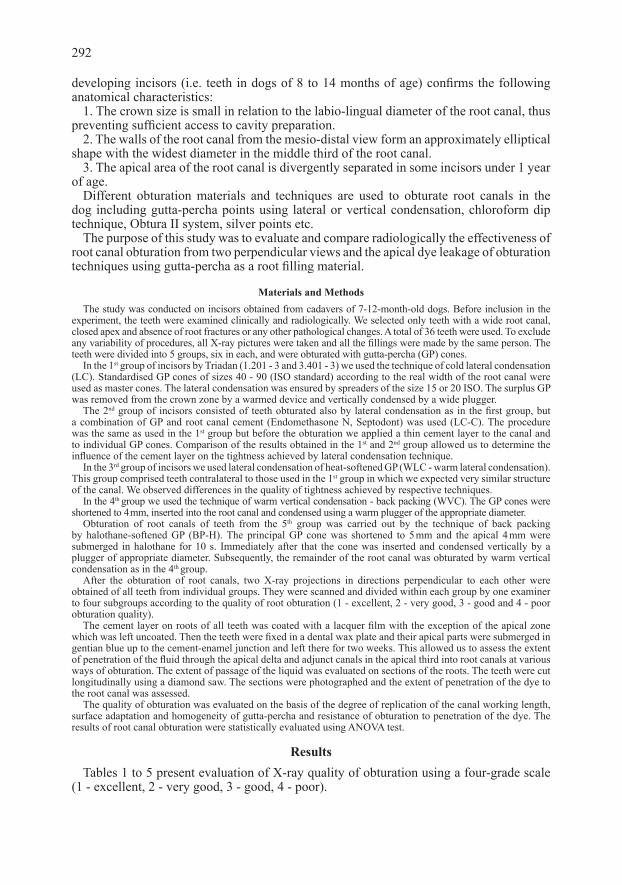
C. Cold lateral condensation using gutta-percha and root cementa - Root canal fill containing considerable volume of root canal cement among gutta-percha
cones with the presence of microgaps
b - Root cement stained by dye confirms insufficient obturation
a)
b)


![Efficacy of gutta-percha solvents used in endodontic ...revodonto.bvsalud.org/pdf/rsbo/v10n4/a09v10n4.pdf · endodontic treatment failure [9]. The clinical diagnosis of the pulp and](https://static.fdocuments.in/doc/165x107/5ed5a14f1b7fdd786a1b5e23/efficacy-of-gutta-percha-solvents-used-in-endodontic-endodontic-treatment-failure.jpg)