COMPARATIVE MORPHOLOGICAL STUDY OF ORAL CAVITY IN … · COMPARATIVE MORPHOLOGICAL STUDY OF ORAL...
6
COMPARATIVE MORPHOLOGICAL STUDY OF ORAL CAVITY IN RABBITS AND GUINEA PIGS Florin STAN Department of Comparative Anatomy, University of Agricultural Sciences and Veterinary Medicine, Faculty of Veterinary Medicine, 3-5 Manastur Street, Cluj-Napoca, Romania Corresponding author email: flodvm@yahoo.com Abstract In recent years, the species belonging to the order Lagomorpha and Rodentia are commonly used both as pets and in biomedical research, including in studies related to the digestive tract. The aim of this study was to perform a detailed anatomical description of the oral cavity of the two species. Due to their size and anatomical conformation is often difficult to make a proper examination of the oral cavity. Dissection was performed on 10 rabbits and 10 Guinea pigs of different sexes and ages. A very important and also quite confusing aspect is related to the dentition (some authors claim that the rabbits are monofyodont). Both species shows aradicular hypsodont dentition, (consisting of a short exposed crown and a long reserve crown with open root), elodont type (continuous growth throughout life). Rabbits are dyphyodont; they have deciduous and permanent sets of teeth compared to Guinea pigs that are monophyodont with a single set of permanent teeth without deciduous precursors. Both species share the same pattern of anisognathism, more pronounced in Guinea pigs, with the maxillary dental arch being wider than the mandibular dental arch. A large diastema separates the incisor and the cheek teeth in each jaw quadrant, being wider in guinea pigs compared to rabbits. Rabbits have one pair of mandibular incisors and two pairs of maxillary incisors with unpigmented enamel, two mandibular and three maxillary premolars and three molar teeth on each side in both the mandible and the maxilla. Guinea pigs have one pair of incisors, one pair of premolars and three pairs of molars on each dental arch. Contrary to rabbits, in Guinea pigs the mandibles (including premolar and molar teeth) are spaced further apart than the maxillae. The masseter muscle is well developed in both species. The temporomandibular joint in Guinea pigs does not subluxate in lateral movement, but allows a large degree of rostrocaudal movement. In rabbits the temporomandibular joint enables large lateral movement and low rostrocaudal movement. This morphological description helps both the clinicians and the researchers, being necessary for a proper understanding of the pathology of oral cavity in rabbits and Guinea pigs. Key words: oral cavity, Guinea pigs, rabbits. INTRODUCTION Guinea pigs belong to the Rodent species which includes over 40% of all mammals. Rabbits, which belong to the Lagomorphs species, differ from guinea pigs by the fact that they have 4 superior incisors and show significant differences in the maxilla and mandible (Crossley 2003; Fischer 2010). Until the second half of the last century, rabbits were classified as a subspecies of Rodents, but considering the differences noted above, they are much more similar to the artiodactyls order (bovines and horses) (Crossley 1995) Nevertheless, these two species share many other anatomical and behavioral characteristics. Rabbits and Guinea pigs are true herbivores, non-ruminant, the main physiological similarity being the particular type of digestion, the so- called hindgut fermentation due to which both species are capable to greatly capitalize the ingested nutrients (Michelle 2012). This physiological particularity is due to two conditions. The first condition is the similar anatomical characteristics - more exactly, the size of the posterior intestine. The second is their small size, which incorporates a big digestive surface compared to their body weight, is consistent with their high metabolic rate and increased food intake. The dietary behavior is similar in the two species: they feed at dawn and at dusk. Both, rabbits and Guinea pigs are strictly herbivores, their dentition and oral cavity muscles adapted to gnawing and crushing the ingested components (Frank 2003) 27 Scientific Works. Series C. Veterinary Medicine. Vol. LX (1) ISSN 2065-1295, ISSN Online 2067-3663, ISSN-L 2065-1295
Transcript of COMPARATIVE MORPHOLOGICAL STUDY OF ORAL CAVITY IN … · COMPARATIVE MORPHOLOGICAL STUDY OF ORAL...

COMPARATIVE MORPHOLOGICAL STUDY OF ORAL CAVITY
IN RABBITS AND GUINEA PIGS
Florin STAN
Department of Comparative Anatomy, University of Agricultural Sciences and Veterinary Medicine, Faculty of Veterinary Medicine, 3-5 Manastur Street, Cluj-Napoca, Romania
Corresponding author email: [email protected]
Abstract In recent years, the species belonging to the order Lagomorpha and Rodentia are commonly used both as pets and in biomedical research, including in studies related to the digestive tract. The aim of this study was to perform a detailed anatomical description of the oral cavity of the two species. Due to their size and anatomical conformation is often difficult to make a proper examination of the oral cavity. Dissection was performed on 10 rabbits and 10 Guinea pigs of different sexes and ages. A very important and also quite confusing aspect is related to the dentition (some authors claim that the rabbits are monofyodont). Both species shows aradicular hypsodont dentition, (consisting of a short exposed crown and a long reserve crown with open root), elodont type (continuous growth throughout life). Rabbits are dyphyodont; they have deciduous and permanent sets of teeth compared to Guinea pigs that are monophyodont with a single set of permanent teeth without deciduous precursors. Both species share the same pattern of anisognathism, more pronounced in Guinea pigs, with the maxillary dental arch being wider than the mandibular dental arch. A large diastema separates the incisor and the cheek teeth in each jaw quadrant, being wider in guinea pigs compared to rabbits. Rabbits have one pair of mandibular incisors and two pairs of maxillary incisors with unpigmented enamel, two mandibular and three maxillary premolars and three molar teeth on each side in both the mandible and the maxilla. Guinea pigs have one pair of incisors, one pair of premolars and three pairs of molars on each dental arch. Contrary to rabbits, in Guinea pigs the mandibles (including premolar and molar teeth) are spaced further apart than the maxillae. The masseter muscle is well developed in both species. The temporomandibular joint in Guinea pigs does not subluxate in lateral movement, but allows a large degree of rostrocaudal movement. In rabbits the temporomandibular joint enables large lateral movement and low rostrocaudal movement. This morphological description helps both the clinicians and the researchers, being necessary for a proper understanding of the pathology of oral cavity in rabbits and Guinea pigs. Key words: oral cavity, Guinea pigs, rabbits. INTRODUCTION Guinea pigs belong to the Rodent species which includes over 40% of all mammals. Rabbits, which belong to the Lagomorphs species, differ from guinea pigs by the fact that they have 4 superior incisors and show significant differences in the maxilla and mandible (Crossley 2003; Fischer 2010). Until the second half of the last century, rabbits were classified as a subspecies of Rodents, but considering the differences noted above, they are much more similar to the artiodactyls order (bovines and horses) (Crossley 1995) Nevertheless, these two species share many other anatomical and behavioral characteristics. Rabbits and Guinea pigs are true herbivores, non-ruminant, the main physiological similarity
being the particular type of digestion, the so-called hindgut fermentation due to which both species are capable to greatly capitalize the ingested nutrients (Michelle 2012). This physiological particularity is due to two conditions. The first condition is the similar anatomical characteristics - more exactly, the size of the posterior intestine. The second is their small size, which incorporates a big digestive surface compared to their body weight, is consistent with their high metabolic rate and increased food intake. The dietary behavior is similar in the two species: they feed at dawn and at dusk. Both, rabbits and Guinea pigs are strictly herbivores, their dentition and oral cavity muscles adapted to gnawing and crushing the ingested components (Frank 2003)
27
Scientific Works. Series C. Veterinary Medicine. Vol. LX (1)ISSN 2065-1295, ISSN Online 2067-3663, ISSN-L 2065-1295

In these conditions, the development of the masseter muscles is also considerable in both species. Another particularity dietary behaviour, directly related to the high necessity of vitamin B and folic acid, is coprophagy, more specific, cecotrophy, present in both rabbits and Guinea pigs (Hoefer 1997; Tynes 2001). Even in the conditions of a modern diet, with higher vitamin and energy intake, this behaviour is not changed, being an instinctive act stimulated by the anal reflex. Precisely, this diet is sometimes responsible for the affections that can occur starting from the oral cavity and on the entire digestive tract (Fischer 2010: Michelle 2012). The morphology of the oral cavity in rabbits and Guinea pigs is the result of evolutionary adjustment to prehension, gnawing and grinding of a natural diet composed mostly of grass. This is rich in phytoliths and silicates which lead to a high level of teeth attrition (Shadle 1936). This aspect cumulated with a low level of nutrients per unit of volume, which leads to a high intake of food, increases the level of attrition. Anatomically and physiologically both species control this bluntness by a permanent growth of teeth. In these conditions, the pathology of the oral cavity in rabbits and Guinea pigs usually is a challenge for practicing physicians (Wagner 1976; Boehmer and Crossley 2009;Michelle 2012). Therefore, for a good understanding of the pathological process, acquiring a solid knowledge of the oral cavity morphology in these species is necessary. The present study achieves a detailed morphological description of the components of the oral cavity in rabbits and Guinea pigs wanting to be helpful for both physicians and researchers. MATERIALS AND METHODS The study was conducted on two lots of 10 rabbits and 10 Guinea pigs. These specimens came from private farms. The two lots are part of a large study of the digestive tract on species from the orders Lagomorphs and Rodent. The subjects were accommodated in proper
conditions, with plenty of food and water. Individual clinical examination revealed no presence of any pathology of the oral cavity. Before euthanasia, in each subject was administered Ketamine 10mg/kg/bw, SC, and euthanasia was performed according to standard procedures, by administration of potassium chlorides 2meq/kg/bw IV. Inspection and gross dissection was performed for each specimen. Each stage of the dissection was photographed and obtained observations were noted. The anatomical differences were also noted and photographed. RESULTS AND DISCUSSIONS In rabbit, the oral cavity appeared elongated, narrow in the rostral portion and slightly enlarged caudally, with a relatively small
opening. The articular process which forms the temporo-mandibular joint is longitudinal, allowing forward/backward moves in vertical plan, and even lateral movement were permitted. The hard palate, narrowed rostral and extended between the molars has shown a variable number (between 18 and 22) of well individualized palatine crests.
Figure 1. The normal shape of upper arcade in rabbit with the presence of 2 sets of incisores. The cheek
teeth are ararnged in parralel rows. Note the divergent orientation of the first ridges and the transversal
position of the intermolar palatinal ridges.
28
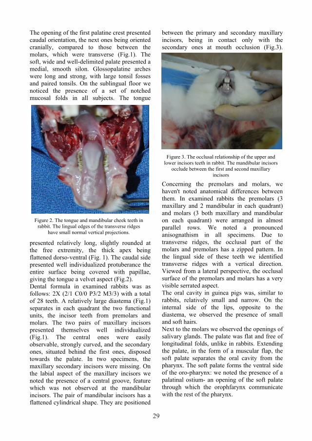
The opening of the first palatine crest presented caudal orientation, the next ones being oriented cranially, compared to those between the molars, which were transverse (Fig.1). The soft, wide and well-delimited palate presented a medial, smooth silon. Glossopalatine arches were long and strong, with large tonsil fosses and paired tonsils. On the sublingual floor we noticed the presence of a set of notched mucosal folds in all subjects. The tongue
presented relatively long, slightly rounded at the free extremity, the thick apex being flattened dorso-ventral (Fig. 1). The caudal side presented well individualized protuberance the entire surface being covered with papillae, giving the tongue a velvet aspect (Fig.2). Dental formula in examined rabbits was as follows: 2X (2/1 C0/0 P3/2 M3/3) with a total of 28 teeth. A relatively large diastema (Fig.1) separates in each quadrant the two functional units, the incisor teeth from premolars and molars. The two pairs of maxillary incisors presented themselves well individualized (Fig.1). The central ones were easily observable, strongly curved, and the secondary ones, situated behind the first ones, disposed towards the palate. In two specimens, the maxillary secondary incisors were missing. On the labial aspect of the maxillary incisors we noted the presence of a central groove, feature which was not observed at the mandibular incisors. The pair of mandibular incisors has a flattened cylindrical shape. They are positioned
between the primary and secondary maxillary incisors, being in contact only with the secondary ones at mouth occlusion (Fig.3).
Concerning the premolars and molars, we haven't noted anatomical differences between them. In examined rabbits the premolars (3 maxillary and 2 mandibular in each quadrant) and molars (3 both maxillary and mandibular on each quadrant) were arranged in almost parallel rows. We noted a pronounced anisognathism in all specimens. Due to transverse ridges, the occlusal part of the molars and premolars has a zipped pattern. In the lingual side of these teeth we identified transverse ridges with a vertical direction. Viewed from a lateral perspective, the occlusal surface of the premolars and molars has a very visible serrated aspect. The oral cavity in guinea pigs was, similar to rabbits, relatively small and narrow. On the internal side of the lips, opposite to the diastema, we observed the presence of small and soft hairs. Next to the molars we observed the openings of salivary glands. The palate was flat and free of longitudinal folds, unlike in rabbits. Extending the palate, in the form of a muscular flap, the soft palate separates the oral cavity from the pharynx. The soft palate forms the ventral side of the oro-pharynx: we noted the presence of a palatinal ostium- an opening of the soft palate through which the orophfarynx communicate with the rest of the pharynx.
Figure 2. The tongue and mandibular cheek teeth in rabbit. The lingual edges of the transverse ridges
have small normal vertical projections.
Figure 3. The occlusal relationship of the upper and lower incisors teeth in rabbit. The mandibular incisors
occlude between the first and second maxillary incisors
29
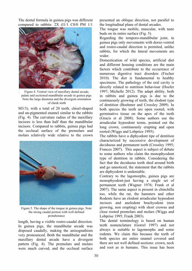
The dental formula in guinea pigs was different compared to rabbits: 2X (I1/1 C0/0 PM 1/1
M3/3), with a total of 20 teeth, chisel-shaped and un-pigmented enamel similar to the rabbits (Fig. 4). The curvature radius of the maxillary incisors is less than half than the mandibular incisors. Compared to rabbits, guinea pigs had the occlusal surface of the premolars and molars relatively wide relative to the crown
length, having a visible rostro-caudal direction. In guinea pigs, the mandibular arcade was disposed caudally, making the anisognathism very pronounced. Both the mandibular and the maxillary dental arcade have a divergent pattern (Fig. 4). The premolars and molars were much curved, and the occlusal surface
presented an oblique direction, not parallel to the longitudinal plane of dental arcades. The tongue was mobile, muscular, with taste buds on its entire surface (Fig. 5). Regarding the temporo-mandibular joint, in guinea pigs only movements with dorso-ventral and rostro-caudal direction is permited, unlike rabbits, for which the lateral movements are wider. Domestication of wild species, artificial diet and different housing conditions are the main factors which contribute to the occurrence of numerous digestive tract disorders (Fischer 2010). The diet is fundamental to healthy specimens. The pathology of the oral cavity is directly related to nutrition behaviour (Hoefer 1997; Michelle 2012). The adapt ability, both in rabbits and guinea pigs is due to the continuously growing of teeth, the elodont type of dentition (Boehmer and Crossley 2009). In both species the teeth are open rooted, with germinative tissue on the apex of the teeth (Gracis et al 2008). Some authors use the arradicular hypsodont terms, pointed out the long crown, continuously erupting and open rooted (Wiggs and Lobprice 1995). The rabbits have a diphyodont tipe of dentition characterized by successive development of deciduous and permanent teeth (Crossley 1995; Frances 2007). This aspect is subject of debate to some authors who claim the monophyodont type of dentition in rabbits. Considering the fact that the deciduous teeth shed around birth and go unnoticed, the statement that the rabbits are diphyodont is undeniable. Contrary to the lagomorphs, guinea pigs are monophyodont-just having a single set of permanent teeth (Wagner 1976; Frank et al 2007). The same aspect is present in chinchilla too, while the rat, the hamsters and other Rodents have an elodont arradicular hypsodont incisors and anelodont brachyodont (non growing, non erupting) with short crowns and close rooted premolars and molars (Wiggs and Lobprise 1995; Frank 2003). The dental terminology is based on human teeth nomenclature (Gorrel 1997) and not always is suitable to lagomorphs and some rodents. We claim this because the teeth of both species are entire enamel covered and there are not well defined sections: crown, neck and root as in humans. This issue has been
Figure 4. Ventral view of maxillary dental arcade, palate and sectioned mandibular arcade in guinea pigs. Note the large diastema and the divergent orientation
of cheek teeth
Figure 5. The shape of the tongue in guinea pigs. Note the strong caudal portion with well defined
protuberance
30

reported by Blood 1999. The terms anatomic crown for the entire tooth, and separate terms for supragingival and subgingival parts, were proposed. The supragingival section was called the clinical crown or exposed crown and the subgingival section was called reserve crown or clinical root. Assigning these terms can be confusing for the most practitioners, so the majority of authors use the human nomenclature. This is in concordance with the terms use in almost veterinary dictionaries (Blood 1999). Although, strictly speaking none of the species mentioned above have real root of the teeth, apex of this, the shape of the root being cylindrical, rather than cone, as in human. The enamel is white in both rabbits and guinea pigs, even though for the majority of Rodents it has a yellow-orange colour (Wiggs 1995, Crossley 2003). Similar to rabbits, in guinea pigs, the enamel is thicker on the vestibular side of the teeth. Because of this, in both species, the teeth are chisel-shaped. Rabbits have two sets of maxillary incisors compared with guinea pigs that have only one set of maxillary incisors, hence the different type of occlusion in rabbits. This is done by positioning the mandibular incisors between the primary and secondary incisors from the maxillary arch. Incisors are strongly curved in both species, but in guinea pigs their length is greater than in rabbits. Growth rate is high, on average 2mm and 2.4mm a week (Shade 1936) and is directly related to the rate of eruption and attrition, hence the need for high-fiber diets. Regarding the premolars and molars in both species, there are no significant anatomical differences. The difference consists in the number of premolars and molars. Rabbits have 3 maxillary, 2 mandibulary premolars and 3 maxillary, 3 mandibulary molars in each quadrant, compared with guinea pigs, which only have 1 maxillary and 1 mandibulary premolar, and 3 mandibulary and maxillary molars in each quadrant. Also, we noted remarkable differences regarding the occlusal surfaces of the teeth. The shape of these surfaces is maintained by the phenomenon of opposed tooth wear due to both diet and chewing movements that rabbits do in the absence of food (Crossley 2003; Frances 2007). Thus we can say that rabbits have a typical
herbivore occusal aspect, with their premolars and molars grouped as a functional unit with a relatively horizontal surface, and transversal enamel crests adapted to shredding and grinding a high-fiber diet. In comparison, guinea pigs had a more oblique occlusal surface than rabbits, with large diastema and multiple ridges of the cheeks (Gracis 2008; Boehmer and Crossley 2009). The clinical significance of this is important, guinea pigs making food deposits in the pouches formed in the cheeks, hampering clinical examination. The anizognathous way of occlusion in guinea pigs, having the mandible wider than the maxilla, together with the convergent aspect of the dental arches, gives the explanation for the strong inclination of the occlusal side of the teeth. Due to presented anatomical particularities it is difficult to achieve a proper clinical examination, both in rabbit and guinea pigs. According to data from the literature only a small percentage of diseases of the oral cavity can be assessed clinically (Gracis 2008; Michelle 2012). Moreover, it is necessary to assess the bone support and the soft tissue that makes up the oral cavity, by imaging methods appropriate to each component. CONCLUSIONS Anatomical data described in this study are a starting point in the interpretation of oral cavity diseases in rabbits and guinea pigs, to help practitioners for a proper evaluation and therapeutic approach in oral cavity pathology. REFERENCES Blood D.C., Studdert V.P., 1999. Ballières
Comprehensive Veterinary Dictionary (ed 2), London,Ballière Tindall.
Boehmer E., Crossley D.A., 2009. Objective interpretation of dental disease in rabbits, guinea pigs and chinchillas Use of anatomical reference lines Tierärztl Prax; 37 (K): 250–260.
Crossley D.A., 1995. Clinical aspects of lagomorph dental anatomy: The rabbit (Oryctolagus cuniculus) J Vet Dent, 12, pp. 137–140.
Crossley D.A., 2003. Oral biology and disorders of lagomorphs. Vet Clin North Am: Exot Anim Pract; 6 (3), 629–659.
Fisher P.G., 2010 Standards of Care in the 21st Century:The Rabbit, Journal of Exotic Pet Medicine, Vol 19, 1, pp 22-35.
31

Frances Margaret Harcourt-Brown 2007. The Progressive Syndrome of Acquired Dental Disease in Rabbits Journal of Exotic Pet Medicine, Rabbits, 16, 3, 146–157.
Frank J.M., Verstraete 2003. Advances in diagnosis and treatment of small exotic mammal dental disease Seminars in Avian and Exotic Pet Medicine Volume 12, Issue 1, January 2003, Pg 37–48.
Gorrel C., 1997. Humane dentistry (letter) J Small Anim Pract, 38 p. 31.
Hoefer H.L., 1997. Gastrointestinal diseases EV Hillyer, KE Quesenberry (Eds.), Ferrets, Rabbits, and Rodents—Clinical Medicine and Surgery, Saunders, Philadelphia, PA , pp. 26–36.
Margherita Gracis, 2008. Clinical Technique: Normal Dental Radiography of Rabbits, Guinea Pigs, and Chinchillas Journal of Exotic Pet Medicine, Dentistry Passerine Birds 17, 2, 78–86.
Michelle L. Campbell-Ward, 2012, Gastrointestinal Physiology and Nutrition in Ferrets, Rabbits, and Rodents (Third Edition) Clinical Medicine and Surgery Pages 183–192.
Shadle A.R., 1936.The arttrition and extrusive growth of the four major incisor teeth of domestic rabbits J Mammal, 17, pp. 15–21.
Tynes V.V., 2001.Managing common gastrointestinal disorders of pet rabbits. Vet Med 96(3):226–233.
Wagner J.E., 1976. Miscellaneous disease conditions of guinea pigs JE Wagner, PJ Manning (Eds.), The Biology of the Guinea Pig, Academic Press, New York, NY, pp. 227–234.
Wiggs B., Lobprise H., 1995. Dental anatomy and physiology of pet rodents and lagomorphs DA Crossley, S Penman (Eds.), Manual of Small Animal Dentistry (ed 2), British Small Animal Veterinary Association, Cheltenham, England pp. 68–73.
32