Colony specificity in Botrylloides leachi. II. Cellular ... · Colony specificity in Botrylloides...
7
ISJ 4: 38-44, 2007 ISSN 1824-307X RESEARCH REPORT Colony specificity in Botrylloides leachi. II. Cellular aspects of the non-fusion reaction L Ballarin, G Zaniolo Department of Biology, University of Padova, Padova, Italy Accepted March 09, 2007 Abstract We continued our study on fusion and non-fusion reactions in the compound ascidian Botrylloides leachi with particular attention to the role of hemocytes in the induction of the strong cytotoxic reaction observed when genetically incompatible colonies encounter each other in cut surface assays. We used antibodies raised against Botryllus phenoloxidase (PO) and pro-inflammatory vertebrate cytokines for immunocytochemical and immunohistochemical analysis on contacting incompatible colonies in order to investigate cellular events during the non-fusion reaction. Results indicate that, analogously to Botryllus schlosseri, morula cells play a key role in the above reaction as they crowded inside the facing ampullae and entered the tunic where they degranulated and induced PO-driven cytotoxicity. Similarly to Botryllus, they also expressed molecules recognised by anti-IL-1-α antibodies, whereas the immunopositivity to anti-TNF-α was unexpectedly restricted to granular cells. Key words: ascidian; Botrylloides; colony specificity; allorecognition; phenoloxidase __________________________________________________________________________________________ Introduction Botryllid ascidians are compound tunicates living in colonies of many zooids embedded in a common tunic and connected by a vascular network which keeps them synchronised. Members of this family share the ability of intraspecific recognition (colony specificity) which discriminates between genetically compatible colonies, allowing them to fuse their tunic and circulation, and incompatible colonies which do not fuse (Taneda et al., 1985; Saito et al., 1994). Non-fusion reaction is often followed by a rejection reaction which leads to the formation of a series of cytotoxic regions along the contact border (points of rejections; Rinkevich, 1992; Saito et al., 1994). In Botryllus schlosseri, a reference species for the study of ascidian colony specificity, the rejection reaction entails the selective chemotactic crowding of morula cells (MC, an ubiquitous cell-type in botryllid ascidians) inside the blind, sausage-like endings of the peripheral tunic vessels (ampullae) of the growing edge (Sabbadin et al., 1992; Rinkevich et al., 1998; Cima et al., 2006) and their later migration through the epithelium of the ampullar tips ___________________________________________________________________________ Corresponding author: Loriano Ballarin Department of Biology, University of Padova Via Ugo Bassi 58/B - 35100 Padova, Italy Email: [email protected] into the partially fused tunic, where they degranulate, thus releasing their vacuolar contents, mainly the enzyme phenoloxidase (PO), contributing to the induction of cytotoxicity (Ballarin et al., 1995, 1998; Cima et al., 2004). The succession of events (chemotaxis, extravasation, degranulation, induction of cytotoxicity) closely resembles inflammation. In Vertebrates, it is induced and modulated by cytokines, mainly IL-1-α and TNF-α, released by leukocytes activated by the recognition of foreign molecules (Abbas et al., 2000). Recently, we demonstrated that molecules recognized by antibodies raised against mammalian IL-1-α and TNF-α are expressed by morula cells in early stages of the rejection reaction of B. schlosseri (Cima et al., 2004). Botrylloides leachi is a compound ascidian, common in the Mediterranean, living in sympatry with B. schlosseri, which can easily be reared in the laboratory. Despite the abundance of this species, colony specificity has been poorly studied until now. In a previous study, we demonstrated that, in the colony allorecognition assay, as defined by Rinkevich (1992), it resembles subpopulation I of Botrylloides from the Mediterranean coast of Israel, as indicated by the induction of megaloampullae (Rinkevich et al., 1994; Simon-Blecher et al., 2006), and occurs in the form of a subcuticular rejection reaction between non-fusible colonies, with a few 38
-
Upload
truongkhanh -
Category
Documents
-
view
214 -
download
0
Transcript of Colony specificity in Botrylloides leachi. II. Cellular ... · Colony specificity in Botrylloides...
ISJ 4: 38-44, 2007 ISSN 1824-307X
RESEARCH REPORT Colony specificity in Botrylloides leachi. II. Cellular aspects of the non-fusion reaction L Ballarin, G Zaniolo Department of Biology, University of Padova, Padova, Italy
Accepted March 09, 2007
Abstract We continued our study on fusion and non-fusion reactions in the compound ascidian Botrylloides
leachi with particular attention to the role of hemocytes in the induction of the strong cytotoxic reaction observed when genetically incompatible colonies encounter each other in cut surface assays. We used antibodies raised against Botryllus phenoloxidase (PO) and pro-inflammatory vertebrate cytokines for immunocytochemical and immunohistochemical analysis on contacting incompatible colonies in order to investigate cellular events during the non-fusion reaction. Results indicate that, analogously to Botryllus schlosseri, morula cells play a key role in the above reaction as they crowded inside the facing ampullae and entered the tunic where they degranulated and induced PO-driven cytotoxicity. Similarly to Botryllus, they also expressed molecules recognised by anti-IL-1-α antibodies, whereas the immunopositivity to anti-TNF-α was unexpectedly restricted to granular cells.
Key words: ascidian; Botrylloides; colony specificity; allorecognition; phenoloxidase __________________________________________________________________________________________ Introduction
Botryllid ascidians are compound tunicates
living in colonies of many zooids embedded in a common tunic and connected by a vascular network which keeps them synchronised. Members of this family share the ability of intraspecific recognition (colony specificity) which discriminates between genetically compatible colonies, allowing them to fuse their tunic and circulation, and incompatible colonies which do not fuse (Taneda et al., 1985; Saito et al., 1994). Non-fusion reaction is often followed by a rejection reaction which leads to the formation of a series of cytotoxic regions along the contact border (points of rejections; Rinkevich, 1992; Saito et al., 1994).
In Botryllus schlosseri, a reference species for the study of ascidian colony specificity, the rejection reaction entails the selective chemotactic crowding of morula cells (MC, an ubiquitous cell-type in botryllid ascidians) inside the blind, sausage-like endings of the peripheral tunic vessels (ampullae) of the growing edge (Sabbadin et al., 1992; Rinkevich et al., 1998; Cima et al., 2006) and their later migration through the epithelium of the ampullar tips ___________________________________________________________________________
Corresponding author: Loriano Ballarin Department of Biology, University of Padova Via Ugo Bassi 58/B - 35100 Padova, Italy Email: [email protected]
into the partially fused tunic, where they degranulate, thus releasing their vacuolar contents, mainly the enzyme phenoloxidase (PO), contributing to the induction of cytotoxicity (Ballarin et al., 1995, 1998; Cima et al., 2004). The succession of events (chemotaxis, extravasation, degranulation, induction of cytotoxicity) closely resembles inflammation. In Vertebrates, it is induced and modulated by cytokines, mainly IL-1-α and TNF-α, released by leukocytes activated by the recognition of foreign molecules (Abbas et al., 2000). Recently, we demonstrated that molecules recognized by antibodies raised against mammalian IL-1-α and TNF-α are expressed by morula cells in early stages of the rejection reaction of B. schlosseri (Cima et al., 2004).
Botrylloides leachi is a compound ascidian, common in the Mediterranean, living in sympatry with B. schlosseri, which can easily be reared in the laboratory. Despite the abundance of this species, colony specificity has been poorly studied until now. In a previous study, we demonstrated that, in the colony allorecognition assay, as defined by Rinkevich (1992), it resembles subpopulation I of Botrylloides from the Mediterranean coast of Israel, as indicated by the induction of megaloampullae (Rinkevich et al., 1994; Simon-Blecher et al., 2006), and occurs in the form of a subcuticular rejection reaction between non-fusible colonies, with a few
38

visible necrotic regions. As happens in B. simodensis (Hirose et al., 1988, 1997), a clear rejection reaction with marked cytotoxicity along the contact border is observable only in cut surface assays (Zaniolo et al., 2006). In this case, MC and some granular cells (Cima et al., 2001) crowd inside the lumen of the ampullae close to the cut surface, enter the tunic, and move towards the contact border. As reported for B. schlosseri, MC degranulate and release the contents of their vacuoles.
In the present work, we continued our study on colony specificity in this species, focussing on the role of hemocytes in the induction of the strong cytotoxic reaction observed when genetically incompatible colonies encounter each other in cut surface assays. To investigate cellular events during the rejection reaction, we employed anti-PO antibodies and antibodies against pro-inflammatory, vertebrate cytokines for immunocytochemical and immunohistochemical analysis of contacting incompatible colonies. Results were compared with the events occuring during the fusion reaction and what is known in B. schlosseri. Materials and Methods Animals
Colonies of B. leachi were collected in the Lagoon of Venice, left to adhere to glass slides (5 x 5 cm) and reared at 20 °C in 20-l aquaria containing filtered seawater (FSW). They were fed with Liquifry Marine (Liquifry Co., Dorking, UK) and algae (Dunaliella sp.). Hemocyte collection
Blood cells were collected from colonies previously rinsed in a solution of 10 mM L-cysteine in FSW, pH 7.2, to prevent cell clotting. The marginal vessel was punctured with a fine tungsten needle and hemocytes were collected with a micropipette in 1.5-ml vials. Cell suspensions were centrifuged for 10 min at 370xg, supernanatants were discarded and pellets resuspended in FSW to obtain a concentration of 107 cell/ml. Fifty μl of hemocyte suspension were put in the centre of culture chambers prepared as already described (Ballarin et al., 1994) and kept upside-down for 30 min to allow the cells to adhere to the coverslips. Histochemical assays on hemocytes
Eosinophily. Fixed blood cells were incubated for 30 min in a solution of 2 % eosin G (Shirae et al., 2002). Cells with affinity for eosin stained red.
PO activity. To reveal PO, fixed hemocytes were incubated for 2 h in a saturated solution of dihydroxyphenyl-L-alanine (L-DOPA) in phosphate-buffered saline (PBS: 8 g/l NaCl, 0.2 g/l KCl, 0.2 g/l KH2PO4, 1.15 g/l Na2HPO4); they were then repeatedly washed in PBS and coverslips were mounted on clean glass slides with Acquovitrex (Carlo Erba, Limito, MI, Italy). Positive sites appeared dark brown.
Quinones. The presence of quinones in hemocytes was revealed by 3-methyl-2-benzothiazolinonehydrazone hydrochloride (MBTH; Winder and Harris, 1991). Briefly, living hemocytes
were incubated for 10 min in a 2-mM solution of MBTH in FSW containing 0.4 % N, N’-dimethylformamide. An intense red stain marked positive sites. Cut surface assay
Colonies were cut with razor blades and pieces of the same size from incompatible and compatible colonies were brought into contact at their cut surface on a supporting glass slide and allowed to adhere for 30 min in a moist chamber. Colonies were then returned to the aquarium and observed under a binocular microscope for up to 48 h, until either non-fusion or fusion reaction occurred. Immunohistochemical and immunocytochemical analyses of contacting colonies
Colony pairs were fixed in 4 % paraformaldehyde plus 0.1 % glutaraldehyde in 0.4 M cacodylate buffer containing 1.7 % NaCl, 1 % sucrose and 1 % caffeine, to prevent the leakage of MC vacuolar contents (Ballarin et al., 1995), dehydrated and embedded in Paraplast Xtra (Oxford Labware, St. Louis MO, USA). Sections (7 μm) were cut with a Leitz 1212 microtome, deparaffined, and incubated overnight with 10 µg/ml primary antibodies raised against B. schlosseri PO (Frizzo et al., 1999), human recombinant IL-1-α and human recombinant TNF-α (Santa Cruz Biotech., Santa Cruz, CA, USA). Slides were then washed in PBS, incubated in biotinylated anti-rabbit-IgG antibody (Santa Cruz Biotech., 50 µg/ml) for 30 min, washed again, and incubated for 30 min in avidin-biotin-peroxidase complex (ABC, Vector Laboratories, Burlingame, CA, USA). After thorough washing in PBS, they were finally incubated for 5 min in a solution 0.02 5% 3,3’ diaminobenzidine (DAB) containing 0.004 % hydrogen peroxide and mounted with Eukitt. Positive sites appeared brown.
For immunocytochemical analysis, hemocytes were activated by incubation for 30 min in 20 ng/ml phorbol myristate acetate (PMA; Sigma, St Louis, MO, USA) and then fixed for 30 min at 4 °C in a solution of 1 % glutaraldehyde and 1 % sucrose in FSW containing 1 % caffeine, and rinsed in PBS. They were then treated with primary and secondary antibodies, followed by incubation in ABC and DAB, as described above. Results Cytochemical and immunocytochemical analysis
MC are large cells, about 15 µm in diameter, which represent the majority of circulating hemocytes and are characterized by the presence of many small vacuoles, about 2 µm in diameter, (Fig. 1a). Among the various hemocyte types, only MC stained with eosin (Fig. 1b) and MBTH (Fig. 1c). In addition, they showed positivity to the assay for PO activity (Fig. 1d). When hemocytes were stimulated with PMA, MC expressed molecules recognized by anti-IL-1-α antibodies (Fig. 1e), whereas only granular cells were positive to anti-TNF-αantibodies (Fig. 1g). The latter, 7-12 µm in diameter, had cytoplasm filled with many small vacuoles (Fig. 1f) which, according to Cima et al. (2001) contain finely granular, weakly electron-dense material.
39

Fig. 1 MC (a-e) and granular cells (f-g) of Botrylloides leachi. Fixed, untreated MC have many small vacuoles (a), are stained by eosin (b) and MBTH (c), show PO activity (d), and are recognized by anti-IL-1-α antibodies (e); granular cells contain many small granules (f) and are stained by anti-TNF-α antibodies (g). Bar = 10 µm. Analysis of non-fusion and fusion reactions
As reported elsewhere (Zaniolo et al., 2006), in the non-fusion reaction, within 24 h colonies fused their tunics in limited regions along the facing border, and hemocytes, mainly MC, crowded inside the ampullae of the contact region (Fig. 2a). Over the next 24 h, many blood cells, particularly MC, leaked from the ampullar lumen and moved towards the colonial border. These MC showed altered morphology, with empty vacuoles of larger size (Fig 2b), surrounded by eosinophilic material (Fig. 2c). Some granular cells also appeared along the contact border (Fig. 2d). All these cells, in later stages, contributed to the formation of a clearly visible, pigmented, necrotic band (Fig. 2e).
In the course of the fusion reaction, tunic fusion occurred within 24 h of contact, and the ampullae grew towards the contact border. Limited cell crowding and no cell leakage was observed. Within 48 h of contact, the ampullar tips touched and fused, establishing a common circulation between the two colonies. Immunocytochemistry during non-fusion and fusion reactions
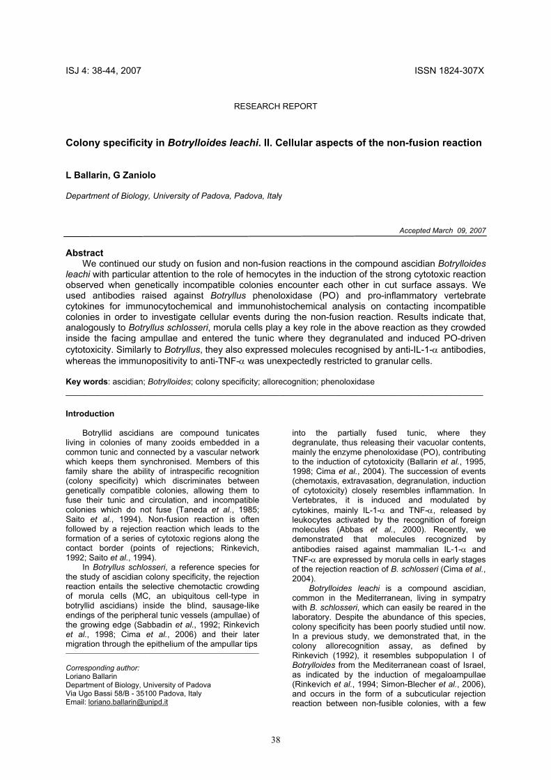
In early stages of the non-fusion reaction, many MC inside the ampullae of the contacting region were positive to anti-PO antibodies (Fig. 3a) whereas, in advanced stages, immunopositivity was observed in limited regions of the tunic, rich in
fibrous material, around MC with altered morphology (Fig. 3b). Before their change in morphology, when still inside the ampullar lumen or just after their migration to the tunic of the contact region, MC were recognized by anti-IL-1-α antibodies (Fig. 3c); this positivity was lost in advanced stages of the non-fusion reaction (Fig. 3d).
Granular cells were the only hemocytes recognized by anti-TNF-α antibodies in early stages of the non-fusion reaction, when still inside the ampullae (Fig. 3e). Some of the tunic cells were also immunopositive (Fig. 3f).
During the fusion reaction, MC were the only cells positive to anti-PO antibodies, whereas no hemocytes showed positivity to anti-cytokine antibodies (Fig. 3g). Some tunic cells were recognized by anti-TNF-α antibodies (Fig. 3h). Discussion
We studied the behaviour of hemocytes during
the non-fusion reaction in B. leachi as compared to fusion. In a previous morphological study, Zaniolo et al. (2006) showed that the non-fusion reaction between contacting growing edges occurs in the form of a subcuticular rejection, closely resembling that described for Botrylloides violaceus, Botrylloides simodensis and Botrylloides fuscus (Hirose et al., 1988, 1997), and leads to the
40

Fig. 2 Non-fusion reaction in Botrylloides leachi, cut surface assay. MC initially crowd inside lumen of facing ampullae (a), then leak from ampullae into tunic and change their morphology (b); once in tunic, some eosinophilic material appears around them (c, arrowheads). Some granular cells are also found along colony border (d, arrows). At end of process, a marked necrotic band formed between colonies (e). Bar = 15 µm for a-d, 1 mm for e. appearance of megaloampullae as described for Botrylloides subpopulation I from the Mediterranean coast of Israel (Rinkevich et al., 1994; Simon-Blecher et al., 2006). To allow better analysis of the role of hemocytes in the non-fusion and fusion reactions, we used the cut surface assay, as defined by Rinkevich (1992), which gives a stronger cytotoxic reaction than the usual colony allorecognition assay (Hirose et al., 1990; Zaniolo et al., 2006).
In the course of the non-fusion reaction, after partial fusion of the tunic, MC selectively crowded inside the ampullae of the contacting region and, later, leaked out into the tunic, to gather along the colony border. In the meantime, a change took place in their morphology, which closely resembled that observed during the non-fusion reaction of B. schlosseri. In the latter species, degranulation occurred, with the consequent release of vacuolar contents, mainly the enzyme PO and its polyphenol substrate, as a consequence of the recognition of allogeneic factors diffusing from the alien colony (Ballarin et al., 1995; Cima et al., 2004). This enzyme is responsible for the cytotoxicity observed along the region where the growing edges meet (Ballarin et al., 1998). As regards B. leachi, our results indicate that: i) PO activity is located in MC, as demonstrated both by the assay with L-DOPA
and the presence of quinones, deriving from the oxidation of polyphenols by PO, and stored inside cell vacuoles, as reported by Shirae et al. (2002) in B. simodensis; ii) MC are the only cells recognized by the anti-PO; iii) immunopositivity to anti-PO antibodies is limited to the early stages of the non-fusion reaction; iv) fibrous tunic material around MC is labelled by the above antibodies in later stages of the non-fusion reaction. On the whole, all these data are consistent with the hypothesis of the occurrence of MC degranulation which, as reported for B. schlosseri (Ballarin et al., 1995, 1998), may be responsible for the induction of the cytotoxicity observed. The phylogenetic closeness of the two species explains the recognition of the B. leachi PO by the antibodies raised against the B. schlosseri protein; similarly, the same antibodies have been reported to recognize B. simodensis MC (Shirae et al., 2002).
In B. schlosseri, activated MC express molecules recognized by antibodies raised against mammalian recombinant IL-1-α and TNF-α (Ballarin et al., 2001). These molecules are also expressed in early stages of the non-fusion reaction (Cima et al., 2004) and are involved in both chemotaxis of MC inside the ampullae of the contacting region and the induction of cytotoxicity (Cima et al., 2004, 2006). In the same way, B. leachi MC are recognized by anti-
41
42

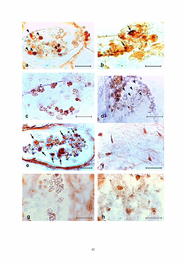
◄Fig. 3 Immunohistochemical analysis of non-fusion (a-f) and fusion (g-h) reactions between contacting colonies of Botrylloides leachi in cut surface assay. a-b: labelling of MC (arrowheads) by anti-PO antibodies inside ampullar lumen (a) and in tunic (b); in latter case, immunopositivity was observed also in fibrous material around altered MC (arrows). c, d: immunopositivity to anti-IL-1-α antibodies. Antibodies recognize MC in early stages of non-fusion reaction (c), whereas MC lose their immunopositivity in later stages (arrowheads) (d). e, f: immunopositivity to anti-TNF-α antibodies. Granular cells (arrows), but not MC (asterisks), are recognized inside ampullae in early stages of non-fusion reaction (e); some tunic cells are also labelled (f). MC are not recognized by anti-IL-1-α antibodies during fusion reaction (g); some tunic cells are stained by anti-TNF-α antibodies (h). Bar = 50 µm.
IL-1-α antibodies, which exerts a role similar to that in B. schlosseri. Conversely, anti-TNF-α recognizes granular cells which, although less abundant than MC, can reach the ampullar lumen and migrate into the tunic, where they probably contribute to the formation of necrotic areas.
Thus, we found an unexpected involvement of this cell type in the non-fusion reaction in B. leachi, which is worthy of further investigation. The reported labelling of tunic cells by anti-TNF-α is observable in both non-fusion and fusion, and suggests an activation of these cells, probably as a consequence of colony injury in the cut surface assay. Analogously to B. schlosseri, we hypothesise that molecules recognized by anti-cytokine antibodies in B. leachi are immunomodulatory molecules (i.e., cytokines, in the broad sense of the term). Although no genes homologous with vertebrate cytokines have been reported in the genome of the ascidian Ciona intestinalis, with the exception of a putative gene for TNF (Azumi et al., 2003), ascidian cytokines could share some conserved domains or epitopes with their Vertebrate counterparts although differing in their aminoacid and nucleotide sequences (Menin et al., 2005). This hypothesis is supported by the observation that, in simpler eukaryotes, such as the ciliate Euplotes raikovi, mammalian IL-2 is interchangeable with the Er-1 pheromone (Vallesi et al., 1998). Future research will be directed towards better insight into the role and nature of these molecules.
References Abbas AK, Lichtman AH, Pober JS. Cellular and
molecular immunology. WB Saunders Company, Philadelphia, 2000.
Azumi K, De Santis R, De Tomaso A, Rigoutsos I, Yoshizaki F, Pinto MR, et al. Genomic analysis of immunity in a Urochordate and the emergence of the vertebrate immune system: “Waiting for Godot”. Immunogenetics 55: 570-581, 2003.
Ballarin L, Cima F, Sabbadin A. Phagocytosis in the colonial ascidian Botryllus schlosseri. Dev. Comp. Immunol. 18: 467-481, 1994.
Ballarin L, Cima F, Sabbadin A. Morula cells and histocompatibility in the colonial ascidian Botryllus schlosseri. Zool. Sci. 12: 757-764, 1995.
Ballarin L, Cima F, Sabbadin A. Phenoloxidase and cytotoxicity in the compound ascidian Botryllus schlosseri. Dev. Comp. Immunol. 22: 479-492, 1998
Ballarin L, Franchini A, Ottaviani E, Sabbadin A. Morula cells as the main immunomodulatory haemocytes in ascidians: evidences from the
colonial species Botryllus schlosseri. Biol. Bull. 201: 59-64, 2001.
Cima F, Perin A, Burighel P, Ballarin L. Morpho-functional characterisation of haemocytes of the compound ascidian Botrylloides leachi (Tunicata, Ascidiacea). Acta Zool. 82: 261-274, 2001.
Cima F, Sabbadin A, Ballarin L. Cellular aspects of allorecognition in the compound ascidian Botryllus schlosseri. Dev. Comp. Immunol. 28: 881-889, 2004.
Cima F, Sabbadin A, Zaniolo G, Ballarin L. Colony specificity and chemotaxis in the compound ascidian Botryllus schlosseri. Comp. Biochem. Physiol. 145: 376-382, 2006
Frizzo A, Guidolin L, Ballarin L, Sabbadin A. Purification and partial characterisation of phenoloxidase from the colonial ascidian Botryllus schlosseri. Mar. Biol. 135: 483-488, 1999.
Hirose E, Saito Y, Watanabe H. A new type of the manifestation of colony specificity in the compound ascidian, Botrylloides violaceus Oka. Biol. Bull. 175: 240-245, 1988.
Hirose E, Saito Y, Watanabe H. Allogeneic rejection induced by cut surface contact in the compound ascidian, Botrylloides simodensis. Invertebr. Reprod. Dev. 17: 159-164, 1990.
Hirose E, Saito Y, Watanabe H. Subcuticular rejection: an advanced mode of the allogeneic rejection in the compound ascidians Botrylloides simodensis and B. fuscus. Biol. Bull. 192: 53-61, 1997.
Menin A, Del Favero M, Cima F, Ballarin L. Release of phagocytosis-stimulating factor(s) by morula cells in a colonial ascidian. Mar. Biol. 148: 225-230, 2005.
Rinkevich B. Aspects of the incompatibility nature in botryllid ascidians. Anim. Biol. 1: 17-28, 1992.
Rinkevich B, Lilker-Levav T, Goren M. Allorecognition/xenorecognition responses in Botrylloides (Ascidiacea) subpopulations from the Mediterranean coast of Israel. J. Exp. Zool. 270: 302-313, 1994.
Rinkevich B, Tartakover S, Gershon H. Contribution of morula cells to allogeneic responses in the colonial ascidian Botryllus schlosseri. Mar. Biol. 131: 227-236, 1998.
Sabbadin A, Zaniolo G, Ballarin L. Genetic and cytological aspects of histocompatibility in ascidians. Boll. Zool. 59: 167-173, 1992.
Saito Y, Hirose E, Watanabe H. Allorecognition in compound ascidians. Int. J. Dev. Biol. 38: 237-247, 1994.
Shirae M, Ballarin L, Frizzo A, Saito Y, Hirose E. Involvement of quinones and phenoloxidase in
43

the allorejection reaction in a colonial ascidian, Botrylloides simodensis: histochemical and immunohistochemical study. Mar. Biol. 141: 659-665, 2002.
Simon-Blecher N, Hanania J, Achituv Y, Rinkevich B. Fine structure of naïve and allogeneic challenged ampullae in Botrylloides subpopulation I from the Mediterranean coast of Israel. Mar. Biol. 148: 987-996, 2006.
Taneda Y, Saito Y, Watanabe H. Self or non-self discrimination in ascidians. Zool. Sci. 2: 433-442, 1985.
Vallesi A, Giuli G, Ghiara P, Scapigliati G, Luporini P. Structure-function relationships of pheromones of the ciliate Euplotes raikovi with mammalian growth factors: cross reactivity between Er-1 and interleukin-2 systems. Exp. Cell. Res. 241: 253-259, 1998.
Zaniolo G, Manni L, Ballarin L. Colony specificity in Botrylloides leachi. I. Morphological aspects. Inv. Surv. J. 3: 125-136, 2006.
Winder J, Harris H. New assay for tyroxine hydroxylase and DOPA oxidase activities of tyrosinase. Eur. J. Biochem. 198: 317-326, 1991.
44