Close Relations between Podocyte Injuries and Membranous ...
50
Instructions for use Title Close Relations between Podocyte Injuries and Membranous Proliferative Glomerulonephritis in Autoimmune Murine Models Author(s) Kimura, Junpei; Ichii, Osamu; Otsuka, Saori; Sasaki, Hayato; Hashimoto, Yoshiharu; Kon, Yasuhiro Citation American Journal Of Nephrology, 38(1), 27-38 https://doi.org/10.1159/000353093 Issue Date 2013-07 Doc URL http://hdl.handle.net/2115/53122 Rights Copyright (c) 2013 S. Karger AG, Basel Type article (author version) File Information manuscript.pdf Hokkaido University Collection of Scholarly and Academic Papers : HUSCAP
Transcript of Close Relations between Podocyte Injuries and Membranous ...

Instructions for use
Title Close Relations between Podocyte Injuries and Membranous Proliferative Glomerulonephritis in Autoimmune MurineModels
Author(s) Kimura, Junpei; Ichii, Osamu; Otsuka, Saori; Sasaki, Hayato; Hashimoto, Yoshiharu; Kon, Yasuhiro
Citation American Journal Of Nephrology, 38(1), 27-38https://doi.org/10.1159/000353093
Issue Date 2013-07
Doc URL http://hdl.handle.net/2115/53122
Rights Copyright (c) 2013 S. Karger AG, Basel
Type article (author version)
File Information manuscript.pdf
Hokkaido University Collection of Scholarly and Academic Papers : HUSCAP

1
Title: The close relations between podocyte injuries and membranous 1
proliferative glomerulonephritis in autoimmune murine models 2
Authors: Junpei Kimura1, Osamu Ichii1, Saori Otsuka1, Hayato Sasaki2, 3
Yoshiharu Hashimoto3, and Yasuhiro Kon1 4
Affiliations: 5
1Laboratory of Anatomy, Department of Biomedical Sciences, Graduate 6
School of Veterinary Medicine, Hokkaido University, Sapporo, Japan 7
2Laboratory of Laboratory Animal Science and Medicine, Department of 8
Disease Control, Graduate School of Veterinary Medicine, Hokkaido 9
University, Sapporo, Japan 10
3Office for Faculty Development and Teaching Enriched Veterinary Medicine, 11
Graduate School of Veterinary Medicine, Hokkaido University, Sapporo, 12
Japan 13
14
Corresponding Author 15
Dr. Y. Kon, D.V.M., Ph.D., Laboratory of Anatomy, Department of Biomedical 16
Sciences, Graduate School of Veterinary Medicine, Hokkaido University, 17
Kita18-Nishi 9, Kita-ku, Sapporo, Hokkaido 060-0818, Japan. Tel.: 18
+81-11-706-5189, Fax: +81-11-706-5189, E-mail: 19

2
1
Running head: Podocyte injuries in murine glomerulonephritis 2
3
Conflict of Interest Statement 4
The authors declare that there is no conflict of interest. 5
6
Keywords: Autoimmune disease, B6.MRL-(D1Mit202-D1Mit403), 7
BXSB/MpJ, Membranous proliferative glomerulonephritis, Podocyte, 8
Systemic lupus erythematosus 9
10
Word count: 5000 words 11
12
3
Abstract 1
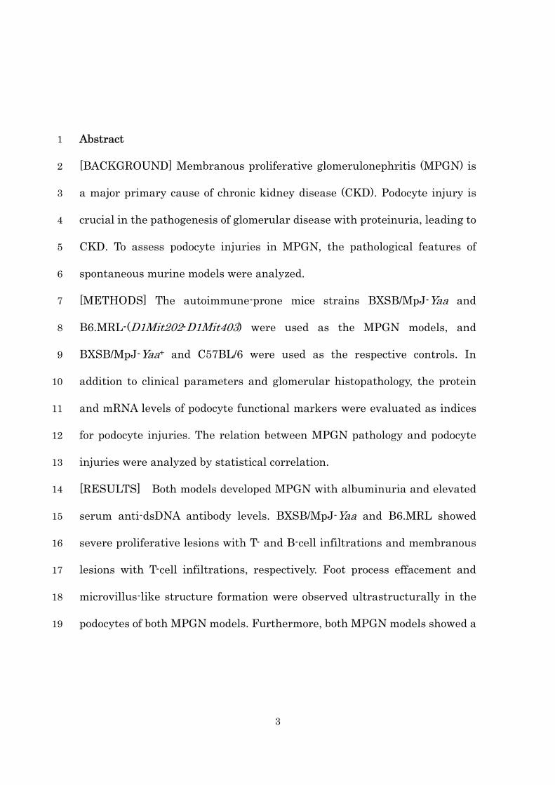
[BACKGROUND] Membranous proliferative glomerulonephritis (MPGN) is 2
a major primary cause of chronic kidney disease (CKD). Podocyte injury is 3
crucial in the pathogenesis of glomerular disease with proteinuria, leading to 4
CKD. To assess podocyte injuries in MPGN, the pathological features of 5
spontaneous murine models were analyzed. 6
[METHODS] The autoimmune-prone mice strains BXSB/MpJ-Yaa and 7
B6.MRL-(D1Mit202-D1Mit403) were used as the MPGN models, and 8
BXSB/MpJ-Yaa+ and C57BL/6 were used as the respective controls. In 9
addition to clinical parameters and glomerular histopathology, the protein 10
and mRNA levels of podocyte functional markers were evaluated as indices 11
for podocyte injuries. The relation between MPGN pathology and podocyte 12
injuries were analyzed by statistical correlation. 13
[RESULTS] Both models developed MPGN with albuminuria and elevated 14
serum anti-dsDNA antibody levels. BXSB/MpJ-Yaa and B6.MRL showed 15
severe proliferative lesions with T- and B-cell infiltrations and membranous 16
lesions with T-cell infiltrations, respectively. Foot process effacement and 17
microvillus-like structure formation were observed ultrastructurally in the 18
podocytes of both MPGN models. Furthermore, both MPGN models showed a 19

4
decrease in immune-positive areas of nephrin, podocin, and synaptopodin in 1
the glomerulus, and in the mRNA expression of Nphs1, Nphs2, Synpo, Actn4, 2
Cd2ap, and Podxl in the isolated glomerulus. Significant negative 3
correlations were detected between serum anti-dsDNA antibody levels and 4
glomerular Nphs1 expression, and between urinary albumin-to-creatinine 5
ratio and glomerular expression of Nphs1, Synpo, Actn4, Cd2ap, or Podxl. 6
[CONCLUSION] MPGN models clearly developed podocyte injuries 7
characterized by the decreased expression of podocyte functional markers 8
with altered morphology. These data emphasized the importance of 9
regulation of podocyte injuries in MPGN.10

5
Introduction 1
Chronic kidney disease (CKD) is one of the most serious public health 2
problems because it is strongly associated with not only end-stage renal 3
disease (ESRD) but also cardiovascular diseases [1]. Thus, understanding 4
the pathophysiology of CKD is important to improve the morbidity and 5
mortality of patients. Recent studies have shown that progressive podocyte 6
injury, also called podocytopathy, is one of the key events in the pathogenesis 7
of major CKD primary diseases such as diabetic nephropathy and renal 8
sclerosis [2,3]. 9
Podocytes are highly differentiated epithelial cells lining the outside of 10
glomerular capillaries, and their foot processes (FPs) regulate the 11
glomerular filtration barrier (blood–urine barrier; BUB) by the formation of 12
a slit diaphragm (SD). Yi et al [4] indicated that a decrease in the expression 13
of SD molecules and effacement of FPs were associated with the development 14
of renal sclerosis. According to Pagtalunan et al [5], the number of podocytes 15
in the glomerulus could be used as indices of podocyte injury in patients with 16
diabetic nephropathy. In a CKD animal study targeting dogs, we have also 17
clarified that the immune-positive levels of glomerular SD molecules, 18
especially nephrin and actinin alpha 4 (ACTN4), negatively correlated with 19

6
serum creatinine levels, and nephrin mRNA expression in the kidneys of 1
CKD groups was significantly lower than that in normal animals and 2
negatively correlated with serum creatinine [6]. Several studies have 3
indicated that the signaling pathway through Notch, transforming growth 4
factor beta (TGF-β), and angiotensin II play crucial roles in podocyte injuries 5
[7–10]. 6
Membranous proliferative glomerulonephritis (MPGN) is one of the 7
major CKD primary diseases and is associated with infections, drugs, and 8
systemic disorders [11]. From early stage MPGN, increased glomerular cells 9
and immune-complex depositions were observed in glomerular lesions with 10
proteinuria caused by the ultrafiltration of plasma proteins [12]. Chronic 11
glomerular lesions with increased urinary protein could trigger the 12
formation of tubulointerstitial lesions and eventually progress to interstitial 13
fibrosis leading to ESRD [12]. Although the appearance of proteinuria 14
indicates BUB disruption in MPGN from the early stage, little is known 15
about podocyte injury in MPGN patients and in model mice. 16
Spontaneous MPGN models, lupus-prone mice such as NZB, (NZB × 17
NZW) F1 hybrid, BXSB/MpJ-Yaa (BXSB-Yaa), and MRL/MpJ-lpr are 18
commonly used. These strains develop systemic autoimmune diseases 19

7
characterized by increased serum autoantibody levels and vasculitis, as well 1
as MPGN [13–15]. BXSB-Yaa carries a mutant gene located on the Y 2
chromosome, designated as Y-linked autoimmune acceleration (Yaa), and 3
males show more severe MPGN than females [16]. We have demonstrated 4
that BXSB-Yaa mice develop glomerular lesions with decreased 5
WT1-positive podocytes leading to proteinuria subsequent to 6
tubulointerstitial lesion formation [17]. Furthermore, we developed a 7
spontaneous CKD model named B6.MRL- (D1Mit202-D1Mit403) (B6.MRL), 8
carrying the C57BL/6 (B6) background and the telomeric regions of 9
chromosome 1 (68–81 cM) derived from lupus-prone MRL/MpJ [18]. This 10
congenic region contains the Fas ligand, interferon activated gene 200 family, 11
and Fc gamma receptor family, which were strongly associated with the 12
developments of autoimmunity and MPGN [18]. With age, female B6.MRL 13
spontaneously develop MPGN similar to human CKD [18-20]. Therefore, we 14
considered that BXSB-Yaa and B6.MRL as appropriate models for the 15
investigation of podocyte injury in MPGN. 16
In this study, the pathological features of MPGN were analyzed using 2 17
murine models, BXSB-Yaa and B6.MRL, with focus on podocyte injuries. The 18
results showed that these MPGN models clearly developed podocyte injuries, 19

8
characterized by the decreased mRNA and protein levels of its functional 1
markers with morphological changes and exacerbation of clinical parameters. 2
Our data emphasized the importance of regulation of podocyte injuries in 3
MPGN. 4
5

9
Materials and Methods 1
Animals and Sample Preparations 2
Experimental animals were handled according to the “Guide for the 3
Care and Use of Laboratory Animals” of Hokkaido University, Graduate 4
School of Veterinary Medicine (approved by the Association for Assessment 5
and Accreditation of Laboratory Animal Care International). Male 6
BXSB-Yaa and female B6.MRL were used as MPGN model mice at age 4 7
months and 9–15 months, respectively. As healthy controls, male 8
BXSB/MpJ-Yaa+ (BXSB) and female B6 mice were used at age 4 and 9 9
months, respectively. B6.MRL was created in our laboratory [18], whereas 10
B6, BXSB, and BXSB-Yaa were purchased from Japan SLC Inc. (Shizuoka, 11
Japan). All mice were maintained under specific pathogen-free conditions. 12
The animals were subjected to deep anesthesia (60 mg/kg pentobarbital 13
sodium administered intraperitoneally), and urine was collected by bladder 14
puncture. After urine collection, the mice were euthanized by exsanguination 15
from the carotid artery, and serum and kidneys were collected. The kidneys 16
were fixed by 4% paraformaldehyde (PFA) in 0.1 M phosphate buffer (PB; pH 17
7.4) or 2.5% glutaraldehyde in 0.1 M PB at 4°C for histopathological analysis. 18
PFA-fixed paraffin sections (2 μm thick) were then prepared and used for 19

10
periodic acid Schiff (PAS) staining or immunostaining. 1
2
Histological Analysis 3
To assess the severity of glomerulonephritis, semiquantitative 4
glomerular damage scoring was performed as previous study [17]. Details of 5
the procedures were described in Supplementary material 1. 6
7
Glomerular Isolation 8
Glomeruli of mice were isolated by a bead perfusion method [21]. Briefly, 9
40 mL of Hanks’ balanced salt solution (HBSS) containing 8 × 10⁷ 10
Dynabeads (Life Technologies, Palo Alto, CA) was perfused from the left 11
ventricle. The kidneys were removed and digested in collagenase (1 mg/mL 12
collagenase A [Roche, Basel, Switzerland] and 100 U/mL deoxyribonuclease I 13
[Life Technologies] in HBSS) at 37°C for 30 min. The digested tissue was 14
gently pressed through a 100-μm cell strainer (BD Falcon, Franklin Lakes, 15
NJ, USA) using a flattened pestle, and the cell suspension was centrifuged at 16
200 × g for 5 min. The cell pellet was resuspended in 2 mL HBSS. Finally, 17
glomeruli containing Dynabeads were gathered by a magnetic particle 18
concentrator (Life Technologies). 19

11
1
Serological and Urinary Analysis 2
For the evaluation of the systemic autoimmune condition, serum levels 3
of anti-double strand DNA (dsDNA) antibody were measured using Mouse 4
Anti-dsDNA Ig’s (Total A+G+M) ELISA kit (Alpha Diagnostic International, 5
San Antonio, TX, USA). For the evaluation of renal function, serum blood 6
urea nitrogen (sBUN) and creatinine (sCre) levels in all animals were 7
measured using Fuji Drichem 7000v (Fujifilm, Tokyo, Japan). Urinary 8
albumin-to-creatinine ratio (uACR) was determined using Albuwell M and 9
The Creatinine Comparison (Exocell, Philadelphia, PA, USA). 10
11
Immunohistochemistry and Histoplanimetry 12
Immunostaining for nephrin, podocin, synaptopodin, CD3, and B220 13
was performed according to the procedure shown in Table 1. Details of the 14
procedures are described in Supplementary material 1. 15
Quantifications of positive immunohistochemical reactions of SD 16
molecules were performed as described previously [6]. Briefly, the 17
glomerulus area and black pixels of positive reaction were measured, and the 18
number of pixels per area was calculated for each SD molecule by means of 19

12
BZII-Analyzer (Keyence, Osaka, Japan). To evaluate T-cell and B-cell 1
infiltration into the glomeruli, the number of CD3- and B220-positive cells 2
was counted. In these measurements, 20 glomeruli were counted in 1 kidney 3
section in each group (n = 5), and the values were expressed as means. 4
5
Immunofluorescence 6
Immunofluorescence for nephrin, podocin, and synaptopodin was 7
performed according to the procedure shown in Table 1 and Supplementary 8
material 1. Finally, the sections were examined under a fluorescence 9
microscope (BZ-9000, Keyence). 10
11
Electron Microscopy 12
Ultrastructural analysis with a transmission electron microscope (TEM) 13
was performed according to the following procedure. After fixation with 2.5% 14
glutaraldehyde in 0.1 M PB for 4 h, small pieces of kidney tissue were fixed 15
with 1% osmium tetroxide in 0.1 M PB for 2 h, dehydrated by graded alcohol, 16
and embedded in epoxy resin (Quetol 812 Mixture; Nisshin EM, Tokyo, 17
Japan). Ultrathin sections (70 nm) were double stained with uranyl acetate 18
and lead citrate. All samples were observed under a JEOL transmission 19

13
electron microscope (JEM-1210; JEOL, Tokyo, Japan). Ultrastructural 1
analysis with a scanning electron microscope (SEM) was performed 2
according to the following procedure. Quarter sizes of glutaraldehyde-fixed 3
kidneys were kept in 2% tannic acid for 1 h at 4°C and postfixed with 1% 4
osmium tetroxide in 0.1 M PB for 1 h. The specimens were dehydrated 5
through graded alcohol, transferred into 3-methylbutyl acetate, and dried 6
using an HCP-2 critical point dryer (Hitachi, Tokyo, Japan). The dried 7
specimens were sputter-coated with Hitachi E-1030 ion sputter coater 8
(Hitachi), and then examined on an S-4100 SEM (Hitachi) with an 9
accelerating voltage of 20 kV. 10
11
Reverse Transcription and Real-time Polymerase Chain Reaction 12
For the examination of mRNA expression, total RNA from isolated 13
glomeruli was purified using RNeasy Mini Kit (Qiagen, Hilden, Germany). 14
Total RNAs were synthesized to cDNAs by a reverse transcription (RT) 15
reaction by using ReverTra Ace reverse transcriptase enzyme (Toyobo, Osaka, 16
Japan) and random dT primers (Promega, Madison, WI). Each cDNA was 17
used for real-time polymerase chain reaction (PCR) with Brilliant III SYBR 18
Green QPCR master mix on Mx3000P (Agilent Technologies, La Jolla, CA, 19

14
USA). The expression levels of genes were normalized to actin beta (Actb) as 1
a housekeeping gene. The appropriate primer pairs are shown in Table 2. 2
3
Statistical Analysis 4
Results were expressed as the mean ± standard error (S.E.) and 5
statistically analyzed using a nonparametric Mann–Whitney U test (P < 6
0.05). The correlation between 2 parameters was analyzed using Spearman’s 7
rank correlation test (P < 0.05). 8
9

15
Results 1
Clinical Parameters of Membranous Proliferative Glomerulonephritis 2
Models 3
As the index of the systemic autoimmune condition, the serum 4
anti-dsDNA level of BXSB-Yaa and B6.MRL was significantly higher than 5
that of BXSB and B6, respectively (Table 3). BXSB-Yaa showed significantly 6
higher anti-dsDNA levels than B6.MRL. In the indices of renal functions, 7
including sBUN, sCre, and uACR, every parameter of BXSB-Yaa and 8
B6.MRL was higher than that of BXSB and B6, respectively. Significant 9
differences between controls and MPGNmodels were observed in uACR. The 10
uACR in BXSB-Yaa was significantly higher than that in B6.MRL. 11
12
Glomerular Histopathology in Membrane ProliferativeGlomerulonephritis 13
Models 14
Both BXSB-Yaa and B6.MRL mice developed membranoproliferative 15
MPGN characterized by glomerular hypertrophy, increased mesangial cells 16
and their matrix, and thickening of the glomerular basement membrane 17
(GBM) (Fig. 1b,d, Table 4). Proliferative and membranous lesions, in 18
particular, were more severe in BXSB-Yaa and B6.MRL, respectively. No 19

16
glomerular lesion was observed in the controls (Fig. 1a,b, Table 4). To assess 1
the glomerular infiltration of immune cells, immunohistochemistry for CD3 2
(T-cell marker) and B220 (B-cell marker) was performed. CD3-positive cells 3
were observed in the glomerulus of BXSB-Yaa and B6.MRL as well as in the 4
renal interstitium (Fig. 1f,h), but not in controls (Fig. 1e,g). Although few 5
B220-positive cells were observed in the glomerulus of BXSB and BXSB-Yaa 6
(Fig. 1i,j), they were scarcely observed in B6 and B6.MRL (Fig. 1k,l). In 7
histoplanimetry, the number of CD3-positive cells in the glomerulus was 8
significantly higher in both MPGN models than in each control (Fig. 1m). In 9
relation to the histological observation, the number of B220-positive cells in 10
the glomerulus was significantly higher in BXSB and BXSB-Yaa than in B6 11
and B6.MRL, respectively. No significant difference was observed between 12
the controls and the MPGN models (Fig. 1m). 13
14
Glomerular Ultrastructure in Membranous ProliferativeGlomerulonephritis 15
Models 16
To analyze the morphological changes of podocytes in MPGN, the 17
glomerular ultrastructure of MPGNmodels was compared with that of the 18
controls by TEM and SEM. Under TEM observation, the controls showed 19

17
clear cytotrabecula (Cyto, which also known as primary process) and 1
cytopodium (FP, which is also known as secondary process) (Fig. 2a,c,f,h). In 2
both MPGN models, FPs showed irregular arrangements with hypertrophy 3
and partial fusions (Fig. 2b,d,e,g,i,j). Furthermore, in MPGN models, the 4
GBM was thickened and wrinkled, and high electron-dense deposits 5
resembling immune complexes were observed in the double-countered GBM 6
of the subendothelial regions (Fig. 2b,d,e,g,i,j). At higher magnifications, the 7
liner SD was clearly observed between FPs of control mice (Fig. 2f,h). In both 8
MPGN models, FP effacement was clearly observed, but the SD and the 9
3-layer structure of the GBM were unclear (Fig. 2g,i,j). 10
Under SEM observation, the width of the cytotrabecula was increased in 11
MPGN models compared with the controls and the FPs were unclear in the 12
former podocytes (Fig. 2k–t). At higher magnifications, the engagements of 13
each FP were irregular, and a microvillus-like process was observed in the 14
surface of podocytes in both MPGN models (Fig. 2q,s,t). In B6.MRL mice 15
podocyte, these morphological changes were observed in TEM and SEM and 16
severer at 15 months than at 9 months. 17
18
Localization and Expression of Slit Diaphragm Molecules in Membranous 19

18
Proliferative Glomerulonephritis Models 1
For the evaluation of podocyte injury, immunohistochemistry and 2
immunofluorescence for SD molecules (nephrin, podocin, and synaptopodin) 3
were performed (Fig. 3). Linear-positive reactions for nephrin, podocin, and 4
synaptopodin were observed along the glomerular capillary rete in controls 5
(Fig. 3a,c,e,g,i,k). In contrast, those in the MPGN models tended to be faint, 6
partially showed granular patterns, and localized to the glomerular edge 7
rather than the center (Fig. 3b,d,f,h,j,l). In immunohistoplanimetry, the 8
relative positive areas of all SD molecules in the glomerulus were 9
significantly smaller in both MPGN models than in controls (Fig. 3m). 10
Furthermore, to evaluate the relation between immune cell infiltration 11
and podocyte injuries, immune cell number (B220- and CD3-positive cells) 12
and the relative SD molecule–positive area (nephrin, podocin, synaptopodin) 13
in the glomerulus were analyzed (Table 5). No significant correlation was 14
detected between both parameters. 15
16
mRNA Expression of Podocyte Functional Markers in Membranous 17
Proliferative Glomerulonephritis Models 18
For further investigation of podocyte injury, the mRNA expression of the 19
19
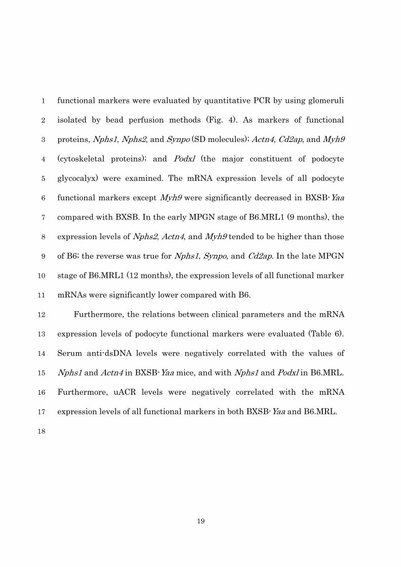
functional markers were evaluated by quantitative PCR by using glomeruli 1
isolated by bead perfusion methods (Fig. 4). As markers of functional 2
proteins, Nphs1, Nphs2, and Synpo (SD molecules); Actn4, Cd2ap, and Myh9 3
(cytoskeletal proteins); and Podxl (the major constituent of podocyte 4
glycocalyx) were examined. The mRNA expression levels of all podocyte 5
functional markers except Myh9 were significantly decreased in BXSB-Yaa 6
compared with BXSB. In the early MPGN stage of B6.MRL1 (9 months), the 7
expression levels of Nphs2, Actn4, and Myh9 tended to be higher than those 8
of B6; the reverse was true for Nphs1, Synpo, and Cd2ap. In the late MPGN 9
stage of B6.MRL1 (12 months), the expression levels of all functional marker 10
mRNAs were significantly lower compared with B6. 11
Furthermore, the relations between clinical parameters and the mRNA 12
expression levels of podocyte functional markers were evaluated (Table 6). 13
Serum anti-dsDNA levels were negatively correlated with the values of 14
Nphs1 and Actn4 in BXSB-Yaa mice, and with Nphs1 and Podxl in B6.MRL. 15
Furthermore, uACR levels were negatively correlated with the mRNA 16
expression levels of all functional markers in both BXSB-Yaa and B6.MRL. 17
18

20
Discussion 1
Podocyte Injuries in Membranous Proliferative Glomerulonephritis 2
Systemic lupus erythematosus (SLE) is a representative autoimmune 3
disease showing the deposition of immune complexes or direct autoantibody 4
deposition to systemic organs, including the kidney, leading to complement 5
activation, Fc receptor ligation, and subsequent inflammation [22]. In 6
particular, SLE-related MPGN (lupus nephritis) is one of the most serious 7
SLE complications since it is a major predictor of poor prognosis [22,23]. In 8
lupus nephritis, it has been suggested that glomerular immune complex 9
depositions cause glomerular lesions such as mesangial cell proliferation and 10
GBM thickening, which lead to BUB disruption, as in other MPGNs such as 11
IgA nephropathy and drug-induced glomerulonephritis [17–20,24]. 12
BXSB-Yaa and B6.MRL are spontaneous murine models of 13
autoimmune-mediated MPGN [17-20]. The present study clarified that both 14
MPGN models had clearly elevated serum dsDNA antibody levels and 15
developed membranoproliferative MPGN with immune complex depositions 16
and altered podocyte morphology, such as FP effacement and the appearance 17
of microvillus-like processes. Furthermore, the glomerular mRNA levels of 18
podocyte functional markers were significantly decreased and negatively 19

21
correlated with uACR in the MPGN models. Little has been known about 1
podocyte injury in MPGN, which is one of the major primary diseases leading 2
to CKD and subsequent ESRD. The present study clarified that the decrease 3
of podocyte functional mRNA expression levels in MPGN closely correlated 4
with the morphological changes of podocytes as well as glomerular 5
dysfunction indicated by increased uACR. Importantly, although the 6
pathological mechanism of podocyte injuries was mainly investigated by 7
using drug-induced glomerular disease models but not spontaneous models 8
such as BXSB-Yaa or B6.MRL. The podocyte injuries in spontaneous models 9
would more clearly reflect those in human clinical cases than the drug 10
induced glomerular disease models. Recent experimental and clinical studies 11
have revealed that podocytes exhibit various structural changes, including 12
FP effacement, cell body attenuation, pseudocyst formation, hypertrophy, 13
cytoplasmic accumulation of lysosomal elements, and detachment from the 14
GBM, under several pathological situations [25]. Among these changes, FP 15
effacement is the most representative change in podocyte shape, which is 16
closely correlated with progressive proteinuria [26, 27]. Furthermore, an 17
increase in podocyte microvilli represents another morphologic alteration 18
during both experimental and human nephrotic syndrome, with unknown 19

22
mechanisms and significance [28]. These findings suggested that 1
immune-mediated MPGNinvolves podocyte injury with morphological 2
change leading to BUB disruption, similar to other glomerular diseases. 3
4
Contribution of Immune Cell Infiltration to Podocyte Injuries in 5
Membranous Proliferative Glomerulonephritis 6
Interestingly, there was a difference in glomerular pathological features 7
between BXSB-Yaa and B6.MRL; proliferative and membranous lesions were 8
more severe in BXSB-Yaa and B6.MRL, respectively. Furthermore, the 9
populations of glomerulus-infiltrating immune cells were different between 10
these models; B-cells were localized in the glomerulus of BXSB-Yaa, but not 11
B6.MRL. Because BXSB also had B-cells in the glomerulus, the BXSB 12
genomic background rather than the Yaa mutation might have a role in the 13
infiltration of B-cells in the glomerulus. Studies on lupus models have 14
demonstrated that infiltrating B-cells in the kidney secrete antibodies with 15
various antigen specificities, contributing to increased in situ immune 16
complexes [29]. Recent reports have suggested that depleting B-cells either 17
before or after disease onset prevented and/or delayed the onset of nephritis 18
in several different lupus model mice [30,31]. Furthermore, lupus-prone 19

23
MRL/MpJ-lpr mice, which express a mutant transgene encoding surface 1
immunoglobulin (meaning that their B-cells are unable to secrete antibodies), 2
still develop nephritis [32]. These reports indicated that B-cells can play 3
some roles not only in antibody productions and the activation of pathogenic 4
T-cells but also in secreting pro-inflammatory cytokines such as tumor 5
necrosis factor alpha (TNF-α) and interleukin 6 (IL-6), contributing to the 6
development of glomerular lesions. However, our study demonstrated that no 7
significant correlation was observed between T-cells or B-cells and SD 8
protein expression levels in the glomerulus of MPGN models. From these 9
findings, the presence and functional activation of infiltrating B-cells in the 10
glomerulus might exacerbate the glomerular proliferative lesions in 11
BXSB-Yaa, not primarily contributing to podocyte injury. 12
13
Putative Mechanism of Podocyte Injuries in Membranous Proliferative 14
Glomerulonephritis 15
Recently, it has been suggested that FP effacement, the response of the 16
podocyte to injury, is dependent on the disruption of the actin cytoskeletal 17
network as an initial event [33,34]. In several glomerular diseases of human 18
and mice, it has been implicated that the molecular framework of process 19

24
consists of actin filaments and cytoskeleton proteins such as ACTN4, 1
CD2-associated protein (CD2AP), and myosin, heavy chain 9, non-muscle 2
(MYH9), and FP effacement is caused by rearrangements of this molecular 3
framework [33-36]. Indeed, our data indicated that the glomerular mRNA 4
expression levels of actin-associated cytoskeletal proteins, including Actn4 5
and Cd2ap, decreased in MPGN models compared with controls. 6
Interestingly, the glomerular Actn4 mRNA expression in both MPGN models 7
strongly and negatively correlated with uACR. In contrast, the glomerular 8
Myh9 expression level of B6.MRL in the early stage (age 9 months) was 9
significantly higher than that of controls. From these findings, altered 10
glomerular mRNA expression levels of cytoskeletal proteins might indicate 11
cytoskeletal rearrangements associated with MPGNprogression and may be 12
associated with the effacement of FPs and formation of microvillus-like 13
processes in podocytes. 14
15
Trigger and Crucial Pathway of Podocyte Injuries in Membranous 16
Proliferative Glomerulonephritis 17
Several factors, including genetic, mechanical, and immunological 18
stresses, and toxins, were suggested to be the causes of FP effacement 19

25
associated with cytoskeletal rearrangement and redistribution of SD 1
proteins [37]. In the present study, MPGN podocytes showed not only 2
morphological changes but also abnormalities in SD protein localization; 3
nephrin, podocin, and synaptopodin were localized to the cell body as well as 4
FP, showing a granular pattern. More recent studies indicated that 5
Rho-kinases play a pivotal role in the organization of the actin cytoskeleton 6
of podocytes [38-40]. Asanuma et al [39] suggested that Rho-kinase regulates 7
actin−myosin-containing stress fibers in the podocyte cell body. 8
Meyer-Schwesinger et al [40] demonstrated that increased activation of 9
Rho-kinases leads to cytoskeletal rearrangement in the course of 10
antibody-mediated podocyte injury, culminating in FP effacement, 11
proteinuria, and detachment into the urine, and that this could be prevented 12
by Rho-kinase inhibition. Furthermore, in vitro and in vivo studies have 13
revealed that proinflammatory cytokines such as IL-1β and TNF-α are 14
involved in Rho-kinase pathway activation [41,42]. From these findings, 15
Rho-kinase abnormality, which is caused by the glomerular exposure of 16
internal or blood proinflamatory cytokines, including IL-1β and TNF-α, 17
might be a trigger for podocyte injury in MPGN. The determination of the 18
immune-mediated pathological pathway of podocyte injuries would lead to 19

26
the regulation of animal and human MPGN. 1
In conclusion, although there was a small difference in glomerular 2
pathological features between BXSB-Yaa and B6.MRL, both MPGN models 3
clearly developed podocyte injuries characterized by decreased expression of 4
functional markers, with morphological changes and aggravation of clinical 5
parameters.6

27
Acknowledgments 1
This work was supported by a Grants-in-Aid for Scientific Research from 2
Graduate School of Veterinary Medicine, Hokkaido University, and 3
Grant-in-Aid for JSPS Fellows (No. 25000961), and Grant-in-Aid for Young 4
Scientist (No. 24688033) and Grant-in-Aid for Scientific Research B (No. 5
24380156) from the Ministry of Education, Culture, Sports, Science, and 6
Technology of Japan. 7
8

28
References 1
1. Tonelli M, Wiebe N, Culleton B, House A, Rabbat C, Fok M, McAlister F, 2
Garg AX. Chronic kidney disease and mortality risk: a systematic review. 3
J Am Soc Nephrol 2006; 17: 2034-2047. 4
2. Diez-Sampedro A, Lenz O, Fornoni A. Podocytopathy in diabetes: a 5
metabolic and endocrine disorder. Am J Kidney Dis 2011; 58: 637-646. 6
3. Kretzler M. Role of podocytes in focal sclerosis: defining the point of no 7
return. J Am Soc Nephrol 2005; 16: 2830-2832. 8
4. Yi F, dos Santos EA, Xia M, Chen QZ, Li PL, Li N. Podocyte injury and 9
glomerulosclerosis in hyperhomocysteinemic rats. Am J Nephrol 2007; 10
27: 262-268. 11
5. Pagtalunan ME, Miller PL, Jumping-Eagle S et al. Podocyte loss and 12
progressive glomerular injury in type II diabetes. J Clin Invest 1997; 99: 13
342-348. 14
6. Ichii O, Yabuki A, Sasaki N, Otsuka S, Ohta H, Yamasaki M, Takiguchi M, 15
Namiki Y, Hashimoto Y, Endoh D, Kon Y. Pathological correlations 16
between podocyte injuries and renal functions in canine and feline 17
chronic kidney diseases. Histol Histopathol 2011; 26: 1243-1255. 18
7. Niranjan T, Bielesz B, Gruenwald A, Ponda MP, Kopp JB, Thomas DB, 19
29
Susztak K. The Notch pathway in podocytes plays a role in the 1
development of glomerular disease. Nat Med 2008; 14: 290-298. 2
8. Schiffer M, Bitzer M, Roberts IS, Kopp JB, ten Dijke P, Mundel P, 3
Böttinger EP. Apoptosis in podocytes induced by TGF-beta and Smad7. J 4
Clin Invest 2001; 108: 807-816. 5
9. Zhang C, Hu JJ, Xia M, Boini KM, Brimson C, Li PL. Redox signaling via 6
lipid raft clustering in homocysteine-induced injury of podocytes. Biochim 7
Biophys Acta 2009; 1803: 482-491. 8
10. Lavoz C, Rodrigues-Diez R, Benito-Martin A, Rayego-Mateos S, 9
Rodrigues-Diez RR, Alique M, Ortiz A, Mezzano S, Egido J, Ruiz-Ortega 10
M. Angiotensin II contributes to renal fibrosis independently of Notch 11
pathway activation. PLoS One 2012; 7: e40490. 12
11. Kawasaki Y. Mechanism of onset and exacerbation of chronic 13
glomerulonephritis and its treatment. Pediatr Int 2011; 53: 795-806. 14
12. Kriz W, LeHir M. Pathways to nephron loss starting from glomerular 15
diseases—insights from animal models. Kidney Int 2005; 67: 404-419. 16
13. Li L, Nukala S, Du Y, Han J, Han J, Liu K, Hutcheson J, Pathak S, Li Q, 17
Mohan C. Murine lupus strains differentially model unique facets of 18
human lupus serology. Clin Exp Immunol 2012; 168: 178-185. 19

30
14. Moser K, Kalies K, Szyska M, Humrich JY, Amann K, Manz RA.. CXCR3 1
promotes the production of IgG1 autoantibodies but is not essential for 2
the development of lupus nephritis in NZB/NZW mice. Arthritis Rheum 3
2012; 64: 1237-1246. 4
15. Okamoto A, Fujio K, Tsuno NH, Takahashi K, Yamamoto K. 5
Kidney-infiltrating CD4+ T-cell clones promote nephritis in lupus-prone 6
mice. Kidney Int 2012; 82: 969-979. 7
16. Subramanian S, Tus K, Li QZ, Wang A, Tian XH, Zhou J, Liang C, Bartov 8
G, McDaniel LD, Zhou XJ, Schultz RA, Wakeland EK. A Tlr7 9
translocation accelerates systemic autoimmunity in murine lupus. Proc 10
Natl Acad Sci U S A 2006; 103: 9970-9975. 11
17. Kimura J, Ichii O, Otsuka S, Kanazawa T, Namiki Y, Hashimoto Y, Kon 12
Y. Quantitative and qualitative urinary cellular patterns correlate with 13
progression of murine glomerulonephritis. PLoS One 2011; 6: e16472. 14
18. Ichii O, Konno A, Sasaki N, Endoh D, Hashimoto Y, Kon Y. Autoimmune 15
glomerulonephritis induced in congenic mouse strain carrying telomeric 16
region of chromosome 1 derived from MRL/MpJ. Histol Histopathol 2008; 17
23: 411-422. 18
19. Ichii O, Konno A, Sasaki N, Endoh D, Hashimoto Y, Kon Y. Altered 19

31
balance of inhibitory and active Fc gamma receptors in murine 1
autoimmune glomerulonephritis. Kidney Int 2008; 74: 339-347. 2
20. Ichii O, Otsuka S, Sasaki N, Yabuki A, Ohta H, Takiguchi M, Hashimoto 3
Y, Endoh D, Kon Y. Local overexpression of interleukin-1 family, member 4
6 relates to the development of tubulointerstitial lesions. Lab Invest 5
2010; 90: 459-475. 6
21. Takemoto M, Asker N, Gerhardt H, Lundkvist A, Johansson BR, Saito Y, 7
Betsholtz C. A new method for large scale isolation of kidney glomeruli 8
from mice. Am J Pathol 2002; 161: 799-805. 9
22. Tsokos GC. Systemic lupus erythematosus. N Engl J Med 2011; 365: 10
2110-2121. 11
23. Waldman M, Appel GB. Update on the treatment of lupus nephritis. 12
Kidney Int 2006; 70: 1403-1412. 13
24. Nowling TK, Gilkeson GS. Mechanisms of tissue injury in lupus nephritis. 14
Arthritis Res Ther 2011; 13: 250. 15
25. Kriz W, Gretz N, Lemley KV. Progression of glomerular diseases: Is the 16
podocyte the culprit? Kidney Int 1998; 54: 687–697. 17
26. Inokuchi S, Shirato I, Kobayashi N, Koide H, Tomino Y, Sakai T. 18
Re-evaluation of foot process effacement in acute puromycin 19

32
aminonucleoside nephrosis. Kidney Int 1996; 50: 1278–1287. 1
27. Shirato I. Podocyte process effacement in vivo. Microsc Res Tech 2002; 57: 2
241-246. 3
28. Hara M, Yanagihara T, Hirayama Y, Ogasawara S, Kurosawa H, Sekine 4
S, Kihara I. Podocyte membrane vesicles in urine originate from tip 5
vesiculation of podocyte microvilli. Hum Pathol 2010; 41: 1265-1275. 6
29. Espeli M, Bokers S, Giannico G, Dickinson HA, Bardsley V, Fogo AB, 7
Smith KG. Local renal autoantibody production in lupus nephritis. J Am 8
Soc Nephrol 2011; 22: 296-305. 9
30. Bekar KW, Owen T, Dunn R, Ichikawa T, Wang W, Wang R, Barnard J, 10
Brady S, Nevarez S, Goldman BI, Kehry M, Anolik JH. Prolonged effects 11
of short-term anti-CD20 B cell depletion therapy in murine systemic 12
lupus erythematosus. Arthritis Rheum 2010; 62: 2443-2457. 13
31. Ramanujam M, Bethunaickan R, Huang W, Tao H, Madaio MP, Davidson 14
A. Selective blockade of BAFF for the prevention and treatment of 15
systemic lupus erythematosus nephritis in NZM2410 mice. Arthritis 16
Rheum 2010; 62: 1457-1468. 17
32. Chan OT, Hannum LG, Haberman AM. A novel mouse with B cells but 18
lacking serum antibody reveals an antibodyindependent role for B cells in 19

33
murine lupus. J Exp Med 1999; 189: 1639-1648. 1
33. Shirato I, Sakai T, Kimura K, Tomino Y, Kriz W. Cytoskeletal changes in 2
podocytes associated with foot process effacement in Masugi nephritis. 3
Am J Pathol 1996; 148: 1283-1296. 4
34. Asanuma K, Kim K, Oh J, Giardino L, Chabanis S, Faul C, Reiser J, 5
Mundel P. Synaptopodin regulates the actin bundling activity of 6
alpha-actinin in an isoform-specific manner. J Clin Invest 2005; 115: 7
1188-1198. 8
35. Durvasula RV, Shankland SJ. Podocyte injury and targeting therapy: an 9
update. Curr Opin Nephrol Hypertens 2006; 15: 1-7. 10
36. Asanuma K, Yanagida-Asanuma E, Takagi M, Kodama F, Tomino Y. The 11
role of podocytes in proteinuria. Nephrology 2007; 12 Suppl 3: S15-20. 12
37. Asanuma K, Mundel P. The role of podocytes in glomerular pathobiology. 13
Clin Exp Nephrol 2003; 7: 255-259. 14
38. Shibata S, Nagase M, Fujita T. Fluvastatin ameliorates podocyte injury 15
in proteinuric rats via modulation of excessive Rho signaling. J Am Soc 16
Nephrol 2006; 17: 754-764. 17
39. Asanuma K, Yanagida-Asanuma E, Faul C, Tomino Y, Kim K, Mundel P. 18
Synaptopodin orchestrates actin organization and cell motility via 19

34
regulation of RhoA signalling. Nat Cell Biol 2006; 8: 485-491. 1
40. Meyer-Schwesinger C, Dehde S, Sachs M, Mathey S, Arefi K, Gatzemeier 2
S, Balabanov S, Becker JU, Thaiss F, Meyer TN. Rho-kinase inhibition 3
prevents proteinuria in immune-complex-mediated antipodocyte 4
nephritis. Am J Physiol Renal Physiol 2012; 303: F1015-1025. 5
41. Kanda T, Wakino S, Homma K, Yoshioka K, Tatematsu S, Hasegawa K, 6
Takamatsu I, Sugano N, Hayashi K, Saruta T. Rho-kinase as a molecular 7
target for insulin resistance and hypertension. FASEB J 2006; 20: 8
169-171. 9
42. Hiroki J, Shimokawa H, Higashi M, Morikawa K, Kandabashi T, 10
Kawamura N, Kubota T, Ichiki T, Amano M, Kaibuchi K, Takeshita A. 11
Inflammatory stimuli upregulate Rho-kinase in human coronary vascular 12
smooth muscle cells. J Mol Cell Cardiol 2004; 37: 537-546. 13

Tables 1
Table 1. Summary of immunostaining conditions 2
Immunohistochemistry Nephrin Podocin Synaptopodin CD3 B220
First antibody Rabbit polyclonal antibodies
(1:500; IBL, Gunma, Japan)
Rabbit polyclonal antibodies
(1:800; IBL)
Mouse monoclonal antibodies
(1:50; Fitzgerald, MA, USA)
Rabbit polyclonal
antibodies
(1:150; Nichirei,
Tokyo, Japan)
Rat monoclonal
antibodies (1:1000;
Cedarlane, Ontario,
Canada)
Second antibody
Biotinylated goat anti-rabbit
IgG antibodies (SABPO kit,
Nichirei)
Biotinylated goat anti-rabbit
IgG antibodies (SABPO kit,
Nichirei)
Simple Stain Mouse
MAX-PO(M) (mouse stain kit,
Nichirei)
Biotinylated goat
anti-rabbit
IgG antibodies
(SABPO kit,
Nichirei)
Biotinylated goat
anti-rat IgG
antibodies (Caltag
Medsystems,
Buckingham, UK)
Immunofluorescence Nephrin Podocin Synaptopodin CD3 B220
First antibody Same as in
immunohistochemistry
Same as in
immunohistochemistry
Same as in
immunohistochemistry / /
Second antibody
Alexa Fluor 546-labeled
donkey anti-rabbit IgG
antibodies (1:500, Life
Technologies)
Alexa Fluor 546-labeled
donkey anti-rabbit IgG
antibodies (1:500, Life
Technologies)
Alexa Fluor 488-labeled
donkey anti-mouse IgG
antibodies (1:500, Life
Technologies)
/ /
3
Table. 2. Summary of specific gene primers for real-time polymerase chain 4
reaction 5
Genes Primer sequence (5-3) Product sizeSpecific function in podocyte
(accession no.) F: forward, R: reverse (bp)
Nphs1 F: ACCTGTATGACGAGGTGGAGAG218 SD protein
(NM_019459) R: TCGTGAAGAGTCTCACACCAG
Nphs2 F: AAGGTTGATCTCCGTCTCCAG 105 SD protein
(NM_130456) R: TTCCATGCGGTAGTAGCAGAC

Synpo F: CATCGGACCTTCTTCCTGTG 90 SD protein
(NM_177340.2) R: TCGGAGTCTGTGGGTGAG
Actn4 F: TCCAGGACATCTCTGTGGAAG 216 Cytoskeletal protein
(NM_021895) R: CATTGTTTAGGTTGGTGACTGG
Cd2ap F: CAAGATGCCTGGAAGACGA 177 Cytoskeletal protein
(NM_009847) R: GCACTTGAAGGTGTTGAAAGAG
Myh9 F: AAGGACCAGGCTGACAAGG 209 Cytoskeletal protein
(NM_022410) R: GTCACGACAAATGGCAGGTC
Podxl F: TCCTAAGGCCGTGTATGAGC 153 Podocyte glycocalyx
(NM_013723) R: GATGCCATGCAGACGATG
1
Table 3. Clinical parameters of membranous proliferative 2
glomerulonephritis model and control mice 3
dsDNA (μg/mL) sBUN (mg/dL) sCre (mg/dL) uACR (μg/mg)
BXSB/MpJ-Yaa+ 4 months 72.35 ± 4.45 29.80 ± 3.50 0.56 ± 0.17 96.02 ± 54.43
BXSB/MpJ-Yaa 4 months 883.07* ± 85.11 38.93 ± 9.92 1.30 ± 1.03 835.20* ± 592.33
C57BL/6 9 months 61.57 ± 19.08 22.88 ± 1.42 0.12 ± 0.01 2.92 ± 0.40
B6.MRL-(D1Mit202-D1Mit403) 9 months 85.01* ± 35.86 24.92 ± 5.42 0.21 ± 0.05 29.02* ± 21.57
15 months 407.55* ± 166.38 30.24 ± 4.63 0.68 ± 0.56 378.61* ± 278.87
Values are mean ± S.E. dsDNA, double-strand DNA; sBUN, serum blood 4
urea nitrogen; sCre, serum creatinine; uACR, urinary albumin-to-creatinine 5
ratio. *Significantly different from each control (Mann–Whitney U test, P < 6
0.05); n ≧ 5. 7

1
Table 4. Quantitative evaluations of glomerular damage 2
glomerulus
damage score cell number in 1
glomerulus
thickness of glomerular basement membrane
(μm)
BXSB/MpJ-Yaa+ 4 month 34.01 ± 10.45 35.12 ± 2.33 1.98 ± 0.11
BXSB/MpJ-Yaa 4 month 180.68 ± 60.77 * 67.09 ± 9.96 * 4.01 ± 1.69 *
C57BL/6 9month 78.11 ± 21.26 41.12 ± 4.27 2.31 ± 0.08
B6.MRL-(D1Mit202-D1Mit403) 15 month 195.96 ± 42.53 * 55.96 ± 3.55 * 5.96 ± 0.98 *
Values are mean ± S.E. *Significantly different from each control (Mann–3
Whitney U test, P < 0.05); n ≧ 5. 4
5
Table 5. Relation between histological parameters and expression of 6
podocyte functional proteins 7
BXSB/MpJ-Yaa B6.MRL-(D1Mit202-D1Mit403)
Parameter/protein Nephrin Podoci
n
Synaptopodin Nephrin Podocin Synaptopodin
CD3-positive
cell number
-0.700 -0.600 -0.700 -0.493 0.309 -0.319
B220-positive
cell number
-0.308 -0.564 -0.564 -0.030 0.277 -0.577
Values are the Spearman's rank correlation coefficients. n ≧ 5. 8

Table 6. Relation between clinical parameters and podocyte functional mRNA expression 1
BXSB/MpJ-Yaa B6.MRLc1(68-81)
SD protein Cytoskeletal protein
Podocyte
glycocalyxSD protein Cytoskeletal protein
Podocyte
glycocalyx
Parameter
/mRNA Nphs1 Nphs2 Synpo Actn4 Cd2ap Myh9 Podxl Nphs1 Nphs2 Synpo Actn4 Cd2ap Myh9 Podxl
sADA -0.569* -0.464 -0.420 -0.662** -0.310 -0.279 -0.437 -0.557* -0.121 -0.439 -0.421 -0.275 -0.379 -0.578*
BUN -0.245 -0.159 -0.203 -0.286 -0.144 -0.256 -0.167 -0.066 -0.170 -0.379 -0.159 -0.352 -0.385 -0.324
sCre -0.295 -0.110 -0.128 -0.195 -0.121 -0.136 -0.191 0.278 0.109 -0.020 0.154 -0.022 -0.199 -0.003
uACR -0.624** -0.512* -0.747** -0.865** -0.624* -0.574* -0.621** -0.505* -0.245 -0.577** -0.469* -0.552** -0.466* -0.49*
Values are the Spearman's rank correlation coefficients. SD, slit diaphragm; sBUN, serum blood urea nitrogen; sCre, 2
serum creatinine; uACR, urinary albumin-to-creatinine ratio. * and **, significantly correlated (Spearman's 3
rank-correlation test, *P < 0.05. **P < 0.01); n ≧ 5. 4

1
Figure legends 1
Figure 1. Histopathology and immune cell infiltration in membranous 2
proliferative glomerulonephritis. 3
(a–d) Histopathology of renal cortices. BXSB/MpJ-Yaa+ (BXSB, 4 months; a), 4
BXSB/MpJ-Yaa (BXSB-Yaa, 4 months; b), C57BL/6 (B6, 9 months; c), and 5
B6.MRL-(D1Mit202-D1Mit403) (B6.MRL, 15 months; d). In the glomerulus, 6
mesangial matrix expansion, mesangial cell proliferation, and 7
periglomerular cell infiltration are observed in the BXSB-Yaa and B6.MRL 8
mice models. Periodic acid Schiff (PAS) staining. Bars = 50 μm. (e–l) 9
Immunohistochemistry of CD3 (e–h) and B220 (i–l). BXSB (e and i), 10
BXSB-Yaa (f and j), B6 (g and k), and B6.MRL (h and k). Bars = 50 μm. 11
CD3-positive cells are observed in the glomerulus and renal interstitium of 12
BXSB-Yaa and B6.MRL (arrows). B220-positive cells are observed in the 13
glomerulus of BXSB and BXSB-Yaa (arrowheads), but not in B6 and B6.MRL. 14
(m) Numbers of CD3-positive and B220-positive cells in the glomerulus. 15
BXSB and BXSB/MpJ-Yaa: 4 months. B6: 9 months. B6.MRL: 15 months. 16
Values are mean ± S.E. *, significantly different (Mann–Whitney U test, P < 17
0.05); n = 5. 18

2
1
Figure 2. Ultrastructural changes of the glomerulus in membranous 2
proliferative glomerulonephritis. 3
(a–j) Ultrastructure of the glomerulus under a transmission electron 4
microscope. BXSB/MpJ-Yaa+ (BXSB, 4 months; a and f), BXSB/MpJ-Yaa 5
(BXSB-Yaa, 4 months; b and g), C57BL/6 (B6, 9 months; c and h), 6
B6.MRL-(D1Mit202-D1Mit403) (B6.MRL, 9 months; d and i), and B6.MRL 7
(15months; e and j). Compared with the podocytes (Pod) of BXSB and B6, 8
showing clear cytotrabecula (Cyto, which is also known as primary process) 9
and cytopodium (foot process, Fp, which is also known as secondary process) 10
(a, c, f, and h), the Fps of BXSB-Yaa and B6.MRL show irregular 11
arrangements with hypertrophy and partial fusions (b, d, e, g, i, and j). The 12
glomerular basement membrane (GBM) is thickened and wrinkled in 13
BXSB-Yaa and B6.MRL (b and e, arrows), and high electron-dense deposits 14
are observed in the double-countered GBM of the subendothelial regions (b 15
and e, asterisks). The slit diaphragm (SD) is a clear linear pattern between 16
the Fps of BXSB and B6 (f and h, arrowheads). In both BXSB-Yaa and 17
B6.MRL, the Fp is effaced, and the SD as well as the 3-layer structure of the 18

3
GBM are unclear (g, i, and j). Ery, erythrocyte. Bars = 1 μm (a–e) and 100 nm 1
(f–j). (k–t) Ultrastructure of the glomerulus under a scanning electron 2
microscope. BXSB (4 months; k and p), BXSB-Yaa (4 months; l and q), B6 (9 3
months; m and r), B6.MRL (9 months; n and s) and B6.MRL (15 months; o 4
and t). The width of Cyto increase in BXSB-Yaa and B6.MRL (l, n, and o) 5
compared with the controls (k and m) and the Fps are unclear in the former 6
Pod. The engagements of each Fp are irregular, and microvillus-like 7
processes (villi) are observed on the surface of Pod in BXSB-Yaa and B6.MRL. 8
Bars = 2 μm. 9
10
Figure 3. Localizations of podocyte functional markers in membranous 11
proliferative glomerulonephritis. 12
(a–l) Immunofluorescence of nephrin (a–d), podocin (e–h), and synaptopodin 13
(i–l). BXSB/MpJ-Yaa+ (BXSB, 4 months; a, e, and i), BXSB/MpJ-Yaa 14
(BXSB-Yaa, 4 months; b, f, and j), C57BL/6 (B6, 9 months; c, g, and k), and 15
B6.MRL-(D1Mit202-D1Mit403) (B6.MRL, 15 months; d, h, and l). The 16
positive reactions for nephrin, podocin, and synaptopodin tended to be faint, 17
partially showed granular patterns, and localized to the glomerular edge 18

4
rather than the center in BXSB-Yaa and B6.MRL glomeruli. Bars = 50 μm. 1
(m) Comparison of SD protein–positive area ratio. The relative 2
immune-positive areas of nephrin, podocin, and synaptopodin in the 3
glomerulus. BXSB and BXSB/MpJ-Yaa: 4 months. B6: 9 months. B6.MRL: 15 4
months. Values are mean ± S.E. *, significantly different from each control 5
mice (Mann–Whitney U test, P < 0.05); n = 5. 6
7
Figure 4. mRNA expression levels of podocyte functional markers in 8
membranous proliferative glomerulonephritis. 9
The relative mRNA expression of podocyte functional markers was analyzed 10
by quantitative real-time PCR, using isolated glomerulus samples. 11
BXSB/MpJ-Yaa+ (BXSB, 4 months), BXSB/MpJ-Yaa (BXSB-Yaa, 4 months), 12
C57BL/6 (B6, 9 months), and B6.MRL-(D1Mit202-D1Mit403) (B6.MRL, 9 13
and 15 months). Each podocyte functional marker was normalized to Actb. 14
Values are mean ± S.E. *, significantly different from each control mice 15
(Mann–Whitney U test, P < 0.05); n ≧ 4. 16
17


Supplementary material 1 1
Histological Analysis 2
Glomerular damage scores were determined according to the following procedures. 3
Briefly, 100 glomeruli per kidney was examined by using PAS-stained sections and 4
scored from 0 to +4 according to the following criteria: 0, no recognizable lesion in 5
glomeruli; +1, a little PAS-positive deposition, mild cell proliferation, mild membranous 6
hypertrophy, and/or partial podocyte adhesion to the parietal layer of the renal 7
corpuscle; +2, segmental or global PAS-positive deposition, cell proliferation, 8
membranous hypertrophy, and/or glomerular hypertrophy; +3, the same as grade 2 with 9
PAS-positive deposition in 50% of regions of glomeruli and/or severe podocyte adhesion 10
to the parietal layer of the renal corpuscle; +4, disappearance of capillary and capsular 11
lumina, global deposition of PAS-positive material, and/or periglomerular infiltration of 12
inflammatory cells and fibrosis, based on the degrees of PAS-positive deposition, cell 13
proliferation, membranous hypertrophy, podocyte adhesion to the parietal layer, 14
disappearance of capillary and capsular lumina, and periglomerular infiltration of 15
inflammatory cells and fibrosis. If, for example, 50 of 100 glomeruli were +1, 25 of 100 16
glomeruli were +2, 20 of 100 glomeruli were +3, and 5 of 100 glomeruli were +4, the 17
semiquantitative score would be {(1 × 50 / 100) + (2 × 25 / 100) + (3 × 5 / 100) + (4 × 5 / 18
100)} × 100 = 180. 19
To assess the glomerular and membranous lesions, cell number in 1 glomerulus and 20
GBM thickness were determined, respectively. The number of cells per glomerulus and 21
thickness of thickest GBM in 1 glomerulus were determined and averaged in at least 10 22
glomeruli of the PAS-stained samples. 23
24

Immunohistochemistry 1
Immunostaining for nephrin, podocin, synaptopodin, CD3, and B220 was performed 2
according to the following procedure. For antigen retrieval, deparaffinized sections were 3
incubated in citrate buffer (pH 6.0) for 20 min at 105°C for nephrin and podocin (Dako 4
Target Retrieval Solution, pH 9; DAKO, Glostrup, Denmark), for 20 min at 105°C for 5
synaptopodin and CD3, or in 0.1% pepsin/0.2 M HCl for 5 min at 37°C for B220. After 6
cooling, slides were soaked in methanol containing 3% H2O2 for 15 min at room 7
temperature. After washing, sections were blocked by 10% normal goat serum for 8
nephrin and podocin, blocking solution A (mouse stain kit; Nichirei, Tokyo, Japan) for 9
synaptopodin, or 0.25% casein/0.01 M PBS for CD3 and B220 for 60 min at room 10
temperature. Then, sections were incubated with rabbit polyclonal antibodies for 11
nephrin (1:500; Immuno-Biological Laboratories, Gunma, Japan), rabbit polyclonal 12
antibodies for podocin (1:800; Immuno-Biological Laboratories), mouse monoclonal 13
antibodies for synaptopodin (1:50; Fitzgerald, MA, USA), rabbit polyclonal antibodies 14
for CD3 (1:150; Nichirei), or rat monoclonal antibodies for B220 (1:1000; Cedarlane, 15
Ontario, Canada) overnight at 4°C. After washing 3 times in phosphate-buffered saline 16
(PBS), sections were incubated with biotin-conjugated goat anti-rabbit IgG antibodies 17
for nephrin, podocin, and CD3 (SABPO kit, Nichirei) or goat anti-rat IgG antibodies for 18
B220 (1:200; Caltag Medsystems, Buckingham, UK) for 30 min at room temperature, 19
washed, and incubated with streptavidin-biotin complex (SABPO kit, Nichirei) for 30 20
min. For synaptopodin, sections were incubated with blocking solution B (mouse stain 21
kit, Nichirei) for 10 min at room temperature, washed, and incubated with Simple Stain 22
Mouse MAX-PO(M) (mouse stain kit, Nichirei) for 10 min. All sections were then 23
incubated with 3,3-diaminobenzidine tetrahydrochloride–H2O2 solution. Finally, the 24

sections were counterstained with hematoxylin. 1
2
Immunofluorescence 3
Immunofluorescence for nephrin, podocin, and synaptopodin was performed according 4
to the following procedure. After the deparaffinization, antigen retrieval was performed 5
by same method as in immunohistochemistry. After being washed, sections were 6
blocked by 5% normal donkey serum for 60 min at room temperature. Incubation with 7
primary antibodies was performed by same method as in immunohistochemistry. After 8
washing with PBS, the sections were incubated with Alexa Fluor 546-labeled donkey 9
anti-rabbit IgG antibodies (1:500, Life Technologies) for nephrin and podocin, or Alexa 10
Fluor 488-labeled donkey anti-mouse IgG antibodies (1:500, Life Technologies) for 11
synaptopodin for 30 min at room temperature and washed again. For nuclear staining, 12
sections were incubated with Hoechst 33342 (1:2000; Dojindo, Kumamoto, Japan) for 5 13
min. 14
15