Human Chorionic Stem Cells: Podocyte Differentiation and ...
36
1 Human Chorionic Stem Cells: Podocyte Differentiation and Potential for 1 the Treatment of Alport Syndrome. 2 3 Dafni Moschidou 1 , Michelangelo Corcelli 1 , Kwan-Leong Hau 1 , Victoria J 4 Ekwalla 1 , Jacques Behmoaras 2 , Paolo De Coppi 3 , Anna L David 1 , George 5 Bou-Gharios 4 , H Terence Cook 2 , Charles D Pusey 2 , Nicholas M Fisk 5 , and 6 Pascale V Guillot 1* . 7 8 1 University College London, Institute for Women’s Health, Maternal and Fetal 9 Medicine Department, London, United Kingdom 10 2 Imperial College London, Faculty of Medicine, Division of Immunity and 11 Inflammation, London, United Kingdom 12 3 University College London, Institute of Child Health, Stem Cells and 13 Regenerative Medicine Department, London, United Kingdom 14 4 University of Liverpool, Institute of Ageing and Chronic Disease, 15 Musculoskeletal Biology Department, Liverpool, United Kingdom 16 5 University of Queensland, UQ Centre for Clinical Research, Brisbane, 17 Queensland, Australia. 18 19 running title: Chorionic Stem Cell Therapy for Alport Syndrome 20 21 * Corresponding author: PVG University College London, Institute for 22 Women’s Health, Department of Maternal and Fetal Medicine, 86-96 Chenies 23 Mews, London, WC1N 1EH, United Kingdom. Email: [email protected], 24 Phone: +44 (0)207 242 9789, Fax: +44 (0)207 404 6181 25 26
Transcript of Human Chorionic Stem Cells: Podocyte Differentiation and ...
1
Human Chorionic Stem Cells: Podocyte Differentiation and Potential for 1
the Treatment of Alport Syndrome. 2
3
Dafni Moschidou1, Michelangelo Corcelli1, Kwan-Leong Hau1, Victoria J 4
Ekwalla1, Jacques Behmoaras2, Paolo De Coppi3, Anna L David1, George 5
Bou-Gharios4, H Terence Cook2, Charles D Pusey2, Nicholas M Fisk5, and 6
Pascale V Guillot1*. 7
8
1 University College London, Institute for Women’s Health, Maternal and Fetal 9
Medicine Department, London, United Kingdom 10
2 Imperial College London, Faculty of Medicine, Division of Immunity and 11
Inflammation, London, United Kingdom 12
3 University College London, Institute of Child Health, Stem Cells and 13
Regenerative Medicine Department, London, United Kingdom 14
4 University of Liverpool, Institute of Ageing and Chronic Disease, 15
Musculoskeletal Biology Department, Liverpool, United Kingdom 16
5 University of Queensland, UQ Centre for Clinical Research, Brisbane, 17
Queensland, Australia. 18
19
running title: Chorionic Stem Cell Therapy for Alport Syndrome 20
21
* Corresponding author: PVG University College London, Institute for 22
Women’s Health, Department of Maternal and Fetal Medicine, 86-96 Chenies 23
Mews, London, WC1N 1EH, United Kingdom. Email: [email protected], 24
Phone: +44 (0)207 242 9789, Fax: +44 (0)207 404 6181 25
26
2
ABSTRACT 27
Alport syndrome is a hereditary glomerulopathy caused by a mutation in type 28
IV collagen genes, which disrupts glomerular basement membrane, leading to 29
progressive glomerulosclerosis and end-stage renal failure. There is at 30
present no cure for Alport syndrome and cell-based therapies offer promise to 31
improve renal function. Here, we found that human first trimester fetal 32
chorionic stem cells (CSC) are able to migrate to glomeruli and differentiate 33
down the podocyte lineage in vitro and in vivo. When transplanted into 7-week 34
old Alport 129Sv-Col4a3tm1Dec/J (-/-) mice, a single intraperitoneal injection of 35
CSC significantly lowered blood urea and urine proteinuria levels over the 36
ensuing two weeks. In addition, nearly two thirds of transplanted -/- mice 37
maintained their weight above the 80% welfare threshold, with both males and 38
females weighing more than aged-matched non-transplanted -/- mice. This 39
was associated with less renal cortical fibrosis and interstitial inflammation 40
compared to non-transplanted mice, as shown by reduction in murine CD4, 41
CD68 and CD45.2 cells. Transplanted CSC homed to glomeruli, where they 42
expressed CR1, VEGFA, SYNAPTOPODIN, CD2AP and PODOCIN at the 43
RNA level, and produced both PODOCIN, CD2AP and COLIVα3 proteins in 44
non-transplanted -/- mice, suggesting indicating that CSC have adopted a 45
podocyte phenotype. Together, these data indicate that CSC may be used to 46
delay progression of renal pathology via a combination of anti-inflammatory 47
effects and potentially replacement of the defective resident podocytes. 48
49
50
3
51
INTRODUCTION 52
53
Alport syndrome (AS) is a genetic chronic kidney disease affecting 1 in 5,000 54
individuals. Initially, AS manifests with haematuria, proteinuria and increased 55
blood pressure, with progressive decline in renal function leading to end-stage 56
renal failure requiring replacement therapy. People with AS also suffer from 57
progressive hearing loss, anterior lenticonus, and macular flecks. AS is 58
caused by mutations in the type IV collagen genes encoding the a3/a4/a5 59
chains, which are produced exclusively by podocytes [1]. These mutations 60
affect the correct assembly of heterotrimeric ColIVa3/a4/a5 chains in the 61
glomerular basement membrane (GBM) and result in the GBM failing to 62
mature from the embryonic ColIVa1/a1/a2 type. Persistence of the immature 63
ColIVa1/a1/a2 GBM leads to its thickening and splitting, causing progressive 64
tubulointerstitial fibrosis and renal failure [1]. The only current treatment for AS 65
is blockade of the renin-angiotensin system, and proposed treatments include 66
collagen receptor blockade, anti-microRNA therapy, and stem cell therapy [2]. 67
The rationale of stem-based therapy is that stem cells isolated from healthy 68
donors will migrate and engraft in renal glomeruli, where they may 69
differentiate into functional podocytes producing new functional GBM. We 70
previously showed that human first trimester fetal blood-derived mesenchymal 71
stem cells (MSC) injected intraperitoneally into fetal mouse recipients 72
migrated to the kidneys where they engrafted in renal glomeruli [3]. However, 73
phenotype rescue by direct cell replacement is challenged by the low level of 74
donor cell engraftment and poor differentiation capacity of the donor cells [4]. 75
4
Col4a3tm1Dec/J mice are deficient in collagen a3(IV) chains, and suffer from 76
progressive glomerulosclerosis, with thickening and lamellation of the 77
glomerular basement membrane GBM, segmental glomerular scarring, 78
tubular atrophy, tubulo-interstitial fibrosis and inflammation [5]. The rate of 79
disease progression depends on the genetic background. On a 129Sv 80
background (129Sv-Col4a3tm1Dec/J mice), inactivation of col4a3 leads to 81
proteinuria by 35 days, elevated blood urea from 50 days onwards, and end 82
stage renal failure by 66 days; whereas on a C57BL/6 background, 83
(C57BL/B6-Col4a3tm1Dec/J mice), these events occur later at 110, 150 and 84
194 days respectively [6]. 85
86
We previously showed that whole bone marrow from wild type Col4a3+/+ (+/+) 87
mice transplanted into Col4a3–/– (-/-) mice produced the missing ColIVa3 88
chain, and contributed to improved renal function. However, transplantation of 89
expanded mesenchymal stem cells (MSC) from +/+ mice into -/-mice failed to 90
improve renal function, suggesting either that culture conditions did not 91
maintain cellular plasticity of bone marrow MSC or that other cells, such as 92
hemopoietic stem cells, were involved in restoring renal function [7]. Using a 93
similar model, Sugimoto et al. reported partial restoration of Col4a3 chain 94
expression, as well as improvement of glomerular structure and kidney 95
histology following wild-type bone marrow transplant [8]. Le Bleu et al. also 96
showed that improvement of renal function in ColIVa3-/- mice was associated 97
with the expression of the missing a3 chain of type IV collagen [9]. In all 98
cases the origin of the cells responsible for the improved renal function was 99
not established, but the results suggest that repair of GBM architecture and 100
5
glomerular integrety is attributable to expression of the collagen type IV a3 101
chain from podocyte-differentiated donor cells. Using a different AS mouse 102
model, i.e. ColIVa5-/- mice, Sedrakyan et al. suggested that mouse stem cells 103
isolated from amniotic fluid delayed interstitial fibrosis and progression of 104
glomerular sclerosis, ameliorating the decline in kidney function [4]. However, 105
donor cells failed to differentiate into podocytes and produce the collagen 106
IVa5 chain, suggesting that improved renal function may have been achieved 107
via production of anti-inflammatory cytokines. 108
The placenta is a potential source of readily obtainable stem cells throughout 109
pregnancy. We recently isolated and characterized human fetal stem cells 110
derived from first trimester chorion (i.e. chorionic stem cells, CSC)10. CSC 111
have a spindle-like morphology, are capable of tri-lineage differentiation 112
(osteogenic, adipogenic and chondrogenic) and demonstrate high tissue 113
repair in vivo [10]. Over 95% of the cell population lack expression of CD14, 114
CD34 and CD45 but express the mesenchymal markers CD105, CD73, 115
CD44, vimentin, CD29, and CD90, with a subset of cells also expressing the 116
pluripotency markers NANOG, SOX2, cMYC, KLF4, SSEA4, SSEA3, TRA-1-117
60, and TRA-1-81 and being able to form embryo bodies containing cells from 118
the three germ lineages. 119
In this study, we provide evidence that human CSC can be differentiated 120
down the podocyte lineage in vitro and in vivo, and delay progression of renal 121
pathology when injected in ColIVa3-/- mice, preventing weigt loss and 122
decreasing levels of cortical fibrosis and interstitial inflammation. 123
124
125
6
MATERIAL AND METHODS 126
127
Animals. 128
Alport (129-Col4a3tm1Dec/J) mutant (Col4a3–/–) and wild type (Col4a3+/+) mice 129
(Jackson Laboratory) were housed in filter cages with a 12:12 hour light-dark 130
cycle (21°C), with water and wet chow (Purina) ad libitum, to avoid 131
dehydration which can affect blood urea measurements. Mice were weaned 132
at 30±1 days and culled at 9 weeks of age. All animals were handled in 133
accordance with good animal practice as defined by the British Home Office 134
Animal Welfare Legislation, and animal work was approved by the Institutional 135
Research Ethics Committee (Imperial College London, UK). 136
137
Glomeruli isolation. 138
Kidneys from wild type and Alport mice were pushed through a series of 139
sieves (pore sizes 150 µm, 106 µm and 45 µm) using the plunger of a 20 ml 140
syringe. Glomeruli retained on the 45 µm sieve were collected into a tube and 141
centrifuged at 1000 RPM for 10 minutes. 142
143
Cell culture. 144
Collection of surplus human chorionic villi samples was approved by the 145
Research Ethics Committees of Hammersmith & Queen Charlotte’s Hospital 146
and of University College London Hospital (UCLH) in compliance with national 147
guidelines (Polkinghorne). CSC were selected by adherence to plastic, further 148
expanded at 10,000 cells/cm2 at 37°C in 5% CO2 incubator, and studied at 149
passage 4-8. Their fetal origin was confirmed by FISH for X and Y 150
7
chromosomes on male samples. The cells were fully characterised as 151
previously reported [10], showing the characteristics of stromal MSC-like cells, 152
i.e. capacity to differentiate down the osteogenic, adipogenic and 153
chondrogenic pathway, and expression of CD73, CD90 and CD105. CSC 154
were cultured for three weeks in Dulbecco’s modified Eagle’s medium-high 155
glucose (Invitrogen) supplemented with 10% fetal bovine serum (BioSera), 156
100 IU/mL penicillin, and 100 µg/mL streptomycin (Invitrogen), i.e. growth 157
medium (D10) on non-coated plastic dishes, or on plastic dishes coated with 158
human type IV collagen (Sigma). 159
Temperature-sensitive conditionally-immortilized human podocyte cell line, 160
derived by Saleem et al. from fresh normal human pediatric kidney 161
specimens, were used as positive controls (gift from Moin Saleem, University 162
of Bristol, Bristol, UK). These cells were originally by incorporating a 163
temperature-sensitive SV40 gene that enables the cells to proliferate at a 164
permissive temperature (33°C) and to differentiate at a non-permissive 165
temperature (37°C), as evidenced by cell morphology and up-regulation of 166
nephrin, synaptopodin, podocin and VEGFA expression [11]. The podocytes 167
were cultured for 21 days at 37°C in 6-well plates in RPMI 1640 medium 168
supplemented with glutamine (Invitrogen, Paisley, UK), 10% fetal calf serum 169
(Biosera, East Sussex, UK), antibiotics and 1% insulin transferrin sodium 170
selenite (Sigma). 171
172
Fluorescence immunostaining and confocal microscopy. 173
CSC were grown exponentially on 10-mm coverslips before being fixed in 4% 174
PFA, 250 mM HEPES (pH 7.6; 10 min, 4°C), re-fixed in 8% PFA, 250 mM 175
8
HEPES (pH 7.6; 50 min, 4°C) and rinsed 3X with PBS. After fixation, the cells 176
were incubated (30 min) with 20 mM glycine in PBS, blocked (1 h) with PBS+ 177
(PBS supplemented with 1% BSA, 0.2% fish skin gelatin, 0.1% casein; pH 178
7.6), incubated (2 h) with Anti- NHPS2 or Podocin (Sigma, 1:1,000) in PBS+, 179
washed (5X over 1.5 h) in PBS+, incubated (1 h) with secondary antibodies 180
(Alexa 488 goat anti rabbit) in PBS+, rinsed (overnight, 4°C) in PBS+, and 181
mounted in VectaShield labelled with DAPI (Vector Labs). Fluorescence 182
confocal laser scanning microscopy images were collected on a Leica TCS 183
SP5 (X400 PL APO oil objective) and transferred to Adobe Photoshop (Adobe 184
Systems). 185
186
Chemotaxis assay. 187
CSC suspension (100 µl of 107 cells/ml in DMEM-0.5% BSA) was placed in 188
the upper compartment of a chemotaxis chamber. Chemoattractants 189
(glomeruli from +/+ or -/- mice, or DMEM-0.5% BSA) were placed in the lower 190
compartment, separated by a 8 µm polycarbonate filter (Neuroprobe). The 191
cells were allowed to undergo chemotaxis (1 hour). The filter was then 192
removed, washed, fixed and stained (1% hematoxylin) (Sigma). Ten random 193
fields were counted at X40 magnification by a blinded observer (triplicates). 194
The migration index (MI) was calculated as the ratio of the number of cells 195
migrating towards the chemoattractant to the number of cells migrating 196
towards media alone. 197
198
Cell transplantation. 199
9
Cells (106 in 10 µl PBS, pooled from 5 different donors to reduce inter-donor 200
differences) were injected intraperitoneally in 7 week-old Col4a3–/– (-/-) or wild 201
Col4a3+/+ (+/+) (n=25 per group). Animal weight was recorded 3 times per 202
week and mice were culled 2 weeks after transplantation or when weight loss 203
exceeded 20% of the maximum previously achieved weight, as mandated by 204
the British Home Office. 205
206
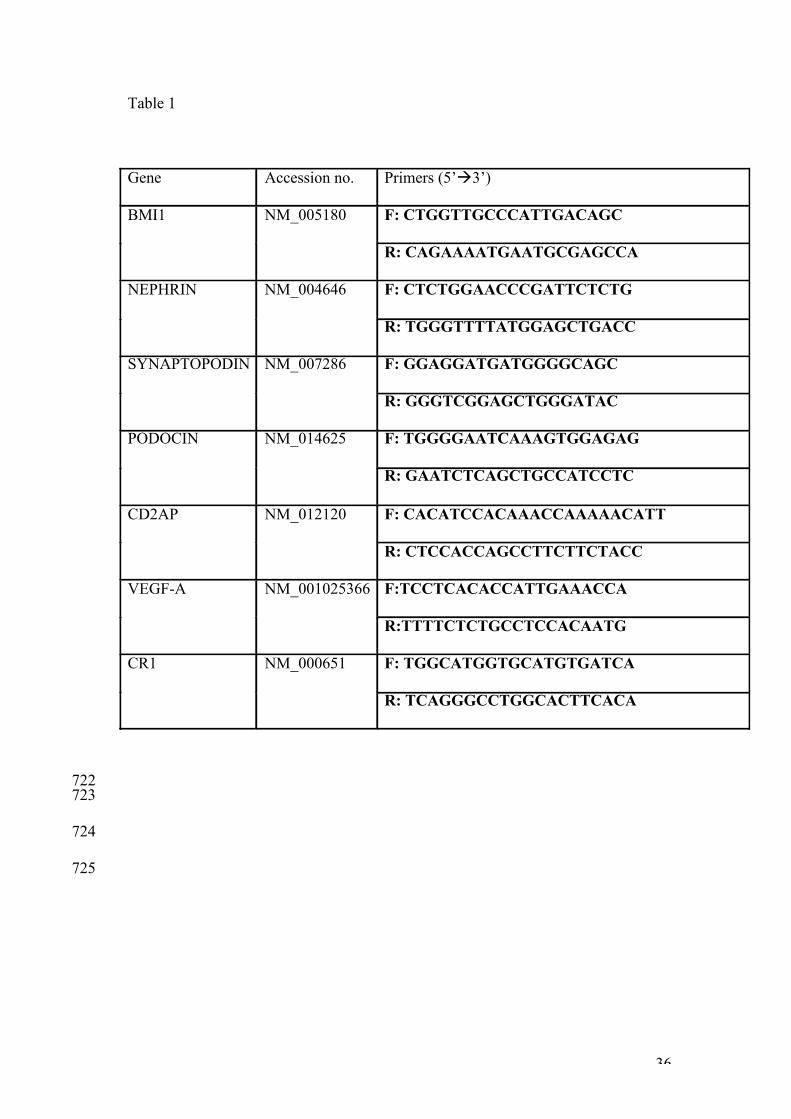
Quantitative real time RT-PCR (QRT-PCR). 207
Total RNA (n=8 mice per group) was extracted from the glomeruli using 208
TRIzol (Invitrogen) and cDNA synthesized using random primers and 1 µl of 209
200 U M-MLV Reverse Transcriptase in the presence of dNTPs (Promega 210
Corp.) (10 min, 75°C; 120 min, 42°C and 10 min, 75°C). QRT-PCR was 211
performed with the ABI Step-One Plus Sequence Detector (Applied 212
Biosystems). We used primers amplifying sequences of the b-actin gene 213
(accession number: NM_001101) present in humans but not in mice to 214
determine the amount of human cellular cDNA in samples (primer specificity 215
confirmed by absence of amplification of mouse cDNA), and primers common 216
to both human and mouse to determine the total cDNA in each sample, as 217
previously described [12]. For both sets, the absence of dimer formation was 218
confirmed using Dissociation Curves 1.0 software (Applied Biosystems). 219
Human:mouse chimerism was estimated as a ratio. Serial dilution of human 220
cDNA in mouse cells formed the calibration curves. The primer sequences are 221
shown in Table 1. 222
223
Histology. 224
10
Fresh kidneys (n=6 mice) were fixed in Bouin’s fixative (Sigma) for 4 hours, 225
dehydrated using serial dilutions of ethanol, embedded in wax, sectioned and 226
stained with Picrosirius red stain. Five random non-overlapping fields were 227
assessed at X200 magnification by a blinded observer, under polarised or 228
white light with an Olympus BX51 microscope. 229
230
Immunohistology. 231
Fresh kidneys were removed and fixed in a solution containing 1% PFA, 232
0.075M L-Lysine, 0.01M Sodium Periodate and 0.037M Phosphate buffer (all 233
reagents from Sigma) for 4 hours at 4°C. Tissues were then stored overnight 234
into 7% sucrose in PBS (Sigma) at 4°C, immersed in OCT compound (VWR) 235
and snap frozen before being stored at -80°C for further analysis. For 236
immunofluorescence, 4 μm sections were air dried (2 hours), fixed in acetone 237
(10 min, 4°C), air dried for a further 2 hours and denatured (1 hour, 4°C) using 238
a solution of 6M Urea and 0.1M Glycine in PBS (pH 3.5, all reagents from 239
Sigma). Slides were then washed with PBS and incubated overnight at 4°C 240
with a Col4a3 primary antibody diluted 1:2000 in 7% non-fat dry milk. The 241
Col4a3 antibody (kindly donated by Dr Dominic Cosgrove, Boys Town 242
National Research Hospital, Omaha, NE) is an affinity purified rabbit 243
polyclonal antibody raised against a peptide mapping the NC1 region of 244
collagen α3 Type IV. This antibody has been tested for cross-reactivity by the 245
provider and reacts with human and mouse [14]. The presence of donor cells 246
was visualised using a rabbit monoclonal antibody raised against human 247
vimentin (Abcam ab137867). Slides were then washed in PBS, incubated at 248
room temperature for 1 hour with a FITC-conjugated secondary antibody, 249
11
washed with PBS and mounted with Vectashield containing DAPI (Vector 250
Laboratories, Peterborough, UK) for visualisation using a confocal laser-251
scanning microscope Leica TCS SP5 (x1000 PL APO oil objective; Leica, 252
Wetzlar, Germany). 253
254
Immunohistochemistry. 255
Immunoperoxidase staining for the macrophage/monocyte T-helper cell 256
marker CD4 (BD Biosciences), the leucocyte marker CD45.2 (eBiosciences) 257
and the macrophge marker CD68 (Abcam) was performed on PLP-fixed 258
kidney cryostat sections (4μm). Sections were incubated overnight at 4°C with 259
primary antibody. For the biotin-conjugated CD45.2 antibody, endogenous 260
biotin and avidin were blocked prior to the addition of the primary antibody, 261
using biotin and avidin block solutions respectively (Vector Labs). Sections 262
were then washed with PBS, incubated with the appropriate HRP-conjugated 263
secondary antibody (Golden Bridge International), washed with PBS and 264
visualised using DAB. The sections were then counterstained with 265
hematoxylin, dehydrated and mounted using DPX (Sigma). 266
267
Blood urea analysis. 268
Blood samples were centrifuged at 1,300 g (10 min, 4°C) and the supernatant 269
was stored at -80°C until analysis. Urea was measured using a urea/ammonia 270
detection kit (R-Biopharm), according to the manufacturers instructions. All 271
samples were analysed at the same time to avoid batch variation. 272
273
Measurement of proteinuria/hematuria. 274
12
Urine was collected from 9 week-old mice and proteinuria was quantitatively 275
measured using mouse albumin and creatinine ELISA (albumin urinary level / 276
creatinine urinary level) (Exocell), according to the manufacturers instructions. 277
assessed for the presence of protein or erythrocytes by dipstick analysis 278
(Siemens Healthcare, Surrey, UK). Results were based on color change and 279
ranged from 0/trace to ++++, according to the manufacturer’s detection guide. 280
Controls included +/+ non-transplanted mice (negative control) and -/- non-281
transplanted mice (positive control). Analysis was performed by two 282
observers blinded as to whether each sample was from transplanted or non-283
transplanted -/- or +/+ groups. 284
285
Western Blotting. 286
Total protein was extracted using RIPAE buffer containing protease inhibitor 287
cocktail and PMSF (Sigma). Protein concentrations were determined using 288
the BCA assay (Thermo-Scientific) with BSA as standard. Proteins were run 289
on 8% SDS-PAGE, transferred to nitrocellulose membranes, blocked with milk 290
and incubated with primary antibodies for human-specific COLIVa3 (160-190 291
kDa), PODOCIN (42 kDa, Santa Cruz) and CD2AP (71 kDa, Millipore). 292
Membranes were incubated with secondary HRP-conjugated anti-goat IgG 293
(Santa Cruz) and proteins detected using enhanced chemiluminescence 294
(Thermo-Scientific). GAPDH was used as a loading control (Millipore). 295
296
Statistical analysis. 297
13
Data are expressed as mean±SEM (standard error) or median and range. 298
Parametric and non-parametric statistics were applied after testing 299
distributions on histograms. P < 0.05 was considered significant. 300
301
302
RESULTS 303
304
CSC cultured on human type IV collagen or co-cultured with glomeruli 305
express podocyte markers and migrate to glomeruli in vitro. 306
CSC were cultured for three weeks in growth medium (D10), either on non-307
coated plastic dishes, on plastic dishes coated with human collagen IV (CSC 308
on COLIV). Alternatively, CSC were co-cultured without cell contact, with 309
freshly-isolated COL4a3-/- and COL4a3+/+ glomeruli (CSC-glomeruli co-310
culture). After 3 weeks of culture, the expression level of various podocyte 311
markers was analysed by QRT-PCR (Figure 1A). The podocyte line 312
expressed NEP HRIN, a gene involved in renal filtration; PODOCIN, 313
expression of which is restricted to mature podocytes; VEGFA, produced 314
during kidney morphogenesis to guide endothelial cells towards glomeruli; 315
SYNAPTOPODIN, an actin-associated gene; CD2AP, which regulates the 316
translocation of dendrin to reorganize podocyte cytoskeleton and stabilize the 317
slit diaphragm; and CR1, complement receptor 1 which protects podocytes 318
from complement attack. The expression of these genes was almost 319
undetectable in CSC cultured alone in growth medium on non-coated dishes. 320
In contrast, the expression of all markers was upregulated when CSC were 321
cultured on dishes coated with human collagen IV. Expression of NEPHRIN, 322
14
SYNAPTOPODIN, and CR1 was further up-regulated when the cells were co-323
cultured with -/- glomeruli, although PODOCIN expression was lower. The 324
expression levels of VIMENTIN and FIBRONECTIN remained unchanged in 325
CSC cultured on Collagen Type IV or by co-culture with glomeruli (data not 326
shown). Confocal immunofluorescnce showed that CSC cultured for 3 weeks 327
on human type IV collagen or co-cultured with -/- glomeruli expressed 328
PODOCIN at a protein level (Figure 1B). These data indicate that CSC have 329
the potential to differentiate down the podocyte lineage. Next, we used a 330
chemotaxis assay where CSC were allowed to migrate for 1h towards freshly 331
isolated Col4a3-/- (-/-) or Col4a3+/+ (+/+) glomeruli ex vivo. CSC did not 332
passively migrate towards growth medium (GM) alone, but showed high 333
chemotaxis towards +/+ glomeruli and significantly greater chemotaxis 334
towards -/- glomeruli (9.5±1.5 MI vs. 21.1±2.6 MI respectively) (Figure 1C). 335
These results indicate that -/- glomeruli produce soluble factors that may 336
stimulate migration and differentiation of CSC, and rationalize the use of CSC 337
for the treatment of AS. 338
339
CSC transplanted into -/- mice engrafted into glomeruli, expressed 340
podocyte markers, and produce PODOCIN and the missing Col4a3 341
protein. 342
We injected 106 CSC intraperitoneally into 7 week old -/- and +/+ mice and 343
assessed the fate of donor cells 2 weeks later. CSC injection was performed 344
at 7 weeks postnatally because analysis of the disease history in -/- mice 345
revealed the presence of blood and protein in urine at this age, whereas the 346
levels of blood urea, which is a robust measure of renal function in mice, was 347
15
still comparable to the values found in +/+ mice. Donor cell engraftment in 348
isolated glomeruli was determined by QRT-PCR using human-specific and 349
non-specific primers for the housekeeping gene b-actin, as previously 350
described13. Results showed that transplanted CSC homed to glomeruli, with 351
10.8 fold higher engraftment in -/- (n=8) compared to +/+ glomeruli (n=8) 352
(Figure 2A). Using human-specific antibody agains vimentin protein, 353
histological analysis of kidney sections from transplanted and non-354
transplanted -/- mice revealed the presence of donor CSC in glomeruli, with 355
some donor cells being also visible outside the the glomeruli of CSC-356
transplanted -/- mice (Figure 2B). Engrafted CSC expressed the podocyte 357
markers CR1, VEGFA, SYNAPTOPODIN and CD2AP (Figure 2C). Human-358
specificity of the primers was verified using RNA from kidneys of -/- non-359
transplanted mice, which showed absence of amplification. At the protein 360
level, engrafted CSC produced PODOCIN, CD2AP and COL4A3, which are 361
absent in non-transplanted -/- and +/+ mice (Figure 2D and 2E). 362
363
Improvement of the -/- phenotype. 364
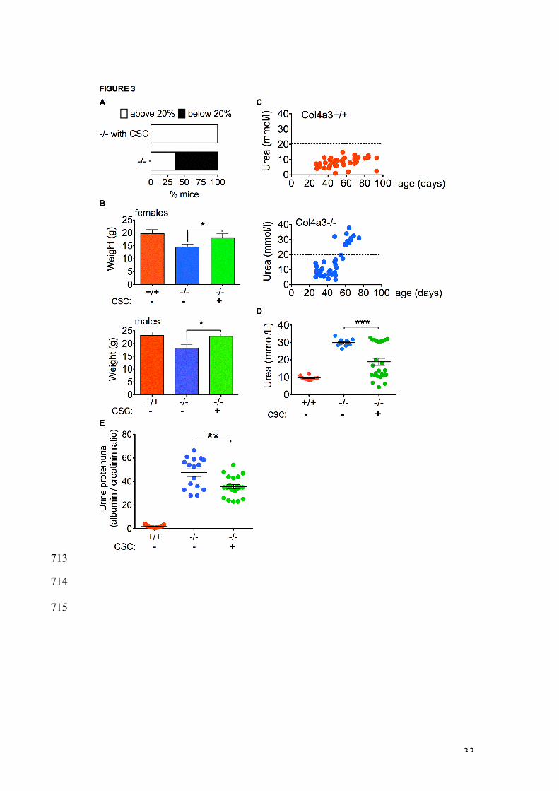
Non-transplanted -/- mice (n=25) progressively lost weight between 7 and 10 365
weeks. By 9 weeks of age, 63% mice dropped their weight below the 20% 366
endpoint level mandated by the British Home Office and had to be culled. In 367
contrast, all CSC-transplanted -/- mice (n=48) maintained their weight until 9 368
weeks (Figure 3A), at which age both transplanted -/- males and females 369
were heavier than age- and sex-matched non-transplanted -/- mice, with their 370
weight being similar to wild-type +/+ mice (Figure 3B). 371
16
Next, we measured blood urea in non-transplanted +/+ and -/- mice over 90 372
days. Levels remained under 20 mmol/l until day 90 in +/+ mice (upper panel 373
of Figure 3C, red dots, n=41), whilst all -/- mice showed elevated blood urea 374
levels by 58 days of age (lower panel of Figure 3C, blue dots, n=45). In 375
contrast, urea levels were lower in 9 week-old -/- mice transplanted with CSC 376
two weeks before than in non-transplanted -/- mice (18.8±2.0 mmol/L, n=25 377
vs. 29.9±0.7 mmol/L, n=10; mean±SEM, P<0.01), with 64% of transplanted -/- 378
mice showing levels similar to +/+ mice (Figure 3D). However, despite 379
transplantation of CSC being associated with lower blood urea levels, 380
proteinuria and hematuria, which were already elevated in -/- mice at the time 381
of cell therapy, remained unchanged following CSC-transplantation (data not 382
shown). Quantification of urine proteinuria (albumin / creatining level) revealed 383
a significant reduction in -/- mice transplanted with CSC compared to their 384
non-transplanted counterparts (35.6±1.8, n=21 vs. 47.5±3.2, n=16; 385
mean±SEM, P<0.01) (Figure 3E). 386
387
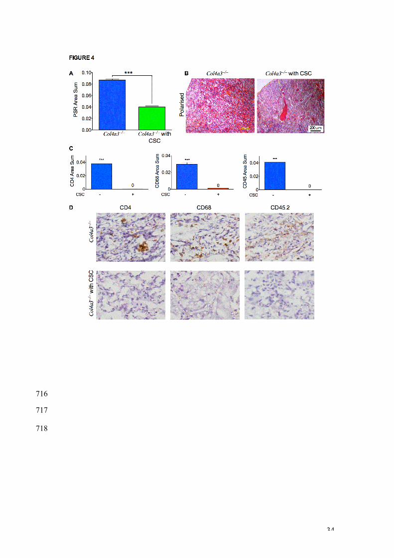
CSC transplantation lowered tubulointerstitial fibrosis and reversed 388
cortical inflammation. 389
We next measured tubulointerstitial fibrosis within the cortex using Picrosirius 390
red staining (PSR). PSR in kidneys from CSC-transplanted -/- mice was lower 391
than levels found in non-transplanted -/- mice (0.089±0.0018, n=6 vs. 392
0.040±0.0015, n=6, P<0.001), indicating a decrease in tubulointerstitial 393
fibrosis (Figure 4A and 4B). Quantification of renal inflammation showed that 394
CSC transplantation reduced the number of glomerular T-helper cells (anti-395
CD4), macrophages (anti-CD68), and haematopoietic cells (anti CD45.2) in -/- 396

17
mice indicating a marked decreased in cortical inflammation (Figure 4C and 397
4D). 398
399
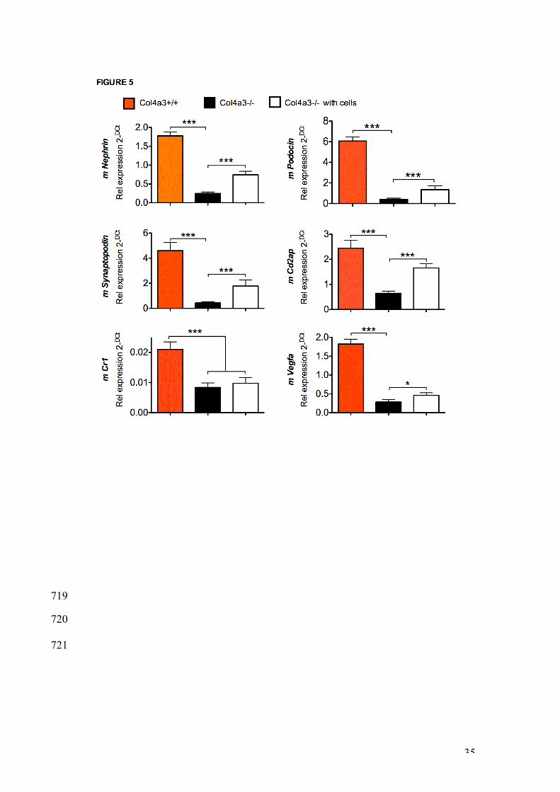
Col4a3 mutation down-regulated endogenous murine podocyte gene 400
expression, and CSC transplantation partially restored renal mRNA 401
expression levels. 402
We next assessed whether the presence of exogenous cells modified gene 403
expression of resident podocytes. Results showed that expression of murine 404
Nephrin, Podocin, Synaptopodin, Cd2ap, Cr1 and Vegfa was higher in the 405
glomeruli of non-transplanted +/+ mice compared to non-transplanted -/- mice 406
(Figure 5). However, CSC transplantation upregulated the renal mRNA 407
expression of Nephrin, Podocin, Synaptopodin, Cd2ap and Vgfa, suggesting 408
that the decrease in glomerular inflammation in transplanted mice was 409
associated with restored podocyte activity. 410
411
412
DISCUSSION 413
414
Several studies have suggested that renal pathology in models of AS can be 415
improved by cell therapy, although the mechanisms mediating these effects 416
remain elusive, with conflicting results possibly attributable to variations in 417
donor cell types. It is therefore essential to identify easily accessible sources 418
of stem cells with high therapeutic potential for the treatment of AS [14]. In this 419
study, we used human first trimester fetal chorionic stem cells (CSC), which 420
are isolated from chorionic villi sampling in ongoing pregnancies and can be 421
18
expanded to high numbers ex vivo, while maintaining tissue repair potential10. 422
For example, when transplanted into collagen type I-deficient mice, they 423
reduced fracture rate and increased bone plasticity; and accelerated skin 424
wound healing [10, 15]. 425
426
Col4a3–/– mice are a model of severe human Alport syndrome. At 7 week of 427
age, -/- Alport mice show high levels of proteinuria, but normal weight and 428
blood urea. Over the next two weeks, blood urea rapidly increases, whilst 429
body weight drops and mice show pronouced interstitial fibrosis and 430
macrophage infiltration. We investigated the capacity of CSC to prevent 431
glomerulopathy in 129Sv-Col4a3-/- mice. We show that 9 week-old -/- mice 432
transplanted with CSC two weeks before have significantly lower blood urea 433
and urine proteinuria, compared to their non-transplanted counterparts. 434
Although animal welfare restrictions prevented us from studying the clinical 435
endpoint of survival, all transplanted -/- mice maintained their weight until 9 436
week of age. This is important because improvement in renal histology is not 437
necessarily associated with delay in death from renal failure [16]. However, 438
the genetic backgroud of the Alport mice has a strong effect on the rate of 439
disease progression [17]. Contrary to 129Sv-Col4a3tm1Dec/J (-/-) mice, which 440
progressively lose weight and do not survive beyond 10 weeks of age, the 441
survival time of homozygous mutant mice is extended to about 14 weeks of 442
age in mice maintained on a mixed genetic background or to 25-30 weeks on 443
the C57BL/6j background. Consequently, we suggest that the elevated blood 444
urea and 20% weight loss we report may be considered as surrogates for 445
end-stage renal failure. We found that proteinuria, which was already elevated 446
19
at the time of cell injection, did not return to normal levels but did not increase 447
further in transplanted -/- mice, whilst it increased in non-transplanted -/- mice 448
. This was not surprising, since structural damage to the GBM would be 449
unlikely to be reversed within 2 weeks. 450
451
Increasing evidence from stem cell transplantation in acquired injury models 452
points to the well characterised anti-inflammatory actions of exogenous stem 453
cells making a major contribution to therapeutic results. For example, murine 454
amniotic fluid cells transplanted into Col4a5-/- mice before the onset of 455
proteinuria have been reported to modify the course of renal fibrosis, despite 456
donor cells failing to differentiate into podocytes or produce collagen IVa54. 457
Here, we found reduced renal fibrosis and cortical inflammation in 458
transplanted mice, which may reflect the anti-inflammatory effect of donor 459
cells or the replacement of defective renal cells. We also found that CSC 460
migrated to the glomeruli, where they persisted over 2 weeks and expressed 461
CR1, VEGFA, SYNAPTOPODIN, CD2AP and PODOCIN at the gene level, 462
and produced PODOCIN, CD2AP and COLIVα3 proteins. which are missing 463
in non-transplanted -/- mice. These data indicate that transplanted CSC have 464
adopted a podocyte phenotype. However, focal staining for Col4a3 in the 465
kidneys does not prove true GBM deposition, and it will be necessary to 466
demonstrate assembly of the correct collagen type IV in the GBM to 467
investigate whether the Col4a3 produced by CSC co-assemble with Col4a4 468
and Col4a5 to improve GBM structure [18]. Similarly to our findings, LeBleu et 469
al. found that wild-type bone marrow-derived cells transplanted into Col4α3 -/- 470
mice improved renal histology and function, with donor cells differentiating into 471
20
VEGF and collagen IV-expressing podocytes [9]; and a recent study by Lin et 472
al. demonstrates that secretion of α3α4α5(IV) heterotrimers is sufficient to 473
slow disease progression by partially restoring the defective collagen network 474
[19]. 475
476
Interestingly, we also found that CSC transplantation stimulated resident 477
podocyte activity, suggesting that the production of Col4a3 from donor cells 478
act as a feed-back to modulate podocyte activity, possibly by releasing trophic 479
factors that promote the differentiation and regeneration of endogenous 480
podocyte progenitors to differentiate into mature podocytes. Although 481
endogenous podocytes remain unable to express the correct form of Collagen 482
type IV, stimulation of podocyte progenitors differentiation into podocytes by 483
donor stem cells may contribute to the amelioration of filtration function [20]. 484
Although it is often assumed that the presence of exogenous cells at the site 485
of injury and their differentiation into target cell phenotypes accounts for the 486
therapeutic effects observed, there is still a lack of evidence for the causality 487
between the two. An increasing number of studies challenge the concept of 488
donor cells acting as a building blocks to replace damaged endogenous cells 489
and data suggest that beyond their potential as a source of cell replacement, 490
stem cells also mediate paracrine treatment. In addition, data suggest that 491
donor cells influence the complex cross-talk between resident cells and 492
extracellular matrix. It is possible that exogenous stem cells reprogramme 493
resident macrophages from an anti-inflammatory to a pro-inflammatory 494
phenotype, as is the case in sepsis [21]. This mechanism might account for 495
the therapeutic effects of wild type bone marrow that we previously reported7. 496
21
For example, blockade of tumour necrosis factor-alpha (TNF-a), a pro-497
inflammatory cytokine, has been shown to ameliorate glomerulosclerosis and 498
proteinuria in AS mice [22]. 499
We believe that CSC may have strong potential for the treatment of 500
glomerulopathies and further studies are indicated to establish the precise 501
mechanism of action of these cells in treatment of AS. 502
503
504
ACKNOWLEDGEMENT: 505
This research was funded by Kidney Research UK, Genzyme Renal 506
Innovations Programme and by the National Institute for Health Research 507
Biomedical Research Centre at Great Ormond Street Hospital for Children 508
NHS Foundation Trust and University College London. ALD is funded by 509
Department of Health through NIHR UCL/UCLH Biomedical Research Centre. 510
We acknowledge support from NIHR Imperial Biomedical Research Centre. 511
512
513
AUTHOR DISCLOSURE STATEMENT: 514
No competing financial interests exist. 515
516
AUTHORS CONTRIBUTION: 517
DM, MC, KLH, VE, PES, JB: Collection and/or assembly of data, data 518
analysis and interpretation; GBG, CDP, ALD, PDC, HTC: Conception and 519
design, financial support, manucript editing; ALD, PDC, NMF: Provision of 520
study material; NMF: data analysis and interpretation, manuscript writing; 521
22
PVG: Conception and design, data analysis and interpretation, financial 522
support, manuscript writing. 523
524
23
REFERENCES 525
526
1. Abrahamson DR, Prettyman AC, Robert B and St John PL. (2003). 527
Laminin-1 reexpression in Alport mouse glomerular basement 528
membranes. Kidney Int 63:826-834. 529
2. Gross O, Perin L and Deltas C. (2014). Alport syndrome from bench to 530
bedside: the potential of current treatment beyond RAAS blockade and 531
the horizon of future therapies. Nephrol Dial Transplant 29:iv124-iv130. 532
3. Guillot PV, Cook TH, Pusey CD, Fisk NM, Harten S, Moss J, Shore I, 533
and Bou-Gharios G. (2008). Transplantation of human fetal 534
mesenchymal stem cells improves glomerulopathy in a collagen type I 535
alpha 2-deficient mouse. J Pathology 214(5):627-636. 536
4. Sedrakyan S, Da Sacco S, Milanesi , Shiri L, Petrosyan A, Varimezova 537
R, Warburton D, Lemley KV, De Filippo RE and Perin L. (2012). 538
Injection of amniotic fluid stem cells delays progression of renal 539
fibrosis. J Am Soc Nephrol 23:661-673. 540
5. Miner JH and Sanes JR. (1996). Molecular and functional defects in 541
kidneys of mice lacking collagen alpha 3(IV): implications for Alport 542
syndrome. J Cell Biol 135:1403-1413. 543
6. Andrews KL, Mudd JL, Li C and Miner JH. (2002). Quantitative trait loci 544
influence renal disease progression in a mouse model of Alport 545
syndrome. Am J Pathol 160:721-730. 546
7. Prodromidi EI, Poulsom R, Jeffery R, Roufosse CA, Pollard PJ, Pusey 547
CD and Cook HT. (2006). Bone marrow derived cells contribute to 548
24
podocyte regeneration and amelioration of renal disease in a mouse 549
model of Alport Syndrome. Stem Cells 24:2448-2455. 550
8. Sugimoto H, Mundel TM, Sund M, Xie L, Cosgrove D and Kalluri R. 551
(2006). Bone-marrow-derived stem cells repair basement membrane 552
collagen defects and reverse genetic kidney disease. Proc Natl Acad 553
Sci (USA) 103:7321-7326. 554
9. LeBleu V, Sugimoto H, Mundel TM, Gerami-Naini B, Finan E, Miller 555
CA, Gattone VH 2nd, Shield CF 3rd, Folkman J and Kalluri R. (2009). 556
Stem cell therapies benefit Alport syndrome. J Am Soc Nephrol 557
20:2359-2370. 558
10. Jones GN, Moschidou D, Puga-Iglesias TI, Kuleszewicz K, Vanleene 559
M, Shefelbine SJ, Bou-Gharios G, Fisk NM, David AL, De Coppi P and 560
Guillot PV. (2012). Ontological differences in first compared to third 561
trimester human fetal placental chorionic stem cells. PLoS One 562
7:e43395. 563
11. Saleem MA, O’Hare MJ, Reiser J, Coward RJ, Inward CD, Farren T, 564
Xing CY, Ni L, Mathieson PW and Mundel P. (2002). A conditionally 565
immortalized human podocyte cell line demonstrating nephrin and 566
podocin expression. J Am Soc Nephrol 13:630–638. 567
12. Guillot PV, Abass O, Bassett JH, Shefelbine SJ, Bou-Gharios G, Chan 568
J, Kurata H, Williams GR, Polak J and Fisk NM. (2008). Intrauterine 569
transplantation of human fetal mesenchymal stem cells reduces bone 570
pathology in osteogenesis imperfecta mice. Blood 111:1717-1725. 571
25
13. Miner, JH and JR Sanes. (1994). Collagen IV a3, a4, and a5 chains in 572
rodent basal lamina: Sequence, distribution, association with laminins, 573
and developmental switches. J. Cell Biol. 127: 879-891. 574
14. Wong C and Rogers I. (2009). Cell therapy for Alport sydrome. J Am 575
Soc Nephrol 20:2279-2281. 576
15. Jones GN, Moschidou D, Abdulrazzak H, Kalirai BS, Vanleene M, 577
Osatis S, Shefelbine, SJ, Horwood NJ, Marenzana M, De Coppi P, 578
Bassett JH, Williams GR, Fisk NM and Guillot PV. (2014). Potential of 579
human fetal chorionic stem cells for the threatment of osteogenesis 580
imperfecta. Stem Cells Dev 23:262-276. 581
16. Ninichuk V, Gross O, Segerer S, Hoffmann R, Radomska E, 582
Buchstaller A, Huss R, Akis N, Schlondorff D and Anders HJ. (2006). 583
Multipotent mesenchymal stem cells reduce interstitial fibrosis but do 584
not delay progression of chronic kidney disease in collagen4A3-585
deficient mice. Kidney Int 70:121-129. 586
17. Cosgrove D, Meehan DT, Grunkemeyer JA, Kornak JM, Sayers R, 587
Hunter WJ and Samuelson GC. (1996). Collagen COL4A3 knockout: a 588
mouse model for autosomal Alport syndrome. Genes Dev 10:2981-589
2992. 590
18. Gross O, Borza DB, Anders HJ, Licht C, Weber M, Segerer S, Torra R, 591
Gubler MC, Heidet L, Harvey S, Cosgrove D, Lees G, Kashtan C, 592
Gregory M, Savige J, Ding J, Thorner P, Abrahamson DR, Antignac C, 593
Tryggvason K, Hudson B, Miner JH. (2009). Stem cell therapy for 594
Alport syndrome: the hope beyond the hype. Nephrol Dial Transplant 595
24:731-734. 596
26
19. Lin X, Suh JH, Go G and Miner JF. (2014). Feasibility of repairing 597
glomerular basement membrane defects in Alport syndrome. J Am Soc 598
Nephrol 25:687-692. 599
20. Lasagni L, Angelotti ML, Ronconi E, Lombardi D, Nardi S, Peired A, 600
Becherucci F, Mazzinghi B, Sisti A, Romoli S, Burger A, Schaefer B, 601
Buccoliero A, Lazzeri E and Romagnani P. (2015). Podocyte 602
regeneration driven by renal progenitors determines glomerular 603
disease remission and can be pharmacologically enhanced. Stem Cell 604
Reports 5:248-263. 605
21. Tyndall A and Pistoia V. (2009). Mesenchymal stem cells combat 606
sepsis. Nat Med 15:109-118. 607
22. Ryu M, Mulay SR, Miosge N, Gross O and Anders HJ. (2012). Tumour 608
necrosis factor-α drives Alport glomerulosclerosis in mice by promoting 609
podocyte apoptosis. J Pathol 226(1):120-131. 610
611
612
613
614
615
616
617
618
619
620
621
27
FIGURE LEGENDS 622
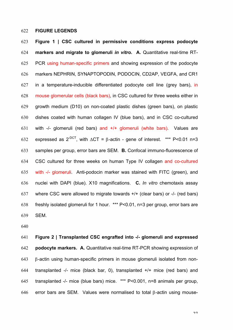
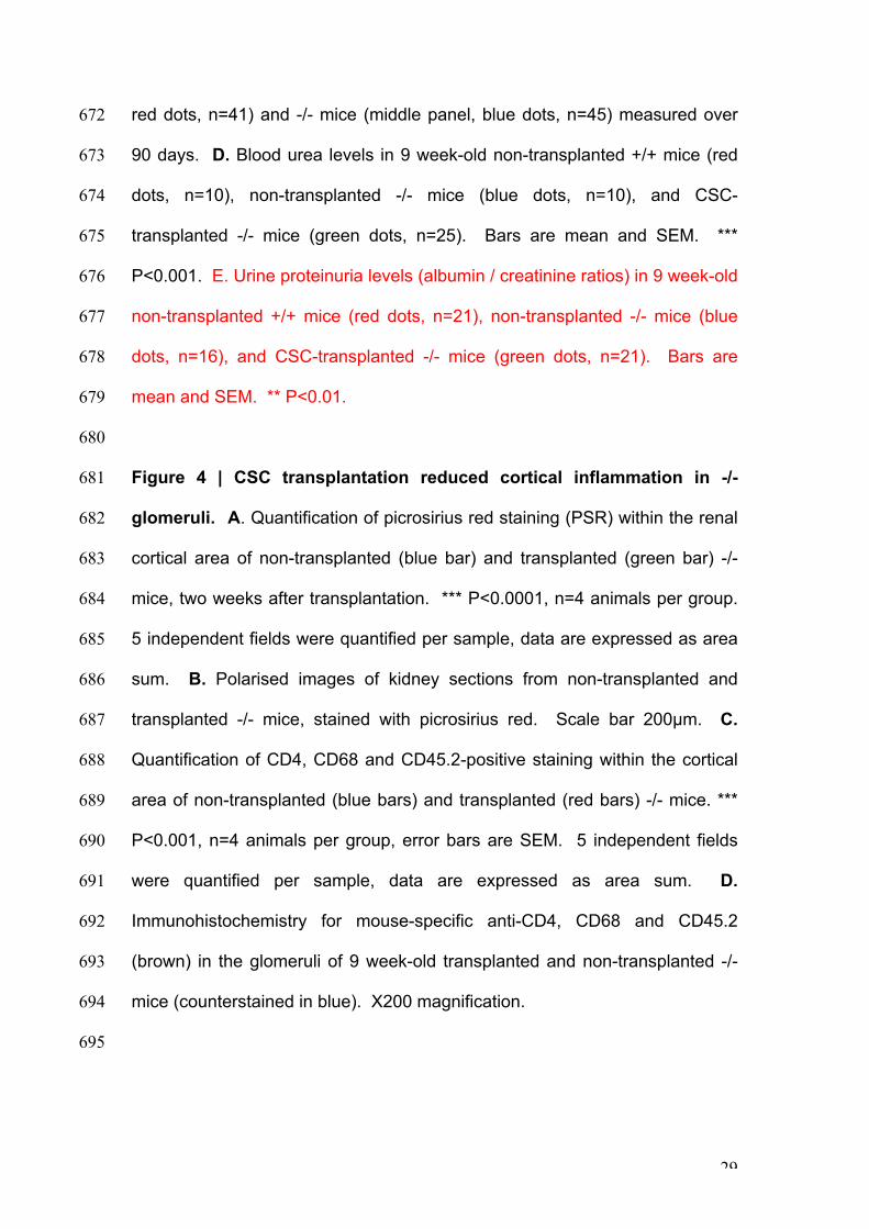
Figure 1 | CSC cultured in permissive conditions express podocyte 623
markers and migrate to glomeruli in vitro. A. Quantitative real-time RT-624
PCR using human-specific primers and showing expression of the podocyte 625
markers NEPHRIN, SYNAPTOPODIN, PODOCIN, CD2AP, VEGFA, and CR1 626
in a temperature-inducible differentiated podocyte cell line (grey bars), in 627
mouse glomerular cells (black bars), in CSC cultured for three weeks either in 628
growth medium (D10) on non-coated plastic dishes (green bars), on plastic 629
dishes coated with human collagen IV (blue bars), and in CSC co-cultured 630
with -/- glomeruli (red bars) and +/+ glomeruli (white bars). Values are 631
expressed as 2-DCT, with DCT = b-actin - gene of interest. *** P<0.01 n=3 632
samples per group, error bars are SEM. B. Confocal immuno-fluorescence of 633
CSC cultured for three weeks on human Type IV collagen and co-cultured 634
with -/- glomeruli. Anti-podocin marker was stained with FITC (green), and 635
nuclei with DAPI (blue). X10 magnifications. C. In vitro chemotaxis assay 636
where CSC were allowed to migrate towards +/+ (clear bars) or -/- (red bars) 637
freshly isolated glomeruli for 1 hour. *** P<0.01, n=3 per group, error bars are 638
SEM. 639
640
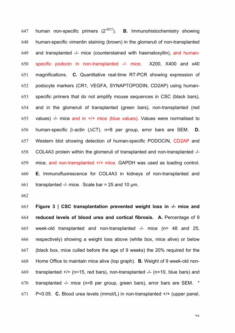
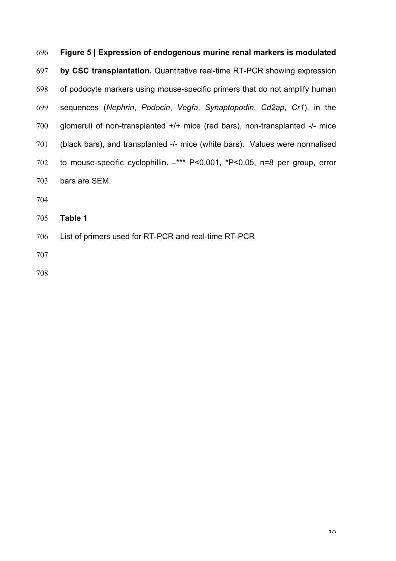
Figure 2 | Transplanted CSC engrafted into -/- glomeruli and expressed 641
podocyte markers. A. Quantitative real-time RT-PCR showing expression of 642
b-actin using human-specific primers in mouse glomeruli isolated from non-643
transplanted -/- mice (black bar, 0), transplanted +/+ mice (red bars) and 644
transplanted -/- mice (blue bars) mice. *** P<0.001, n=8 animals per group, 645
error bars are SEM. Values were normalised to total b-actin using mouse-646
28
human non-specific primers (2-DCT). B. Immunohistochemistry showing 647
human-specific vimentin staining (brown) in the glomeruli of non-transplanted 648
and transplanted -/- mice (counterstained with haematoxyllin), and human-649
specific podocin in non-transplanted -/- mice. X200, X400 and x40 650
magnifications. C. Quantitative real-time RT-PCR showing expression of 651
podocyte markers (CR1, VEGFA, SYNAPTOPODIN, CD2AP) using human-652
specific primers that do not amplify mouse sequences in CSC (black bars), 653
and in the glomeruli of transplanted (green bars), non-transplanted (red 654
values) -/- mice and in +/+ mice (blue values). Values were normalised to 655
human-specific b-actin (DCT). n=8 per group, error bars are SEM. D. 656
Western blot showing detection of human-specific PODOCIN, CD2AP and 657
COL4A3 protein within the glomeruli of transplanted and non-transplanted -/- 658
mice, and non-transplanted +/+ mice. GAPDH was used as loading control. 659
E. Immunofluorescence for COL4A3 in kidneys of non-transplanted and 660
transplanted -/- mice. Scale bar = 25 and 10 µm. 661
662
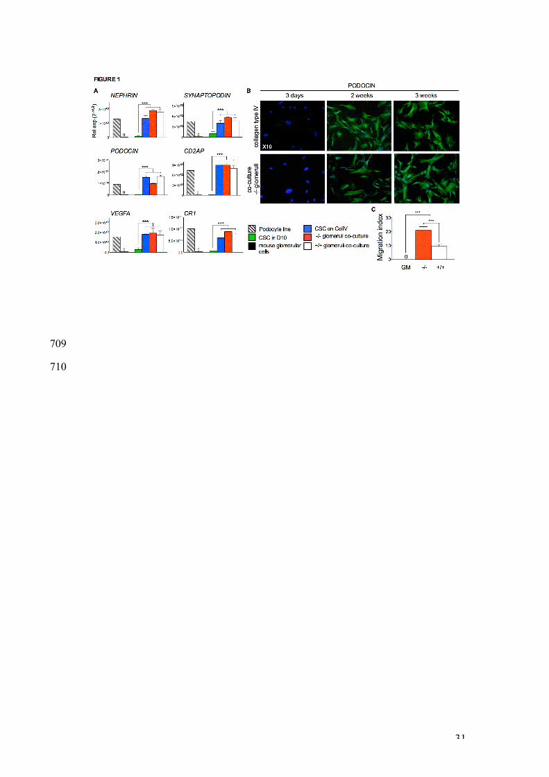
Figure 3 | CSC transplantation prevented weight loss in -/- mice and 663
reduced levels of blood urea and cortical fibrosis. A. Percentage of 9 664
week-old transplanted and non-transplanted -/- mice (n= 48 and 25, 665
respectively) showing a weight loss above (white box, mice alive) or below 666
(black box, mice culled before the age of 9 weeks) the 20% required for the 667
Home Office to maintain mice alive (top graph). B. Weight of 9 week-old non-668
transplanted +/+ (n=15, red bars), non-transplanted -/- (n=10, blue bars) and 669
transplanted -/- mice (n=8 per group, green bars), error bars are SEM. * 670
P<0.05. C. Blood urea levels (mmol/L) in non-transplanted +/+ (upper panel, 671
29
red dots, n=41) and -/- mice (middle panel, blue dots, n=45) measured over 672
90 days. D. Blood urea levels in 9 week-old non-transplanted +/+ mice (red 673
dots, n=10), non-transplanted -/- mice (blue dots, n=10), and CSC-674
transplanted -/- mice (green dots, n=25). Bars are mean and SEM. *** 675
P<0.001. E. Urine proteinuria levels (albumin / creatinine ratios) in 9 week-old 676
non-transplanted +/+ mice (red dots, n=21), non-transplanted -/- mice (blue 677
dots, n=16), and CSC-transplanted -/- mice (green dots, n=21). Bars are 678
mean and SEM. ** P<0.01. 679
680
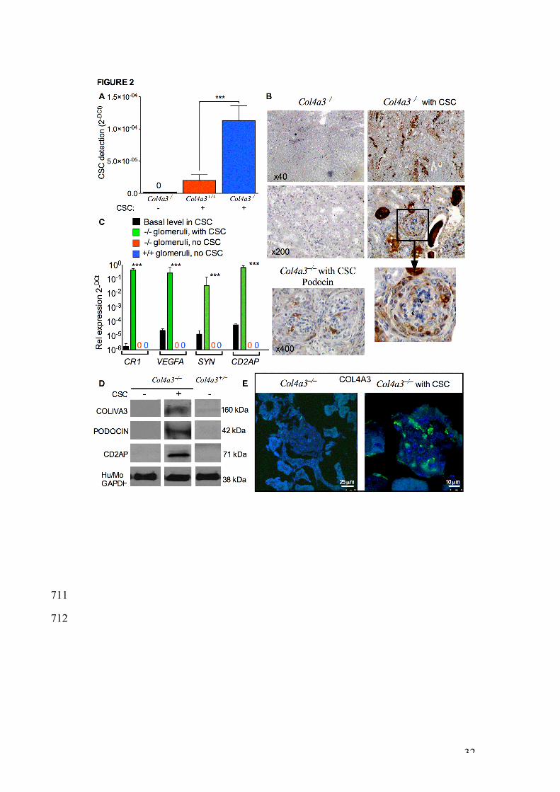
Figure 4 | CSC transplantation reduced cortical inflammation in -/- 681
glomeruli. A. Quantification of picrosirius red staining (PSR) within the renal 682
cortical area of non-transplanted (blue bar) and transplanted (green bar) -/- 683
mice, two weeks after transplantation. *** P<0.0001, n=4 animals per group. 684
5 independent fields were quantified per sample, data are expressed as area 685
sum. B. Polarised images of kidney sections from non-transplanted and 686
transplanted -/- mice, stained with picrosirius red. Scale bar 200μm. C. 687
Quantification of CD4, CD68 and CD45.2-positive staining within the cortical 688
area of non-transplanted (blue bars) and transplanted (red bars) -/- mice. *** 689
P<0.001, n=4 animals per group, error bars are SEM. 5 independent fields 690
were quantified per sample, data are expressed as area sum. D. 691
Immunohistochemistry for mouse-specific anti-CD4, CD68 and CD45.2 692
(brown) in the glomeruli of 9 week-old transplanted and non-transplanted -/- 693
mice (counterstained in blue). X200 magnification. 694
695
30
Figure 5 | Expression of endogenous murine renal markers is modulated 696
by CSC transplantation. Quantitative real-time RT-PCR showing expression 697
of podocyte markers using mouse-specific primers that do not amplify human 698
sequences (Nephrin, Podocin, Vegfa, Synaptopodin, Cd2ap, Cr1), in the 699
glomeruli of non-transplanted +/+ mice (red bars), non-transplanted -/- mice 700
(black bars), and transplanted -/- mice (white bars). Values were normalised 701
to mouse-specific cyclophillin. *** P<0.001, *P<0.05, n=8 per group, error 702
bars are SEM. 703
704
Table 1 705
List of primers used for RT-PCR and real-time RT-PCR 706
707
708
31
709
710
32
711
712
33
713
714
715
34
716
717
718
35
719
720
721
36
Table 1
Gene Accession no. Primers (5’à3’)
BMI1 NM_005180 F: CTGGTTGCCCATTGACAGC
R: CAGAAAATGAATGCGAGCCA
NEPHRIN NM_004646 F: CTCTGGAACCCGATTCTCTG
R: TGGGTTTTATGGAGCTGACC
SYNAPTOPODIN NM_007286 F: GGAGGATGATGGGGCAGC
R: GGGTCGGAGCTGGGATAC
PODOCIN NM_014625 F: TGGGGAATCAAAGTGGAGAG
R: GAATCTCAGCTGCCATCCTC
CD2AP NM_012120 F: CACATCCACAAACCAAAAACATT
R: CTCCACCAGCCTTCTTCTACC
VEGF-A NM_001025366 F:TCCTCACACCATTGAAACCA
R:TTTTCTCTGCCTCCACAATG
CR1 NM_000651 F: TGGCATGGTGCATGTGATCA
R: TCAGGGCCTGGCACTTCACA
722 723
724
725