Chapter 5 Hydrodynamics of Sniffing by Crustaceans
18
Chapter 5 Hydrodynamics of Sniffing by Crustaceans Mimi A.R. Koehl Abstract Chemical signals are dispersed in aquatic environments by turbulent water currents. The first step in smelling these signals is the capture of odor molecules from the water around an organism. Olfactory antennules of crustaceans are used to study the physical process of odor capture because they are external organs protruding into the water where researchers can measure how they interact with their fluid environment. The antennules of lobsters, crabs, and stomatopods, which bear chemosensory hairs (“aesthetascs”), flick through the water. For any array of small hairs, there is a critical velocity range above which the array is “leaky” and fluid can flow between the hairs, and below which fluid barely moves through the spaces between the hairs. When antennules flick they move faster than the critical velocity and water flows into the spaces between aesthetascs. In contrast, during the return stroke the antennule moves more slowly than the critical velocity and the water sampled during the flick is trapped between the aesthetascs until the next flick. Odorant molecules in the water trapped between the aesthetascs during the return stroke and interflick pause diffuse to the surfaces of the aesthetascs, before the next flick traps a new parcel of water. Therefore, each antennule flick is a “sniff,” taking a discrete sample of the odor plume in space and time. 5.1 Introduction Many animals communicate via chemical signals (odors) released into the surrounding fluid (water or air). The first step in smelling chemical signals is the capture of odor molecules from the fluid around an organism. Therefore, to under- stand how organisms capture odors, scientists need to investigate how olfactory organs interact with the water or air around them. The olfactory antennules of M.A.R. Koehl (*) Department of Integrative Biology, University of California at Berkeley, Berkeley, CA 94720-3140, USA e-mail: [email protected] T. Breithaupt and M. Thiel (eds.), Chemical Communication in Crustaceans, DOI 10.1007/978-0-387-77101-4_5, # Springer Science+Business Media, LLC 2011 85
Transcript of Chapter 5 Hydrodynamics of Sniffing by Crustaceans

Chapter 5Hydrodynamics of Sniffing by Crustaceans
Mimi A.R. Koehl
Abstract Chemical signals are dispersed in aquatic environments by turbulentwater currents. The first step in smelling these signals is the capture of odormolecules from the water around an organism. Olfactory antennules of crustaceansare used to study the physical process of odor capture because they are externalorgans protruding into the water where researchers can measure how they interactwith their fluid environment. The antennules of lobsters, crabs, and stomatopods,which bear chemosensory hairs (“aesthetascs”), flick through the water. For anyarray of small hairs, there is a critical velocity range above which the array is“leaky” and fluid can flow between the hairs, and below which fluid barely movesthrough the spaces between the hairs. When antennules flick they move faster thanthe critical velocity and water flows into the spaces between aesthetascs. In contrast,during the return stroke the antennule moves more slowly than the critical velocityand the water sampled during the flick is trapped between the aesthetascs until thenext flick. Odorant molecules in the water trapped between the aesthetascs duringthe return stroke and interflick pause diffuse to the surfaces of the aesthetascs,before the next flick traps a new parcel of water. Therefore, each antennule flick is a“sniff,” taking a discrete sample of the odor plume in space and time.
5.1 Introduction
Many animals communicate via chemical signals (odors) released into thesurrounding fluid (water or air). The first step in smelling chemical signals is thecapture of odor molecules from the fluid around an organism. Therefore, to under-stand how organisms capture odors, scientists need to investigate how olfactoryorgans interact with the water or air around them. The olfactory antennules of
M.A.R. Koehl (*)Department of Integrative Biology, University of California at Berkeley,Berkeley, CA 94720-3140, USAe-mail: [email protected]
T. Breithaupt and M. Thiel (eds.), Chemical Communication in Crustaceans,DOI 10.1007/978-0-387-77101-4_5, # Springer Science+Business Media, LLC 2011
85

crustaceans provide useful systems for studying the physical process of odor capturebecause they are external organs protruding into the water where researchers can seehow they interact with their fluid environment.
The olfactory organs of malacostracan crustaceans (e.g., lobsters, shrimp, crabs,stomatopods) are the lateral branches (called “lateral filaments”) of the antennules,which bear chemosensory hairs (called “aesthetascs”) (Fig. 5.1a, c, e) (reviewed by
Fig. 5.1 Examples of arthropods with hair-bearing olfactory appendages. The boxes aroundantennules in (a, c, and e) indicate the region of the antennule diagrammed in (b, d, and f ),respectively. (a) Spiny lobster, Panulirus argus. (b) Magnified view of a section of the lateralfilament of a P. argus antennule. The lateral filament flicks downward, with the aesthetascs at anangle of ~35! to the direction of motion (Gleeson et al. 1993). (c) Stomatopod (“mantis shrimp”),Squilla empusa. (d) Magnified view of a section of the aesthetasc-bearing filament of a stomatopodantennule, Gonodactylus mutatus. The antennule flicks laterally, with the aesthetascs perpendicularto the direction of movement (Mead et al. 1999). (e) Blue crab, Callinectes sapidus. (f ) Magnifiedview of the tip of the antennule of a C. sapidus. The antennule can flick in many directions, but theaesthetascs point in the direction of motion during a flick (Martinez, Lee, and Koehl, unpublisheddata). (g) Head of a male silkworm moth, Bombyx mori, showing the olfactory antennae. When themale fans his wings, air moves from front to back across the antennae (Loudon and Koehl 2000)(figure reprinted from Koehl 2001a, with kind permission of Springer Science+Business Media)
86 M.A.R. Koehl

Ache 1982; Koehl 2006) (see Hallberg and Skog, Chap. 6). Although nonaesthetascchemosensory hairs on antennules or legs of some species can also be involved inolfaction (reviewed in Koehl 2006), I will focus here on the aesthetasc-bearinglateral filaments of the antennules to explain the physics of odor capture. Odormolecules in the water around an animal must reach the surfaces of those aesthe-tascs to be sensed, so understanding the fluid mechanics of arrays of hairs is criticalto deciphering how these crustaceans catch chemical signals.
The diversity of antennulemorphology and deployment is intriguing. For example,the arrangement of aesthetascs on the lateral filaments differs between species,ranging from the complex arrays on lobster antennules (Fig. 5.1b) to the simplerows on stomatopod antennules (Fig. 5.1d) and the dense brushes on crab antennules(Fig. 5.1f). Do these differences in morphology affect odor capture? Further-more, many malacostracans flick the lateral filaments of their antennules throughthe surrounding water (Fig. 5.2). How does flicking affect water motion around theaesthetascs, and thus odor capture?
My interest in the hydrodynamics of molecule capture by flicking crustaceanantennules grew from our studies of the hydrodynamics of hair-bearing suspension-feeding appendages and the physical mechanisms they use to capture food particlesfrom the surrounding water (reviewed in Koehl 1995). Those studies, whichrevealed how difficult it is to get water to flow between tiny hairs, sparked mycuriosity about how the chemosensory hairs on insect antennae (Fig. 5.1g) (Loudonand Koehl 2000) and crustacean antennules (Fig. 5.1b, d, f) (Koehl 2001a) cancapture molecules from the surrounding fluid. The basic physical rules we discov-ered about how arrays of hairs interact with fluids (Cheer and Koehl 1987a; Koehl1992) predicted that the size and spacing of aesthetascs as well as the velocity ofantennule flicking should make a big difference to the flow of odor-bearing waterinto arrays of these chemosensory hairs (Koehl 1996).
Another research path also led me to crustacean antennules. Years of fieldresearch in coastal marine habitats made me realize the importance of understand-ing the physical environment of an organism on the spatial and temporal scales
Fig. 5.2 Diagram of the spiny lobster, Panulirus argus, flicking the aesthetasc-bearing lateralfilaments of its olfactory antennules. Drawing by Jorge A. Varela Ramos
5 Hydrodynamics of Sniffing by Crustaceans 87

experienced by that organism (which are not necessarily the scales at which wehumans experience the environment). For example, the hydrodynamic forces thatcan rip a sea anemone off a wave-swept shore depend on the instantaneous watervelocities and accelerations it encounters just a few centimeters above the sub-stratum as it sits among its neighbors, not on the much faster freestream waterflow across the habitat (Koehl 1977). The waterborne chemical signals thatbenthic crustaceans encounter in their natural habitats are dispersed from odorsources by messy turbulent water currents. Early models of how animals searchfor the source of a chemical signal assumed that such turbulent odor plumes arediffuse clouds that become wider and more dilute with distance from the source(reviewed in Koehl 2006). My field experience studying flow microhabitats,however, led me to ask what patterns of odor concentration are actually inter-cepted by crustaceans navigating in marine habitats. To answer this question Iwould have to figure out what the instantaneous odor concentrations are in thesmall slices of water sampled by the olfactory antennules as they flick in naturalenvironments.
5.2 Physical Mechanisms of Odor Capture
What are the physical mechanisms that olfactory antennules use to capture chemicalsignals from the surrounding water? Odor molecules diffuse in a fluid via Brownianmotion. The time required for molecules to travel through a fluid by Brownianmotion increases as the square of the distance (Vogel 1994), therefore moleculardiffusion is only important in moving odors over very short distances (e.g., from thewater surrounding an aesthetasc to the receptors). Turbulent water currents in theenvironment transport chemical signals from a source to a crustacean’s olfactoryantennule, while small-scale water motion near the aesthetascs carries signal-ladenwater close enough to the surfaces of these chemosensory hairs that odor moleculescan diffuse to the olfactory receptors (Koehl 1996, 2001a, b, 2006). Thus, under-standing how water samples are moved into the spaces between aesthetascs is animportant part of deciphering the process of capturing chemical signals from theenvironment.
5.2.1 Antennule Flicking
A number of researchers have suggested that when malacostracan crustaceansflick the lateral filaments of their antennules, they increase the penetration ofambient water into the spaces between aesthetascs, and thus bring odor-carryingwater closer to the receptor cells in those chemosensory hairs (Snow 1973;Schmitt and Ache 1979; Atema 1985; Gleeson et al. 1993; Koehl 1995, 1996).
88 M.A.R. Koehl
Early evidence for this idea was provided by Schmitt and Ache (1979), who foundthat the response to changes in odor concentration by olfactory receptor neurons inlobster antennules was enhanced if the antennule flicked. The idea that thisenhanced response was due to improved water flow into the aesthetasc arraywas supported by Moore et al. (1991), who found that when they squirted wateronto lobster antennules (to mimic flicking), the penetration into the aesthetascarray of tracer molecules carried in the water was increased. How does water flowthrough an aesthetasc array during a flick, and how does it depend on antennulemorphology and motion?
5.2.2 Fluid Flow Through Arrays of Hairs
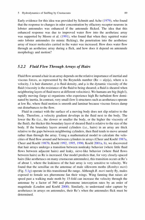
Fluid flow around a hair in an array depends on the relative importance of inertial andviscous forces, as represented by the Reynolds number (Re ¼ ulr/m), where u isvelocity, l is hair diameter, r is fluid density, and m is the dynamic viscosity of thefluid (viscosity is the resistance of the fluid to being sheared; a fluid is sheared whenneighboring layers of fluid move at different velocities). We humans are big (high l),rapidly moving (large u) organisms who experience high Re turbulent flow domi-nated by inertia. In contrast, very small (low l) structures such as aesthetascs operateat low Re, where fluid motion is smooth and laminar because viscous forces dampout disturbances to the flow.
Fluid in contact with the surface of a moving body does not slip relative to thebody. Therefore, a velocity gradient develops in the fluid next to the body. Thelower the Re (i.e., the slower or smaller the body, or the higher the viscosity ofthe fluid), the thicker this boundary layer of sheared fluid is relative to the size of thebody. If the boundary layers around cylinders (i.e., hairs) in an array are thickrelative to the gaps between neighboring cylinders, then fluid tends to move aroundrather than through the array. Using a mathematical model to calculate the velo-cities of fluid flow around and between cylinders in arrays (Cheer and Koehl 1987a,Cheer and Koehl 1987b; Koehl 1992, 1995, 1996; Koehl 2001a, b), we discoveredthat hair arrays undergo a transition between nonleaky behavior (where little fluidflows between adjacent hairs) and leaky, sieve-like behavior (where fluid flowsbetween hairs) as Re is increased. Our model predicts that, for very closely-spacedhairs (like aesthetascs on many crustacean antennules), this transition occurs at Re’sof about 1, where the leakiness of the hair array is very sensitive to velocity. Wefound that the sensillae on the antennae of male silkworm moths (Bombyx mori)(Fig. 5.1g) operate in this transitional Re range. Although B. mori rarely fly, malesexposed to female sex pheromone fan their wings. Wing fanning that raises airspeed past a walking male moth by 15-fold can increase the velocity through theantennae by a factor of 560 and pheromone interception rates by an order ofmagnitude (Loudon and Koehl 2000). Similarly, to understand odor capture byaesthetascs in arrays on antennules, their Re’s when the antennules flick must bedetermined.
5 Hydrodynamics of Sniffing by Crustaceans 89

5.3 Hydrodynamics of Flicking Antennules
We made high-speed videos of flicking antennules of lobsters (Goldman and Koehl2001), shrimp (Mead 1998), stomatopods (Mead et al. 1999), and crabs (Koehl2001a). By digitizing the position of an antennule lateral filament in each videoframe, we determined its velocity during flicks and return strokes. For all theseanimals the flick down stroke or outstroke was much faster than the return stroke,and the Re’s of the aesthetascs were in the range where the leakiness transitionshould occur. Our models predicted that water should flow between the aesthetascsduring the rapid flick, but not during the slower return stroke.
5.3.1 Dynamically-Scaled Physical Models of AntennulesReveal When Fluid Flows into Arrays of Aesthetascs
We tested these predictions using dynamically-scaled physical models of lobster(Reidenbach et al. 2008), crab (Waldrop, Reidenbach, and Koehl, unpublisheddata), and stomatopod (Mead and Koehl 2000) antennule lateral filaments.The relative magnitudes of flow velocities measured at different positions inthe fluid around a dynamically-scaled model are the same as the relative magni-tudes of flow velocities measured at comparable positions around a real antennule(e.g., Koehl 2003). Therefore, we can use dynamically-scaled models to work outthe detailed flow velocity maps around and through arrays of aesthetascs (watervelocities that would be very difficult to measure around such tiny chemosensoryhairs on real flicking antennules). Dynamically-scaled models are geometrically-similar to real antennules and operate at the same Re’s. We used large (higher l)models, but operated them at the same Re’s as flicking antennules by movingthem more slowly (lower u) through mineral oil, a fluid that is more viscous(higher m) than water.
We used a technique called “particle image velocimetry” (PIV) to determine theflow velocity maps around and through aesthetasc arrays. We marked the oil withneutrally-buoyant particles, and visualized one plane of fluid at a time by illuminat-ing it with a thin sheet of laser light. By moving a video camera at the same speed asa towed antennule model, we could record fluid motion relative to the aesthetascs.Analyzing these videos, we calculated the water velocity vector fields relative toreal antennules during their rapid flick and slower return stroke. Such PIV studies ofdynamically-scaled physical models revealed that water does flow through theaesthetasc array during the rapid flick down stroke, but not during the slower returnstroke for spiny lobsters (Koehl 2001a, b; Reidenbach et al. 2008), stomatopods(Mead and Koehl 2000; Mead and Caldwell, Chap. 11), and crabs (Waldrop,Reidenbach and Koehl, unpublished data).
90 M.A.R. Koehl

5.3.2 Water Flow Through Aesthetasc Arraysof Lobsters and Crabs
The water velocity profile around the lateral filament of the olfactory antennule ofthe spiny lobster, Panulirus argus, is very different during the flick downstroke thanduring the return stroke (Fig. 5.3). Although the velocity of the flick is about fourtimes the speed of the return stroke, the water velocity between the aesthetascsduring the flick (Fig. 5.3, middle panel), is roughly twenty five times faster thanduring the return (Fig. 5.3, right panel) because the flick Re is above the Re of theleakiness transition and the return Re is below it (Reidenbach et al. 2008). We foundthat the flick down stroke lasts just long enough to allow complete replacement ofthe water in the spaces between the aesthetascs. In contrast, the water between theaesthetascs during the return stroke is essentially trapped there. By working withphysical models, we could manipulate the morphology and orientation of an
Fig. 5.3 Lateral filament of the antennule of the spiny lobster, Panulirus argus. The photographon the left is a scanning electron micrograph (SEM) of a portion of the lateral filament, showing thechemosensory hairs (aesthetascs) and guard hairs attached to the stalk of the filament (segments ofwhich are visible at the top of the photograph). The dashed line shows where a cross-section istaken through the lateral filament, and diagrams of that cross-section are shown in the middle andleft panels of this figure. These diagrams are maps of water velocities relative to the lateralfilament when it rapidly flicks downward (middle panel) and slowly returns upward (rightpanel). The stalk of the lateral filament is labeled “antennule” and the direction of its motion isindicated by the large black arrow. The large red arrow indicates the direction of water motionrelative to the antennule lateral filament. The white box outlines the region occupied by the array ofaesthetascs, and the position of the guard hairs is labeled. The scale to the right of the diagramsindicates water velocity: red areas with yellow velocity vectors show the fastest flow relative to theantennule, yellow/green areas with shorter velocity vectors indicate less rapid flow, and blue areaswithout velocity vectors are regions of the slowest water movement. The downstroke is about 4times faster than the return stroke, but the velocity of the water between the aesthetascs isapproximately 25 times faster during the downstroke than during the return stroke (Reidenbachet al. 2008) (SEM by J. Goldman; water velocity maps calculated from PIV measurements arounddynamically-scaled physical models of the antennules; modified after Reidenbach et al. 2008)
5 Hydrodynamics of Sniffing by Crustaceans 91
antennule to explore how such features affect water flow near aesthetascs. Theseexperiments revealed that the complex zigzag arrangement of aesthetascs on theantennules of spiny lobsters (Fig. 5.3, left panel) and their orientation relative to theflicking direction produce uniform flow velocities along the length of the aesthe-tascs when the antennule flicks.
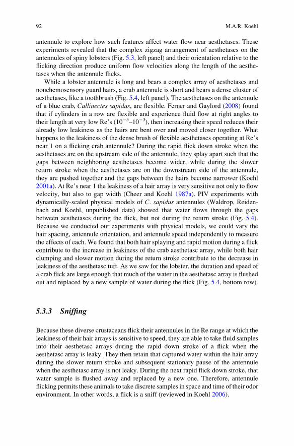
While a lobster antennule is long and bears a complex array of aesthetascs andnonchemosensory guard hairs, a crab antennule is short and bears a dense cluster ofaesthetascs, like a toothbrush (Fig. 5.4, left panel). The aesthetascs on the antennuleof a blue crab, Callinectes sapidus, are flexible. Ferner and Gaylord (2008) foundthat if cylinders in a row are flexible and experience fluid flow at right angles totheir length at very low Re’s (10#5–10#3), then increasing their speed reduces theiralready low leakiness as the hairs are bent over and moved closer together. Whathappens to the leakiness of the dense brush of flexible aesthetascs operating at Re’snear 1 on a flicking crab antennule? During the rapid flick down stroke when theaesthetascs are on the upstream side of the antennule, they splay apart such that thegaps between neighboring aesthetascs become wider, while during the slowerreturn stroke when the aesthetascs are on the downstream side of the antennule,they are pushed together and the gaps between the hairs become narrower (Koehl2001a). At Re’s near 1 the leakiness of a hair array is very sensitive not only to flowvelocity, but also to gap width (Cheer and Koehl 1987a). PIV experiments withdynamically-scaled physical models of C. sapidus antennules (Waldrop, Reiden-bach and Koehl, unpublished data) showed that water flows through the gapsbetween aesthetascs during the flick, but not during the return stroke (Fig. 5.4).Because we conducted our experiments with physical models, we could vary thehair spacing, antennule orientation, and antennule speed independently to measurethe effects of each. We found that both hair splaying and rapid motion during a flickcontribute to the increase in leakiness of the crab aesthetasc array, while both hairclumping and slower motion during the return stroke contribute to the decrease inleakiness of the aesthetasc tuft. As we saw for the lobster, the duration and speed ofa crab flick are large enough that much of the water in the aesthetasc array is flushedout and replaced by a new sample of water during the flick (Fig. 5.4, bottom row).
5.3.3 Sniffing
Because these diverse crustaceans flick their antennules in the Re range at which theleakiness of their hair arrays is sensitive to speed, they are able to take fluid samplesinto their aesthetasc arrays during the rapid down stroke of a flick when theaesthetasc array is leaky. They then retain that captured water within the hair arrayduring the slower return stroke and subsequent stationary pause of the antennulewhen the aesthetasc array is not leaky. During the next rapid flick down stroke, thatwater sample is flushed away and replaced by a new one. Therefore, antennuleflicking permits these animals to take discrete samples in space and time of their odorenvironment. In other words, a flick is a sniff (reviewed in Koehl 2006).
92 M.A.R. Koehl

Fig. 5.4 Lateral filament of the antennule of the blue crab,Callinectes sapidus. The photograph onthe top left is a SEM of the lateral filament, showing the chemosensory hairs (aesthetascs) attachedto the stalk of the filament. The proximal and distal ends of the aesthetascs are labeled. The dashedline shows where a cross-section is taken through the lateral filament, and a diagram of that cross-section is shown just to the left of the SEM. Since the aesthetascs splay apart during the rapid flickdownstroke and collapse together during the slower return stroke, we have transformed the actualcoordinates of the aesthetascs into a rectilinear grid (the “aesthetasc coordinates” shown on the leftof the top row) so that the comparison of the flow between them during the down and return strokesis easier to see. The diagrams in the middle row are maps of water velocities relative to the lateralfilament when it rapidly flicks (left diagram) and slowly returns (right diagram). These velocitymaps are plotted on “aesthetasc coordinates”. In the flick downstroke diagram, the antennule isshown moving from left to right, so the water flow relative to the antennule is right to left. In thereturn stroke diagram, the antennule is moving right to left, so the flow relative to the antennule isleft to right. The scale to the right of these flow maps indicates water velocity: areas of white andpale gray show the fastest flow relative to the antennule, while the darkest areas are regions of theslowest water movement. The graphs at the bottom of the figure show the water velocity relative tothe aesthetascs at different positions along the length of the aesthetascs for the flick down stroke(left) and return stroke (right). The line across each graph indicates the water speed necessary forthe water in the middle of the aesthetasc array to be washed out of the hair array during the stroke.During the down stroke, most of the water between the aesthetascs is flushed out of the array,whereas during the return stroke, the water is trapped between the aesthetascs (SEM byM. Martinez; water velocity maps calculated from PIV measurements around dynamically-scaledphysical models of the antennules by L. Waldrop, M. Reidenbach, and M. Koehl)
5 Hydrodynamics of Sniffing by Crustaceans 93
5.4 How Odor Plumes are Sampled by Flicking Antennules
When the lateral filament of an antennule flicks, it samples a small slice of the waterin a crustacean’s environment. What are the patterns of odor concentrations in thewater samples captured by flicking antennules as the animals move through habitatsexposed to ambient water motion?
5.4.1 Odor Concentrations in Turbulent Odor Plumes
Water currents in aquatic habitats are turbulent. As a chemical signal from an odorsource is carried across the environment by a turbulent current, the signal-bearingwater is also stirred into the surrounding odor-free water by swirling eddies.Although early models of how animals search for the source of a chemical signalassumed that such odor plumes are diffuse clouds that become wider and moredilute with distance from the source (reviewed in Koehl 2006), scientists are nowable to map the fine-scale spatial distribution of odor concentrations in turbulentmoving water using a technique called “planar laser-induced fluorescence” (PLIF).If a chemical cue is labeled with a fluorescent dye and allowed to ooze from asource in a flume (a long tank in which water flows), investigators can see how thatdye is dispersed by turbulent water currents or waves by illuminating a slice of theodor plume with a sheet of laser light. The laser light makes the dye glow, and thebrightness of the dye is proportional to the odor concentration.
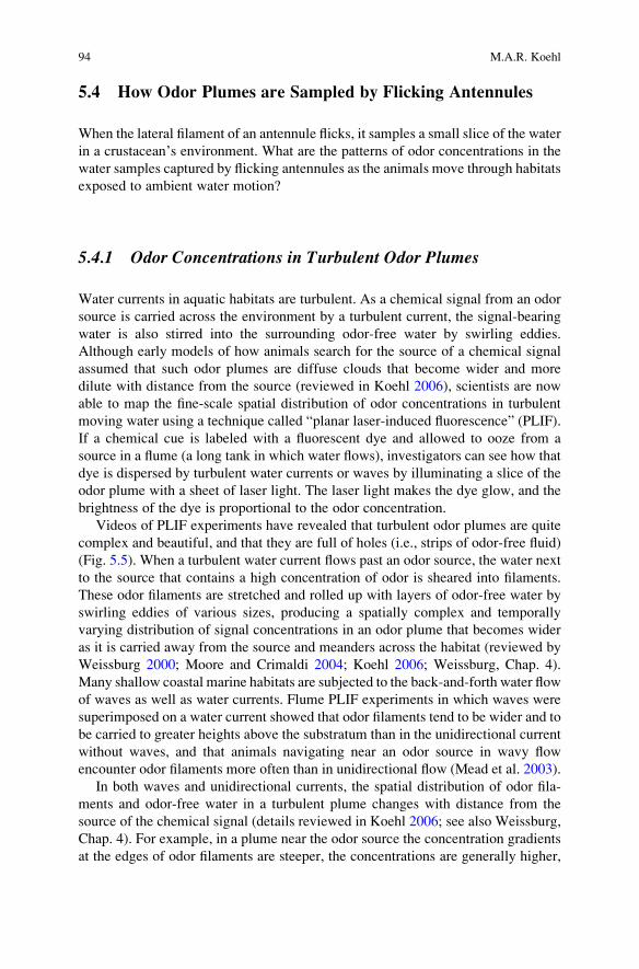
Videos of PLIF experiments have revealed that turbulent odor plumes are quitecomplex and beautiful, and that they are full of holes (i.e., strips of odor-free fluid)(Fig. 5.5). When a turbulent water current flows past an odor source, the water nextto the source that contains a high concentration of odor is sheared into filaments.These odor filaments are stretched and rolled up with layers of odor-free water byswirling eddies of various sizes, producing a spatially complex and temporallyvarying distribution of signal concentrations in an odor plume that becomes wideras it is carried away from the source and meanders across the habitat (reviewed byWeissburg 2000; Moore and Crimaldi 2004; Koehl 2006; Weissburg, Chap. 4).Many shallow coastal marine habitats are subjected to the back-and-forth water flowof waves as well as water currents. Flume PLIF experiments in which waves weresuperimposed on a water current showed that odor filaments tend to be wider and tobe carried to greater heights above the substratum than in the unidirectional currentwithout waves, and that animals navigating near an odor source in wavy flowencounter odor filaments more often than in unidirectional flow (Mead et al. 2003).
In both waves and unidirectional currents, the spatial distribution of odor fila-ments and odor-free water in a turbulent plume changes with distance from thesource of the chemical signal (details reviewed in Koehl 2006; see also Weissburg,Chap. 4). For example, in a plume near the odor source the concentration gradientsat the edges of odor filaments are steeper, the concentrations are generally higher,
94 M.A.R. Koehl

Fig. 5.5 Frames of videos ofa robotic spiny lobster,Panulirus argus, flicking areal antennule lateral filamentat a position 1 m downstreamfrom an “odor” (i.e.,fluorescent dye) source in aflume in which a turbulentwater current of 0.10 m/s wasflowing. The top image showshow a sheet of laser lightreflected off a mirror on thefloor of the flume illuminatesthe water both above andbelow a flicking antennule.The light swirls in the waterare filaments of dye, and theirbrightness is proportional toconcentration. The middleand lower images showclose-up views of a lateralfilament during the rapid flickdownstroke. In the middleimage, filaments of dye canbe seen flowing into theaesthetasc array. In contrast,in the lower image, theantennule encounters anodor-free “hole” during thedown stroke (frames of videostaken by M. Koehl andJ. Koseff)
5 Hydrodynamics of Sniffing by Crustaceans 95

the odor filaments and the gaps between them tend to be narrower, and the variationin concentration between filaments is greater than they are in that plume at a greaterdistance downstream from the odor source. Furthermore, the frequency of encoun-ters with odor filaments at the edge of an odor plume is lower than along its midline,although the odor concentrations can be similar. Therefore, the fine-scale patternsof odor concentration in the water contain information about position relative to thesource of that odor. Can a flicking antennule capture these fine-scale aspects of odorplume structure?
5.4.2 Patterns of Odor Concentrations Capturedby Flicking Antennules
A flicking antennule samples only the thin slice of water through which it sweeps.If that slice of water can be illuminated by a sheet of laser light, PLIF can be used tomeasure the pattern of odor concentrations in the water in the aesthetasc array of thelateral filament of a crustacean antennule. A challenge to this approach is getting acrustacean to flick its antennule in the plane of laser light. We overcame thischallenge by using a robotic lobster to flick real antennule lateral filaments in asheet of laser light shining through a turbulent dye plume in a flume (Koehl et al.2001) (Fig. 5.5, top). We used fresh antennules from spiny lobsters, P. argus, andthe robot flicked them using the kinematics we had measured for antennules of thatspecies (Goldman and Koehl 2001). We made high-speed videos of the robot-flicked antennules, and in each video frame we measured the brightness of dyewithin the aesthetasc array to determine how those dye (i.e., odor) concentrationschanged over time.
Our PLIF experiments using the robotic lobster revealed a number of surprises.For example, since an odor plume is full of aroma-free holes, the flicking antennuleof a lobster standing in the middle of a plume sometimes encounters filaments ofchemical signal (Fig. 5.5, middle), and sometimes it does not (Fig. 5.5, bottom).Furthermore, when a flicking antennule does run into odor filaments, water and thefine filaments of dye (i.e., odor) it carries flow through the spaces between aesthe-tascs during the rapid down stroke without being stirred up (Fig. 5.5, middle). Thenthe spatial pattern of odor concentration peaks and valleys that happen to be in theaesthetasc array at the very end of the down stroke are trapped there during the slowreturn stroke and the stationary pause before the next flick. The odor filaments in theplume around the antennule flow past the lateral filament in the ambient current, butthe stripes of chemical signal and of odor-free water within the aesthetasc array stayin place until the next flick, when the old water sample is flushed away and a newsample is trapped between the aesthetascs (Koehl et al. 2001; Koehl 2006). PLIFmeasurements of the odor samples captured by flicking stomatopod antennulesyielded similar results (Mead et al. 2003; Caldwell and Mead, this volume).These experiments indicate that each time the lateral filament of an antennule
96 M.A.R. Koehl

flicks, it captures a snapshot of the fine-scale odor concentration patterns in a smallslice of the odor plume.
5.5 Flux of Odorant Molecules to Aesthetasc Surfaces
Chemical signals in the water trapped in an aesthetasc array disperse across thesmall distances between these chemosensory hairs via molecular diffusion. Calcu-lation of the diffusion of odorant molecules carried in the water between aesthetascson spiny lobster antennules during the slow return stroke (Fig. 5.3, right panel) andinterflick pause indicate that the duration of the return stroke and the pause beforethe next flick is long enough for odor molecules in that water sample to diffuse toaesthetasc surfaces (Reidenbach et al. 2008). Similarly, a mathematical model ofthe advection (i.e., transport by moving water) and diffusion (i.e., Brownianmotion) of odorant molecules in aroma filaments encountered by flicking stomato-pod antennules showed that the flux (number arriving per area per time) to aesthe-tasc surfaces of molecules in odor filaments that have been carried into the spacesbetween the aesthetascs is high (Stacey et al. 2002). In contrast, the model showedthat the flux of signal molecules from odor filaments carried past antennules in theambient current during the slow return stroke (when water does not flow betweenthe aesthetascs) is very low. These calculations, in combination with the PLIFmeasurements described above, indicate that a sample of the odor plume is capturedwithin the aesthetasc array during the flick down stroke, and the odorant moleculesin that trapped sample diffuse to the chemosensory aesthetascs before that sample isshed and the next sample taken by the subsequent flick.
5.6 Effects of Ambient Flow, Locomotion, and Sizeon Odor Sampling
Since the leakiness of an array of chemosensory hairs depends on Re, odorsampling by antennules can be affected by the fluid velocity (u) relative to theantennule and by the diameter of the aesthetascs (l). Therefore, ambient water flowand animal locomotion (both affecting u), as well as body size (affecting l) caninfluence odor capture by antennules.
The ways in which crustaceans deploy their antennules in ambient currents canaffect water flow through the aesthetasc arrays. In our flume experiments withlobsters (Koehl et al. 2001), an ambient water current of 10 cm/s did not force waterand odor filaments into the aesthetasc arrays on antennules held parallel to the flowdirection, whereas water and odor samples did move into the arrays during flickdown strokes of 6 cm/s (during a down stroke where the water flow relative to theantennule is perpendicular to the long axis of the antennule). This suggests that if
5 Hydrodynamics of Sniffing by Crustaceans 97

crustaceans hold their antennules perpendicular to ambient currents with theaesthetascs facing upstream, water should penetrate into the hair array if theambient flow is fast enough. Are there ambient current velocities above whichcrustaceans cease antennule flicking because the water motion in the environmentdrives fluid through their aesthetasc arrays? If so, can the animals track an odorplume to its source as well as they do when they can sniff (i.e., take a discrete odorsample in space and time with each antennule flick)?
Water also moves relative to the antennules of crustaceans when they run(e.g., crabs, 11 cm/s, Martinez et al. 1998) or swim (e.g., mysids, 10–18 cm/s,Cowles and Childress 1988; amphipods 4–14 cm/s, Sainte-Marie 1986; isopods,8–30 cm/s, Alexander and Chen 1990) through the water. What are the orientationsof the antennules when crustaceans locomote, and how does the water movementrelative to them affect the leakiness of their arrays of aesthetascs?
Since the leakiness of a hair array depends on size (l), an intriguing aspect ofodor capture by crustacean antennules is the ontogeny of sniffing. Malacostracancrustaceans grow from microscopic larvae into large adults. How do antennulemorphology and kinematics change during the ontogeny of a crustacean as itchanges size, and how does that affect how they take odor samples from thesurrounding water? Comparison of different sizes of stomatopods (Mead et al.1999) and lobsters (Goldman and Koehl 2001) revealed that small animals havelarger aesthetascs relative to body size than do big animals, and move rapidlyenough during the flick downstroke that they can sniff. Future research shouldextend these studies to smaller sizes to explore how the morphology and kinematicsof the antennules of microscopic larvae and tiny juveniles affect how they sampletheir odor environment. Is there a lower limit to antennule size for sniffing to bepossible?
Our predictions about how Re affects flow through arrays of chemosensoryhairs, and thus odor capture, suggest other comparative studies. For example,how do the fluid dynamics of odor sampling by the olfactory organs of the larvaeand juveniles mentioned above compare with those of other small crustaceans, suchas the planktonic copepods that follow scent trails to find mates (e.g., Weissburget al. 1998), or the deep sea amphipods that use odors to locate carrion (e.g., Premkeet al. 2003)? How do their swimming behaviors or feeding currents affect flowacross their olfactory organs, and do they flick? Antennule “sweeps” have beenreported from lysianassid amphipods and it was suggested that sweeps facilitatewater exchange around the aesthetasc-bearing callynophores (Kaufmann 1994).
5.7 Crustaceans as Model Systems to Study Odor Capture
There are a number of advantages of using crustaceans as systems to study thephysical process of odor capture. Antennules are olfactory organs that protrude intothe environment, so their interactions with the surrounding odor-bearing fluid aremuch easier to study than are the fluid mechanics of chemosensory surfaces hidden
98 M.A.R. Koehl

within internal nasal passages, such as those of vertebrates. Furthermore, thediversity of antennule morphologies shown by different species of crustaceansenables us to explore the functional consequences of different “designs” of externalolfactory organs. Many other types of invertebrate animals have external chemo-sensory organs that bear hair-like sensillae (reviewed in Koehl 2001a, 2006), so theprinciples about odor capture learned by studying crustacean antennules can beuseful for understanding the function of these other “noses” as well. Anotheradvantage of the species of crustaceans that we have been using as study organisms(e.g., lobsters, crabs, stomatopods) is that they live in accessible shallow marinehabitats where we can measure the hydrodynamic conditions that they experiencein their natural habitats and that disperse the chemical signals they encounter. Suchinformation enables us to design biologically relevant laboratory studies of anten-nule function.
Since crustaceans are used to study the neurobiology of olfaction and thebehavioral uses of chemical signaling, they provide a system with the potential ofenabling us to relate the biophysics of odor capture to how animals process thatinformation, and to how they react to the spatio-temporal patterns of odor informa-tion they capture.
5.8 Future Directions
Several important questions about crustacean odor capture that remain unansweredprovide promising directions for future research. One such question is whethercrustaceans “use” the fine-scale spatio-temporal information their antennules arephysically able to capture when they flick in turbulent odor plumes. This questioncan be addressed at the neurobiological level (Do fine-scale patterns of odorconcentrations captured by the antennules affect patterns of neuron firing in theolfactory lobe?) and at the behavioral level (Do fine-scale patterns of odor concen-trations captured by the antennules affect plume-searching behavior?). For exam-ple, to determine whether different spatio-temporal odor-concentration patternsalter neuron firing, the standard olfactometers (e.g., Y-tube flow set-ups; see alsoFig. 10.2 in Thiel, Chap. 10) used to deliver odors to neurobiological preparationscould be replaced by odor-delivery systems that mimic the realistic spatial andtemporal patterns of odor delivery that antennules experience when flicking atdifferent positions in an odor plume. Similarly, video records of the movementsof crustaceans searching in odor plumes visualized by PLIF (Fig. 5.5) enable us tocorrelate the fine-scale patterns of concentrations of signal captured by right andleft antennules on each flick with the subsequent behaviors of the animals (Meadet al. 2003; Mead and Caldwell, Chap. 11). Such studies of what antennulesactually sample as animals navigate in odor plumes should also enable us to workout search algorithms that were not possible to recognize when whole plumes werevisualized and flicking was ignored.
5 Hydrodynamics of Sniffing by Crustaceans 99

While the olfactory antennules of large malacostracan crustaceans have beenused as research systems to elucidate mechanisms by which chemical signals in theenvironment are sampled, less is known about the hydrodynamics of the chemo-sensory hairs on other parts of crustacean bodies. The approaches used and theprinciples elucidated by studying flow through arrays of aesthetascs on antennulescan also shed light on how the morphology of other types of chemosensory hairs, aswell as their arrangement in arrays and their positions on the body affect their odor-capturing performance. Another important avenue for future neurobiological andbehavioral research is to explore how information from chemosensors on the bodyand legs is coupled with information sampled by the antennules to inform ananimal’s behavior.
Comparisons of the morphology and kinematics of aquatic olfactory organs withthose that operate in air (such as the antennules of terrestrial hermit crabs or theantennae of insects) would provide an interesting test of our ideas about mechanismsof sniffing. The density (r) of water is about 1,000 times higher than that of air, butthe viscosity (m) is only about 56 times greater (Vogel 1994), so a structure of a givensize must move about 18 times faster in air than in water to achieve the same Re.Therefore, I would expect the leakiness transition for an array of chemosensory hairsof a given size to occur at higher speeds in air. A more striking difference betweenwater and air, however, is that the diffusivity (D, a molecule’s propensity to diffuse ina particular fluid) of molecules in air is about 10,000 times greater than in water(Vogel 1994). Because odors can move a given distance via molecular diffusionthrough air much more rapidly than through water, the filaments in odor plumes arewider (reviewed in Koehl 2006), and the time required for odor molecules to diffusefrom sampled air to the surfaces of chemosensory sensillae is much shorter in air thanin water. Analysis of the aerodynamics of odor capture by the antennules of intertidalor terrestrial crustaceans (e.g., brachyuran or hermit crabs) could be done usingapproaches similar to those described in this chapter to determine how antennulemorphology and kinematics determine if and how they sniff.
5.9 Summary and Conclusions
Chemical signals are dispersed in aquatic environments by turbulent water currents.The lateral filaments of the antennules of malacostracan crustaceans, which beararrays of chemosensory hairs (aesthetascs), are an important research system forstudying the physics of how olfactory organs bearing hair-like olfactory sensillacapture such chemical signals from the environment. On the scale of an antennulean odor plume is not a diffuse cloud, but rather is a series of fine filaments of aroma-bearing water swirling in scent-free water; the spatio-temporal patterns of thesefilaments depend on distance from the odor source. When a lobster, stomatopod, orcrab flicks the lateral filament of its antennule, water and any odor filaments it iscarrying flow through the spaces between the aesthetascs during the rapid downstroke, but not during the slower return stroke or during the stationary pause before
100 M.A.R. Koehl
the next flick. For any array of small hairs, there is a critical velocity range abovewhich the array is “leaky” and fluid can flow between the hairs, and below whichfluid barely moves through the spaces between the hairs. The striking difference inflow through aesthetasc arrays during the down stroke vs. the upstroke occursbecause the down stroke velocities are above and the return stroke velocities arebelow the speeds at which this transition in leakiness occurs. Odorant molecules inthe water trapped between the aesthetascs during the return stroke and pause diffuseto the surfaces of the aesthetascs, before the next flick traps a new parcel of water.Therefore, each antennule flick is a “sniff,” taking a discrete sample of the odorplume in space and time. Our work has shown that the size and arrangement ofchemosensory hairs on olfactory organs like antennules or antennae, as well as theirvelocity relative to the surrounding fluid, affect the temporal patterns of odordelivery to the chemosensory hairs. Thus, the physics of odor capture providesthe first step in filtering olfactory information from the environment.
Acknowledgments My research reported here was supported by grants from the James S.McDonnell Foundation and from the Office of Naval Research (USA), a John D. and CatherineT. MacArthur Foundation Fellowship, and the Virginia G. and Robert E. Gill Chair (University ofCalifornia, Berkeley). I thank the coauthors on my papers cited in this chapter for the discussionsand collaborations that led to the ideas presented here.
References
Ache BW (1982) Chemoreception and thermoreception. In: Atwood HL, Sandeman DC (eds) Thebiology of the crustacea. Academic Press, New York, pp 369–393
Alexander DE, Chen T (1990) Comparison of swimming speed and hydrodynamic drag in twospecies of Idotea (Isopoda). J Crust Biol 10:406–412
Atema J (1985) Chemoreception in the sea: Adaptations of chemoreceptors and behavior toaquatic stimulus conditions. Soc Exp Biol Symp 39:387–423
Cheer AYL, Koehl MAR (1987a) Fluid flow through filtering appendages of insects. I.M.A.J Math Appl Med Biol 4:185–199
Cheer AYL, Koehl MAR (1987b) Paddles and rakes: fluid flow through bristled appendages ofsmall organisms. J Theor Biol 129:17–39
Cowles DL, Childress JJ (1988) Swimming speed and oxygen consumption in the bathypelagicmysid Gnathophausia ingens. Biol Bull 175:111–121
Ferner MC, Gaylord B (2008) Flexibility foils filter function: structural limitations on suspensionfeeding. J Exp Biol 211:3563–3572
Gleeson RA, Carr WES, Trapido-Rosenthal HG (1993) Morphological characteristics facilitatingstimulus access and removal in the olfactory organ of the spiny lobster, Panulirus argus:insight from the design. Chem Senses 18:67–75
Goldman JA, Koehl MAR (2001) Fluid dynamic design of lobster olfactory organs: High-speedkinematic analysis of antennule flicking by Panulirus argus. Chem Senses 26:385–398
Kaufmann RS (1994) Structure and function of chemoreceptors in scavenging lysianassoidamphipods. J Crust Biol 14:54–71
Koehl MAR (1977) Effects of sea anemones on the flow forces they encounter. J Exp Biol69:87–105
Koehl MAR (1992) Hairy little legs: feeding, smelling, and swimming at low Reynolds number.Fluid dynamics in biology. Contemp Math 141:33–64
5 Hydrodynamics of Sniffing by Crustaceans 101

Koehl MAR (1995) Fluid flow through hair-bearing appendages: feeding, smelling, and swimmingat low and intermediate Reynolds number. In: Ellington CP, Pedley TJ (eds) Biological fluiddynamics. Soc Exp Biol Symp 49, pp 157–182
Koehl MAR (1996) Small-Scale fluid dynamics of olfactory antennae. Mar Fresh Behav Physiol27:127–141
Koehl MAR (2001a) Fluid dynamics of animal appendages that capture molecules: arthropodolfactory antennae. In: Fauci L, Gueron S (eds) Computational modeling in biological fluiddynamics. IMA Series # 124, pp 97–116
Koehl MAR (2001b) Transitions in function at low Reynolds number: hair-bearing animalappendages. Math Methods Appl Sci 24:1523–1532
Koehl MAR (2003) Physical modelling in biomechanics. Phil Trans Roy Soc B 358:1589–1596Koehl MAR (2006) The fluid mechanics of arthropod sniffing in turbulent odor plumes. Chem
Senses 31:93–105Koehl MAR, Koseff JR, Crimaldi JP, McCay MG, Cooper T, Wiley MB, Moore PA (2001)
Lobster sniffing: antennule design and hydrodynamic filtering of information in an odor plume.Science 294:1948–1951
Loudon C, Koehl MAR (2000) Sniffing by a silkworm moth: wing fanning enhances air penetra-tion through and pheromone interception by antennae. J Exp Biol 203:2977–2990
Martinez MM, Full JR, Koehl MAR (1998) Underwater punting by an intertidal crab: A novel gaitrevealed by the kinematics of pedestrian locomotion in air vs. water. J Exp Biol201:2609–2623
Mead KS (1998) The biomechanics of odorant access to aesthetascs in the Grass Shrimp,Palaemonetes vulgaris. Biol Bull 195:184–185
Mead KS, Koehl MAR (2000) Stomatopod antennule design: The asymmetry, sampling effi-ciency, and ontogeny of olfactory flicking. J Exp Biol 203:3795–3808
Mead KS, Koehl MAR, O’Donnell MJ (1999) Stomatopod sniffing: the scaling of chemosensorysensillae and flicking behavior with body size. J Exp Mar Biol Ecol 241:235–261
Mead KS, Wiley MB, Koehl MAR, Koseff JR (2003) Fine-scale patterns of odor encounter by theantennules of mantis shrimp tracking turbulent plumes in wave-affected and unidirectionalflow. J Exp Biol 206:181–193
Moore PA, Atema J, Gerhardt GA (1991) Fluid dynamics and microscale chemical movement inthe chemosensory appendages of the lobster, Homarus americanus. Chem Senses 16:663–674
Moore P, Crimaldi J (2004) Odor landscapes and animal behavior: tracking odor plumes indifferent physical worlds. J Mar Syst 49:55–64
Premke K, Muyakshin S, Klages M, Wegner J (2003) Evidence for long-range chemoreceptivetracking of food odour in deep-sea scavengers by scanning sonar data. J Exp Mar Biol Ecol285:283–294
Reidenbach MA, George NT, Koehl MAR (2008) Antennule morphology and flicking kinematicsfacilitate odor sampling by the spiny lobster, Panulirus argus. J Exp Biol 211:2849–2858
Sainte-Marie B (1986) Feeding and swimming of lysianassid amphipods in a shallow cold-waterbay. Mar Biol 91:219–229
Schmitt BC, Ache BW (1979) Olfaction: responses of a decapod crustacean are enhanced byflicking. Science 205:204–206
Snow PJ (1973) The antennular activities of the hermit crab, Pagurus alaskensis (Benedict). J ExpBiol 58:745–765
Stacey M, Mead KS, Koehl MAR (2002) Molecule capture by olfactory antennules: mantisshrimp. J Math Biol 44:1–30
Vogel S (1994) Life in moving fluids. Princeton University Press, PrincetonWeissburg MJ (2000) The fluid dynamical context of chemosensory behavior. Biol Bull
198:188–202Weissburg MJ, Doall MH, Yen J (1998) Following the invisible trail: mechanisms of chemosen-
sory mate tracking by the copepod Temora. Phil Trans Roy Soc B 353:701–712
102 M.A.R. Koehl