Understanding Skin Cancer Causes & Types Risks Factors Understanding UVB Sun exposure Prevention.

2011-2012
Update on Cancer ResearchCauses and Prevention
Update on C
ancer Research
Causes and P
revention

2
Update on Cancer ResearchCauses and Prevention
2011-2012

Update on Cancer Research
Update on Cancer Research is published
by the Department of Resource Development
at the Weizmann Institute of Science
P.O.Box 26, Rehovot, Israel 76100
Tel: 972 8 934 4582
e-mail:[email protected]

Update on Cancer ResearchCauses and Prevention
Table of Contents
Introduction: Research on Cancer Causes and Prevention .............................2From Inflammatory Lung Disease to Lung Cancer .............................................6Fusion Genes as Leukemic Agents ..........................................................................8Three Pathways to Breast Cancer Diagnosis ............................................................10
Cellular Communication and Breast Cancer ........................................................12
Down Syndrome and Leukemia ........................................................................14
A Molecular Link between Cell Death, Cancer, and Diabetes ............................16
Tumor Death by Design ...................................................................................18
Arresting Cancer Growth ...................................................................................20
Novel Risk Biomarkers for Early Detection and Prevention of Lung Cancer .......22
The Many Faces of the p53 Tumor Suppressor .....................................................24
Switching Cancer Cells Off .................................................................................26
A Holistic Approach to Studying Tumorigenesis .....................................................28
Necrosis Gone Awry: Pathways to Cancer ..............................................................30
In Appreciation of Donors to Cancer Research .............................................32
2011-2012

2
The history of medicine teaches us that the
best way to fight disease is to prevent it
altogether. A prime example is heart disease;
there was a dramatic decrease in death from
heart disease in the second half of the 20th
century, half of which was due to prevention.
Unfortunately, there is very little in way of
prevention when it comes to cancer—a global
health concern, with an estimated 13 million
people diagnosed with cancer worldwide every
year and eight million deaths annually. So
what can we do to avoid the onset of cancer?
Weizmann Institute scientists are looking
for answers. By conducting in-depth basic
research into the processes that transform
healthy cells into cancerous ones and the
genes and mechanisms involved, researchers
at the Weizmann Institute seek to identify
ways to target the mutated genes and faulty
mechanisms as a means to curing cancer. Their
findings are likely to enable risk assessment
and preventive strategies, as well as methods
for early detection of cancer, which will greatly
help increase the success rate of therapy.
The Weizmann Institute has a long-standing
record as a major contributor to scientific
understanding of cancerous transformations. First
on the list is one of the world pioneers of cancer
research, the late Prof. Isaac Berenblum, whose
studies at the Weizmann Institute in the first
decades following Israel’s independence formed
the foundation for cancer research in Israel. It was
Prof. Berenblum who formulated the two-stage
theory of cancer development in the late 1940s.
Although it is clear today that cancer progresses
in multiple stages, Prof. Berenblum was the
first to demonstrate that for cancer to develop,
certain events must occur in an ordered manner.
Research on Cancer Causes and Prevention at the Weizmann Institute of Science
Introduction:

3
In the early 1960s, Prof. Leo Sachs found that
irradiation and certain chemicals can induce the
transformation of healthy cells into cancerous
ones in a petri dish, paving the way to new
in vitro experimental approaches to study the
cellular changes that allow tumor growth.
He also found that leukemia cells can be
made to behave again like normal cells and
stop multiplying by adding a key regulator of
development. This insight showed that cancer
cells can be “reprogrammed,” and also led to the
formation of a new method for treating leukemia.
Like chemical carcinogens and irradiation,
v i ruses too can induce cancer, but
they may also have therapeutic uses.
Prof. Ernest Winocour found that some viruses
selectively kill cells in the early stages of the
malignant transformation. Understanding
the basis of this selectivity may lead to the
exploitation of “good” viruses for ridding the
body of cells at high risk of becoming cancerous.
With the dawn of the era of DNA cloning in
the 1980s, Profs. David Givol and Eli Canaani
revealed that rearrangements in the structure of
the genome due to chromosomal translocations
play an important role in cancer development.
Most impressive, it was Prof. Canaani who
demonstrated that chronic myelogenous
leukemia (CML) was the outcome of a particular
chromosome translocation that leads to the faulty
fusion of two parts from different chromosomes
and results in the expression of a new fused
cancerous protein. This fused protein is the
target for Imatinib (Gleevec/Glivec), a drug
that treats the disease and is now commonly
prescribed to cancer patients throughout the
world. Since Prof. Canaani’s discovery, 400
more chromosomal translocations and fused
4
genes were identified in various cancers and
are now the subjects of intense research.
Weizmann Institute researchers have also
shed light on the regulatory processes that
play a role in cancer induction. One such
line of research involves the growth factors,
hormone-like proteins that facilitate cell growth
and are critical in tumor progression. It was
Prof. Sachs that, in the 1960s, discovered the
family of growth factors known as colony
stimulating factors, responsible for inducing
the growth and differentiation of specific types
of blood cells. In later years, Prof. Yosef Yarden
uncovered the family of growth factors called
Neuregulins (NRGs) and further showed that
one of the cellular proteins that mediate the
activities of the Neuregulins, the HER2 protein,
which is typically over-expressed in relatively
aggressive breast tumors, acts as an amplifier of
growth activity in breast cancer cells. Another
contribution to the field of growth factors came
from the research of Prof. Givol, who cloned
the fibroblast growth factor receptors (FGFRs),
known to be involved in lung and bladder cancer.
Of note, Prof. Michael Sela pioneered the
synergy concept in cancer therapy, in which
an antibody that inhibits an epidermal growth
factor receptor’s growth-promoting signals is
used in combination with chemotherapy. This
led to the drug Erbitux, used in the treatment of
colon cancer, as well as head and neck cancers.
Another important step in understanding
cancer induction came from the research on
the major tumor suppressor p53, the most
studied protein in cancer research. This effort
includes the seminal work of Profs. Givol,
Moshe Oren, and Varda Rotter in the 1980s.
They were among the first researchers to clone
and characterize the p53 gene, with Prof. Givol
successfully cloning the active full-length human
variant. This protein, suggested earlier by
Prof. Rotter to be a general tumor cell marker,
was shown by Profs. Oren and Rotter to actively
prevent cancer. It has since been found that
p53 is dysfunctional in almost all cancer types.
Another major contribution to the field of
tumor suppressors came from the research of
5
Prof. Adi Kimchi, who discovered the prominent
family member DAP-kinase and showed that
when it is inactive in cancer cells, they are
unable to undergo programmed cell death.
Her studies on the regulation and molecular
mode of action of DAP-kinase established
the current use of this gene as a prognostic
tool in many types of human cancers.
The cell’s main mechanism to prevent genetic
mutations is the DNA repair system. It was the
research of Prof. Zvi Livneh and Dr. Tamar Paz-
Elizur on this system that revealed that low activity
of OGG1, an enzyme involved in the repair of
oxidative DNA damage, is a risk factor for lung
cancer and head and neck cancers. This insight
led them to develop tests for measuring the level
of DNA repair enzymes in human cells, which can
be utilized as a preventative diagnostic measure.
An additional diagnostic tool came from
the laboratory of Prof. Hadassa Degani. Her
research of hormonal regulation of blood
vessel growth in breast cancer led to the
development of a magnetic resonance imaging
(MRI) technique for the early detection of breast
tumors, now in common practice worldwide.
These examples represent only a fraction of the
scientific contributions of the Weizmann Institute
to the understanding of the basic processes that
underlie cancerous transformations. This booklet
showcases some of the recent findings of a
selection of Weizmann scientists in this field, who,
in many cases, are using their new insights to
devise innovative therapies and preventative tools.
6
The lungs, which are among the most
immunologically active organs, maintain the
body’s first line of defense against numerous
pathogens and air-borne particulates,
including dust and tobacco smoke. However,
this organ can only withstand so much ill
treatment before succumbing to the induced
immunological responses that contribute
to the development of health problems
such as inflammation and lung cancer.
The latter can be induced directly by carcinogens,
such as tobacco smoke, or indirectly during
the onset of various inflammatory processes,
primarily those associated with chronic obstructive
pulmonary disease (COPD). One possibility by
which COPD instigates cancer is by “switching
off” tumor suppressor genes, essential in the
prevention of tumor formation and metastasis.
Prof. Ronen Alon’s research focuses on how
immune cells respond to pathogens and
migrate out of the blood stream to induce
inflammation and eradicate infectious agents.
In one of his latest collaborative studies,
focused on the interplay between tobacco
smoke and COPD, he has shed new light on
the activities of the tumor suppressor DAP-
kinase. This protein, which prompts damaged
cells to commit suicide, is missing in many
tumor cell lines, including certain lung cancers.
As part of this joint study with Prof. Adi Kimchi
from the Department of Molecular Genetics,
Prof. Alon and his colleagues designed an
experiment to explore how animal models that
lack DAP-kinase cope in a COPD environment,
induced by either bacterial insult or tobacco
smoke. They found that these animal models
From Inflammatory Lung Disease to Lung Cancer
Prof. Ronen AlonDepartment of Immunology
DAP-kinase, and possibly other
tumor suppressors, may have
both a direct and indirect role
in lung cancer prevention.

7
exhibit profoundly higher sensitivity to both
tobacco smoke and bacterial insults. It appears
that DAP-kinase helps protect various types
of lung cells from COPD-like inflammation.
The scientists have further suggested that
DAP-kinase’s protective effect is achieved in
the following manner: In response to a local
insult, both the lung airways and white blood
cells populating the lung express DAP-kinase,
which inhibits the pro-inflammatory factors
released by these various lung cells on site.
Thus, DAP-kinase, and possibly other tumor
suppressors, may have both a direct and
indirect role in lung cancer prevention: It
can drive newly mutated cells to commit
suicide; and it can suppress inflammation of
non-mutated neighboring cells and immune
cells, thereby preventing the formation of
the supportive environment cancers need to
develop into aggressive tumors. Prof. Alon’s
findings further emphasize that restraining lung
inflammation can protect lung cells from incurring
cancerous mutations and, once transformed,
from developing into aggressive tumors.
This research avenue will help in the identification
of risk factors that predispose humans to
COPD and specific lung cancers, as well as
assist in the elucidation of the links between
inflammation and cancer. Defining these factors
is essential for pinpointing novel targets for
early diagnostics and therapeutic intervention,
first in animal models and later on in humans.
Prof. Alon’s research is supported by the Flight Attendant Medical Research Institute grants. He is the incumbent of the Linda Jacobs Professorial Chair in Immune and Stem Cell Research.

8
Prognosis of childhood acute leukemia has
improved dramatically over the past half a
century. However, for 20 to 25 percent of these
cases the projection is still quite dismal. Among
that group are a significant number of infants
from birth to one year of age. The vast majority
of these infants carry in their leukemia cells a
fused gene caused by erroneous switching of
DNA segments between two chromosomes. It
is this new fusion gene that produces a cancer-
causing hybrid protein. One of the genes that
have been found in such fused constructs is MLL.
Prof. Eli Canaani, who was the first to prove the
link between hybrid proteins and cancer, has
been investigating the role of the MLL gene in
acute leukemias. He has shown that the MLL
gene may fuse to at least 50 partner genes,
each scenario leading to a potent oncogene.
MLL fusions have been found in approximately
10 percent of human leukemias and account
for more than 70 percent of childhood acute
leukemias; they are also prevalent among
patients that develop acute leukemia after
being previously treated for other malignancies.
Intensive investigations in Prof. Canaani’s
laboratory and others have established the mode
of operation of the MLL-fusion oncogenes:
Gene expression comprises three main steps—
initiation, elongation, and termination. The fused
proteins interfere with the stage of elongation
during the process of transcription, changing
the recruitment of elongation factors to target
genes, thereby leading to excessive expression.
Prof. Canaani and his group have moved one
step closer to understanding the nature of
MLL-related leukemias by identifying all of
the approximately 170 primary gene targets
of MLL-AF4, the predominant MLL fusion
protein in infant acute leukemia. They have
Fusion Genes as Leukemic Agents
Prof. Eli CanaaniDepartment of Molecular Cell Biology
Prof. Canaani and his group
have moved one step closer to
understanding the nature of
MLL-related leukemias.

9
shown that among MLL’s targets are at least
four genes that are strongly expressed in
the majority of MLL-associated leukemias.
The scientists have further revealed that the
proteins expressed by these target genes are
essential for the proliferation and spread of
the leukemic cells in the bone marrow of
animal models transplanted with the cells.
These results offer new targets for therapeutic
intervention in MLL-associated acute leukemias.
Prof. Canaani’s research is supported by the Kirk Center for Childhood Cancer and Immunological Disorders; the Sergio Lombroso Award for Cancer Research; the Spitz Family Philanthropic Fund; and the U.S.-Israel Binational Science Foundation. He is the incumbent of the Harry Kay Professorial Chair of Cancer Research.
In the 1980s, Prof. Canaani isolated
a fused protein that triggers chronic
myelogenous leukemia (CML), the
first discovery of cancer produced by
protein fusion. This finding provided
the foundation for the development of
Imatinib (known as Gleevec in the U.S.
and Glivec in Europe), the first drug
based on the molecular understanding
of a specific cancer. Produced by
Novartis, it was approved in 2001 by
the FDA and is now routinely prescribed
around the world to patients with CML.
10
Three Pathways to Breast Cancer Diagnosis
Prof. Hadassa DeganiDepartment of Biological Regulation
Among Prof. Degani’s many
contributions to science and
medicine are three breast cancer
diagnostic tools, all based on
magnetic resonance imaging.
Prof. Hadassa Degani has dedicated the
last 25 years to characterizing hormonal
regulation of breast cancer and developing
mechanisms of endocrine therapy. Among her
many contributions to science and medicine
are three breast cancer diagnostic tools, all
based on magnetic resonance imaging (MRI).
In the late 1990s, Prof. Degani showed that
MRI, in combination with the injection of a
contrast agent, can be used to construct a 3D
image of blood flow in breast tissue to detect
regions with heightened activity, caused by
the formation of new blood vessels—a marker
of cancer growth. In 2003, her method,
nicknamed “3TP,” received U.S. Food and
Drug Administration clearance for use in
the detection of breast cancer, as well as
prostate cancer. It is now employed by doctors
worldwide to detect cancer and distinguish
between malignant tumors and benign lumps.
Prof. Degani has now gone one step further
by developing a completely non-invasive,
sensitive breast cancer screening method. Her
new invention relies on the fact that breast
tumors decrease the natural diffusion of fluid
in the mammary ductal tubes. This is because
the vast majority of breast tumors form on
the inner lining of the tubes, obstructing
fluid movement in the tubes as they grow.
Prof. Degani’s new MRI-based technique
provides a 3D map of the self-diffusion of
water in the ductal trees, with clogged regions
pointing to the location of tumors. Since cell
growth obstruction of the ductal tubes occurs
at an early stage of tumor development, this
detection method raises the possibility of

11
Prof. Degani’s research is supported by the Center for Health Sciences, funded by the Dwek Family Biomedical Research Fund and the Maria and Bernhard Zondek Hormone Research Fund; the Jeanne and Joseph Nissim Foundation for Life Sciences Research; the Estate of Ernst and Anni Deutsch; the Junior Cancer League; the Estate of Ruth Rubin; Lord David Alliance, U.K.; the Israel Science Foundation; and the National Institutes of Health, U.S. She is the incumbent of the Fred and Andrea Fallek Professorial Chair in Breast Cancer Research.
increasing the probability of curing the disease.
To test the new method, Prof. Degani and
Dr. Edna Furman-Haran, head of the Weizmann
Institute’s functional MRI (fMRI) unit, are now
conducting a clinical study, taking place on
campus, in collaboration with the breast imaging
unit at Meir Medical Center, Israel, headed by
Dr. Myra Feinberg-Shapiro. The study involves
more than 120 female volunteers, split into three
groups: with no known lumps, with a benign
lump, and with a malignant tumor. The initial
results indicate that the MRI method accurately
distinguishes between the three tissue types.
To complement the two abovementioned
tools, Prof. Degani, in collaboration with
Prof. David Milstein (Department of Organic
Chemistry) and Prof. Joel Sussman (Department
of Structural Biology), is also developing a
molecular imaging method for detecting the
“estrogen receptor,” a cellular protein that,
in cancer cells, interacts with the hormone
estrogen to promote growth. This receptor
is over-expressed in many breast tumors and
serves as a marker for endocrine therapy. Using
this method, doctors will be able to better
characterize the type of cancer a patient has,
knowledge needed to personalize treatment
to ensure the best possible medical outcome.

12
Chemical communication between cells
and transfer of information inside them are
essential for cell survival and function. It is
by these means that cells receive instructions
from their surroundings. In the absence of
such communication, important processes
such as cell division, secretion of certain
substances, or even cell death would not occur.
A major hallmark of cancer is the ability of
individual cells to free themselves from having
to abide by the instructions they receive.
Prof. Ari Elson’s studies examine the molecules
that participate in this flow of information
in cells. In particular, his team focuses on
the enzymes that add or remove small
phosphorous-containing chemical groups from
proteins, thus affecting their structure and
function. Irregularities in these processes are
among the best-known and most intensively
characterized causes of disease, including
cancer. One of the molecules that Prof. Elson
is studying is called PTPe, which, in the context
of breast cancer, helps tumor cells survive.
Almost all instances of breast cancer originate
in the lobules or ducts of the mammary glands.
In one study, Prof. Elson’s group described
how PTPe contributes to the ability of another
protein to transform healthy mammary cells
into tumor cells. Interestingly, absence of
PTPe resulted in mammary cancers that were
less destructive, indicating that its function
is important for the malignant process.
Advanced breast cancer often causes metastases
in bone that lead to a decrease in bone
mass and an increase in the susceptibility
for fractures, as well as an imbalance in
various metabolic processes. A recent study by
Prof. Elson’s group has led him to believe that PTPe
also plays a role in this type of metastasis. This
study relates to the activity of osteoclasts, bone
cells whose function is to remove bone tissue.
Cellular Communication and Breast Cancer
Prof. Ari ElsonDepartment of Molecular Genetics
Prof. Elson has shown that the
PTPe protein contributes to the
ability of another protein to
transform healthy mammary cells
into tumor cells.
13
Prof. Elson’s team found that, in mice that lack
PTPe, the osteoclasts do not receive their orders
to remove the bone tissue and, as a result,
these mice exhibit increased bone mass. Based
on these findings, his group has suggested
that mice lacking PTPe might be protected
from bone loss caused by bone metastases of
breast cancer. The scientists now aim to provide
more precise quantification of this result and
to examine the possibility that inhibiting PTPe
in osteoclasts can be used as a method for
combating metastatic breast cancer in bone.
Lacking PTPe may also be good for your figure.
Prof. Elson’s studies show that female mice
lacking PTPe are relatively resistant to weight
gain when fed a high-fat diet, revealing that
PTPe participates in the signals that regulate body
weight. Therefore, in addition to breast cancer
and bone loss, pharmacological inhibition of
PTPe may also be useful for combating obesity.
Prof. Elson’s research is supported by the M.D. Moross Institute for Cancer Research; the Kekst Family Institute for Medical Genetics; the Yeda-Sela Center for Basic Research; the Maurice and Vivienne Wohl Charitable Foundation; the Fritz Thyssen Stiftung; the Estate of Fannie Sherr; the European Union Seventh Framework Programme for Research and Technological Development; the Israel Cancer Research Fund; the Israel Science Foundation; the Minerva Foundation; the Osteogenesis Imprefecta Foundation; and the U.S. Department of Defense Breast Cancer Research Program. He is the Director of the Ekard Research School of Biological Science and the Lorry I. Lokey Research School of Biochemical Science, and is the incumbent of the Marshall and Renette Ezralow Professorial Chair.
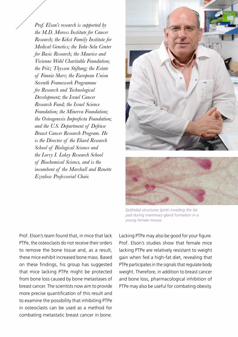
Epithelial structures (pink) invading the fat pad during mammary gland formation in a young female mouse.

14
Down syndrome, also called Trisomy 21, is the
most common cause of human birth defects.
This severe developmental disorder results
from an extra copy of chromosome 21 in
the cells’ genetic makeup. Unfortunately, it
appears that Trisomy 21 patients are also 500
times more likely to contract childhood acute
megakaryoblastic leukemia than the general
population. This leukemia is characterized by the
uncontrolled proliferation of megakaryoblasts,
the forefathers of platelets. In many cases, it
strikes in infancy and then passes, but it can
return later on with much harsher consequences.
What causes acute megakaryoblastic leukemia
to be particularly prevalent among Down
syndrome patients? Previous research in
Prof. Yoram Groner’s lab had pointed to a possible
culprit: the protein Runx1. This regulatory
protein binds to DNA segments and, thereby,
controls the expression of a large number of
genes. The damning evidence against Runx1
included its location on chromosome 21 and the
fact that quantities of an “abridged” version
of Runx1, which muddles the normal function
of this protein, are found in many leukemias.
Prof. Groner’s working hypothesis is that when
an extra copy of the Runx1 gene is present
in megakaryoblasts, as is the case in Down
syndrome patients, its expression levels are
not regulated correctly, leading, in turn, to
faulty expression of its target genes, a change
that brings about a shift from regulated
differentiation to uncontrolled proliferation.
The first step to proving this hypothesis was
to verify Runx1’s role in megakaryoblast
differentiation. And, indeed, in a collaborative
study with the group of Dr. Amos Tanay from
the Department of Computer Science and
Applied Mathematics, Prof. Groner’s team
was able to show that Runx1 is a key player in
Down Syndrome and Leukemia
Prof. Yoram GronerDepartment of Molecular Genetics
Prof. Groner is investigating
why acute megakaryoblastic
leukemia is particularly
prevalent among Down
syndrome patients.

15
differentiation, binding to over 10,000 sites on
the genome at various stages in the process.
To play so many different roles, Runx1 teams
up with other regulators of gene expression,
including one called GATA1, which is often
faulty in Down syndrome patients with leukemia.
The scientists now believe that mishaps in the
cooperation between the two regulators might
contribute to the development of leukemia.
This hypothesis has gained strength in a recent
study in Prof. Groner’s lab, which entailed
the creation of transgenic animal models
with faulty versions of both the Runx1 and
GATA1 genes. The scientists observed that
these animal models display the symptoms of
Down syndrome and leukemia. The researchers
now plan to further explore how the two
proteins interact and, it is hoped, to finally
clear up the mystery of the disappearing-
recurring leukemia in Down syndrome.
Prof. Groner’s research is supported by the Dr. Miriam and Sheldon G. Adelson Medical Research Foundation; the Irving B. Harris Foundation; the Leona M. and Harry B. Helmsley Charitable Trust; the European Union Seventh Framework Programme for Research and Technological Development; Foundation Jerome Lejeune; and the Israel Science Foundation. He is the Director of the M.D. Moross Institute for Cancer Research, the Kekst Family Institute for Medical Genetics, and the David and Fela Shapell Family Center for Genetic Disorders Research, and is the incumbent of the Dr. Barnet Berris Professorial Chair of Cancer Research.

16
A Molecular Link between Cell Death, Cancer, and Diabetes
Prof. Atan GrossDepartment of Biological Regulation
Prof. Gross’s research may
help in the design of drugs
that induce mitochondria
apoptosis in cancerous cells or
correct mitochondria fat/sugar
metabolism in obese or diabetic
individuals.

17
When cells incur too much damage, reaching
the point where repair is no longer an option,
they make the fatal decision to “commit
suicide,” a process known as “programmed
cell death” or “apoptosis.” This process is one
of the body’s safeguards against disease, as it
ensures that malignant cells are removed from
the general population. Apoptosis is particularly
important when it comes to cancer: Cells must
circumvent it to ensure uncontrolled proliferation.
Apoptosis is brought about either by closing
down central command— the nucleus— or by
shutting off the energy plants—the mitochondria,
specialized cellular organs entrusted with the
important task of converting nutrients into the
chemical energy required for the cell’s ongoing
operations. The mitochondrion is also the place
where cellular respiration, i.e., the conversion
of oxygen into carbon dioxide, takes place.
Prof. Atan Gross is mapping the activities of
key apoptotic factors and their role in other
cellular processes. He has shown that one such
factor, a protein called “BID,” plays a critical
role in inducing apoptosis in the mitochondria.
His exploration of BID’s activities has also led to
the discovery of a novel mitochondrial protein,
“mitochondrial carrier homolog 2,” or “MTCH2”
for short. This protein acts as a receptor for BID;
from its post on the mitochondria membrane,
it recruits passing BID molecules and enables
them to execute apoptosis in the mitochondria.
It seems, however, that MTCH2 has
other qualifications listed in its resume.
Prof. Gross believes that assisting in mitochondria
apoptosis is only MTCH2’s “night job,”
and that its primary occupation may be
transporting metabolites—molecules involved
in cellular metabolism, such as sugars and
fats—across the mitochondria membrane. This
hypothesis is very plausible given the great
similarity between the structure of MTCH2
and that of other mitochondrial transporters.
Prof. Gross and his team’s investigation
of MTCH2’s “day job” has already yielded
interesting results; they have unraveled an
intriguing connection to the metabolism of fats
and sugars in the mitochondria. For example,
they found that when the MTCH2 gene is
silenced in obese animal models, the animals
remain slim even when on a high-fat diet. This
finding provides new insight into obesity and
metabolic diseases such as Type II diabetes.
Prof. Gross’s research has potential biomedical
implications. If clinicians could regulate the
production and activity of MTCH2, they
would be able, for instance, to induce
mitochondria apoptosis in cancerous
cells or correct mitochondria fat/sugar
metabolism in obese or diabetic individuals.
Prof. Gross’s research is supported by the Dr. Josef Cohn Minerva Center for Biomembrane Research; the Jeanne and Joseph Nissim Foundation for Life Sciences Research; the Pearl Welinsky Merlo Foundation; the Victor Pastor Fund for Cellular Disease Research; the German-Israeli Cooperation Program in Cancer Research; the German-Israeli Foundation for Scientific Research and Development; the Israel Cancer Association; the Israel Science Foundation; and the U.S.-Israel Binational Science Foundation.

18
Programmed cell death is the principal mechanism
by which cells are physically eliminated in
our body. Cancer cells must turn off this
internal system in order to proliferate, and
that is why finding a way of turning it back
on only in these cells represents the most
attractive means of combating tumor growth.
A cell has several end-of-life options from which to
choose. It can “commit suicide” (i.e., apoptosis),
resort to “self-cannibalism” (i.e., autophagy),
opt for immune system-assisted suicide (i.e.,
programmed necrosis), or initiate a combination
thereof. The cell’s decision is dependent on the
external and internal stimuli it receives. Each
death module is characterized by a specific set of
morphological features and signaling pathways.
Prof. Adi Kimchi is studying the molecular
mechanisms underlying these programmed cell
death options. Her research is providing critical
knowledge about how cell fate decisions are made
and the way by which specific alterations in these
mechanisms promote cancer development.
Her group’s investigations have already led
to the identification and comprehensive
characterization of several new components
of the cell death network in mammalian cells,
namely, the “Death Associated Proteins” (DAPs).
Among the newly identified DAPs is “DAP-
kinase,” which Prof. Kimchi’s team exposed as a
potent tumor suppressor. DAP-kinase is typically
missing or inactive in many tumors, including
various hematological cancers such as B-cell
lymphomas and many types of carcinomas and
sarcomas. This scientific development highlights
the importance of investigating the programmed
cell death machinery in understanding cancer.
Following her discovery of DAP-kinase,
Prof. Kimchi and her team have taken a leading
role in exposing the nature of the cell death
pathways in which it is involved. In one recent
Tumor Death by DesignProf. Adi KimchiDepartment of Molecular Genetics
Prof. Kimchi’s research is
providing critical knowledge
about how cell fate decisions
are made and the way by
which specific alterations in
these mechanisms promote
cancer development.

19
study, the scientists revealed that another tumor
suppressor, “Beclin-1,” is, in fact, a direct
target of DAP-kinase. Beclin-1 is an essential
autophagy component, but it is usually inhibited
by its regulator, the protein Bcl-2. DAP-kinase
prevents Beclin-1 from associating with Bcl-2,
thereby freeing Beclin-1 to activate autophagy.
In parallel to investigating specific cell death
pathways, Prof. Kimchi’s group has also
been developing a novel holistic approach
for analyzing the complexity of the entire
programmed cell death network. By applying this
approach to cells exposed to chemotherapeutic
drugs, Prof. Kimchi’s team discovered several
novel principles delineating the structural and
functional organization of the programmed
cell death machinery, including specific links
between apoptosis and autophagy. These
findings open exciting future directions in
personalized cancer therapy where it will
be possible, through model simulations, to
identify ways of overcoming a specific cancer’s
resistance to cell death by switching to alternative
pathways still operational inside the tumor.
Prof. Kimchi’s research is supported by the European Union Seventh Framework Programme for Research and Technological Development; the Flight Attendant Medical Research Institute grants; the German-Israeli Cooperation Program in Cancer Research; and the Israel Science Foundation. She is the incumbent of the Helena Rubinstein Professorial Chair in Cancer Research.

20
Cellular senescence, which occurs when cells
keep functioning but stop reproducing, is
one of the natural mechanisms that limits the
proliferative potential of cells and can be a potent
barrier to tumorigenesis. Dr. Valery Krizhanovsky
was one of the leading scientists in the research
group that showed that it occurs as part of a
number of responses to stress or injury, and that
senescent cells are eliminated by the immune
system to limit cancer and tissue damage.
One of Dr. Krizhanovsky’s scientific interests
is the interplay between senescence, the p53
tumor suppressor protein, and cancer. Mutations
that inactivate the p53 network occur in most
human cancers. The p53 protein protects against
cancer by inducing cells to “commit suicide”
(i.e., apoptosis); by preventing damaged cells
from reproducing; or by promoting senescence.
In his postdoctoral work, Dr. Krizhanovsky and
Arresting Cancer GrowthDr. Valery KrizhanovskyDepartment of Molecular Cell Biology
Dr. Krizhanovsky and his
colleagues have suggested
that the route to converting an
ordinary cell into a cancerous
cell requires bypassing two
important checkpoints: p53-
mediated apoptosis and
senescence.
a team of researchers set out to discover how
p53 works to control liver cancer. He helped
create an ingenious “reversible” gene knockout
that enabled them to turn the p53 gene’s output
off and on. Not surprising, they found that liver
tumors with their p53 suppressed grew quickly
when transplanted into mice, and that the tumors
receded dramatically when the p53 functions
were turned on, even for a short time. But they
were surprised to find that tumors were cleared
not by apoptosis but through cells becoming
senescent and subsequently being removed
by the immune system, revealing a previously
undescribed tumor suppression mechanism.
Another cancer research avenue focuses on
induced pluripotent stem cells (IPS cells).
Dr. Krizhanovsky and other researchers have
pointed out the striking similarities between
cancer cells and IPS cells. Just like embryonic

21
Dr. Krizhanovsky’s research is supported by the Abisch Frenkel Foundation for the Promotion of Life Sciences; the Henry S. and Anne Reich Family Foundation; the Simms/Mann Family Foundation; the Estate of David Arthur Barton; the European Union Seventh Framework Programme for Research and Technological Development; the Israel Science Foundation; and the U.S.-Israel Binational Science Foundation. He is the incumbent of the Carl and Frances Korn Career Development Chair in Life Sciences.
stem cells, IPS cells can self-renew and are able to
give rise to all tissue types of the body, but they
can be produced from adult cells, which could
eliminate the need for embryonic stem cells and
immunologically matched transplant donors.
IPS cells were first produced by controlling four
transcription factors, but recent studies have
shown that inhibiting p53 enables the production
of IPS cells with only two of the four factors. This
finding led Dr. Krizhanovsky and his colleagues
to suggest that, perhaps, the route to converting
an ordinary cell into either a cancerous or IPS cell
requires bypassing two important checkpoints:
p53-mediated apoptosis and senescence.
Dr. Krizhanovsky is currently working to
understand the role of cellular senescence in tissue
damage, cancer, various diseases, and aging.
Apoptosis and senescence prevent normal cells from becoming IPS and cancer cells

22
When our cars break down, we visit the
mechanic, and when the water pipes clog
up, we phone the plumber. Our cells too have
a repair crew, entrusted with the important
job of fixing damaged DNA. This complex,
multi-layered system is vital for ensuring the
integrity of the genetic material, which is
“attacked” about 20,000 times a day—one
hit every four seconds—by sunlight, tobacco
smoke, metabolic byproducts, and other
environmental agents. It also assists in the correct
duplication of the entire 3 billion “letters” of
the human genome during cell replication.
It is, therefore, not surprising that the DNA
repair system is essential for protecting
genes from incorporating disease-causing
mutations, particularly cancerous ones. This
fact is highlighted by the finding that mutations
in the DNA repair system are implicated in
many hereditary cancers. And this is also
true of non-hereditary ones. Prof. Zvi Livneh
and Dr. Tamar Paz-Elizur, for example, have
shown that reduced activity of one of the
members of the DNA repair apparatus, the
enzyme “8-oxoguanine DNA glycosylase,
”is a risk factor for lung and head and neck
cancers, and may therefore be used as a
biomarker for predisposition of these cancers.
Prof. Livneh and Dr. Paz-Elizur are now using their
extensive knowledge on DNA repair enzymes
to pioneer a method that will assist physicians
in the risk assessment and early detection of
lung cancer, one of the deadliest forms of
the disease. The idea behind the detection
method is straightforward: An imbalance in the
activity of the enzymes that repair DNA damage
associated with lung cancer in a blood sample
will indicate an increased risk of the disease.
Prof. Livneh, Dr. Paz-Elizur, and their team have
already completed the development of assays
that accurately measure the levels of three such
Novel Risk Biomarkers forEarly Detection and Preventionof Lung Cancer
Prof. Zvi LivnehDepartment of Biological Chemistry
Prof. Livneh and Dr. Paz-Elizur
are using their extensive
knowledge on DNA repair
enzymes to pioneer a method
that will assist physicians in
the risk assessment and early
detection of lung cancer.

23
DNA repair enzyme biomarkers. To adapt the
method to a clinical setting, they switched
from a manually operated procedure to an
automated one, which has greatly increased
the scope of analysis, testing efficiency, and
measurement accuracy. One great benefit
of the new setup is that it is now possible to
simultaneously test for a number of DNA repair
enzymes using the same blood sample without
significantly affecting the overall throughput—a
major step forward in translating the findings
from the lab to the public health arena.
The novel risk assessment method is currently
being used in a study involving 200 blood
samples—100 from lung cancer patients
and 100 from healthy subjects—collected for
the purpose of examining the involvement
of the DNA repair system in lung cancer.
The scientists are also in the process of
examining the relation between the novel
DNA repair biomarkers and early detection
of lung cancer using spiral CT imaging.
Prof. Livneh’s research is supported by the M.D. Moross Institute for Cancer Research; the J & R Center for Scientific Research; the Helen and Martin Kimmel Institute for Stem Cell Research; the Leona M. and Harry B. Helmsley Charitable Trust; the Flight Attendant Medical Research Institute grants; the Israel Science Foundation; and the National Institutes of Health, U.S. He is Dean of Biochemistry, the Director of the Y. Leon Benoziyo Institute for Molecular Medicine, and is the incumbent of the Maxwell Ellis Professorial Chair in Biomedical Research.

24
Profs. Moshe Oren and Varda Rotter are pioneers
of research into the molecular mechanisms of
cancer and the p53 tumor suppressor gene. This
gene is known as the “guardian of the genome,”
because it puts the “brakes” on cancer when
the cell’s DNA is damaged. However, when these
brakes are not functioning properly, the road
to cancer remains open, and there is nothing
to stop a normal cell from transforming into
a full‐blown cancerous one. Most alarming,
scientists have found that p53 is mutated in
over half of all cases of human cancer, making
it the most frequently altered gene in tumors.
After 30 years of rigorous research and numerous
discoveries into this enigmatic protein, there
is still much more to learn regarding p53’s
cancer-related activities. One example relates
to the association between inflammation
and cancer. This link was discovered long
ago; yet, recent research implies that the
contribution of chronic inflammation to tumor
growth is more than previously estimated.
Using genetically engineered mouse models,
Profs. Oren and Rotter found that the presence
of p53 mutations greatly accelerates the
emergence of inflammation-associated
cancer in the gut—the tumors that arise
are more invasive in comparison with those
that lack p53 mutations. This finding may
have future practical implications in view of
ongoing attempts by several pharmaceutical
companies to develop drugs that abolish
the unwelcome activities of mutant p53.
New revelations have put forward the idea
that p53 also plays a role in the life of stem
cells. It is well accepted that p53 is central for
the maintenance of genomic stability, and the
expression of the deregulated or mutant p53
gene leads to tumor development. However,
recent findings have suggested that mutated
The Many Faces of the p53 Tumor Suppressor
Profs. Moshe Oren and Varda Rotter Department of Molecular Cell Biology
Recent findings have suggested
that the mutated p53 tumor
suppressor gene may cause
normal stem cells to transform
into cancer stem cells.
25
p53 may also cause normal stem cells to
transform into cancer stem cells. Indeed, by
using in vitro models of stem cell generation,
Prof. Rotter’s team has found that stem cells
with defective p53 diverted into aggressive
cancer cells. This observation further highlights
the notion that p53 must commence its
vigilant status as protector of the cells from
the time of their earliest emergence as stem
cells and remain attentive throughout the life
cycles of the body’s countless cell population.
Prof. Oren’s research is supported by the Dr. Miriam and Sheldon G. Adelson Medical Research Foundation; the Robert Bosch Foundation; the Estate of Harold Z. Novak; the European Union Seventh Framework Programme for Research and Technological Development; the Flight Attendant Medical Research Institute grants; the Ministry for Science and Culture of Lower Saxony; and the National Institutes of Health, U.S. He is the incumbent of the Andre Lwoff Professorial Chair in Molecular Biology.
Prof. Rotter’s research is supported by the Leir Charitable Foundations; the Centre Leon Berard; the Jeanne and Joseph Nissim Family Foundation for Life Sciences; the Estate of John M. Lang; Donald Schwarz, Sherman Oaks, CA; the European Union Seventh Framework Programme for Research and Technological Development; the Flight Attendant Medical Research Institute grants; and the Israel Science Foundation. She is the director of the Women’s Health Research Center, and is the incumbent of the Norman and Helen Asher Professorial Chair of Cancer Research.

26
As an organism grows and develops, its
cells must divide to allow for this growth.
However, in order for the organism to function
properly, its cells must also know when to
stop growing. For example, when an organ
has reached what is considered its “normal
size,” the growth must cease. The ability to
stop growing is particularly pertinent when it
comes to cancer, which can be viewed as the
outcome of cells that have lost this function.
Scientists have recently identified a new
regulatory system that controls cell growth
and organ size. This pathway consists of cellular
proteins, beginning with those located on the
cell surface that receive messages from other
cells, continuing with proteins within the cell that
receive the messages and relay them onward,
and ending with the “Yap” effector—a protein
entrusted with the important job of turning
on and off genes that regulate cell growth.
Prof. Yosef Shaul’s interest in the regulatory
activities involving cell growth has led him to
take a closer look at the new pathway. His
team’s investigation revealed the intricate
nature of Yap’s activities: It seems that the
Yap protein can actually change its role. In
response to DNA damage, Yap is modified in
a way that, instead of inducing cell growth
in cancerous cells (and healthy ones), it starts
promoting the complete opposite—cell
death (i.e., apoptosis). In doing so, Yap helps
protect the body from cancerous growth.
Interestingly, Prof. Shaul’s team has shown that
Yap’s conversion to an inducer of apoptosis can
be stimulated by irradiation (i.e., radiotherapy),
a method commonly used in cancer treatment.
This finding opens the way to the possibility
of adjusting cancer radiotherapy protocols so
as to increase Yap-mediated tumor cell death.
The research team is now analyzing the
Switching Cancer Cells Off
Prof. Yosef ShaulDepartment of Molecular Genetics

27
various genes that are induced by Yap under
DNA damage due to radiation therapy and,
in addition, is working to further reveal
the molecular mechanisms behind Yap’s
“switching” phenomenon. They are also
studying another effector in the pathway, a
protein called “Taz,” to determine how its
modification may also result in a role shift.
Prof. Shaul believes that once his team
determines the molecular mechanisms
underlying Yap’s activities, they will be
able to further sensitize tumor cells to
radiotherapy, and maybe even formulate new
approaches for switching tumor cells off.
Prof. Shaul’s investigation
revealed that, in response
to DNA damage, the Yap
protein is modified in a way
that, instead of inducing cell
growth in cancerous cells
(and healthy ones), it starts
promoting cell death.
Prof. Shaul’s research is supported by the M.D. Moross Institute for Cancer Research; the Y. Leon Benoziyo Institute for Molecular Medicine; the J & R Center for Scientific Research; the Phyllis and Joseph Gurwin Fund for Scientific Advancement; the Leona M. and Harry B. Helmsley Charitable Trust; the Israel Cancer Research Fund; and the Minerva Foundation. He is the Director of the Leo and Julia Forchheimer Center for Molecular Genetics and the incumbent of the Oscar and Emma Getz Professorial Chair.

28
Tumor cells often proliferate by suppressing
the expression of the body’s normal cancer-
fighting genes. Since the completion of the
Human Genome Project, scientists such as
Dr. Amos Tanay and his interdisciplinary group of
computer scientists, biologists, mathematicians,
and physicists have begun to concentrate on
understanding the intricate relationship between
genes, the mechanisms that interpret them, and
the physical processes governing how they are
expressed, a field of research known as epigenetics.
Dr. Tanay and his team combine extensive
computational work with the development of
new experimental systems to study genomes and
epigenomes as they change in cancer. They are
particularly interested in the dynamics of DNA
methylation: the addition of a methyl group
(one carbon and three hydrogen atoms) to a
gene, which is associated with the reduction,
or even silencing, of the gene’s expression.
The importance of DNA methylation in cancer
was highlighted in Dr. Tanay’s recent in vitro
study of prostate cancer cells. He and his team
found that silent accumulation of changes in
DNA methylation may be progressively eroding
the cell’s capability to respond to signals, thereby
impairing the natural defense program of the
cell against malignant processes. Interestingly,
they obtained similar results when studying
another epigenetic “silencing” feature, called
polycomb repressive complexes, which set
prostate cancer cells apart from their normal
counterparts. The team’s data suggests that
the two silencing mechanisms act in parallel
to reprogram the cancer epigenome and
provide researchers with a mathematical
model by which such reprogramming
can be understood and challenged.
Dr. Tanay and his group are also shedding light
on how genetic regions that lie outside the
known protein-coding regions contribute to
tumorigenesis. In a recent study with researchers
A Holistic Approach to Studying TumorigenesisDr. Amos TanayDepartment of Computer Scienceand Applied Mathematics
In a recent international
collaborative study, Dr. Tanay’s
lab explained how a particular
genomic region that is far
from any known protein
coding gene can genetically
increase the risk for many
cancers, including prostate,
breast, and colon.

29
and cancer specialists at Harvard, MIT, and
UCLA, Dr. Tanay’s lab explained how a particular
genomic region that is far from any known
protein coding gene can genetically increase
the risk for many cancers, including prostate,
breast, and colon tumors. Modern genetic
studies have revealed numerous regions that
are associated with increased risk for cancer
but are remote from classical cancer-related
genes. The mechanisms associating these
regions with target genes are rarely understood
and so the ability to develop new therapies
and diagnosis based on such genetic studies
remained limited. The team is now developing
high throughput DNA sequencing methods for
globally measuring and modeling the physical
proximity among remote genetic elements,
allowing interpretation of results from genetic
studies and better understanding of the many
faces of cancer genomes and epigenomes.
Dr. Tanay’s research is supported by Pascal and Ilana Mantoux, Israel;the European Union Seventh Framework Programme for Research and Technological Development; and the Israel Science Foundation.

30
The group of related proteins known as the
“tumor necrosis factor” (TNF) family plays an
important role in keeping us healthy. These
natural immune hormones are crucial in the
inflammatory process and other immune defense
mechanisms. As their name implies, some TNF
family members have the ability to destroy tumors.
When functioning normally, the TNF family
members execute their role by recognizing
infected or tumor cells and initiating a
number of immune mechanisms that act
to induce the cells’ destruction, thereby
restricting the spread of diseased cells.
Prof. David Wallach was not only one of the first
researchers to isolate TNF; he was also the first
to isolate the TNF receptors, proteins located
on the cell membrane that transfer TNF’s cell
death messages into the cell. The receptors
Necrosis Gone Awry:Pathways to Cancer
Prof. David WallachDepartment of Biological Chemistry
The studies of Prof. Wallach
suggest that deficiency in the
caspase-8 protein contributes
to the development of certain
kinds of cancer.
isolated by Prof. Wallach are now used in
medication for effective treatment of chronic
inflammatory diseases to which TNF contributes.
One of Prof. Wallach’s research aims is to
elucidate the mechanisms that regulate the
function of two cellular molecules activated
by TNF: caspase-8 and NIK. Both an absence
of caspase-8, which plays a crucial role in the
initiation of cell death, and excessive activity
of signaling molecules like NIK are known
to contribute to the emergence of tumors.
In addition to mediating cell death, caspase-8
was found to be frequently deficient in certain
kinds of cancer, including neuroblastoma and
small cell lung carcinoma. The studies of
Prof. Wallach’s group suggest that caspase-8
deficiency contributes to the development
of these cancers both by its effects on cells,

31
resulting in enhanced mutations, and by
facilitating the production of inflammatory
mediators. Further clarification of the
irregularities of cell cancer function resulting
from caspase-8 deficiency will, it is hoped,
indicate ways to supplement for its absence.
In regard to NIK, Prof. Wallach’s group found
that it is involved with the activation of proteins
that control cell growth, cell survival, and
inflammation. Mutations resulting in NIK over-
activation lead to enhanced cellular activities
that greatly contribute to the development of
several cancers, including multiple myeloma
and pancreatic carcinoma. Prof. Wallach’s
current NIK studies are aimed at exploring
the mechanisms of NIK action and developing
drugs that will inhibit the function of NIK.
Prof. Wallach’s research is supported by the M.D. Moross Institute for Cancer Research; the David and Fela Shapell Family Center for Genetic Disorders Research; the Leona M. and Harry B. Helmsley Charitable Trust; the Flight Attendant Medical Research Institute grants; and Ares Trading S.A. He is the incumbent of the Joseph and Bessie Feinberg Professorial Chair.
TNF binds to the TNF Receptor, thereby initiating signaling cascades that affect a variety of cell functions, including the expression of certain genes within the nucleus.

32
In AppreciationThe Weizmann Institute of Science gratefully
acknowledges the invaluable assistance of our
many generous donors worldwide who have
contributed to cancer research and have joined
us on this extraordinary journey of discovery
for the benefit of all mankind. As so many
have participated in this endeavor, we list
here only the institutes, centers, and major
funds providing support for cancer research.
The M. D. Moross Institute for Cancer
Research, directed by Prof. Yoram Groner from
the Department of Molecular Genetics, serves
as the central and primary entity in facilitating
collaborations among the more than 50 research
groups at the Weizmann Institute working on
cancer-related topics, and fostering expansion
into clinical research efforts and applications.
Many of the studies benefitting from the support
of the Moross Institute have generated valuable
insights into cancer diagnostics and therapies.

33
Additionally, a network of institutes, centers, and research schools support Weizmann Institute research on specific aspects of cancer research:
The professorial and career development chairs, major endowed funds, and awards that support Weizmann Institute cancer investigations are:
Arthur and Rochelle Belfer Institute of Mathematics and Computer Science
Y. Leon Benoziyo Institute for Molecular Medicine
Crown Human Genome Center
Dolfi and Lola Ebner Center for Biomedical Research
Ekard Research School of Biological Science
Leo and Julia Forchheimer Center for Molecular Genetics
J &R Center for Scientific Research
Kahn Family Research Center for Systems Biology of the Human Cell
Kekst Family Institute for Medical Genetics
Helen and Martin Kimmel Institute for Stem Cell Research
Kirk Center for Childhood Cancer and Immunological Disorders
Lorry I. Lokey Research School of Biochemical Science
Gabrielle Rich Center for Transplantation Biology Research
David and Fela Shapell Family Center for Genetic Disorders Research
Yad Abraham Research Center for Cancer Diagnostics and Therapy
Willner Family Center for Vascular Biology
Esthy and James Adler Fund for Cancer Research
Lord David Alliance, CBE
Lila and Israel Jacob Alter Scholarship
Robert Armour Family Career Development Chair in Cancer Research in Perpetuity
Ernst and Kaethe Ascher Career Development Chair
Norman and Helen Asher Professorial Chair of Cancer Research
Jacob and Sonia Hager Axelrad Endowed Doctoral Scholarship Fund for Cancer Research
Arthur and Rochelle Belfer Building for Biomedical Research
Louis Bergman Endowment Fund in Cancer and Basic Research
Dr. Barnet Berris Professorial Chair of Cancer Research
Esther Sarah Bildner Cancer Research Fund
Adolfo and Evelyn Blum Career Development Chair of Cancer Research in Perpetuity
Lois and Morton Bookey Cancer Research Fund

34
Isaac and Elsa Bourla Professorial Chair of Cancer Research
Jean-Jacques Brunschwig Fund for the Molecular Genetics of Cancer
Fritzi Busch-Druckmann Cancer Research Fund
Arthur Konviser Memorial Scholarship in Cancer Research
E. Theodore Cohn and Evelyn Cohn Endowment for Immunology
Jean and Sanford Colen Cancer Research Endowment Fund
Scholarship for Biomedical Research in Memory of Richard J. Daley ll
Henry H. Drake Professorial Chair of Immunology
Georg F. Duckwitz Professorial Chair of Cancer Research
Dukler Fund for Cancer Research
Fred and Andrea Fallek Professorial Chair in Breast Cancer Research
Herman and Mina Feller Fund for General Science & Cancer Research
Romy Goldmintz Endowment Fund for Cancer Research
Goldrich Philanthropic Fund
Samuel K. & Elizabeth L. Goldstein Cancer Research Fund
Harold & Jean Grossman Postdoctoral Fellowship in Cancer Research
Richard Gundelfinger Cancer Research Fund
Paul B. & Vera J. Hachenburger Memorial Fund in Cancer and Heart Research
Julius Harband Fund for Cancer Research
Havas Endowed Fund
Herbsztein Fund for Cancer Research
Barry Hershman & Barbara Isaacson Scholarship for Biomedical and Cancer Research
Sarah Hoffman Endowment Fund
Lawrence G. Horowitz Professorial Chair
Harry Kay Professorial Chair of Cancer Research
Marvelle Koffler Program for Breast Cancer Research
Carl and Frances Korn Career Development Chair in the Life Sciences
Henry J. Leir Professorial Chair
La Fondation Raphael et Regina Levy
Yale S. Lewine and Ella Miller Lewine Professorial Chair for Cancer Research
David Lewis Charitable Fund
Terri & Barry Lind Scholarship for New Scientists Specializing in Cancer Research
Sergio Lombroso Award for Cancer Research
Georges Lustgarten Cancer Research Fund

35
Sheldon J. Mandell Scholarship in Cancer Research
Nora Menasce Scholarships in Cancer Research
Arnold Meyer Endowed Cancer Research Fund
Henri Meyer Cancer Research Fund
Max, Helene, Arthur & Thekla Muller Cancer Research Fund
Fondation Nelfort
Professor Erwin Neter Professorial Chair of Cell and Tumor Biology
Hazel Olzman Bellin Memorial Endowed PhD Scholarship in Cancer Research
Marc & Elaine Oppenheimer Family Fund for Breast Cancer Research
Joni Perlman Rosenberg Scholarship Fund for the Support of Cancer Research
Prime Properties Ltd.
Harry, Lillian and Sylvan Ray Memorial Scholarship in Cancer Research
Recanati Career Development Chair of Cancer Research in Perpetuity
Reich Postdoctoral Fellowship in Cancer Research
Sophie M.T. and Richard S. Richards Professorial Chair in Cancer Research
Bianca and Edwin Rindsberg Cancer Research Fund
John Roberts Professorial Chair of Cancer Research
Steven B. Rubenstein Research Fund for Leukemia and Other Blood Disorders
Helena Rubinstein Postdoctoral Fellowship in Biomedical Sciences and Cancer Research
Helena Rubinstein Professorial Chair in Cancer Research
Olin-Sang Professorial Chair of Leukemia Research
Hattie and Arnold Segal Endowed Fund for Cancer and Multiple Sclerosis Research
Ruth and Leonard Simon Children’s Scholarship in Leukemia and Cancer Research
Ruth and Leonard Simon Professorial Chair of Cancer Research
Robert L. Solnick Memorial Endowment Fund for Cancer Research
Talisman Foundation
Tauro Career Development Chair in Biomedical Research
Dr. Dvora and Haim Teitelbaum Endowment Fund
Weizmann Canada Leukemia Research Fund in honor of Dr. Michael Baker
Ida (Ivy) Wharton and Ruth Ivenar Scholarships in Cancer Research

36
The gifts that help support Weizmann Institute cancer research are the following:
Abisch Frenkel Foundation for the Promotion of Life Sciences
H. Apfelbaum (Berchem, Belgium)
Edith C. Blum Foundation
Joseph and Bessie Feinberg Foundation
Martin Fox (USA)
Eugenie Fromer (New York, NY)
Gephen Trust (Switzerland)
Bernard M. Jaffe (Bellingham, WA)
Junior Cancer League
Laub Fund for Oncogene Research
Shirley Levy (Beverly Hills, CA)
Ruby MacFarlane Trust
Victor Pastor Fund for Cellular Disease Research
Clotilde Pontecorvo (Rome, Italy)
Richard and Christine Purchas Charitable Trust
Edith Reich (New York, NY)
Margot Reis (Wilmington, DE)
Harry M. Ringel Memorial Foundation
Michel Rubinstein (Paris, France)
Silvia Schnur (Scarsdale, NY)
Edouard Seroussi (Tel Aviv, Israel)
Skirball Foundation
Ms. Esther Smidof (Geneva, Switzerland)
Stiftung Tauro
Edna Weiss (Los Angeles, CA)
Maurice and Vivienne Wohl Charitable Foundation

37
We also acknowledge the estates of the following donors:
Unis Ankir
Olga Klein-Astrachan
Nathan A. Baily
Sylvia Bennett
Margot Bernstein
Norman Davis
Maria Derfler
Ernst and Anni Deutsch
Victor Eliachar
Abraham Finkelstein
Alice Gardos Schwarz
Yaacov Goldenberg
Lisa Goodstein
David Guerchenson
Goldie Kaback
Marvin Klein
Rosalie Kochansky-Katz
Kay Lasney
Rebecca Malakoff
Hilda Natanson
Oscar Schickler
Howard Schumir
Alice Schwartz
Pearl K. Schwartz Endowment Fund for Cancer Research
David Semate
Paul Sparr Foundation
Norma Stiefel
George Talis
Ilse R. Vasco