B.sc.(Micro+Biotech) II Animal & Plant Physiology Unit 2.2 Urinary System
Upload
rai-universityCategory
view
122download
2
Cell biology and Genetics
UNIT- 1

CELL
The cell (from Latin cella, meaning "small room”) is the basic structural, functional, and biological unit of all known living organisms. Cells are the smallest unit of life that can replicate independently, and are often called the "building blocks of life". The study of cells is called cell biology.
1

The Cell TheoryThe 3 Basic Components of the Cell Theory were now
complete:
1. All organisms are composed of one or more cells. (Schleiden & Schwann)(1838-39)
2. The cell is the basic unit of life in all living things. (Schleiden & Schwann)(1838-39)
3. All cells are produced by the division of preexisting cells. (Virchow)(1858)

Modern Cell Theory Modern Cell Theory contains 4 statements, in addition to
the original Cell Theory: The cell contains hereditary information(DNA) which is passed on
from cell to cell during cell division.
All cells are basically the same in chemical composition and metabolic activities.
All basic chemical & physiological functions are carried out inside the cells.(movement, digestion,etc)
Cell activity depends on the activities of sub-cellular structures within the cell(organelles, nucleus, plasma membrane)

How Has The Cell Theory Been Used? The basic discovered truths about cells, listed in
the Cell Theory, are the basis for things such as: Disease/Health/Medical Research and Cures(AIDS, Cancer,
Vaccines, Cloning, Stem Cell Research, etc.)

Characteristics of All Cells A surrounding membrane
Protoplasm – cell contents in thick fluid
Organelles – structures for cell function
Control center with DNA
2
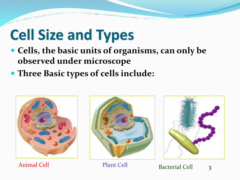
Cell Size and Types Cells, the basic units of organisms, can only be
observed under microscope
Three Basic types of cells include:
Animal Cell Plant Cell Bacterial Cell 3


Prokaryotes – The first Cells
Cells that lack a nucleus or membrane-bound organelles
Includes bacteria
Simplest type of cell
Single, circular chromosome
First cell type on earth
Cell type of Bacteria and Archaea
Nucleoid = region of DNA concentration

Eukaryotes
Cells that HAVE a nucleus and membrane-bound organelles
Includes protists, fungi, plants, and animals
More complex type of cells

Ultra structure of cell Ultrastructure (or ultra-structure) is the
nanostructure of a biological specimen, such as a cell,tissue, or organ, at scales smaller than can be viewedwith light microscopy. It is viewed with ultramicroscopy or electron microscopy.
Such cellular structures as organelles, which allow thecell to function properly within its specifiedenvironment, can be examined at the ultrastructurallevel.

CELL: The cell is the living functional unit of all organisms.
An organism may be composed of one cell only (Unicellular) e.g. Bacteria and Algae or of several cells (Multicellular) e.g. Man. The cell exists in two forms:
1.Eukaryotic cell, which has a nucleus that is enclosed in a nuclear envelope and several membrane- limited compartments e.g. the human cell.
2.Prokaryotic cell which has no nucleus and is devoid of membrane-limited compartments e.g. the bacterial cell.

NUCLEUS This is the largest organelle of the cell often located in
the central part of the cytoplasm and enclosed in a double-layered nuclear membrane. Its shape usually corresponds to the shape of the cell in which it is found.
It contains a nucleolus/nucleoli (which produces ribosomal subunits) and chromatin (DNA).
The latter is the genetic material implicated in cell division and in the synthesis of several molecules particularly proteins.

The contents of thenucleus are present as aviscous, amorphousmass of materialenclosed by a complexnuclear envelope thatforms a boundarybetween the nucleusand cytoplasm.
4

Included within the nucleus of a typical interphase (i.e.,nonmitotic)cell are:(1) the chromosomes, which are present as highly
extended nucleoprotein fibers, termed chromatin;
(2) one or more nucleoli, which are irregularly shaped electron-dense structures that function in the synthesis of ribosomal RNA and the assembly of ribosomes
(3) the nucleoplasm, the fluid substance in which the solutes of the nucleus are dissolved; and
(4) the nuclear matrix, which is a protein-containing fibrillar network.
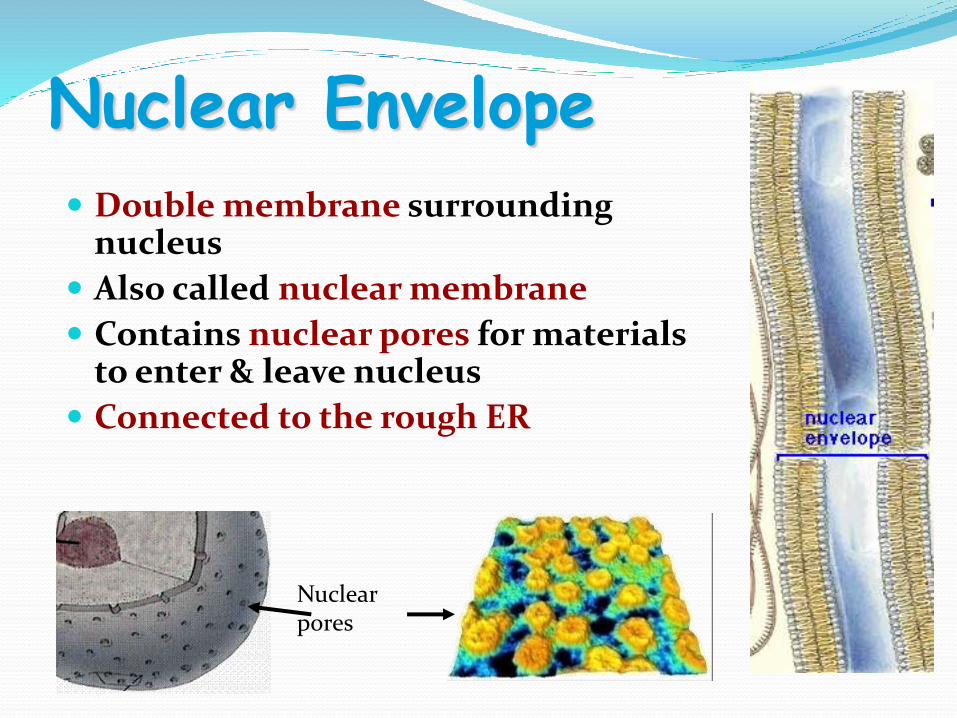
Nuclear Envelope
Double membrane surrounding nucleus
Also called nuclear membrane
Contains nuclear pores for materials to enter & leave nucleus
Connected to the rough ER
Nuclear pores

The Nuclear Envelope The separation of a cell’s genetic material from the
surrounding cytoplasm may be the single most important feature that distinguishes eukaryotes from prokaryotes, which makes the appearance of the nuclear envelope a landmark in biological evolution.
The nuclear envelope consists of two cellular membranes arranged parallel to one another and separated by 10 to 50 nm
The membranes of the nuclear envelope serve as a barrier that keeps ions, solutes, and macromolecules from passing freely between the nucleus and cytoplasm.
The two membranes are fused at sites forming circular pores that contain complex assemblies of proteins. The average mammalian cell contains several thousand nuclear pores.

The Structure of the Nuclear Pore Complex and Its Role inNucleocytoplasmic Exchange
The nuclear envelope is the barrierbetween the nucleus and cytoplasm,and nuclear pores are the gatewaysacross that barrier. Unlike the plasmamembrane, which prevents passage ofmacromolecules between thecytoplasm and the extracellular space,the nuclear envelope is a hub of activityfor the movement of RNAs and proteinsin both directions between the nucleusand cytoplasm.
The replication and transcription ofgenetic material within the nucleusrequire the participation of largenumbers of proteins that aresynthesized in the cytoplasm andtransported across the nuclearenvelope.
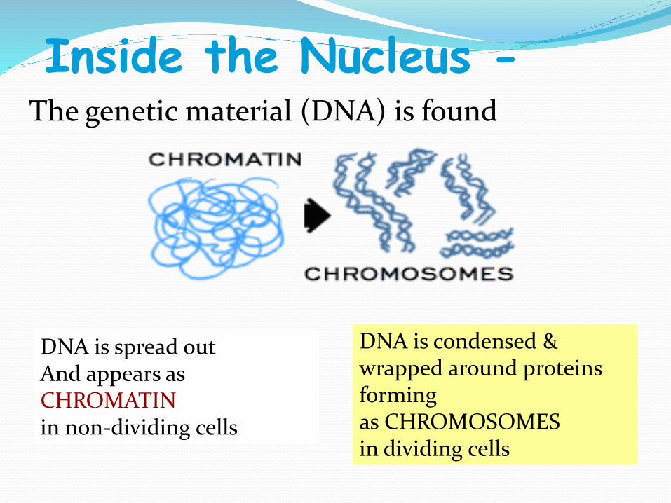
Inside the Nucleus -The genetic material (DNA) is found
DNA is spread out And appears as CHROMATINin non-dividing cells
DNA is condensed & wrapped around proteins forming as CHROMOSOMES in dividing cells

What Does DNA do?DNA is the hereditary
material of the cell
Genes that make up the DNA molecule code for different proteins
Mitochondria
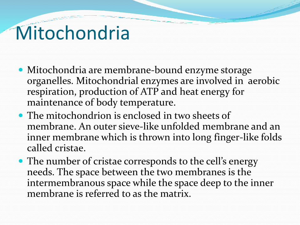
Mitochondria are membrane-bound enzyme storage organelles. Mitochondrial enzymes are involved in aerobic respiration, production of ATP and heat energy for maintenance of body temperature.
The mitochondrion is enclosed in two sheets of membrane. An outer sieve-like unfolded membrane and an inner membrane which is thrown into long finger-like folds called cristae.
The number of cristae corresponds to the cell’s energy needs. The space between the two membranes is the intermembranous space while the space deep to the inner membrane is referred to as the matrix.
The Matrix also contains chromosomes DNA, ribosomes, messenger RNA and Transfer RNA which are utilized in the synthesis of small amount of proteins for use within the matrix.
However the bulk of the proteins required in the mitochondrion is synthesised in the cytosol. The mitochondrial matrix also contains granules which store calcium ions.
The mitochondrion produces about 100 molecules of ATP per second.

Mitochondrion(plural = mitochondria)
“Powerhouse” of the cell
Generate cellular energy (ATP)
More active cells like muscle cells have MORE mitochondria
Both plants & animal cells have mitochondria
Site of CELLULAR RESPIRATION (burning glucose)
5
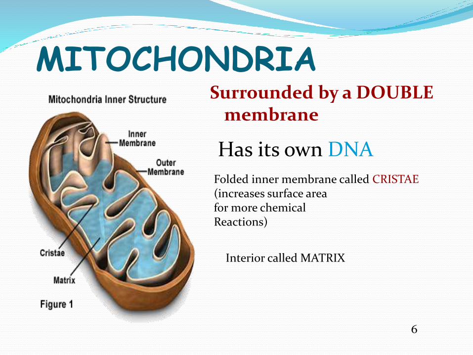
MITOCHONDRIASurrounded by a DOUBLE
membrane
Folded inner membrane called CRISTAE(increases surface areafor more chemical Reactions)
Has its own DNA
Interior called MATRIX
6

Interesting Fact --- Mitochondria Come
from cytoplasm in the EGG cell during fertilization
Therefore …
You inherit your mitochondria from your mother!

Muscle tissues are most commonly affected by mitochondrial deficiency diseases because of their high-energy metabolism.
Most mitochondrial diseases often result from chromosomal defect in the nucleus or in the mitochondrion.
Hereditary mitochondrial diseases are usually maternal in origin because only very few paternal mitochondria are left in the zygote following fertilization.

Rod shape
Cell Powerhouse
Mitochondrion( mitochondria )
7

THE ENDOPLASMIC RETICULUM (ER)
This organelle is made up of anastomosing network of intercommunicating channels/cisternae/sacs enclosed in a continuous membrane.
ER occurs in two forms, namely Rough and Smooth which are also interconnected. While the cisternae of smooth ER are tubular in shape, those of Rough ER are flattened. The roughness on the surface of rough ER is due to the adsorption of polyribosome on their outer surface.
Polyribosome also impacts the basophilic staining characteristic on RER. Furthermore, its membrane is continuous with that of the nuclear envelope.
Distribution and Functions of RER
RER is prominent in protein synthesising cells such as; Pancreatic acinarcells, cells of the endocrine glands, plasma cells, fibroblast etc.
Proteins synthesised in RER are stored in Lysosomes or granules; stored temporarily before exocytosis or used as integral membrane proteins.

Smooth Endoplasmic Reticulum (SER)
This is ER not bund to polyribosomes but continuous with RER and are less abundant is cell containing RER.
Distribution and Functions of SER
SER is found in all cells where they are involved in: ‘The synthesis phospholipids and cholesterol used in all cellular membranes including membranes of organelles.
They occur in abundance in other cells where they are involved in: Sequestration and release of Calcium ions a vital process in muscular contraction Biosynthesis of Lipids required for synthesis of steroid hormones
Detoxification of potentially harmful compounds such as alcohol and barbiturates

RIBOSOMES Ribosomes are small,
electron-dense particles not enclosed in membrane and are located in the cytosol.
Measuring about 20-30 nanometer they are basophilic and stained by all basic dyes.

Ribosome is composed of rRNA and about 80 different proteins. It usually occur in two subunits, large and small subunits.
The rRNA of the ribosome is synthesized in the nucleus while its protein is synthesized in the cytosol.
Ribosomes are involved in protein synthesis. While cytosolic proteins (free proteins) are synthesized by polyribosomes, secretory and endoplasmic reticulum proteins are synthesized on the membrane of rough endoplasmic reticulum.

THE CYTOPLASM
The fluid component of the cytoplasm is the Cytosol(pH 7.2) while the metabolically active contents of the cytoplasm are the Organelles.
Apart from being metabolically active, organelles are permanent residents of the cell which would survive cell division i.e. they reappear in the daughter cells following cell division.
Organelles occur in two forms, freely within the cytosol or enclosed in membrane. The cytoplasm also contains substances which are not metabolically active.
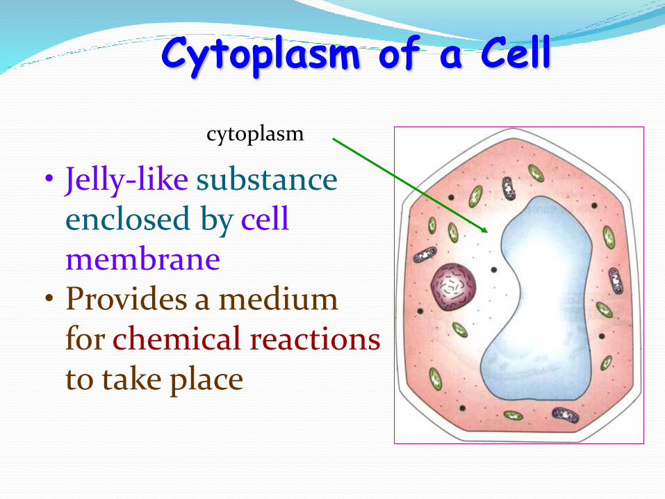
• Jelly-like substance enclosed by cell membrane
• Provides a medium for chemical reactionsto take place
Cytoplasm of a Cell
cytoplasm
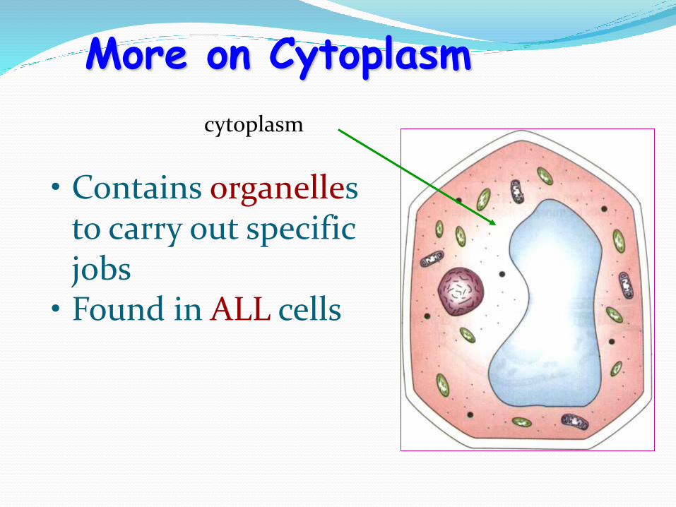
• Contains organelles to carry out specific jobs
• Found in ALL cells
More on Cytoplasm
cytoplasm

Chloroplasts Chloroplasts are large green organelles that are found
only in the cells of plants and algae, not in the cells ofanimals or fungi.
These organelles have an even more complex structurethan mitochondria: in addition to their two surroundingmembranes, chloroplasts possess internal stacks ofmembranes containing the green pigment chlorophyll
When a plant is kept in the dark, its greenness fades; whenput back in the light, its greenness returns. This suggeststhat the chlorophyll—and the chloroplasts that contain it

Animals and plants all need energy to live, grow, andreproduce.
Animals can use only the chemical energy they obtain byfeeding on the products of other living things. But plantscan get their energy directly from sunlight, andchloroplasts are the organelles that enable them to do so.From the standpoint of life on Earth, chloroplasts carry outan even more essential task than mitochondria: theyperform photosynthesis—that is, they trap the energy ofsunlight in chlorophyll molecules and use this energy todrive the manufacture of energy-rich sugar molecules.
In the process they release oxygen as a molecular by-product.
Plant cells can then extract this stored chemical energywhen they need it, by oxidizing these sugars in theirmitochondria, just as animal cells can. Chloroplasts thusgenerate both the food molecules and the oxygen that allmitochondria use.

Chloroplasts Found only in producers
(organisms containing chlorophyll)
Use energy from sunlight to make own food (glucose)
Energy from sun stored in the Chemical Bonds of Sugars

Chloroplasts Surrounded by DOUBLE
membrane
Outer membrane smooth
Inner membrane modified into sacs called Thylakoids
Thylakoids in stacks called Grana & interconnected
Stroma – gel like material surrounding thylakoids

Like mitochondria, they are surrounded by two membranes, an outer membrane that is permeable to small molecules and ions, and an inner membrane that encloses the internal compartment.
This compartment contains many flattened, membrane-surrounded vesicles or sacs, the thylakoids, usually arranged in stacks called grana.

Embedded in the thylakoid membranes (commonly called lamellae) are the photosynthetic pigments and the enzyme complexes that carry out the light reactions and ATP synthesis.
The stroma (the aqueous phase enclosed by the inner membrane) contains most of the enzymes required for the carbon assimilation reactions.
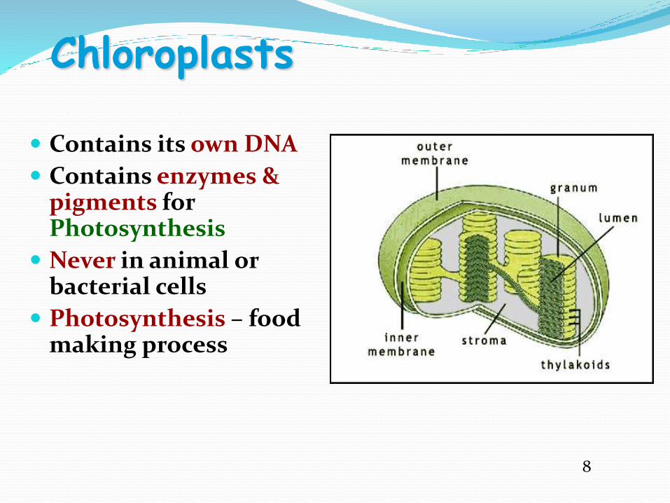
Chloroplasts
Contains its own DNA
Contains enzymes & pigments for Photosynthesis
Never in animal or bacterial cells
Photosynthesis – food making process
8

Cytoskeleton Helps cell maintain cell
shape
Also help move organellesaround
Made of proteins
Microfilaments are threadlike & made of ACTIN
Microtubules are tubelike& made of TUBULIN

Centrioles
Found only in animal cells
Paired structures near nucleus
Made of bundle of microtubules
Appear during cell division forming mitotic spindle
Help to pull chromosome pairs apart to opposite ends of the cell
9

Golgi Bodies
Look like a stack of pancakes
Modify, sort, & package
molecules from ER
for storage OR transport out of cell

Lysosomes• Contain digestive
enzymes
• Break down food, bacteria, and worn out cell parts for cells
• Programmed for cell death (AUTOLYSIS)
• Lyse (break open) & release enzymes to break down & recycle cell parts)

Lysosome Digestion
• Cells take in food by phagocytosis
• Lysosomes digest the food & get rid of wastes

Cilia & Flagella:
Made of protein tubes called microtubules
Microtubules arranged (9 + 2 arrangement)
Function in moving cells, in moving fluids, or in small particles across the cell surface
10

Cilia & Flagella
Cilia are shorter and more numerous on cells
Flagella are longer and fewer (usually 1-3) on cells
11

Vacuoles
Fluid filled sacks for
storage
Small or absent in
animal cells
Plant cells have a
large Central
Vacuole
No vacuoles in
bacterial cells
12

Vacuoles
In plants, they store Cell Sap
Includes storage of sugars,proteins, minerals, lipids,wastes, salts, water, andenzymes

Contractile Vacuole
Found in unicellular protists like paramecia
Regulate water intake by pumping out excess (homeostasis)
Keeps the cell from lysing (bursting)
51
Contractile vacuole animation13

Chloroplasts Found only in producers
(organisms containing chlorophyll)
Use energy from sunlight to make own food (glucose)
Energy from sun stored in the Chemical Bonds of Sugars

Chloroplasts Surrounded by DOUBLE
membrane
Outer membrane smooth
Inner membrane modified into sacs called Thylakoids
Thylakoids in stacks called Grana & interconnected
Stroma – gel like material surrounding thylakoids
14

Like mitochondria, they are surrounded by two membranes, an outer membrane that is permeable to small molecules and ions, and an inner membrane that encloses the internal compartment.
This compartment contains many flattened, membrane-surrounded vesicles or sacs, the thylakoids, usually arranged in stacks called grana.

Embedded in the thylakoid membranes (commonly called lamellae) are the photosynthetic pigments and the enzyme complexes that carry out the light reactions and ATP synthesis.
The stroma (the aqueous phase enclosed by the inner membrane) contains most of the enzymes required for the carbon assimilation reactions.

Chloroplasts:
Contains its own DNA
Contains enzymes & pigments for Photosynthesis
Never in animal or bacterial cells
Photosynthesis – food making process

Vacuoles:
Fluid filled sacks for storage
Small or absent in animal cells
Plant cells have a large Central Vacuole
No vacuoles in bacterial cells

Vacuoles
In plants, they store Cell Sap
Includes storage of sugars, proteins, minerals, lipids, wastes, salts, water, and enzymes

Contractile Vacuole
Found in unicellular protists like paramecia
Regulate water intake by pumping out excess (homeostasis)
Keeps the cell from lysing (bursting)
59
Contractile vacuole animation

Microtubules: Microtubules are a component of the cytoskeleton,
found throughout the cytoplasm.
These tubular polymers of tubulin can grow as long as 50 micrometres, with an average length of 25 µm, and are highly dynamic.
The outer diameter of a microtubule is about 24 nm while the inner diameter is about 12 nm. They are found in eukaryotic cells and are formed by the polymerization of a dimer of two globular proteins, alpha and beta tubulin.

Microtubules are very important in a number of cellular processes. They are involved in maintaining the structure of the cell and, together with microfilaments and intermediate filaments, they form the cytoskeleton. They also make up the internal structure of cilia and flagella.
They provide platforms for intracellular transport and are involved in a variety of cellular processes, including the movement of secretory vesicles, organelles, and intracellular substances (see entries for dynein and kinesin).
They are also involved in cell division (mitosis and meiosis), including the formation of mitotic spindles, which are used to pull apart eukaryotic chromosomes.

Microtubules are nucleated and organized in microtubule organizing centres (MTOCs), such as the centrosome or the basal bodies found in cilia and flagella. These MTOCs may or may not possess centrioles.
There are many proteins that bind to microtubules, including motor proteins such as kinesin and dynein, severing proteins like katanin, and other proteins important for regulating microtubule dynamics

Microfilaments: Microfilaments or actin filaments are the thinnest
filaments of the cytoskeleton, a structure found in the cytoplasm of eukaryotic cells.
These linear polymers of actin subunits are flexible and relatively strong, resisting buckling by multi-piconewtoncompressive forces and filament fracture by nanonewtontensile forces.
Microfilaments are highly versatile, functioning in cytokinesis, amoeboid movement, and changes in cell shape. In inducing this cell motility, one end of the actin filament elongates while the other end contracts, presumably by myosin II molecular motors.

Additionally, they function as part of actomyosin-driven contractile molecular motors, wherein the thin filaments serve as tensile platforms for myosin's ATP-dependent pulling action in muscle contraction and pseudopod advancement.
Microfilaments have a tough, flexible framework which helps the cell in movement.
Peroxisomes and Glyoxysomes: Peroxisomes, also known as microbodies, are single
membrane cellular organelles. They are spherical or oval in shape and contain the enzyme catalase.
Catalase protects the cell from the toxic effects of H2O2, by converting it to H2O and O2.
Peroxisomes are also involved in the oxidation of long chain fatty acids (> C18), and synthesis of plasmalogens and glycolipids.

Plants contain glyoxysomes, a specialized type ofperoxisomes, which are involved in the glyoxylate pathway
Peroxisome biogenesis disorders (PBDs), are a group of rarediseases involving the enzyme activities of peroxisomes.The biochemical abnormalities associated with PBDsinclude increased levels of very long chain fatty acids anddecreased concentrations of plasmalogens.
The most severe form of PBDs is Zellweger syndrome, acondition characterized by the absence of functionalperoxisomes.
The Victims of this disease may die within one year afterbirth

Different type of cells and tissue A tissue is a group of cells that have a similar shape and
function. Different types of tissues can be found indifferent organs. In humans, there are four basic typesof tissue: epithelial, connective, muscular, andnervous tissue. There may be various sub-tissueswithin each of the primary tissues.
1. Epithelium - lines and covers surfaces
2. Connective tissue - protect, support, and bind together
3. Muscular tissue - produces movement
4. Nervous tissue - receive stimuli and conduct impulses
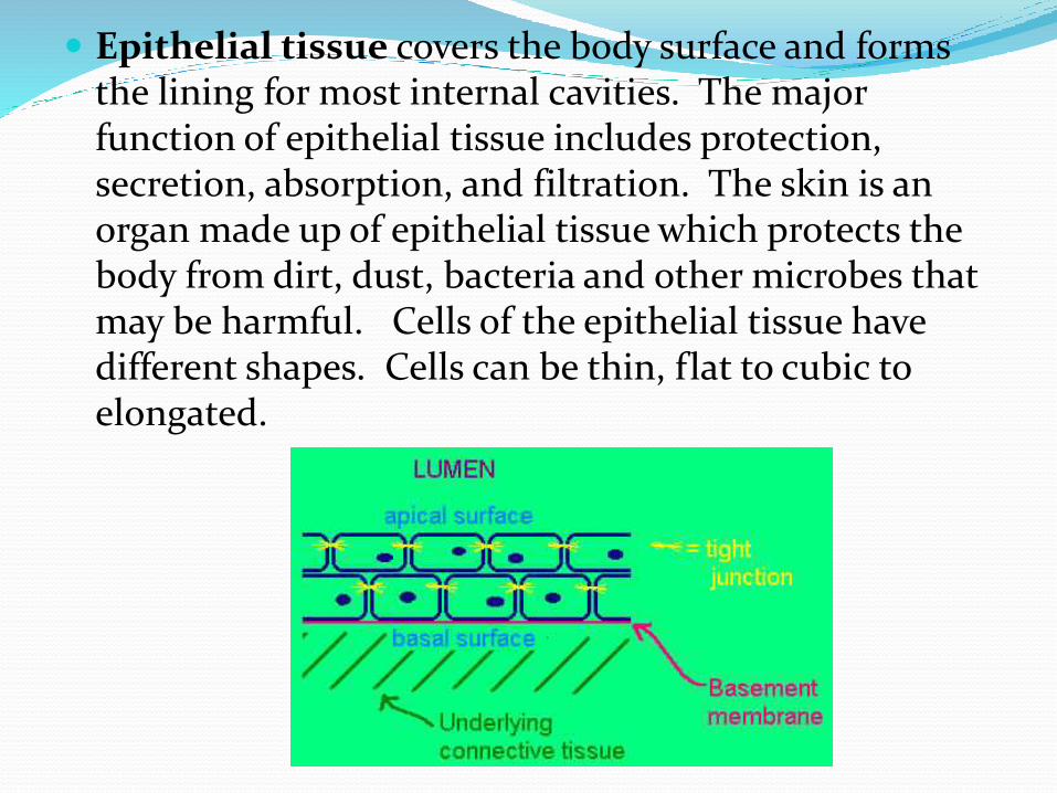
Epithelial tissue covers the body surface and forms the lining for most internal cavities. The major function of epithelial tissue includes protection, secretion, absorption, and filtration. The skin is an organ made up of epithelial tissue which protects the body from dirt, dust, bacteria and other microbes that may be harmful. Cells of the epithelial tissue have different shapes. Cells can be thin, flat to cubic to elongated.

The functions of epithelial tissues are:
Protection-- as a barrier between the outer world (or inner spaces) and our bodies.
Secretion-- when our bodies need to release material, like hormones into the blood, this tissue has to allow for such material to pass through. Often, it is the cells within the epithelial tissue that make the material for secretion.
Absorption-- epithelial tissue facing our digestive tract has to be very good at absorbing nutrients from the digestive tract lumen in order for us to get what we need from what we eat.
Excretion-- epithelial tissue even lines the excretory lumina, like the tracts from the kidneys through to the urethra.
15

Connective tissue is the most abundant and the mostwidely distributed of the tissues. Connective tissuesperform a variety of functions including support andprotection. The following tissues are found in thehuman body, ordinary loose connective tissue, fattissue, dense fibrous tissue, cartilage, bone, blood, andlymph, which are all considered connective tissue.
16

There are three types ofmuscle tissue: skeletal,smooth, andcardiac. Skeletal muscle is avoluntary type of muscletissue that is used in thecontraction of skeletalparts. Smooth muscle isfound in the walls of internalorgans and blood vessels. Itis an involuntary type. Thecardiac muscle is found onlyin the walls of the heart andis involuntary in nature.
17

Nerve tissue is composed of specialized cells which not only receive stimuli but also conduct impulses to and from all parts of the body. Nerve cells or neurons are long and string-like.
18
In tissues the simplest combination is called a membrane, or a sheet of tissues which cover or line the body surface or divide organs into parts. Examples include the mucous membrane which lines body cavities. Tissues combine to form organs. An organ is a part of the body which performs a definite function. The final units of organization in the body are called systems. A system is a group of organs each of which contributes its share to the function of the body as a whole.

References Images references:
1. http://micro.magnet.fsu.edu/primer/museum/janssen.htmla
2. http://en.wikipedia.org/wiki/White_blood_cell
3. http://papapipi.com/tag/plant-cell
4. http://www.uic.edu/classes/bios/bios100/summer2002/nucleus.jpg
5-12 http://biology4isc.weebly.com/cell-organelles.html
13-18 Cell and Molecular Biology, 6th Ed By Karp
Reading references:
Cell and Molecular Biology, 6th Ed By Karp
Molecular Cell Biology by Lodish 5th Edition