B.Sc. Microbiology/Biotech II Cell biology and Genetics Unit 2 cell cycle
Upload
rai-universityCategory
view
72download
0
Cell biology and geneticsCell Membrane (Transport)
Unit 3

Cell Membrane (Transport)Cell Membrane and Cell Wall:• ALL cells have a cell membrane made of proteins and lipids
Cell Membrane
lipid bilayer
protein channel
protein pump
Layer 1
Layer 2
• SOME cells have cell membranes and cell walls – ex: plants, fungi and bacteria
Cell Membrane
Cell Wall
1

• Plant cells have a cell wall made of cellulose – that cellulose is fiber in our diet
• Bacteria and fungi also have cell walls, but they do not contain cellulose
• Cell membranes and cell walls are porous allowing water, carbon dioxide, oxygen and nutrients to pass through easily

Function of the Cell Membrane:• Cell membrane separates the components of a cell
from its environment—surrounds the cell• “Gatekeeper” of the cell—regulates the flow of
materials into and out of cell—selectively permeable• Cell membrane helps cells maintain homeostasis—
stable internal balance
2
• The plasma membrane surrounding animal cells is where the exchange of substances inside and outside of cells takes place.
• Some substances need to move from the extracellular fluid outside cells to the inside of the cell, and some substances need to move from the inside of the cell to the extracellular fluid.
5

• Some of the proteins that are stuck in the plasma membrane help to form openings (channels) in the membrane.
• Through these channels, some substances such as hormones or ions are allowed to pass through.
• They either are “recognized” by a receptor (a protein molecule) within the cell membrane, or they attach to a carrier molecule, which is allowed through the channels.
6
• Because the plasma membrane is choosy about what substances can pass through it, it is said to be selectively permeable.
• Selectively permeable or semi permeable means that only certain substances are able to pass through the membrane.
7

Transport of substances
Macromolecules transport
Endocytosis
Phagocytosis Pinocytosis
Exocytosis
Micromolecules transport
Passive transport
Simple diffusion
Facilitated diffusion
Active transport
8

Micromolecules transport
• In this type of transport, molecules cross the membrane either directly via the lipid layer or with the help of some specific proteins.
• There are two types of transport:Passive transportActive transport
9

10
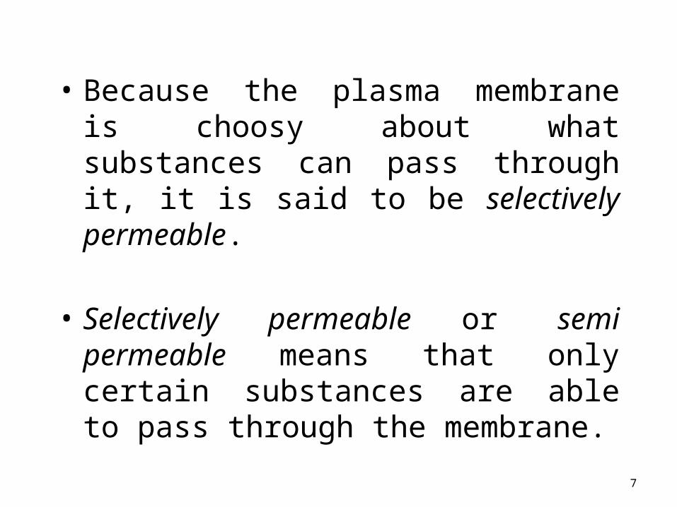
Passive TransportSimple Diffusion
• Requires NO energy
• Molecules move from area of HIGH to LOW concentration

11
Diffusion through a Membrane
Cell membrane
•Solute moves DOWN concentration gradient (HIGH to LOW)•Example: Oxygen or water diffusing into a cell and carbon dioxide diffusing out.

• Diffusion is the movement of small particles across a selectively permeable membrane like the cell membrane until equilibrium is reached.
These particles move from an area of high concentration to an area of low concentration.
outside of cell
inside of cell
DIFFUSION
HIGH to LOW concentration
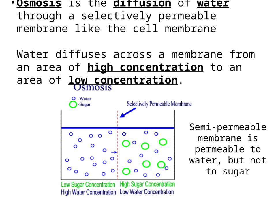
• Osmosis is the diffusion of water through a selectively permeable membrane like the cell membrane
Water diffuses across a membrane from an area of high concentration to an area of low concentration.
Semi-permeable membrane is
permeable to water, but not to sugar

15
Osmosis• Diffusion of water
across a membrane• Moves from HIGH
water potential (low solute) to LOW water potential (high solute)
Diffusion across a membrane
Semipermeable membrane

Hypertonic Solutions: contain a high concentration of solute relative to another solution (e.g. the cell's cytoplasm). When a cell is placed in a hypertonic solution, the water diffuses out of the cell, causing the cell to shrivel.
Hypotonic Solutions: contain a low concentration of solute relative to another solution (e.g. the cell's cytoplasm). When a cell is placed in a hypotonic solution, the water diffuses into the cell, causing the cell to swell and possibly explode.
Isotonic Solutions: contain the same concentration of solute as another solution (e.g. the cell's cytoplasm). When a cell is placed in an isotonic solution, the water diffuses into and out of the cell at the same rate. The fluid that surrounds the body cells is isotonic.

17
Passive Transport
Facilitated diffusion
Doesn’t require energy
Uses transport proteins to move high to low concentrationExamples: Glucose or amino acids moving from blood into a cell.
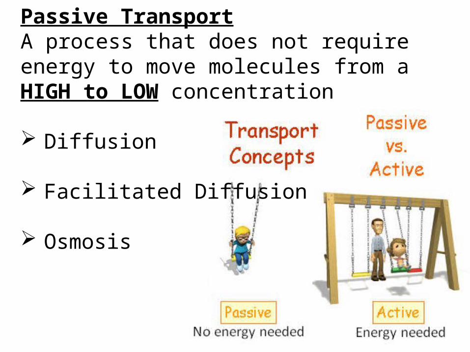
Passive TransportA process that does not require energy to move molecules from a HIGH to LOW concentration
Diffusion
Facilitated Diffusion
Osmosis

19
Proteins Are Critical to Membrane Function

Permeability• The permeability of a membrane is the rate of
passive diffusion of molecules through the membrane. These molecules are known as permeant molecules.
• Permeases• Any of the proteins that mediate the transport of vario
us molecules across biological membranes.
20

Transport protein
Carrier proteins mediated transport
• Carrier proteins are also called as (transporters or permease)
• non- covalently bind to specific molecules to be transported
• Undergo conformation change• Example: glucose mediated transporter(GLUT)

Channel protein mediated transport
• It forms open pores through the membrane• Allow free diffusion of any molecules with
appropriate size and shape• Concern with specifically with inorganic ion
transport called ion channels.– Highly specific– Not permanently open– Voltage gated channels– Ligand gated channels– Aqueaporins( water transport)
24
Types of Transport Proteins• Channel proteins are embedded in the
cell membrane & have a pore for materials to cross
• Carrier proteins can change shape to move material from one side of the membrane to the other

Cannel protein Carrier protein
Down to their concentration gradient
Can be uniport or symport or antiport(co-transport)
Gated or non-gated protein Single solute is pass at one time
Many be passive Many be active or passive
Faster rate Slower rate of transport

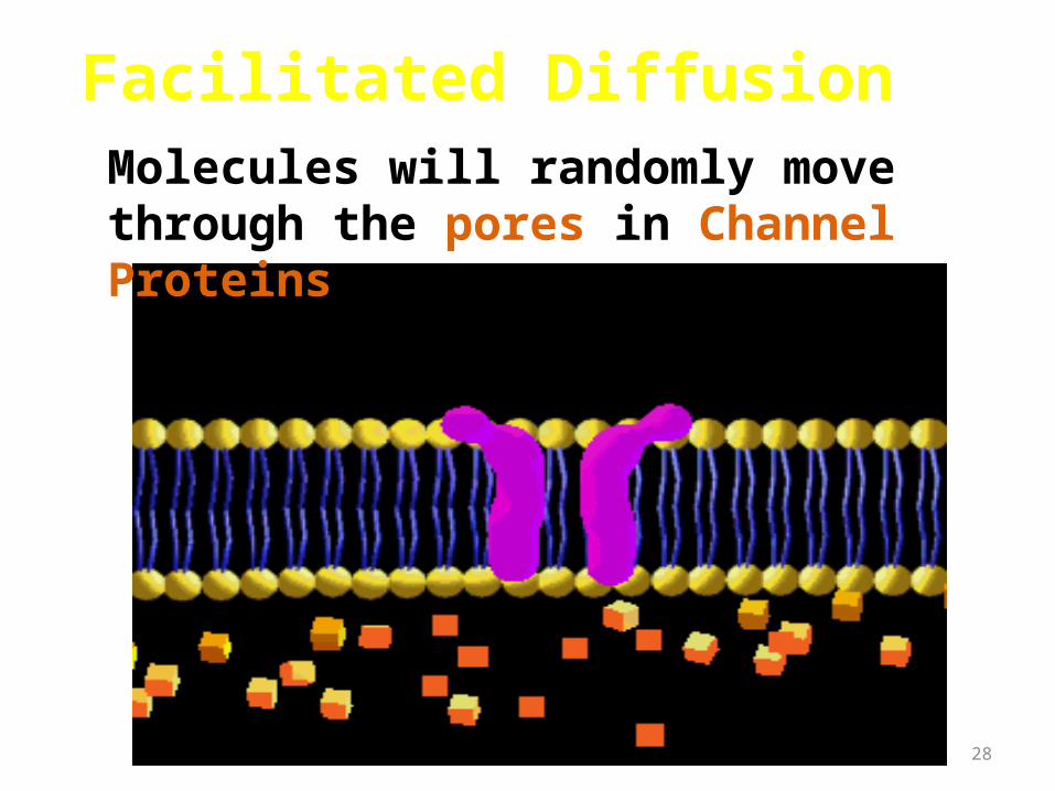
• Facilitated Diffusion is the movement of larger molecules like glucose through the cell membrane – larger molecules must be “helped”
Proteins in the cell membrane form channels for large molecules to pass through
Proteins that form channels (pores) are called protein channels
outside of cell
inside of cell
Glucose molecules
28
Facilitated Diffusion
Molecules will randomly move through the pores in Channel Proteins.

29
Facilitated Diffusion
• Some Carrier proteins do not extend through the membrane.
• They bond and drag molecules through the lipid bilayer and release them on the opposite side.

30
Carrier Proteins• Other carrier
proteins change shape to move materials across the cell membrane
31
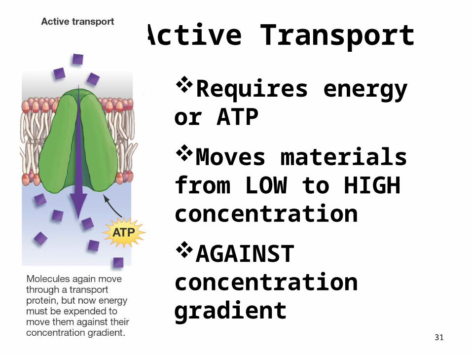
Active Transport
Requires energy or ATP
Moves materials from LOW to HIGH concentration
AGAINST concentration gradient

Active TransportActive transport is the movement of molecules from LOW to HIGH concentration.
Energy is required as molecules must be pumped against the concentration gradient.
Proteins that work as pumps are called protein pumps.
Ex: Body cells must pump carbon dioxide out into the surrounding blood vessels to be carried to the lungs for exhale. Blood vessels are high in carbon dioxide compared to the cells, so energy is required to move the carbon dioxide across the cell membrane from LOW to HIGH concentration.
outside of cell
inside of cell
Carbon Dioxide molecules

Primary active transport
• Primary active transport involves using energy (usually through ATP hydrolysis) at the membrane protein itself to cause a conformational change that results in the transport of the molecule through the protein.
33
34
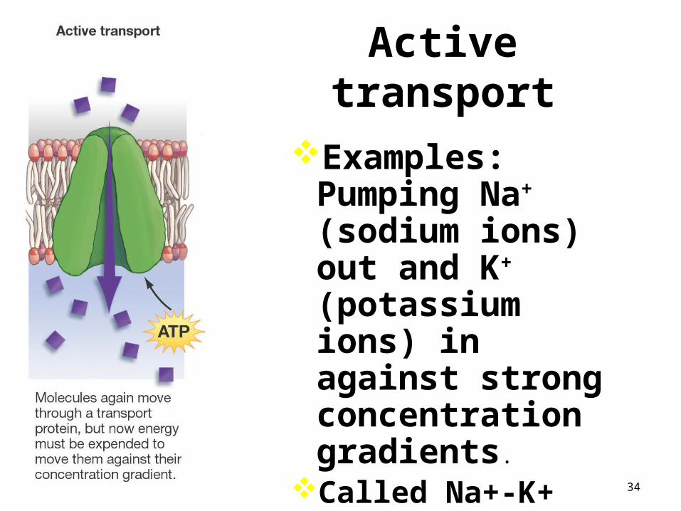
Active transport
Examples: Pumping Na+ (sodium ions) out and K+ (potassium ions) in against strong concentration gradients.
Called Na+-K+ Pump

Secondary active transport
• Secondary active transport involves using energy to establish a gradient across the cell membrane, and then utilizing that gradient to transport a molecule of interest up its concentration gradient.
• An example of this mechanism is as follows: • E. coli establishes a proton (H+) gradient across the cell membrane
by using energy to pump protons out of the cell. Then those protons are coupled to lactose at the lactose permease transmembrane protein. The lactose permease uses the energy of the proton moving down its concentration gradient to transport lactose into the cell.
35
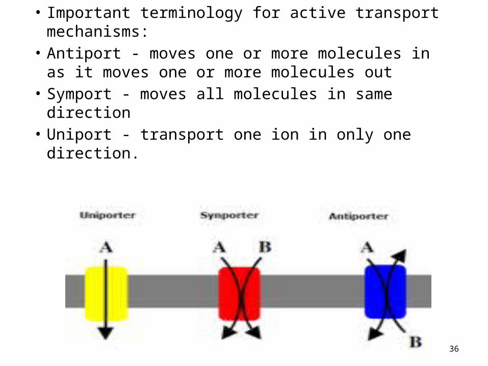
• Important terminology for active transport mechanisms:
• Antiport - moves one or more molecules in as it moves one or more molecules out
• Symport - moves all molecules in same direction• Uniport - transport one ion in only one direction.
36

Macromolecules transport
• Sometimes, the molecules are just too big to easily flow across the plasma membranes or dissolve in the water so that they can be filtered through the membrane.
• Two types are:• Endocytosis• Exocytosis
37

Transport of macromolecule across PM
• Vesicles are used to transport large particles across the PM.– Requires energy
• Types:– Exocytosis– Endocytosis
• Phagocytosis, pinocytosis, receptor-mediated

39
Moving the “Big Stuff” inside the cell
•Large molecules move materials into the cell by endocytosis.
•The process by which the cell membrane of the cell folds inwards to ingest material is called endocytosis

Vesicle forming
Endocytosis
Endocytosis can occur in three ways• Phagocytosis ("cell eating")• Pinocytosis ("cell drinking")• Receptor-mediated endocytosis

Endocytosis
• Endocytosis– PM sinks inward, pinches off and forms a vesicle– Vesicle often merges with Golgi for processing and
sorting of its contents

Endocytosis - terms
• Phagocytosis – cell eating– Membrane sinks in and captures solid particles for
transport into the cell – Examples:
• Solid particles often include: bacteria, cell debris, or food
• Pinocytosis – cell drinking– Cell brings in a liquid

Endocytosis - comments
• Phagocytosis and pinocytosis are not selective– Membrane sinks inward and captures whatever
particles/fluid present.– Vesicle forms and merges with the Golgi body…

44
Pinocytosis
• Cell forms an invagination
• Materials dissolve in water to be brought into cell
• Called “Cell Drinking”

Receptor Mediated Endocytosis
• Receptor Mediated Endocytosis is a highly specific form of endocytosis.
– Receptor proteins on the outside of the cell bind specific substances and bring them into the cell by endocytosis
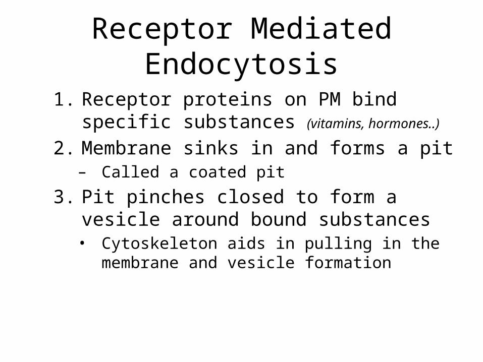
Receptor Mediated Endocytosis
1. Receptor proteins on PM bind specific substances (vitamins, hormones..)
2. Membrane sinks in and forms a pit– Called a coated pit
3. Pit pinches closed to form a vesicle around bound substances• Cytoskeleton aids in pulling in the membrane and
vesicle formation

47
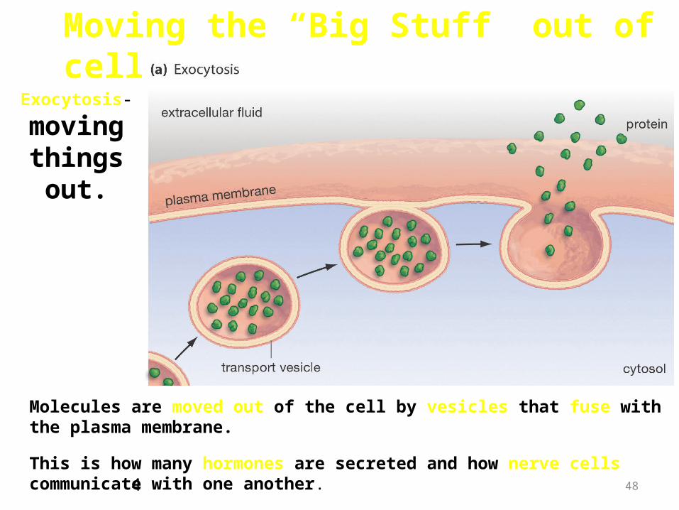
Exocytosis The opposite of endocytosis is exocytosis. Large molecules that are manufactured in the cell are released through the
cell membrane.
Inside Cell Cell environment3
48
Moving the “Big Stuff” out of cell
Molecules are moved out of the cell by vesicles that fuse with the plasma membrane.
Exocytosis-
moving things out.
This is how many hormones are secreted and how nerve cells communicate with one another. 4

Food is moved into the cell by Endocytosis
Wastes are moved out of the cell by Exocytosis
• Endocytosis and Exocytosis is the mechanism by which very large molecules (such as food and wastes) get into and out of the cell

Ex: White Blood Cells, which are part of the immune system, surround and engulf bacteria by endocytosis.

Bulk Flow
• Exocytosis– Cytoplasmic vesicle merges with the PM
and releases its contents– Example:
• Golgi body vesicles merge with the PM an release their contents
• How nerve cells release neurotransmittors

APOPTOSIS

INTRODUCTION
Cell death by injury-Mechanical damage
-Exposure to toxic chemicals Cell death by suicide
-Internal signals-External signals
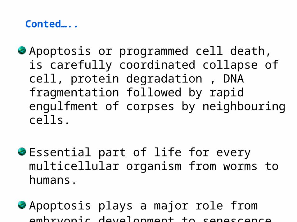
Conted…..
Apoptosis or programmed cell death, is carefully coordinated collapse of cell, protein degradation , DNA fragmentation followed by rapid engulfment of corpses by neighbouring cells.
Essential part of life for every multicellular organism from worms to humans.
Apoptosis plays a major role from embryonic development to senescence.

Why should a cell commit suicide?
Apoptosis is needed for proper developmentExamples: – The resorption of the tadpole tail – The formation of the fingers and toes of the fetus – The sloughing off of the inner lining of the uterus
– The formation of the proper connections between neurons in the brain
Apoptosis is needed to destroy cells
Examples: – Cells infected with viruses – Cells of the immune system – Cells with DNA damage – Cancer cells

What makes a cell decide to commit suicide?
Withdrawal of positive signalsexamples : – growth factors for neurons – Interleukin-2 (IL-2)
Receipt of negative signals examples : – increased levels of oxidants within the cell – damage to DNA by oxidants – death activators :
• Tumor necrosis factor alpha (TNF-) • Lymphotoxin (TNF-β) • Fas ligand (FasL)
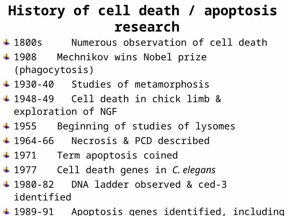
History of cell death / apoptosis research
1800s Numerous observation of cell death1908 Mechnikov wins Nobel prize (phagocytosis) 1930-40 Studies of metamorphosis1948-49 Cell death in chick limb & exploration of NGF1955 Beginning of studies of lysomes1964-66 Necrosis & PCD described1971 Term apoptosis coined1977 Cell death genes in C. elegans1980-82 DNA ladder observed & ced-3 identified1989-91 Apoptosis genes identified, including bcl-2,
fas/apo1 & p53, ced-3 sequenced(Richerd et.al., 2001)

Necrosis vs. Apoptosis
• Cellular condensation• Membranes remain intact• Requires ATP• Cell is phagocytosed, no
tissue reaction • Ladder-like DNA
fragmentation• In vivo, individual cells appear
affected
• Cellular swelling• Membranes are broken• ATP is depleted• Cell lyses, eliciting an
inflammatory reaction • DNA fragmentation is
random, or smeared• In vivo, whole areas of the
tissue are affected
Necrosis Apoptosis

Apoptosis• Apoptotic cells can be recognized by stereotypical morphological
changes: the cell shrinks, shows deformation and looses contact to its neighbouring cells. Its chromatin condenses and marginates at the nuclear membrane, the plasma membrane is blebbing or budding, and finally the cell is fragmented into compact membrane-enclosed structures, called 'apoptotic bodies' which contain cytosol, the condensed chromatin, and organelles.
• The apoptotic bodies are engulfed by macrophages and thus are removed from the tissue without causing an inflammatory response.
• Those morphological changes are a consequence of characteristic molecular and biochemical events occurring in an apoptotic cell, most notably the activation of proteolytic enzymes which eventually mediate the cleavage of DNA into oligonucleosomal fragments as well as the cleavage of a multitude of specific protein substrates which usually determine the integrity and shape of the cytoplasm or organelles

NECROSIS
• Apoptosis is in contrast to the necrotic mode of cell-death in which case the cells suffer a major insult, resulting in a loss of membrane integrity, swelling and disrupture of the cells.
• During necrosis, the cellular contents are released uncontrolled into the cell's environment which results in damage of surrounding cells and a strong inflammatory response in the corresponding tissue

NECROSIS Vs APOPTOSIS
Wilde, 19995
References • Images references:• 1-5 Cell and Molecular Biology, 6th Ed By Karp
• Reading references:• Cell and Molecular Biology, 6th Ed By Karp• Molecular Cell Biology by Lodish 5th Edition