Biotechnological Potential of the Brazilian Caatinga Biome · Cebil “Angico-vermelho”...
17
_____________________________________________________________________________________________________ *Corresponding author: E-mail: [email protected]; Advances in Research 5(1): 1-17, 2015, Article no.AIR.17426 ISSN: 2348-0394 SCIENCEDOMAIN international www.sciencedomain.org Biotechnological Potential of the Brazilian Caatinga Biome Iasmim Lucas da Silva 1 , Luana Cassandra Breitenbach Barroso Coelho 2* and Leonor Alves de Oliveira da Silva 1 1 Department of Antibiotics, Biological Sciences Center, Federal University of Pernambuco, Arthur de Sá, S/N, City University, Recife-PE, 50670-901, Brazil. 2 Department of Biochemistry, Biological Sciences Center, Federal University of Pernambuco, Avenue Moraes Rego, S/N, City University, Recife-PE, 50670-420, Brazil. Authors’ contributions This work was carried out in collaboration between all authors. Authors ILS and LAOS managed the literature searches and wrote the first draft of the manuscript. Author LCBBC designed the study and managed the study performed. All authors read and approved the final manuscript. Article Information DOI: 10.9734/AIR/2015/17426 Editor(s): (1) Monica Butnariu, Department of Chemistry & Biochemistry, Banat’s University of Agricultural Sciences and Veterinary Medicine from Timisoara, Romania. Reviewers: (1) Anonymous, Malaysia. (2) Tunira Bhadauria, Department of Zoology, Kanpur University, India. (3) Anonymous, China. Complete Peer review History: http://www.sciencedomain.org/review-history.php?iid=1154&id=31&aid=9431 Received 13 th March 2015 Accepted 22 nd April 2015 Published 26 th May 2015 ABSTRACT The Caatinga biome is a unique Brazilian biome predominant in the Northeast of Brazil and situated in the Semiarid Region. The rhizosphere comprises the narrow zone of soil that is directly influenced by the roots of plants and associated soil microorganisms. It is a dynamic environment with maximum microbial activity due to the presence of root exudates and radicular secretions representing the major carbon source readily available to microorganisms. The typical rhizospheric community in the Caatinga biome comprises microorganisms with different types of metabolism and adaptive responses to changes depending on soil temperature, plant species, nutritional status, age, stress, illness, and other factors. Assays for a variety of soil enzymes give an indication of the functional diversity assumed by the microbes present. A useful characteristic of the rhizosphere isolates is the ability of the rhizobacteria to excrete enzymes such as cellulases and L- asparaginases. Among the important species found in the Caatinga biome is Poincianella Review Article
-
Upload
nguyenphuc -
Category
Documents
-
view
216 -
download
0
Transcript of Biotechnological Potential of the Brazilian Caatinga Biome · Cebil “Angico-vermelho”...

_____________________________________________________________________________________________________ *Corresponding author: E-mail: [email protected];
Advances in Research 5(1): 1-17, 2015, Article no.AIR.17426
ISSN: 2348-0394
SCIENCEDOMAIN international www.sciencedomain.org
Biotechnological Potential of the Brazilian Caatinga Biome
Iasmim Lucas da Silva1, Luana Cassandra Breitenbach Barroso Coelho2*
and Leonor Alves de Oliveira da Silva1
1Department of Antibiotics, Biological Sciences Center, Federal University of Pernambuco, Arthur de Sá, S/N, City University, Recife-PE, 50670-901, Brazil.
2Department of Biochemistry, Biological Sciences Center, Federal University of Pernambuco,
Avenue Moraes Rego, S/N, City University, Recife-PE, 50670-420, Brazil.
Authors’ contributions
This work was carried out in collaboration between all authors. Authors ILS and LAOS managed the literature searches and wrote the first draft of the manuscript. Author LCBBC designed the study and
managed the study performed. All authors read and approved the final manuscript.
Article Information
DOI: 10.9734/AIR/2015/17426 Editor(s):
(1) Monica Butnariu, Department of Chemistry & Biochemistry, Banat’s University of Agricultural Sciences and Veterinary Medicine from Timisoara, Romania.
Reviewers: (1) Anonymous, Malaysia.
(2) Tunira Bhadauria, Department of Zoology, Kanpur University, India. (3) Anonymous, China.
Complete Peer review History: http://www.sciencedomain.org/review-history.php?iid=1154&id=31&aid=9431
Received 13th March 2015 Accepted 22
nd April 2015
Published 26th
May 2015
ABSTRACT
The Caatinga biome is a unique Brazilian biome predominant in the Northeast of Brazil and situated in the Semiarid Region. The rhizosphere comprises the narrow zone of soil that is directly influenced by the roots of plants and associated soil microorganisms. It is a dynamic environment with maximum microbial activity due to the presence of root exudates and radicular secretions representing the major carbon source readily available to microorganisms. The typical rhizospheric community in the Caatinga biome comprises microorganisms with different types of metabolism and adaptive responses to changes depending on soil temperature, plant species, nutritional status, age, stress, illness, and other factors. Assays for a variety of soil enzymes give an indication of the functional diversity assumed by the microbes present. A useful characteristic of the rhizosphere isolates is the ability of the rhizobacteria to excrete enzymes such as cellulases and L-asparaginases. Among the important species found in the Caatinga biome is Poincianella
Review Article
Silva et al.; AIR, 5(1): 1-17, 2015; Article no.AIR.17426
2
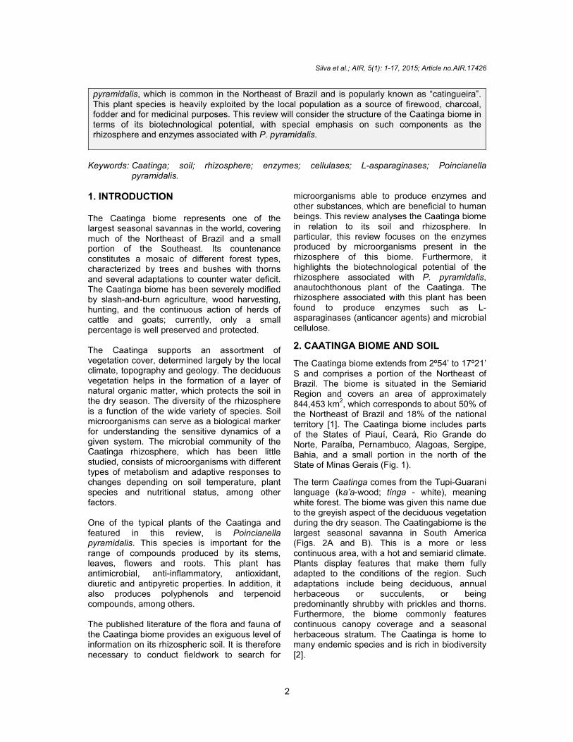
pyramidalis, which is common in the Northeast of Brazil and is popularly known as “catingueira”. This plant species is heavily exploited by the local population as a source of firewood, charcoal, fodder and for medicinal purposes. This review will consider the structure of the Caatinga biome in terms of its biotechnological potential, with special emphasis on such components as the rhizosphere and enzymes associated with P. pyramidalis.
Keywords: Caatinga; soil; rhizosphere; enzymes; cellulases; L-asparaginases; Poincianella
pyramidalis.
1. INTRODUCTION The Caatinga biome represents one of the largest seasonal savannas in the world, covering much of the Northeast of Brazil and a small portion of the Southeast. Its countenance constitutes a mosaic of different forest types, characterized by trees and bushes with thorns and several adaptations to counter water deficit. The Caatinga biome has been severely modified by slash-and-burn agriculture, wood harvesting, hunting, and the continuous action of herds of cattle and goats; currently, only a small percentage is well preserved and protected. The Caatinga supports an assortment of vegetation cover, determined largely by the local climate, topography and geology. The deciduous vegetation helps in the formation of a layer of natural organic matter, which protects the soil in the dry season. The diversity of the rhizosphere is a function of the wide variety of species. Soil microorganisms can serve as a biological marker for understanding the sensitive dynamics of a given system. The microbial community of the Caatinga rhizosphere, which has been little studied, consists of microorganisms with different types of metabolism and adaptive responses to changes depending on soil temperature, plant species and nutritional status, among other factors. One of the typical plants of the Caatinga and featured in this review, is Poincianella pyramidalis. This species is important for the range of compounds produced by its stems, leaves, flowers and roots. This plant has antimicrobial, anti-inflammatory, antioxidant, diuretic and antipyretic properties. In addition, it also produces polyphenols and terpenoid compounds, among others. The published literature of the flora and fauna of the Caatinga biome provides an exiguous level of information on its rhizospheric soil. It is therefore necessary to conduct fieldwork to search for
microorganisms able to produce enzymes and other substances, which are beneficial to human beings. This review analyses the Caatinga biome in relation to its soil and rhizosphere. In particular, this review focuses on the enzymes produced by microorganisms present in the rhizosphere of this biome. Furthermore, it highlights the biotechnological potential of the rhizosphere associated with P. pyramidalis, anautochthonous plant of the Caatinga. The rhizosphere associated with this plant has been found to produce enzymes such as L-asparaginases (anticancer agents) and microbial cellulose.
2. CAATINGA BIOME AND SOIL
The Caatinga biome extends from 2º54’ to 17º21’ S and comprises a portion of the Northeast of Brazil. The biome is situated in the Semiarid Region and covers an area of approximately 844,453 km
2, which corresponds to about 50% of
the Northeast of Brazil and 18% of the national territory [1]. The Caatinga biome includes parts of the States of Piauí, Ceará, Rio Grande do Norte, Paraíba, Pernambuco, Alagoas, Sergipe, Bahia, and a small portion in the north of the State of Minas Gerais (Fig. 1).
The term Caatinga comes from the Tupi-Guarani language (ka’a-wood; tinga - white), meaning white forest. The biome was given this name due to the greyish aspect of the deciduous vegetation during the dry season. The Caatingabiome is the largest seasonal savanna in South America (Figs. 2A and B). This is a more or less continuous area, with a hot and semiarid climate. Plants display features that make them fully adapted to the conditions of the region. Such adaptations include being deciduous, annual herbaceous or succulents, or being predominantly shrubby with prickles and thorns. Furthermore, the biome commonly features continuous canopy coverage and a seasonal herbaceous stratum. The Caatinga is home to many endemic species and is rich in biodiversity [2].

Silva et al.; AIR, 5(1): 1-17, 2015; Article no.AIR.17426
3
Fig. 1. Boundary of the Caatinga biome (solid line) superimposed over a map of the Northeast of Brazil showing state boundaries (dashed lines)
(A) (B)
Fig. 2. Caatinga vegetation during the dry season in the Sertão sub-region of the Northeast
of Brazil, Sousa, Paraíba State (A) and transition from rainy to dry season in the Agreste sub-region of the Northeast of Brazil, Brejo da Madre de Deus, Pernambuco State (B)
The Fabaceae family is one of the most representative of the Caatinga, comprising approximately 300 endemic species distributed among three subfamilies, Faboideae, Caesalpinioideae and Mimosoideae [3]. Forty-two species have been studied in terms of pollination and reproductive systems [4]. Only a few species have been analysed for their ability to fix nitrogen. Such systems highlight the importance of relationships among plants and their symbiotic microorganisms [4-6]. Different parts of plants of the Caatinga biome, such as leaves, flowers, fruits, stems and roots,
are made use of in various ways for folk medicine. These materials are commonly prepared as teas, syrups, macerations, poultices, and powders for inhalation (Table 1). Such widespread use in folk medicine has prompted research into the commercial and therapeutic potential of species from the Caatinga biome. Such research has played a key role in the discovery of new medicinal products [7]. Studies on the density and diversity of microorganisms in soils are scarce in areas of dry tropical conditions, especially in Caatinga soils. Various chemical and physical factors,

Silva et al.; AIR, 5(1): 1-17, 2015; Article no.AIR.17426
4
which include the availability of nutrients, organic matter, soil moisture and temperature, influence microorganisms in the soil [8]. In arid environments, all these factors are generally unfavorable for microbial growth in soil [9].
The characteristics of the Caatinga biome include high solar radiation, low cloud cover, high mean annual temperature, and very low rates of
relative humidity, high evapotranspiration potential, and most especially, low and irregular rainfall in most areas. According to Brinkmann et al. [10] rainfall constitutes one of the main limiting factors for availability of dry biomass in this area and, therefore, is closely linked to vegetation. Therefore, the Semiarid Region is a sensitive indicator of climate change.
Table 1. Plant species endemic to the Caatinga biome and their applications
References: [11,12,13,14,15,16,17,18,19]
Species Common names Utilization
Anadenanthera colubrinavar. Cebil
“Angico-vermelho” Reforestation, firewood and charcoal production. Shell: production of tannins, treatment of respiratory problems, inflammation, diarrhea, cough, bronchitis, flu, and toothache.
Amburana cearenses “Imburana-de-cheiro” Carpentry, cookery and perfumery. Medicinal properties to treat flu, cold, asthma, cramps, stroke, and body aches. The bark and seeds produce phenolic glycosides, flavonoids, phenolic acids, sucrose, glycosyl, glycosides and phytosterols.
Bromelia laciniosa “Macambira” Produces flavonoids. Leaves are food for animals and humans. Recommended for gastrointestinal problems, fever, jaundice, hepatitis and dandruff. Powdered leaves used in cooking as a source of protein.
Cereus jamacaru “Mandacuru” Used as expectorant, diuretic, anthelmintic and a cardiotonic agent.
Mimosa hostilis “Juremapreta” Bark and leaves used to treat burns and skin problems, in addition to presenting antimicrobial, cell regenerative and astringent effects and used as a pectoral painkiller.
Poincianella pyramidalis “Catingueira” Production of chemical compounds, used as popular medicine, with antimicrobial activity.
Spondias tuberosa “Umbuzeiro” Food resource for producing candy, ice cream and juices. Rich in ascorbic acid and mineral salts (calcium, potassium and magnesium); source of tannins and proteins.
Ziziphus joazeiro “Juazeiro” In folk medicine, used for cicatrizing, treating dermatitis and mycoses. The cortex of stalk and leaves is rich in saponins and used in the manufacture of dandruff shampoo and hair tonic as well as being used to wash fabrics, cotton and glass objects. In addition, the zest of the stem cortex, when dried and powdered, is used as toothpaste.

Silva et al.; AIR, 5(1): 1-17, 2015; Article no.AIR.17426
5
The soils of the Semiarid Region and Caatinga biome, produced by weathering of rocks of the Precambrian period, have a crystalline nature, while some areas feature sedimentary deposits. Thus, the predominant clay found is montmorillonite, a clay mineral with Grumosol or Vertisol characteristics, which is very common in level areas (pediplain) of the Caatinga [20]. The soils of the Caatinga biome have geomorphological expressions such as latosols, which are fairly uniform with high levels of weathering; argisols, characterised by high concentrations of clay which may contain yellow, red, gray and reddish-yellow colours; planosols, imperfectly or poorly drained soils showing abrupt transitions; luvisols, usually fairly deep to shallow soils containing clay; and neosols or undeveloped soils [5,6]. The Northeast of Brazil contains many areas with vertisols, clayey to loamy soils which experience changes in volume and is extremely hard with many cracks when dry. Vertisolsare found in areas of Caatinga, such as flat or gently undulating areas with depressions or sites of old ponds [20]. The Caatinga is currently in a severe process of desertification, caused mainly by deforestation and misuse of natural resources. Desertification results in reduced crop production and leads to changes in the interactions occurring in the soil, with consequent and often irreversible loss of biodiversity of plants typical of the biome. The microbial diversity of this soil is atypical to that of other biomes, as it developed under conditions of extreme temperatures and in clayey and stony soils which may have been influenced or not by local human actions [5,6]. The Department of Antibiotics of the Federal University of Pernambuco (UFPE) is isolating, qualifying and quantifying the microbial community from rhizospheric soil of “catingueira” (P. pyramidalis) to analyse the metabolites produced by its rhizobacteria and its biotechnological potential. The results obtained indicate that the rhizobacteria isolated have the ability to produce enzymes of industrial and pharmaceutical interest.
3. RHIZOSPHERE Soil is a complex environment with many important consequences for nature and humans. This system is characterized by a variety of physical, chemical and biological processes, which are often influenced by environmental
factors. Soil is responsible for providing many of the nutrients in food and acts as a natural system, playing an important role in the water cycle and nutrient turnover. Soil is an abiotic and biotic habitat that includes organisms such as plants, animals and humans as well as microorganisms such as fungi, bacteria and viruses [21]. Microorganisms play important ecological functions such as recycling and maintaining ecosystem health. They can facilitate the absorption of nutrients by plants, help in processes such as fixation of atmospheric nitrogen, change the availability and toxicity of metals, and promote bio-control and bioremediation, as well as the growth of plants. These microorganisms can occur in association with mineral particles and organic matter, and in the rhizosphere of plants [22]. The term “rhizosphere” (Fig. 3) refers to the area of soil affected by the roots of plants, and therefore, has high microbial activity. The rhizosphere has a high concentration of organic nutrients, which come from the roots and favor the development of microbiota. Roots influence the rhizosphere by releasing dead cells, mucilage, exudates, vitamins, carbohydrates, enzymes and other compounds into the soil [21].
Fig. 3. Rhizosphere of chamomile (Matricaria recutita)
The composition of the microbial mass of a particular rhizosphere may be influenced by numerous factors such as the species involved, plant age, types of root exudates, soil conditions and status imposed by the environment; the effects associated with the plant are highly selective [22,23]. The soil in the root region controls the growth and development of plants and the association with rhizosphere microflora promotes several beneficial activities. The

Silva et al.; AIR, 5(1): 1-17, 2015; Article no.AIR.17426
6
rhizosphere is important in processes related to the exchange of O2 and CO2 mineralization, plant nutrition, gradients between soil layers, nitrification, and symbiosis, among others [24-26]. Knowledge of soil microorganisms, besides being fundamental to the taxonomic survey of species, can lead to the discovery of metabolic processes used by these organisms. Such processes may be important not only for environmental interactions but also for biotechnological applications of the macromolecules produced, such as enzymes [27,28]. The rhizospheric and endophytic microorganisms of soil of the Caatinga biome include arbuscular mycorrhizal fungi (AMF), members of the Glomeromycota phylum, actinobacteria of the Streptomyces genus and symbiotic or rhizospheric bacteria of β - proteobacteria, belonging to the Burkholderia genus, and species such as Rhizobium tropici, Bradyrhizobium elkanii and Burkholderia sp. [27-29]. The project Biodiversity and Bioprospecting of Microorganisms from Caatinga, conducted by the Embrapa Environmental Agency in Jaguariuna, State of São Paulo (2011), involves studies such as rhizosphere isolation and enzymatic analysis of rhizospheric soil samples collected from P. pyramidalis. The microorganisms in the samples have been shown to be able to synthesize enzymes for biotechnological uses in the cellulolytic and pharmaceutical industries. Microorganism’s producers of L-asparaginase and cellulase belonged to the genera Bacillus, Pseudomonas and Corynebacterium. An example of microorganisms that live associated with roots is the plant growth-promoting rhizobacteria (PGPR) that inhabit the soil and are often isolated with the rhizosphere of various plants. The effects of PGPR on roots and plant development are profound, including production and excretion of enzymes (ammonia lyase, chalcone synthase, peroxidase, proteases, cellulases and L-asparaginases), in addition to their beneficial effects on seed germination, seedling emergence and plant growth [23,30,31]. Rhizobacteria, as beneficial microorganisms, may be symbiotic or free-living saprophytes. The most well studied species include Pseudomonas fluorescens, P. putida, Azospirillum brasilense, Serratia marcescens, Bacillus subtilis, B. megaterium, Rhizobium spp., Bradyrhizobium
spp., Arthrobacter spp., Enterobacter spp. and Azotobacter spp. [32].
Rhizobacteria are also responsible for promoting the production of plant hormones, such as auxins and gibberellins, metabolites of bacterial origin thatare known to have a significant effect on plant growth. However, it has been reported that rhizobacteria of the genus Bacillus are capable of producing concentrations of hormones that cause deleterious effects on plants, while also have the potential to produce cellulase, xylanase and L-asparaginase [30,33,34]. Another mechanism often studied in rhizobacteria is the solubilizing action of mineral phosphates and production of indole acetic acid (IAA) [35].
Membrane-signaling proteins allow bacteria to receive a range of signals from plants. Such signals may be a result of the plant responding to some type of bacterial infection or an attempt to coordinate bacterial gene expression. Many of these associations are species-specific to each plant, resulting in high microbial diversity for each rhizospheric substrate. The changes promoted by the plant can have direct effects on soil microorganisms present in the rhizosphere and their potential growth. Rhizobacteria are responsible for significant microbial processes that occur in the rhizosphere such as pathogenesis, production of antibiotics, geochemical cycle of minerals and plant colonization [36,37].
Many microorganisms in the soil have the ability to produce extracellular enzymes that degrade high molecular weight biomolecules that they would be unable to absorb directly. Enzymes are part of the cell contents released after cell death due to cell lysis or changes in permeability. Plant physiologists have shown that plant roots excrete enzymes into the rhizosphere for nutritional purposes or for destruction of cell membranes. The roots of plants are sources of catalase, tyrosinase, L-asparaginase, urease, amylase, invertase, protease, and lipase, among other enzymes [21] (Tables 2 and 3). Soil enzymes can act as indicators of soil quality, being sensitive to soil management and directly related to the transformation of nutrients [51] and the microbial community [52]. Quantification of soil enzymatic activity can provide information on changes in metabolic processes, contributing to a better understanding of the effects of microbiological management practices. It can also lead to insights for the biotechnological use of organisms present in the soil.

Silva et al.; AIR, 5(1): 1-17, 2015; Article no.AIR.17426
7
Table 2. Microorganisms producing bacterial L-asparaginase and their characteristics
Microorganisms Characteristics Aspergillus niger Production of extracellular enzymes, organic acids,
biotransformation of xenobiotics, bioremediation and pre-treatment.
Bacillus subtilis Protease, chitinase and lipopeptide antimicrobials are among the metabolites responsible for anti-fungal and anti-bacterial activities of B. subtilis strains.
Escherichia coli Enteropathogenic bacterium, producer of several enzymes, including type 1 and type 2 L-asparaginase. A precursor to the creation of drugs.
Erwinia chrysanthemi Bacterium responsible for the production of the pharmaceuticals used against acute lymphoblastic leukemia (ALL).
Pseudomonas aeruginosa Nosocomial pathogenic opportunist, present in the soil as rhizobacteria and responsible for producing enzymes including L-asparaginase.
Streptomyces gulbargensis Actinobacterium, responsible for producing L-asparaginase. References: [38,39,40,41,42,43,44,45]
Table 3. Microorganisms producing bacterial cellulases and their characteristics
Microorganisms Characteristics Aspergillus niger Producer of hydrolytic enzymes used in a variety of industrial
products such as those made by the food, feed, pulp, paper and textile industries.
Bacillus pumilus Producer of enzymes, growth promoters and cellulolyticsused in plant disease biocontrol and sustainable agriculture.
Clostridium cellulolyticum Used for isobutanol production. High ethanol productivity from cellobiose, cellulose and grasses.
Clostridium thermocellum Produces ethanol and organic acids. The bacterium is highly cellulolytic, degrading cellulose through the action of a multi-enzymatic complex tetrad called cellulosome.
Trichoderma reesei Producer of five cellulolytic enzymes, two cellobiohydrolases and three endoglucanases. Used in manufacturing and to make industrial products. References: [38,40,46,47,48,49,50]
There is a need to focus such research on Caatinga soil, which has been little studied to the present.
4. ENZYMES
Enzymes, known as biological catalysts, are mainly of a protein nature with the function of metabolic control. They are highly specific protein biocatalysts, usually consisting of long chains of amino acids with extremely versatile and highly stereospecific peptide bonds. Their catalytic power depends on different substrate concentrations, effects of pH values, temperature and ionic strength in the medium [53,54]. Enzymes, obtained from different sources, can be highly purified by conventional protein or affinity methods [55].
Enzymes promote reaction speeds much higher than reactions obtained in the presence of conventional chemical catalysts; this behavior allows for reductions in the final cost of the process and prevents the formation of undesirable byproducts. Furthermore, due to their high specificity and greater process efficiency, enzymes allow biodegradation, reducing the amount of waste generated [56,57]. In this context, biotechnological and biomedical applications represent a valuable and promising option for exploitation of various types of reactions [58]. Enzymatic reactions are of paramount importance to living organisms. An important feature of such reactions is that they occur at specific sites within the enzyme molecule, called the active site, which binds to the substrate.

Silva et al.; AIR, 5(1): 1-17, 2015; Article no.AIR.17426
8
Increasing the reaction speed does not change its balance, showing that enzymes differ from other catalysts by the high specificity of enzyme-substrate reactions. The use of enzymes on an industrial level is of great utility since they can optimize and promote the quality of products or make their preparation more economically viable. This ability is because enzymes act on substances that make up a particular product, and for each compound, there are specific catalysts for that reaction [54].
Brazil is a major importer of industrial enzymes, despite the fact that the country provides subsidies for enzyme production from natural products. The potential of Brazil to become a major producer of industrial enzymesis evidenced by the great biological diversity, still little explored, of plants, animals and microorganisms (bacteria, fungi, viruses) which can be found in its many different ecosystems and biomes. Such biological diversity can serve as a source for obtaining new enzyme producers of industrial interest. The large concentration of organic matter and crop residues produced in Brazil are low-cost substrates for fermentation [59].
4.1 Cellulases Cellulose (Fig. 4) is composed of subunits of D-glucose linked by β-1,4glycosidic bonds. Cellulases are enzymes that act on crude fiber, converting the cellulose into glycosidic components. These are the second largest group of commercially exploited carbohydrases, mainly due to their high efficiency and specificity of degradation. The conversion of cellulose to simple forms of carbon can be useful in bioenergy production [60].
The β-1,4 bond, located on the cellulose chain is the result of a 180° rotation of the plan of alternating units of glucose, resulting in a balanced molecular chain that makes possible the formation of a molecular structure with fibrous and crystalline structures of high strength
and tension. The cellulose chain consists of amorphous, reducing and non-reducing regions [61].
According to Zhang et al. [62], the amorphous region is characterised by its easy hydrolysis. This is the result of this region having few interactions with hydrogen, making it easy to hydrate and more accessible to enzymes. The enzyme complexes produced by various microorganisms have shown the ability to catalyze the hydrolysis of both crystalline and amorphous cellulose into soluble, low molecular weight sugars such as glucose and cellobiose.
Cellulases are present in plant cell walls, produced by tunicates, and synthesized by a wide variety of microorganisms, including fungi and bacteria. These microorganisms can be aerobic, anaerobic, mesophilic or thermophilic. Among them, the genera Clostridium, Cellulomonas, Thermomonospora, Trichoderma and Aspergillus are the most studied and the best producers of cellulases [61,63]. Cellulases degrade different sequences, structures and hydrolytic mechanisms such as the configuration of the anomeric carbon, inverting or retaining structure [64]. Basic and applied studies on cellulolytic enzymes have demonstrated their biotechnological potential in various industries including food, animal feed, brewing and wine, agriculture, biomass refining, paper and cellulose, textile, as well as laundry and ethanol production [63].
Cellulases have unique conformations for each microorganism without changing their functionality. They can be classified according to their site of action in the cellulosic substrate and are divided into three groups of enzymes, namely: endo - (1,4) - β - D - glucanase (EC 3.2.1.4),exo - (1,4 ) - β - D - glucanase (EC 3.2.1.91), and β - glucosidase (EC 3.2.1.21), which together constitute the complex of cellulase enzymes. Synergism of such enzymes converts the insoluble cellulosic substrates into soluble sugars [62,65].
Fig. 4. Molecular structure of cellulose

Silva et al.; AIR, 5(1): 1-17, 2015; Article no.AIR.17426
9
Endoglucanases (EC 3.2.1.4) are responsible for initiating hydrolysis which internally attacks pulp fibers at random but mostly in the amorphous part of the fiber. They release oligosaccharides of various sizes, also referred as carboxymethylcellulase and cellulases. Fig. 5 features an example of an endoglucanase from the bacterium Thermotoga maritima. Endoglucanase act solely in the amorphous portion of cellulose; however, when reducing the cellulose chain, their activity decreases. Their natural action substrate is generally cellulose and xyloglucane, having variable specificity compared to carboxymethyl cellulose (CMC), β-glucan, avicel (crystalline cellulose) and xylan [62].
Fig. 5. Structure of the endoglucanase produced by the microorganism
Thermotoga maritima Exoglucanases (EC 3.2.1.91) act on the reducing and non-reducing portions of cellulose, cleaving the microcrystalline cellulose and shortening the polysaccharide chains [66]. Exoglucanases are classified according to their enzymatic reactions: 1,4-β-D-glucan hydrolases (EC. 3.2.1.74) and cellobiohydrolases (EC 3.2.1.91), divided into types I and II. The 1,4-β-D-glucan hydrolases, also known as exo-β-glucosidase, are responsible for the hydrolysis of cellulose, directly releasing glucose polymer. Type I cellobiohydrolase enzyme (Fig. 6A) acts by hydrolyzing reducing terminals, while type II (Fig. 6B) hydrolyses non-reducing terminals. These enzymes generally suffer inhibition from their hydrolysis product (cellobiose); also, they have limited specificity on substrates of carboxymethylcellulose and hydroxethylcellulose [67].
β-glucosidase (EC 3.2.1.21) which hydrolyses cellobiose is responsible for the removal of glucose from non-reducing terminals. A representation of this enzyme, produced by B. polymixa can be seen in Fig. 7. The absence of exocellulases impairs the process of biomass
saccharification [62]. According to Kuhad et al. [63], anaerobic microorganisms do not release enzymes into the extracellular medium but rather into the cellulosome present in their walls, producing low concentrations of enzymes. Therefore, the result of degradation comes from fermentation processes with ethanol, CO2 and organic acids. Microorganisms with oxidative metabolism produce large amounts of enzymes. When these enzymes are secreted into a culture media containing various sources of cellulose (CMC, Avicel) they can then be recovered in the supernatant. This process produces high yields, characteristic of aerobic metabolism. It is important to note that most cellulolytic bacteria in the soil or associated with the rhizosphere of plants (e.g., Bacillus, Micromonospora, Themobifida) are producers of endospores and secondary metabolites, which are unique abilities that confer selective advantages in nature [65]. According to a study by Soares Jr. et al. [68], bacteria isolated from inhospitable soils such as the Caatinga biome and Antarctica showed significant production of cellulases. The isolates revealed preferentially endoglycolytic activity according to temperature and substrate enrichment. Furthermore, the identification of some isolates by partial sequencing of the 16S rRNA gene indicated that Pedobacter, Chryseobacterium and Flavobacterium bacteria were the major genera of cellulolytic bacteria isolated from Antarctic soil; the phylum of bacterial Firmicutes (e.g., Bacillus) were most commonly isolated from Caatinga samples; Actinobacteria were present in both types of soil (e.g., Mycobacterium and Arthrobacter).
4.2 L-asparaginases The enzyme L - asparaginase (EC 3.5.1.1, L - asparagine amidohydrolase, L - ASNase), which mainly occurs in microorganisms and plants, is responsible for catalyzing L-asparagine (L-Asn), thereby producing L-aspartic acid (L-Asp) and ammonia. In addition, but to a lesser extent, the hydrolysis of L-glutamine (L-Gln) to L-glutamate (L-Glu) rapidly exhausts the mix of cell extracts of asparagine in the body [69]. L-asparaginasehas been used since 1960 to treat acute lymphoblastic leukemia and other leukemia diseases such as myelosarcoma, acute myelocytic leukemia, lymphosarcoma and Hodgkin's disease (Fig. 8).

Silva et al.; AIR, 5(1): 1-17, 2015; Article no.AIR.17426
10
A)
B)
Fig. 6. (A) Representation of structures of type I cellobiohydrolase from Trichoderma harzianum and (B) type II cellobiohydrolase
from Humicola insolens
Fig. 7. Representation of the structure of β-glucosidase produced by the microorganism
Bacillus polymixa The enzyme is responsible for depleting asparagine in tumor cells, which are unable to produce this amino acid; hence, its absence causes the death of malignant cells [70,71]. The L-asparaginase complex has been isolated and characterized from various microorganisms, including many Gram-negative bacteria, mycobacteria (Gram-positive aerobic, non-motile, acid-resistant bacteria), yeasts, fungi, plants and some vertebrate plasma. Among such sources are Escherichia coli, Erwinia cartovora,
Enterobacter aerogenes, Candida utilis, Staphylococcus aureus, Thermus thermophilus, as well as Pisum sativum, Aspergillus tamari, Aspergillus terreus and Pseudomonas stutzeri [72].
According to substrate specificity, bacterial L-amidohydrolases fall into two broad classes. The first class is represented by EcA asparaginases (Escherichia coli L-asparaginase), ErA (Erwinia chrysanthemi L-asparaginase) and WsA (Wolinella succinogenes L-asparaginase), in which enzymes primarily use L-asparagine as a substrate. The second class, also named glutaminase asparaginases, is responsible for hydrolyzing L-asparagine and L-glutamine with comparable efficiency, and is represented by PGA (derived from Pseudomonas 7A) and AGA (from Acinetobacter glutaminasificans) [45].
Type I L-asparaginases are expressed constitutively in the cytoplasm and catalyze the hydrolysis of both L-Asn and L-Gln amino acids; type II L-asparaginases are expressed under anaerobic conditions in the periplasmic space of the bacterial membranes and exhibit higher specificity for L-Asn-hydrolysis [69].
Bacterial L-asparaginases (L-asparagine, L-amidohydrolase EC 3.5.1.1) are enzymes of high therapeutic value due to their use in the treatment of neoplasia. Escherichia coli L-asparaginase (Fig. 9) produces two distinct L-asparaginases, whose most significant difference is their affinity for the substrate L-asparagine. The enzyme with the greater affinity is L-asparaginase II or EC-2. This form is tetrameric and is located in the periplasmic space, between the bacterial plasma membrane and the cell envelope. It displays high affinity and is particularly effective for use in certain types of cancer therapies [45,69].
The effectiveness of antileukemic activity of L-asparaginases depends on various enzymatic properties such as the Michaelis kinetic constants (KM), the pH of maximum activity and the presence and stability of various chelating agents such as EDTA and others. Some agents, such as 2 - mercaptoethanol glutathione increase the activity of the enzyme. Variation in anti-tumor activity has also been associated with the affinity of the enzyme for its substrate. According to changes in the culture medium, such as pH, concentrations and level of oxygen transfer, as well as the microorganism present, synthesis of L-asparaginase may vary [69].

Silva et al.; AIR, 5(1): 1-17, 2015; Article no.AIR.17426
11
Fig. 8. Crystal structure of the Archaeal
asparagine synthetase
The current use of asparaginase obtained from Escherichia coli has caused many toxic side effects when administered continuously [73,70] and involves a high cost of drug production. Therefore, researchers have tried different microorganisms and substrate inducers of this enzyme with lower toxicity. The main side effects include neurological, haematological, gastrointestinal, pancreatic, hepatic, renal and metabolic disorders [74,75]. Yeasts, such as Saccharomyces, Hansenula, Cryptococcus, Candida, Sporobolomyces, Rhodotorula and Pichiaproduce two types of biologically and genetically distinct L-asparaginases. One is secreted into the periplasmic space and acts in the hydrolysis of asparagine external to the cell, while the other is an enzyme of internal activity [76]. Plant cell asparagine is the main metabolite responsible for the storage and transport of nitrogen used in the biosynthesis of proteins. In leguminous plants, most enzymes relate to metabolic pathways of atmospheric nitrogen assimilation. According to Bell and Adams [77] the activity of L-asparaginase in soil around roots of Pinuspinaster and P. irradiar highlighted the need for and importance of further studies based on plant roots in different biomes. This is especially true in the Caatinga biome, understudied in comparison to other ecosystems. 5. “CATINGUEIRA”, Poincianella
pyramidalis Poincianella pyramidalis (Tul.) L.P. Queiroz (Fig. 10) was previously denominated Caesalpinia pyramidalis Tul. A sample of the collected material is archived as voucher specimen number 88494, IPA, at the herbarium “Dárdano de Andrade Lima” (Empresa
Pernambucana de Pesquisa Agropecuária, Recife, Brazil). The plant belongs to the family Fabaceae, subfamily Caesalpinioideae, genus Poincianella, and is considered a typical and representative species of the Semiarid Region of the Northeast of Brazil. This species, popularly known as “catingueira”, “pau-de-porco” and “mussitaiba” [78] is commonly found on stony soils of the Caatinga biome, and may be associated with various plant species. This plant occurs in the States of Piauí, Ceará, Rio Grande do Norte, Paraíba, Pernambuco, Alagoas, Sergipe and Bahia and is considered endemic to the Caatinga [16]. P. pyramidalis is characterised by the absence of spines and grows on average to between 4-6 m high, though it can reach up to 12 m. This plant usually achieves this height when it grows in areas with good water gradient and deep soils, which produces a straight trunk. On drier sites and shallow soils, the species reaches 0.80-1.00 m high and features a twisted trunk. Its bark, as an adult, is predominantly brown with patches of yellow or green, light gray, and white. The leaves are bipinnate, flowers are yellow and arranged in short branches; the fruit is a flat pod, and seeds are thrown long distances by dehiscence of the pod (Fig. 11) [16]. P. pyramidalis peels are used in folk medicine for intestinal treatments such as dysentery and diarrhea processes, while the yellow flowers are used to treat respiratory infections [19]. There have been several studies which show the species to feautre anti-inflammatory, antioxidant and antimicrobial properties [79-81], besides being endowed with diuretic and antipyretic properties [78]. P. pyramidalis wood supplies firewood, charcoal and stakes. Its logs and branches, due to the large amount of lignin and cellulose they contain, can produce fuel alcohol and metallurgical coke; also, the wood ash has a high content of potassium, used in soap making [16]. Several metabolites have been isolated from P. pyramidalis, such as lupeol, β-sitosterol, chalcone, kaempferol, apigenin, lignane, and methyl stigamasterolgallate. The presence of diterpenes, flavonoids and other phenolic compounds are characteristic of this genus. Phenylpropanoids, biflavonoids, lignans, triterpenes, and gallic acid have also been isolated from P. pyramidalis [82-84].

Silva et al.; AIR, 5(1): 1-17, 2015; Article no.AIR.17426
12
Fig. 9. Molecular structure of Escherichia coli
L-asparaginase, types I and II, respectively
Fig. 10. P. pyramidalis in the Agreste sub-region of Brejo da Madre de Deus, State of
Pernambuco. Photographed at the Nilo Coelho Monumental Sculpture Park
Compounds obtained from a trunk extract of P. pyramidalis using chloroform include 4,4'-dihydroxy-2'-methoxichalcone, syringaresinol, and methyl gallate. Assays using crude ethyl acetate extracts obtained from the leaves and roots of P. pyramidalis and tested against strains of Staphylococcus aureus and Escherichia coli showed zones of inhibition for S. aureus species. P. pyramidalisis used in reforestation projects in areas of the Caatinga biome and has been
shown to grow in all environments of the biome, either rocky or wet, thereby favoring recolonization by other types of plants: woody and herbaceous species, grasses or cacti [78]. P. pyramidalis was the plant species chosen as the focus of this review, due to the production of various types of metabolites (plant extracts) and chemicals (polyphenols and terpenoids). Our results of the analyses of the microbial mass present in the rhizosphere of this plant show its potential for the production of L-asparaginase and cellulase.
Fig. 11. Aspects of leaves, immature fruits, stems and flowers of
Poincianella pyramidalis
6. CONCLUSION This review considers the Caatinga biome, a Brazilian vegetation type unique in the world. The biome is composed of various rhizospheric communities featuring different types of metabolism, which vary according to soil temperature, plant species, nutritional status, plant age as well as stress and disease resistance, among other factors. A characteristic of isolates from the rhizosphere of the Caatinga biome is the ability of the rhizobacteria to excrete enzymes, such as L-asparaginases and cellulases. These enzymes are used as antitumor drugs and industry products, including cleaning, paper, textile and beverage products. Further studies of the microbiota of the Caatinga biome are needed to better evaluate their biotechnological value. Our examination of the bacteria isolated from the rhizosphere of one common species, P. pyramidalis, highlights the usefulness of enzymes excreted by the bacteria as well as the biotechnological potential of the Caatinga biome since such enzymes can be produced in a low-cost and environmentally friendly process.

Silva et al.; AIR, 5(1): 1-17, 2015; Article no.AIR.17426
13
ACKNOWLEDGMENTS The authors acknowledge the Conselho Nacional de Desenvolvimento Científico e Tecnológico (CNPq) for a Research Grant and fellowship to LCBBC. In addition, we are grateful to Scott Vinson Heald for his kind English review. COMPETING INTERESTS The authors declare that there are no conflicts of interest pertaining to the material in this manuscript.
REFERENCES 1. IBGE. Instituto Brasileiro de Geografia e
Estatística. Mapa de Biomas e de Vegetação. Rio de Janeiro; 2013.
2. Silva MV, Macedo AJ, Paiva PMG, Coelho LCBB, Baumvol IJR. A Caatinga e seu Potencial Biotecnológico. 1th ed. Editora Universitária UFPE, Pernambuco; 2013.
3. Giulietti AM, Du Bocage Neta AL, Castro AAJF, Gamarra-Rojas CFL, Sampaio EVSB, Virgínio JF, Queiroz LP, Figueiredo MA, Rodal MJN, Barbosa MRV, Harley RM. Diagnóstico da vegetação nativa do bioma Caatinga, in: Silva, JMC, Tabarelli M, Fonseca MT, Lins LV. Biodiversidade da Caatinga: áreas e ações prioritárias para a conservação.1th ed. MMA, UFPE. Embrapa Semi-Árido, Brasília; 2004.
4. Machado IC, Lopes AV, Sazima M. Plant sexual systems and a review on breeding system studies in the Caatinga, a Brazilian tropical dry Forest. Ann Bot. 2006;97(2): 277-287.
5. Freitas ADS, Sampaio EVSB, Fernandes AR, Santos CERS. Biological nitrogen fixation in legume trees of the Brazilian Caatinga. J Ari Environ. 2010;74(3):344-349.
6. Teixeira FCP, Borges WL, Xavier GR, Rumjanek NG. Characterization of indigenous rhizobia from caatinga. Braz J Microbiol. 2010;41(1):201-208.
7. Silva MIG, Melo CTV, Vasconcelos LF, Carvalho AMR, Souza FCF. Bioactivity and potential therapeutic benefits of some medicinal plants from the Caatinga (semi-arid) vegetation of Northeast Brazil: A review of the literature. Braz J Pharmacogn. 2012;22(1):193-207.
8. Moura MC, Pontual EV, Paiva PMG, Coelho, LCBB. An outline to corrosive
bacteria. Microbial pathogens and strategies for combating them: science, technology and education. 1th ed. Badajoz: Formatex Research Centre, Spain; 2013.
9. Gorlach-Lira K, Coutinho HDM. Population dynamics and extracellular enzymes activity of mesophilic and thermophilic bacteria isolated from semi-arid soil of northeastern Brazil. Braz J Microbiol. 2007; 38(1):135-141.
10. Brinkmann K, Dickhoefer U,Schlecht E, Buerkert A. Quantification of above ground range land productivity and anthropogenic degradation on the Arabian Peninsula using Landsat imagery and field inventory data. Rem Sens Environ. 2011;15(2):465-474.
11. Barrandeguy ME, Prinz K, García MV, Finkeldey R. Development of microsatellite markers for Anadenanthera colubrina var. Cebil (fabaceae), a native tree from South America. Am J Bot. 2012;99(9):372-374.
12. Lima RF, Alves EP, Rosalen PL, Ruiz ALTG, Duarte MCT, Góes VFF, Medeiros ACD, Pereira JV, Godoy GP, Costa EMMB. Antimicrobial and antiproliferative potential of Anadenanthera colubrina (Vell.) Brenan. Evid-Based Compl Alt. 2014;2014(1):1-7.
13. Pereira EPL, Ribeiro PR, Loureiro MB, Castro RD, Fernandez LG. Effect of water restriction on total phenolics and antioxidant properties of Amburana cearensis (Fr. Allem) A.C. Smith cotyledons during seed imbibition. Acta Physiol Plant. 2014;36(5):1293-1297.
14. Oliveira-Júnior RG, Oliveira AP, Guimarães AL, Araújo ECC, Braz-Filho R, Øvstedal DO, Fossen T, Almeida JRGS. The first flavonoid isolated from Bromelia laciniosa (Bromeliaceae). J Med Plants Res. 2014;8(14):558-563.
15. Vatta AF, Kandu-Lelo C, AdemolaI O, Eloff JN. Direct anthelmintic effects of Cereus jamacaru (Cactaceae) on trichostrongylid nematodes of sheep: In vivo studies. Vet Parasitol. 2011;180(3-4):279-286.
16. Maia GN. Catingueira. In: Maia, G. N. Caatinga: árvores e arbustos e suas utilidades. 1th ed. Leitura e Arte, São Paulo; 2004.
17. Abd-Alla MH, El-Sayed El-SA M. RasmeyAbdel-H. Biosynthesis of L-Glutaminase by Streptomyces Variabilis ASU319 isolated from rhizosphere of

Silva et al.; AIR, 5(1): 1-17, 2015; Article no.AIR.17426
14
Triticum vulgaris. Univ J Microbiol Research. 2013;1(3):27-35.
18. Albuquerque UP, Medeiros PM, Almeida ALS. Monteiro JM, Neto EMFL, Melo JG, Santo JP. Medicinal plants of the caatinga (semi-arid) vegetation of NE Brazil: A quantitative approach. J Ethnopharmacol. 2007;114(3):352-354.
19. Santos JP, Araújo EL, Albuquerque UP. Richness and distribution of useful woody plants in the semiarid region of northeastern Brazil. J Arid Environ. 2008; 72(5):652-663.
20. Leal IR, Silva JMC, Tabarelli M, Lacher, TE. Changing the course of biodiversity conservation in the Caatinga of northeastern Brazil. Conservation Biology. 2005;19(3):701-706.
21. Melo IS, Azevedo JL, Microbiologia ambiental. 2th ed. Jaguariúna: Embrapa Meio Ambiente, São Paulo; 2008.
22. Marschner P, Crowley D, Yang CH. Development of specific rhizosphere bacterial communities in relation to plant species, nutrition and soil type. Plant and Soil. 2004;261(1-2):199-208.
23. Ahemad M, Kibret M. Mechanisms and applications of plant growth promoting rhizobacteria: Current Perspective. J. King Saud Univ. 2014;26(1)1-20.
24. Rasche F, Hodl V, Poll C, Kandeler E, Gerzabek MH, Van Elsas JD, Sessitsch A. Rhizosphere bacteria affected by transgenic potatoes with antibacterial activities compared with the effects of soil, wild-type potatoes, vegetation stage and pathogen exposure. FEMS Microbiol Ecol. 2006;56(1):219-235.
25. Berg G, Smalla K. Plant species and soil type cooperatively shape the structure and function of microbial communities in the rhizosphere. FEMS Microbiol Ecol. 2009; 68(1):1-13.
26. Gomes FS, Pontual EV, Coelho LCBB, Paiva PMG. Saprophytic, symbiotic and parasitic bacteria: Importance to environment, biotechnological applications and biocontrol. Adv Res. 2014;2(5):250-265.
27. Souto PC, Souto JS, Miranda JRP, Santos RV, Alves AR. Soil microbial community and mesofauna under dry forest vegetation in the Semi-arid region of Paraíba, Brazil. R Bras Ci Solo, 2008;32(1):151-160.
28. Mello CMA, Silva IR, Pontes JS, Goto BT, Silva GA, Maia LC. Diversity of arbuscular mycorrizal fungi in an area of Caatinga,
PE, Brazil. Acta Bot Bras. 2012;26(4):938-943.
29. Freitas ADS, Borges WL, Andrade MMM, Sampaio EVSB, Rosália CESS, Passos SR, Xavier GR, Mulato BM, Lyra MCCP. Characteristics of nodule bacteria from Mimosa spp grown in soils of the Brazilian semiarid region. Afr J Microbiol Res. 2014;8(8)788-796.
30. Abd-Alla MH, El-Sayed, El-SA, M. Rasmey Abdel-H. Biosynthesis of L-Glutaminase by Streptomyces variabilis ASU319 isolated from rhizosphere of Triticum vulgaris. Univ J Microbiol Research. 2013;1(3):27-35.
31. Singhal B, Swaroop K. Optimization of culture variables for the production of L- asparaginase from Pectobacterium carotovorum. A J Biotechnol. 2013;12(50): 6959-6967.
32. Hayat R, Ali A, Amara U, Khalid R, Ahmed I. Soil beneficial bacteria and their role in plant growth promotion: A review. Ann Microbiol. 2010;60(4);579-598.
33. Akhtar N, Sharma A, Deka D, Jawed M, Goyal D, Goyal A. Characterization of cellulose producing Bacillus sp. for effective degradation of leaf litter biomass. Environ Prog Sustainable Energy. 2013; 32(4):1195-1201.
34. Mittal A, Nagar S, Gupta VK. Production and purification of high levels of cellulase-free bacterial xylanase by Bacillus sp. SV-34S using agro-residue. Ann Microbiol. 2013;63(3):1157-1167.
35. Karadeniz A, Topcuoglu SF, Inan S. Auxin, gibberellin, cytokinin and abscisic acid production in some bacteria. World J Microbiol Biotechnol. 2006;22(10):1061-1064.
36. Vasconcellos RLF, Silva MCP, Ribeiro CM, Nogueira CEJBN. Isolation and screening for plant growth-promoting (PGP) actinobacteria from Araucaria angustifolia rhizosphere soil. Sci Agri. 2010;67(6):743-746.
37. Torres-Cortés G, Millán V, Fernández-González AJ, Ramírez-Saad HC, Fernández-López M, Toro N, Martínez-Abarca F. Bacterial community in the rhizosphere of the cactus species Mammillaria carnea during dry and rainy seasons assessed by deep sequencing. Plant Soil. 2012;357(1-2):275-288.
38. Souza WR, Gouvea PF, Savoldi M, Malavazi I, Bernardes LAS, Goldman MHS, Vries RP, Oliveira JVC, Goldman GH. Transcriptome analysis of Aspergillus

Silva et al.; AIR, 5(1): 1-17, 2015; Article no.AIR.17426
15
niger grown on sugarcane bagasse. Biotechnol Biofuels. 2011;4(40):1-17.
39. Frisvad JC, Larsen TO, Thrane U, Meijer M, Varga J, Samson RA, Nielsen KF. Fumonisin and ochratoxin production in industrial Aspergillus niger strains. PLoS One. 2011;6(8):1-6.
40. Orberá TM, Serrat MJ, Ortega E. Potential applications of Bacillus subtilis strain SR/B-16 for the control of phytopathogenic fungi in economically relevant crops. Biotecnol App. 2014;31(1):13-17.
41. Kim Yu-K, Lee Shin-C, ChoYoung-Y, Oh Hyun-J, Ko YH. Isolation of cellulolytic Bacillus subtilis strains from agricultural environments. Int Schol Res Network. 2012;2012(1):1-9.
42. Vrooman LM, Stevenson KE, Supko JG, O'Brien J, Dahlberg SE, Asselin BL, Athale UH, Clavell LA, Kelly KM, Kutok JL, Laverdière C, Lipshultz SE, Michon B, Schorin M, Relling MV, Cohen HJ, Neuberg DS, Sallan SE, Silverman LB. Post induction dexamethasone and individualized dosing of Escherichia coli L-Asparaginase each improve outcome of children and adolescents with newly diagnosed acute lymphoblastic leukemia: Results from a randomized study—Dana-Farber Cancer Institute ALL Consortium. J Clin Oncol. 2013;31(9):1202-1210.
43. Karamitros CS, Labrou NE. Extracellular expression and affinity purification of L-asparaginase from E. chrysanthemi in E. coli. Sustain Chem Process. 2014;2(16):1-8.
44. Rai PS. Study on production, purification and characterization of L-asparaginase from Escherichia coli and Pseudomonas aeruginosa. I J Pharm Chem Biol Sci. 2013;3(3):565-570.
45. Amena S, Vishalakshi N, Prabhakar M, Dayanand A, Lingappa K. Production, purification and characterization of L-asparaginase from Streptomyces gulbargensis. Braz J Microbiol. 2010;41(1): 173-178.
46. Saha N, Wirth S, Ulrich A. Cellulolytic bacterial biodiversity in long-term manure experimental sites. Afr J Agr Res. 2013; 8(3):299-307.
47. Higashide W, Li Y, Yang Y, Liao JC. Metabolic engineering of Clostridium cellulolyticum for production of isobutanol from cellulose. Appl Environ Microbiol. 2011;77(8):27272733.
48. Lil Y, Tschaplinski TJ, Engle NL, Hamilton CY, Rodriguez Jr M, Liao JC, Schadt CW, Guss AM, Yang Y, Graham DE. Combined inactivation of the Clostridium cellulolyticum lactate and malate dehydrogenase genes substantially increases ethanol yield from cellulose and switchgrass fermentations. Biotechnol Biofuels. 2012;5(2)1-13.
49. Feinberg L, Foden J, Barrett T, Davenport KW, Bruce D, Detter C, Tapia R, Han C, Lapidus A, Lucas S, Cheng J, Pitluck S, Woyke T, Ivanova N, Mikhailova N, Land M, Hauser L, Argyros DA, Goodwin L, Hogsett D, Caiazza N. Complete genome sequence of the cellulolytic thermophile Clostridium thermocellum DSM1313. J Bacteriol. 2011;193(11):2906-2907.
50. Abdeljabbar DM, Song HJ, Link AJ. Trichoderma reesei cellobiohydrolase II is associated with the outer membrane when over expressed in Escherichia coli. Biotechnol Lett. 2012;34(1):91-96.
51. Yang L, Li T, Li F, Lemcoff JH, Cohen S. Fertilization regulates soil enzymatic activity and fertility dynamics in a cucumber field. SciHortic. 2008;116:21-26.
52. Araújo SF, Monteiro RTR. Indicadores biológicos de qualidade do solo. Biosci J. 2007;23(3):66-75.
53. Madigan MT, Martinko JM, Parker J. Brock biology of microorganisms. In International Microbiology. 12th ed. Pearson Benjamin Cummings, San Francisco; 2008.
54. Nelson DL, Cox MM. Lehninger Principles of Biochemistry. 5th ed. Artmed, Porto Alegre; 2011.
55. Coelho LCBB, Santos AFS, Napoleão TH, Correia MTS, Paiva PMG. Protein Purification by Affinity Chromatography, in: Ahmad, R. 1th ed. Protein Purification, chapter 3. Rijeka: INTECH Open Access Publisher, Croatia; 2012.
56. Coelho LCBB, Leite KM, Napoleão TH, Pontual EV, Gomes FS, Carvalho EVMM, Paiva PMG. Fish waste as source of protease inhibitor and antibacterial agent. Marine Biology. 1th ed. Nova Science Publishers, New York; 2011.
57. Leite KM, Pontual EV, Napoleão TH, Gomes FS, Maciel de Carvalho EVM, Paiva PMG, Coelho LCBB. Trypsin inhibitor and antibacterial activities from liver of tilapia fish (Oreochromis niloticus). Advances in Environmental Research. 1th ed. Nova Science Publishers, New York; 2012.

Silva et al.; AIR, 5(1): 1-17, 2015; Article no.AIR.17426
16
58. Mendes AA, Oliveira PC, Castro HF, Giordano RLC. Application of chitosan as support for immobilization of enzymes of industrial interest. Quim. Nova. 2011;34(5): 831-840.
59. Bon EPS, Ferrara MA, Corvo ML. Enzimas em biotecnologia – produção, aplicações e mercado. 5th ed.Interciência, Rio de Janeiro; 2008.
60. Soares Jr FL, Dias ACF, Fasanella CC, Taketani RG, Lima AOS, Melo IS, Andreote FD. Endo- and exoglucanase activities in bacteriafrom mangrove sediment. Braz J Microbiol. 2013;44(3): 969-976.
61. Sandgren M, Sahlberg J, Mitchinson C. Structural and biochemical studies of GH family 12 cellulases: Improved thermal stability, and ligand complexes. Prog Biophys Mol Biol. 2005;89(3):246-291.
62. Zhang PYH, Himmel ME, Mielenz JR. Outlook for cellulase improvement: screening and selection strategies. Biotechnol Adv. 2006;24(5):452-481.
63. Kuhad RC, Gupta R, Singh A. Microbial cellulases and their industrial applications. Enzyme Res. 2011;2011(1):1-10.
64. Gloster TM, Farid MI, Macauley K, Eklo JM, Roberts S, Turkenburg JP, Bjornvad ME, Jorgensen PL, Johansen KSD. Characterization and three-dimensional structures of two distinct bacterial xyloglucanases from Families GH5 and GH12. J Biol Chem. 2007;282(26):19177-19189.
65. Deswal D, Khasa, YP, Kuhad RC. Optimization of cellulase production by a brown rot fungus Fomitopsis sp. RCK2010 under solid state fermentation. Bioresour Technol. 2011;102(10):6065-6072.
66. Bayer EA, Belaich JP, Shoham Y, Lamed R. The cellulosomes: Multienzyme machines for degradation of plant cell wall polysaccharides. Annu Rev Microbiol. 2004;58(1):521-554.
67. Sukumaram RK, Singhania RR, Pandey AJ. Microbial cellulases – Production, applications and challenges. SciInd Res. 2005;64(11):832-844.
68. Soares Jr.FL, Melo IS, Dias ACF, Andreote FD. Cellulolytic bacteria from soils in harsh environments. World J Microbiol Biotechnol. 2012;28(5):2195-2203.
69. Ahmad N, Pandit NP, Maheshwari SK. L-asparaginase gene - a therapeutic approach towards drugs for cancer cell. Int J Biosci. 2012;2(4):1-11.
70. Prakasham RS, Rao CS, Rao RS, Lakshmi GS, Sarma PN. L-asparaginase production by isolated Staphylococcus sp. J Appl Microbiol. 2007;102(5)1382-1391.
71. Credali A, Garcia-Calderon M, Dam S, Perry J, Diaz-Quintana A, Parniske M, Wang TL, Stougaard J, Vega JM, Marquez AJ. The K
+ dependent asparaginase,
NSE1, is crucial for plant growth and seed production in Lotus japonicas. Plant Cell Physiol. 2013;54(1):107-118.
72. Pawar PB, Joshi KG, Khobragade RM, Deshmukh AM, Adhapure NN. Screening, optimization of medium and solid state fermentation for L-asparaginase production. Global J Bio-Science Biotechnol. 2014;3(1)91-96.
73. Kotzia GA, Labrou NE. Cloning, expression and characterization of Erwinia carotovora L-asparaginase. J Biotechnol. 2005:119(4)309-323.
74. Pui CH, Evans WE N. Engl. Treatment of acute lymphoblastic leukemia. J Med. 2006;354(2):166-78.
75. Balakrishnan K, Nair A, Kumar R. Screening of microbial isolates for the fermentative production of L-asparaginase in submerged fermentation. J Pharm Sci. 2013;2(3):1-6.
76. Nagarethinam S, Nagappa AN NU, Rao JV, Vanathi BM. Microbial L asparaginase and its future prospects. Asian J Med Res. 2012;1(4):159-168.
77. Bell TL, Adams MA. Ecophysiology of ectomycorrhizal fungi associated with Pinus spp. in low rainfall areas of Western Australia. Plant. Ecolog. 2004;171(1-2):35-52.
78. Saraiva AM, Saraiva CL, Gonçalves AM, Soares RR, Mendes FO, Cordeiro RP, Xavier HS, Pisciottano MNC. Antimicrobial activity and bioautographic study of antistaphylococcal components from Caesalpinia pyramidalis Tull. Braz J Pharm Sci. 2012,48(1):1-8.
79. Salvat A, Antonacci L, Fortunato RH, Suarez EY, Godoy HM. Antimicrobial activity in methanolic extracts of several plant species from northern Argentina. Phytomed. 2004;11(2-3):230-234.
80. Da Silva CHTP, Sobrinho TJSP, Castro VTNA, Lima DCA, De Amorim ELC. Antioxidant capacity and phenolic content of Caesalpinia pyramidalis Tul. And Sapium glandulosum (L.) Morong from northeastern Brazil. Molecules. 2011;16(6): 4728-4739.

Silva et al.; AIR, 5(1): 1-17, 2015; Article no.AIR.17426
17
81. Santana DG, Santos CA, Santos ADC, Nogueira PCL, Thomazzi SM, Estevam CS, Antoniolli AR, Camargo EA. Beneficial effects of the ethanol extract of Caesalpinia pyramidalis Tul. on the inflammatory response and abdominal hyperalgesia in rats with acute pancreatitis. J Ethnopharmacol. 2012;142(2):44-455.
82. Bahia MV, Santos JB, David JP, David JM. Biflavonoids and other phenolics from Caesalpinia pyramidalis (Fabaceae). J Braz Chem Soc. 2005;16(6):1402-1405.
83. Bahia MV, David JP, David JM. Occurrence of biflavones in leaves of Caesalpinia pyramidalis specimens. Quim Nova. 2010;33(6):1297-1300.
84. Sá MCA, Peixoto RM, Krewer CC, Almeida JRGS, Vargas AC, Costa MM. Antimicrobial activity of Caatinga biome ethanolic plant extracts against gram negative and positive bacteria. Rev Bras Cienc Vet. 2011;18(2-3):62-66.
_________________________________________________________________________________ © 2015 Silva et al.; This is an Open Access article distributed under the terms of the Creative Commons Attribution License (http://creativecommons.org/licenses/by/4.0), which permits unrestricted use, distribution, and reproduction in any medium, provided the original work is properly cited.
Peer-review history: The peer review history for this paper can be accessed here:
http://www.sciencedomain.org/review-history.php?iid=1154&id=31&aid=9431