Bio/Mucoadhesive drug deivery system
39
Presented by: Swapnil singh M.S. (Pharm.) Semester II Dept. of Pharmaceutics NIPER Mohali
-
Upload
swapnil-singh -
Category
Science
-
view
80 -
download
2
Transcript of Bio/Mucoadhesive drug deivery system

Presented by:
Swapnil singh
M.S. (Pharm.) Semester II
Dept. of Pharmaceutics
NIPER Mohali

2
Adhesion can be defined as the bond produced by contact between a pressure sensitive adhesive and a surface
Ability to stick adhere or hold
Bioadhesion is defined as the state in which two materials, at least one of which is biological in nature, are held together, if it is mucus then called as mucoadhesion

3
These dosage forms are readily localized in the region applied to improve and enhance bioavailability of drugs
Facilitate intimate contact of the formulation with the underlying absorption surface
Allows modification of tissue permeability for absorption of macromolecules, such as peptides and proteins
Prolongs residence time at the site of application thus decreases dosing frequency
Local delivery to the intestine and proximal small intestine
Effective for delivery of drugs with narrow absorption window

4
Buccal delivery system
Sub-lingual delivery system
Nasal delivery system
Occular delivery system
Gastro intestinal delivery system
Rectal delivery system
Vaginal delivery system

5
Rapid adherence to the mucosal layer without any
change in the physical property of the delivery matrix
Minimum interference to th release of the active
agent
Biodegradable without producing any byproducts
Enhance the penetration of the active agent

6
Mucoadhesive inner layers called mucosa inner epithelial cell lining is covered with viscoelastic fluid
Secreted by Goblet cells lining the epithelia or by special exocrine glands
Composed of water and mucin (an anionic polyelectrolyte)
Thickness varies from 40 µm to 300 µm
General composition of mucus
Water…………………………………..95%
Glycoproteins and lipids……….0.5-5%
Mineral salts………………………..1%
Free proteins…………………………0.5-1%
7
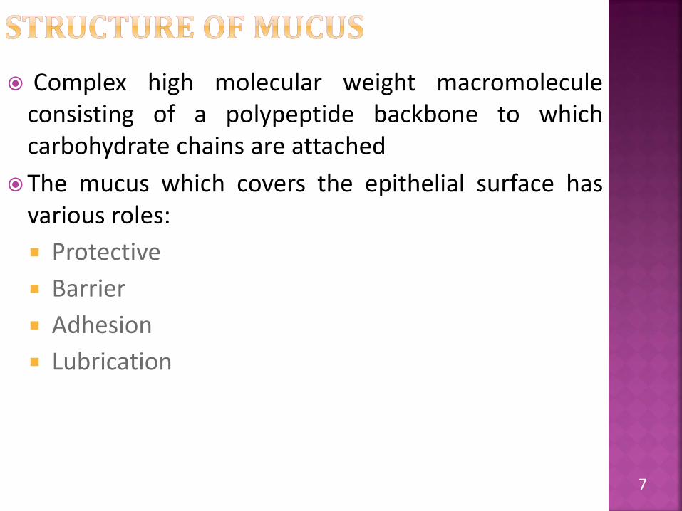
Complex high molecular weight macromoleculeconsisting of a polypeptide backbone to whichcarbohydrate chains are attached
The mucus which covers the epithelial surface hasvarious roles:
Protective
Barrier
Adhesion
Lubrication

8
Mucus secreting cells
Structure of mucin

9

10
The mechanism responsible in the formation of mucoadhesive bonds are not fully known, however most researcher has described it as a three step process
Step 1 : Wetting and swelling of the polymer(contact stage)
Step 2 : Interpenetration between the polymer chains and the mucosal membrane
Step 3 : Formation of bonds between the entangled chains (both known as consolidation stage)

11
Wetting and swelling step occurs when polymer spreads over the surface of mucosal membrane to develop intimate contact
Swelling of polymer occur because the components of polymer have an affinity for water
12
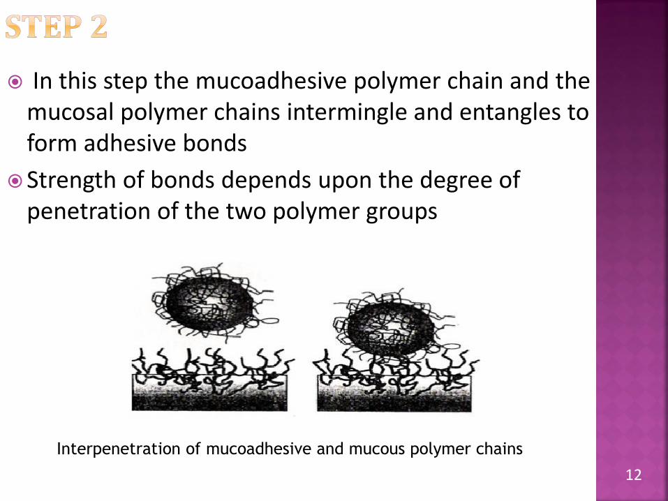
In this step the mucoadhesive polymer chain and the mucosal polymer chains intermingle and entangles to form adhesive bonds
Strength of bonds depends upon the degree of penetration of the two polymer groups
Interpenetration of mucoadhesive and mucous polymer chains

13
This step involves formation of weak chemical bonds between the entangled polymer chains
Bonds includes primary bonds such as covalent bonds and secondary interactions such as vanderWaals and hydrogen bonds
14
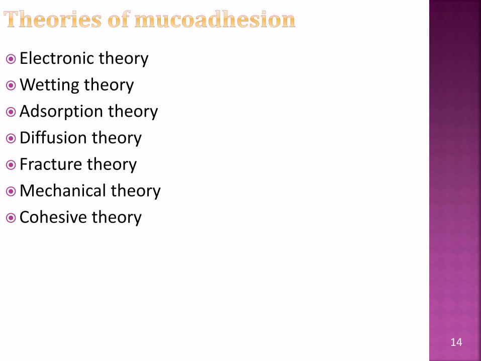
Electronic theory
Wetting theory
Adsorption theory
Diffusion theory
Fracture theory
Mechanical theory
Cohesive theory

15
Electronic theory
This theory considers that both mucoadhesiveand biological materials possess opposing electrical charges; when both materials come in contact with each other they form double electronic layer at interface leading to mucoadhesion
Wetting theory
Best applied to liquid or low viscosity bioadhesives
Postulates that if the contact angle of liquids on the substrate surface is lower, then there is a great affinity for the liquid to the substrate surface. This affinity can be measured by contact angle

16
Wetting theory calculates the contact angle and work of adhesion (Wa), given by Dupre’s eq.
Ƴb & Ƴt are surface tension of polymer and substrate respectively and Ƴbt is interfacial tension
The adhesive work done is a sum of the surface tensions of the two adherent phases, less the interfacial tensions apparent between both phases
t
17
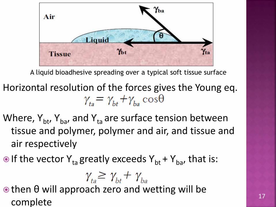
Horizontal resolution of the forces gives the Young eq.
Where, Ƴbt, Ƴba, and Ƴta are surface tension between tissue and polymer, polymer and air, and tissue and air respectively
If the vector Ƴta greatly exceeds Ƴbt + Ƴba, that is:
then θ will approach zero and wetting will be complete
A liquid bioadhesive spreading over a typical soft tissue surface

18
Adsorption theoryThe mucoadhesive device adheres to mucus by
secondary chemical interactions such as vanderWaals,hydrogen bonding or hydrophobic interaction
Such forces are considered most important in adhesiveinteraction because, although they are individuallyweak, a great number of interactions can result instrong adhesionDiffusion theory
Diffusion theory describes that polymeric chainsfrom the bioadhesive interpenetrate into glycoproteinmucin chains and reach a sufficient depth within theopposite matrix to allow formation of asemipermanent bond

19
The depth of 0.2-0.5 µm is required to produce an efficient mucoadhesive bond

20
Fracture theory:
This theory describes the force required for the separation of two surfaces after adhesion
Where σ is the fracture strength, ε fracture energy, E young modulus of elasticity, and L the critical crack length
It is used for rigid or semi-rigid bioadhesive materials, in which the polymer chains do not penetrate into the mucus layer

21
Mechanical theory
Mechanical theory considers adhesion to be due to the filling of the irregularities on a rough surface by a mucoadhesive liquid. Moreover, such roughness increases the interfacial area available to interactions
Cohesive theory
It proposes that the phenomena of bioadhesionare mainly due to the intermolecular interactions amongst like-molecules

22
Polymer related factors:
Molecular weight
Conc. of polymer
Flexibility of polymer chains
Presence of functional group
Spatial conformation
Cross linking density
Environment related factors:
pH of polymer substrate interface
Applied strength
Physiological factors:
Mucin turn over
Disease state

23
They are water soluble and water insoluble polymers which are swellable networks joined by cross linking agent
Characteristic of ideal polymer
Degradation products should be non toxic and non absorbable from GIT
Good spreadibility, wetting, swelling and biodegradable properties
Optimum molecular weight
Non irritant to mucous membrane
Form a strong non-covalent bond with mucinepithelial cell surface
24
Adhere quickly to moist tissue
Allow easy incorporation of the drug
Stable, cost effective and approved by regulatory authorities

25
According to source
Natural and semisynthetic Synthetic
Agarose Carbopol
Chitosan Polycarbophil
Gelatin Polyacrylic acid
Hyaluronic acid Methacrylic acid
Carrageenan Polyacrylates
Pectin PVA
CMC PVP
Thiolated CMC Ethylhexa acrylate
Sodium CMC Thiolated polymer
Hydroxyethylcellulose
Hydroxypropylcellulose
HPMC

26
According to water solubility
According to charge
Soluble Insoluble
CMC, Sodium CMC, HEC, HPMC,
MC, PVA, PVP, etc.
Carbopol, Polycarbophil,
Polyacrylic acid, PEG, etc.
Charged Uncharged
Aminodextran, Chitosan,
Carbopol, SodiumAlginate,
Pectin, SodiumCMC, etc.
Starch, HPC, PEG, PVA, PVP, etc.

27
Route Formulation
Oral(Buccal and Sublingual) Tablet, patch, gel, ointment
Nasal Gel, microspheres, dry powder
Ocular Insert, gel
Gastrointestinal Gel, tablet, microsphere,
capsule
Skin/Transdermal Patch, liposome
Vaginal Gel, microsphere, tablet
Rectal Gel
28
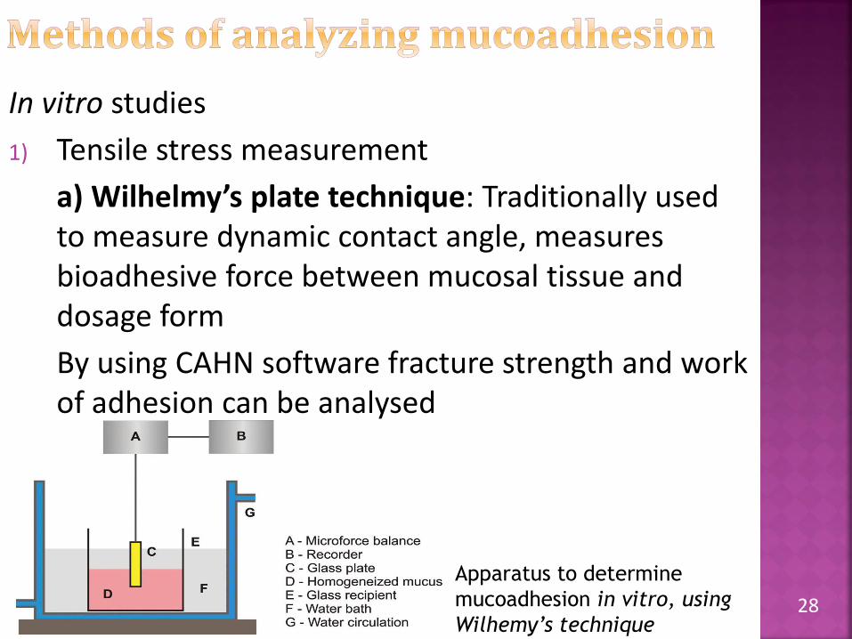
In vitro studies
1) Tensile stress measurement
a) Wilhelmy’s plate technique: Traditionally used to measure dynamic contact angle, measures bioadhesive force between mucosal tissue and dosage form
By using CAHN software fracture strength and work of adhesion can be analysed
Apparatus to determine
mucoadhesion in vitro, using
Wilhemy’s technique

29
2) Everted gut sac procedure(ex vivo)
Can be applied to liposomes, microspheres and nanoparticles

30
3) Colloidal gold staining method: Employs red colloidal gold particles stabilised by adsorbed mucin forming mucin-gold conjugates
Upon interaction with conjugates bioadhesivehydrogel develop red colour on surface
Interaction can be quantified by :
Measuring red colour intensity on Hydrogel
Measuring decrease in concentration of conjugates at 525nm

31
Tests measuring mucoadhesive strength
Depending on the direction in which themucoadhesive is separated from the substrate,iti spossible to obtain the detachment, shear, andrupture tensile strengths
The force most frequently evaluated in such tests isrupture tensile strength
Generally, the equipment used is a texture analyzerIn this test,
The force required to remove the formulation from amodel membrane is measured

32
Bioadhesion test using the texture analyzer
Microbalance methodWilhemy’s plate technique, or the microforce balance technique, can also be modified in order to measure the specific adhesion force of microparticles

33
The general problem of adhesion force and from the rheological methods is that the mucoadhesive response is seen macroscopically while the interactions occur at a microscopic level
Following methods are used to study molecular interactions
a) Dielectric Spectroscopy: Study of material response to the application of an electrical field
the impedance or permittivity of the sample is obtained and the property of charges changing in the system can be determined
b) Zeta potential: Mucin particles are suspended in an appropriate buffer and mixed with a solution of the polymer, If the zeta potential value of the mucin particles changes, this can suggest greater affinity between polymer and mucinparticles

34
c) Optical biosensor: One molecule is immobilized other remains in solution, The molecules in solution, when binding to the immobilized molecules, alter the refraction index of the medium and this change is detected by the screening of a laser beam
d) Falling Liquid Film Method :
In the case of particulate systems, the amount remaining on the mucous membrane can be counted with the aid of a coulter counter, For semi-solid systems, the non adhered mucoadhesive can be quantified by high performance liquid chromatography
This methodology allows the visualization of formation of liquid-crystalline mesophase on the mucous membrane

35
In vivo Techniques
1) GI transit using radio-opaque technique: It involves use of radio opaque markers, e.g., barium sulphate, encapsulated in BDDS
Mucoadhesive labelled with Cr51, Tc99, In113 Have been also used
2)Gamma scintigraphy: Information are obtained noninvasively
Provides various information like:
Dosage form across different regions of GI tract
Time and site of disintegration
Site of absorption
Effect of food and disease

36

37

38
Flavia Chiva Carvalho, Marcos Luciano Bruschi, Raul Cesar Evangelista, Maria Palmira Daflon Gremiao ; Mucoadhesivedrug delivery systems; Brazilian Journal of Pharmaceutical Sciences vol. 46, 2010;1-18.
Formulation and in vitro evaluation of mucoadhesive buccaltablets of Timolol maleate;Int J Pharm Biomed Res 2010, 1(4), 129-134
Smart, J. D., Kellaway, I. W., Worthington, H. E. C., An in-vitro investigation of mucosa-adhesive materials for use in controlled drug delivery, J. Pharm. Pharmacol., 1984,295-299
Pranshu tangri: mucoadhesive drug delivery: mechanism and methods of evaluation, Int J. of Pharma and biosciences: vol. 2, 2011, 458-461

39