Preparation and Characterization of Mucoadhesive …dasan.sejong.ac.kr/~sko/paper/2016 Preparation...
5
Preparation and Characterization of Mucoadhesive Buccal Nanoparticles Using Chitosan and Dextran Sulfate Ji Woon Suh, † Ji-Soo Lee, † Sanghoon Ko, ‡ and Hyeon Gyu Lee* ,† † Department of Food and Nutrition, Hanyang University, 17 Haengdang-dong, Seongdong-gu, Seoul 133-791, Republic of Korea ‡ Department of Food Science and Technology, Sejong University, 98 Gunja-dong, Gwangjin-gu, Seoul 143-747, Republic of Korea ABSTRACT: The aim of this study was to formulate buccal mucoadhesive nanoparticles (NPs) using the natural mucoadhesive polymers. The natural mucoadhesive polymers chitosan (CS) and dextran sulfate sodium salt (DS) were used to prepare mucoadhesive NPs using the ionic gelation method. As the molecular weight of DS decreased, the amount of mucin and the number of buccal cells adsorbed on DS increased. The CS/DS NPs ranged from 100 to 200 nm in diameter. The adhesive interactions of CS/DS NPs with mucin were not significantly different from those of CS/sodium triphosphate pentabasic (TPP) NPs; however, CS/DS NPs exhibited 5 times greater mucoadhesive activity to buccal cells compared to control CS/TPP NPs in ex vivo adhesion tests. These results indicate that the buccal mucoadhesive properties of NPs can be improved using natural mucoadhesive polymers. KEYWORDS: mucoadhesive, dextran sulfate, chitosan, nanoencapsulation ■ INTRODUCTION Mucoadhesion is defined as the adhesive capacity of synthetic or natural polymers to the mucous gel layer covering mucosal membranes. 1 It is generally believed that prolonging the residence time on the adsorbing membrane by mucoadhesion provides optimal conditions for effective delivery. Therefore, mucoadhesive systems have been studied as an approach to increase the residence time of the dosage form on the absorbing mucosal surface and to localize drugs or biological nutrients in a particular region. 2 Mucoadhesion also allows for the diffusion and penetration of mucoadhesive materials into the mucous layer, resulting in improved absorption and bioavailability. 3 Buccal delivery provides a number of unique advantages, including excellent accessibility, bypass of the first pass metabolism, and avoidance of presystemic elimination within the gastrointestinal tract. However, the major limitations of absorption across the oral mucosa include exposure to salivary flow, shearing forces as a result of tongue movements and swallowing, and short residence time. These limitations could, in principle, be effectively alleviated by the use of a buccal mucoadhesive delivery system. Buccal mucoadhesive delivery systems have been fabricated by several different approaches. Most studies have focused on buccal mucoadhesive tablets, patches, films, gels, and ointments for drug delivery to treat oral disease; these patches are based on synthetic mucoadhesive polymers. 4 Tiyaboonchai et al. 5 used polyethylenimine-dextran sulfate nanoparticles (NPs) for the buccal mucoadhesive delivery of Punica granatum peel extract. This strategy resulted in prolonged release and significant antibacterial activity against oral pathogens that cause plaque, cavities, and oral malodor. For a mucoadhesive delivery system to be used in food applications, an encapsulation technique is necessary to protect the cargo from potentially hostile characteristics of the external environ- ment, such as pH, temperature, and moisture. In addition, encapsulation can mask unwanted flavor and taste. NPs also have sensory advantages relevant to the food industry, because nanosized particles cannot be felt by the mouth as a result of their reduced particle size; in contrast, microsized particles can be felt. 6,7 However, only a few studies have used a buccal mucoadhesive NP system for biological nutrients, such as curcumin and plant phenolic extracts, and reported improved absorption and bioavailability. These studies were based on natural biopolymers for their application in the food industry. For optimal formulation of mucoadhesive delivery systems, appropriate mucoadhesive polymers are required. The binding capacity of polymers to the mucosal membrane has been reported to be influenced by ionic bonds, hydrophobic interactions, covalent bonds, and physical entanglement. 2,8,9 Mucoadhesive properties have also been reported to be affected by a number of factors, such as molecular weight (MW), flexibility, hydrogen-bonding capacity, cross-linking density, charge, concentration, and polymer swelling. 4 Various poly- mers, including chitosan (CS), alginate, dextran sulfate sodium salt (DS), and carrageenan, have been reported as mucoadhe- sive biopolymers. 10 Particularly, mucoadhesive biopolymers CS and DS can also be used for nanoencapsulation. The positive charge of CS binds to a negatively charged mucous surface as a result of ionic bonds. Mü ller et al. 11 observed that CS-modified nano- suspensions exhibited increased retention time in the gastro- intestinal tract. In addition, the sulfate functional groups of DS form strong hydrogen-bonding interactions with mucin. 12 Tiyaboonchai et al. 5 demonstrated the ability of DS to adhere to the mucosal surface. Although nanoencapsulation using CS and DS could enhance cellular uptake of curcumin, 13 the use of Received: February 22, 2016 Revised: May 2, 2016 Accepted: May 24, 2016 Published: May 24, 2016 Article pubs.acs.org/JAFC © 2016 American Chemical Society 5384 DOI: 10.1021/acs.jafc.6b00849 J. Agric. Food Chem. 2016, 64, 5384-5388
-
Upload
hoangnguyet -
Category
Documents
-
view
226 -
download
0
Transcript of Preparation and Characterization of Mucoadhesive …dasan.sejong.ac.kr/~sko/paper/2016 Preparation...
Preparation and Characterization of Mucoadhesive BuccalNanoparticles Using Chitosan and Dextran SulfateJi Woon Suh,† Ji-Soo Lee,† Sanghoon Ko,‡ and Hyeon Gyu Lee*,†
†Department of Food and Nutrition, Hanyang University, 17 Haengdang-dong, Seongdong-gu, Seoul 133-791, Republic of Korea‡Department of Food Science and Technology, Sejong University, 98 Gunja-dong, Gwangjin-gu, Seoul 143-747, Republic of Korea
ABSTRACT: The aim of this study was to formulate buccal mucoadhesive nanoparticles (NPs) using the natural mucoadhesivepolymers. The natural mucoadhesive polymers chitosan (CS) and dextran sulfate sodium salt (DS) were used to preparemucoadhesive NPs using the ionic gelation method. As the molecular weight of DS decreased, the amount of mucin and thenumber of buccal cells adsorbed on DS increased. The CS/DS NPs ranged from 100 to 200 nm in diameter. The adhesiveinteractions of CS/DS NPs with mucin were not significantly different from those of CS/sodium triphosphate pentabasic (TPP)NPs; however, CS/DS NPs exhibited 5 times greater mucoadhesive activity to buccal cells compared to control CS/TPP NPs inex vivo adhesion tests. These results indicate that the buccal mucoadhesive properties of NPs can be improved using naturalmucoadhesive polymers.
KEYWORDS: mucoadhesive, dextran sulfate, chitosan, nanoencapsulation
■ INTRODUCTION
Mucoadhesion is defined as the adhesive capacity of syntheticor natural polymers to the mucous gel layer covering mucosalmembranes.1 It is generally believed that prolonging theresidence time on the adsorbing membrane by mucoadhesionprovides optimal conditions for effective delivery. Therefore,mucoadhesive systems have been studied as an approach toincrease the residence time of the dosage form on the absorbingmucosal surface and to localize drugs or biological nutrients in aparticular region.2 Mucoadhesion also allows for the diffusionand penetration of mucoadhesive materials into the mucouslayer, resulting in improved absorption and bioavailability.3
Buccal delivery provides a number of unique advantages,including excellent accessibility, bypass of the first passmetabolism, and avoidance of presystemic elimination withinthe gastrointestinal tract. However, the major limitations ofabsorption across the oral mucosa include exposure to salivaryflow, shearing forces as a result of tongue movements andswallowing, and short residence time. These limitations could,in principle, be effectively alleviated by the use of a buccalmucoadhesive delivery system.Buccal mucoadhesive delivery systems have been fabricated
by several different approaches. Most studies have focused onbuccal mucoadhesive tablets, patches, films, gels, and ointmentsfor drug delivery to treat oral disease; these patches are basedon synthetic mucoadhesive polymers.4 Tiyaboonchai et al.5
used polyethylenimine−dextran sulfate nanoparticles (NPs) forthe buccal mucoadhesive delivery of Punica granatum peelextract. This strategy resulted in prolonged release andsignificant antibacterial activity against oral pathogens thatcause plaque, cavities, and oral malodor. For a mucoadhesivedelivery system to be used in food applications, anencapsulation technique is necessary to protect the cargofrom potentially hostile characteristics of the external environ-ment, such as pH, temperature, and moisture. In addition,encapsulation can mask unwanted flavor and taste. NPs also
have sensory advantages relevant to the food industry, becausenanosized particles cannot be felt by the mouth as a result oftheir reduced particle size; in contrast, microsized particles canbe felt.6,7 However, only a few studies have used a buccalmucoadhesive NP system for biological nutrients, such ascurcumin and plant phenolic extracts, and reported improvedabsorption and bioavailability. These studies were based onnatural biopolymers for their application in the food industry.For optimal formulation of mucoadhesive delivery systems,
appropriate mucoadhesive polymers are required. The bindingcapacity of polymers to the mucosal membrane has beenreported to be influenced by ionic bonds, hydrophobicinteractions, covalent bonds, and physical entanglement.2,8,9
Mucoadhesive properties have also been reported to be affectedby a number of factors, such as molecular weight (MW),flexibility, hydrogen-bonding capacity, cross-linking density,charge, concentration, and polymer swelling.4 Various poly-mers, including chitosan (CS), alginate, dextran sulfate sodiumsalt (DS), and carrageenan, have been reported as mucoadhe-sive biopolymers.10
Particularly, mucoadhesive biopolymers CS and DS can alsobe used for nanoencapsulation. The positive charge of CS bindsto a negatively charged mucous surface as a result of ionicbonds. Muller et al.11 observed that CS-modified nano-suspensions exhibited increased retention time in the gastro-intestinal tract. In addition, the sulfate functional groups of DSform strong hydrogen-bonding interactions with mucin.12
Tiyaboonchai et al.5 demonstrated the ability of DS to adhereto the mucosal surface. Although nanoencapsulation using CSand DS could enhance cellular uptake of curcumin,13 the use of
Received: February 22, 2016Revised: May 2, 2016Accepted: May 24, 2016Published: May 24, 2016
Article
pubs.acs.org/JAFC
© 2016 American Chemical Society 5384 DOI: 10.1021/acs.jafc.6b00849J. Agric. Food Chem. 2016, 64, 5384−5388
CS and DS to enhance the mucoadhesive activity of NPs hasnot been fully explored.For a buccal mucoadhesive NP system for a biological
nutrient to be used in the food industry, the naturalmucoadhesive polymer is preferable to wall material of theNP system with regard to safety. Hence, the aim of this studywas to formulate buccal mucoadhesive NPs using naturalmucoadhesive polymers CS and DS. The physical properties ofthe NPs, including particle size, polydispersity index (PDI), andderived count rate (DCR), were investigated. In addition, theeffect of CS and DS concentrations on the mucoadhesiveactivity of the NPs was evaluated using in vitro and ex vivoadhesion tests.
■ MATERIALS AND METHODSMaterials. CS (water-soluble, MW of 1000−3000, 24 cps, 95%
deacetylated) was obtained from Kittolife Co. (Seoul, Korea). DS(MWs of 15 000, 40 000, and 200 000 kDa), sodium triphosphatepentabasic (TPP), and magnesium chloride were purchased fromSigma-Aldrich Chemical Co. (St. Louis, MO). All other chemicalswere of reagent grade, and all solvents were of high-performance liquidchromatography (HPLC) grade.Preparation of NPs. Two types of NPs, CS/DS and CS/TPP,
were obtained by ionic gelation of CS with DS and TPP,respectively.14 CS was dissolved in distilled water at a concentrationof 1−2 mg/mL. The CS/DS NPs were prepared by adding 3 mL ofDS solution (0.2−0.6 mg/mL) into CS solution for 10 min undermagnetic stirring (1000 rpm, WiseStir MS-MP8, Wise LaboratoryInstruments, Wertheim, Germany). The CS/TPP NPs (controlgroup) were prepared by adding 1 mL of TPP solution (0.3−0.6mg/mL) into CS solution for 10 min under magnetic stirring.Physical Properties of the NPs. The physical properties of the
CS/DS and CS/TPP NPs, including particle size, PDI, ζ potential, andDCR, were determined by dynamic light scattering with a MalvernZetasizer Nano ZS instrument (Malvern Instruments, Ltd., Malvern,Worcestershire, U.K.). All measurements were performed in triplicateat multiple narrow modes at 25 ± 1 °C.Determination of In Vitro Mucoadhesive Properties. The
mucoadhesive properties of the NPs to mucin were investigated asdescribed previously,15 but with minor modifications. Briefly, 0.6 mLof mucin solution (0.5 mg/mL) was mixed with 0.6 mL ofnanosuspension and incubated at 37 °C in a shaking water bath for1 h. After centrifugation at 14000g for 40 min, the supernatant wascollected and the amount of free mucin was measured using theBradford protein assay. The supernatant was incubated with Bradfordreagent for 5 min, after which the absorbance was measured at 595 nmwith a spectrophotometer (Biomate 3S, Thermo Scientific, Waltham,MA). The amount of mucin-adsorbed NPs was calculated as thedifference between the total amount of mucin added and the residualmucin in the supernatant. The mucin concentration was calculatedfrom a standard curve of mucin in a concentration from 0 to 5 mg/mL.16
Determination of Ex Vivo Mucoadhesive Properties. The exvivo mucoadhesive properties of the NPs were determined by themethods of Kockkisch et al.17 and Patel et al.,18 with minormodifications. All procedures for collecting buccal cells were approvedby the Hanyang University Institutional Review Board. Buccal cellswere collected from 10 healthy male and female volunteers by gentlyscraping the oral cavity with a tongue depressor. Donors wereprohibited from eating or drinking for 1 h prior to buccal cellcollection. Cells were immediately suspended in 0.25 M aqueoussucrose solution, and the cell suspension was then added to 0.5% (w/v) trypan blue solution. The cell concentration was determined using ahemocytometer and standardized to 48 × 106 cells per test. All cellswere stored at 4 °C and used within 4 h of collecting.The cells were separated from supernatants by centrifugation at
700g for 5 min and then reacted with samples (polymers or NPs) inphosphate buffer in a 30 °C shaking incubator for 30 min. The 0.1%
Alcian blue was added, and incubation for 1 h at 30 °C was carried out.The cells were then washed using 0.25 M sucrose solution. Thecomplexed dye with polymer-treated cells was eluted by immersion in5 M magnesium chloride for 1 h at 30 °C. Cells were then spun bycentrifugation at 700g for 15 min, and the absorbance of thesupernatant was measured at 605 nm using a spectrophotometer. Acontrol was performed following the same procedure without a testsample. Results are expressed as a percentage reduction compared tothe control.
Statistical Analysis. Statistical analyses were conducted usingSPSS (Statistical Package for the Social Sciences, SPSS, Inc., Chicago,IL). One-way analyses of variance (ANOVA) were performed toinvestigate the significance of differences between conditions (p <0.05). All experiments were performed at least in triplicate, and resultsare presented as means ± standard deviations (SDs).
■ RESULTS AND DISCUSSIONEffect of the DS MW on In Vitro and Ex Vivo
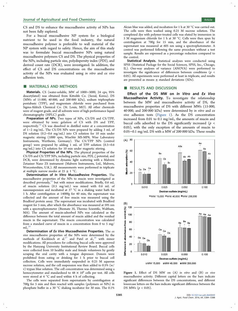
Mucoadhesive Activity. To investigate the relationshipbetween the MW and mucoadhesive activity of DS, themucoadhesive properties of DS with different MWs (15 000,40 000, and 200 000 kDa) were determined by in vitro and exvivo adhesion tests (Figure 1). As the DS concentrationincreased from 0.01 to 0.1 mg/mL, the amounts of mucin andbuccal cells adsorbed to the DS significantly increased (p <0.05), with the only exception of the amounts of mucin to0.05−0.1 mg/mL DS with a MW of 200 000 kDa. These results
Figure 1. Effect of DS MW on (A) in vitro and (B) ex vivomucoadhesive activity. Different capital letters on the bars indicatesignificant differences between the DS concentrations, and differentlowercase letters on the bars indicate significant difference between theDS MWs (p < 0.05).
Journal of Agricultural and Food Chemistry Article
DOI: 10.1021/acs.jafc.6b00849J. Agric. Food Chem. 2016, 64, 5384−5388
5385
could be explained by the hydrogen bond between the sulfategroups in DS and the glycoprotein component of the mucus.4
Hydrogen bonding is an important factor in mucoadhesion ofpolymer, and the hydrogen-bonding potential can be influencedby the flexibility of the polymer.4 Therefore, as the polymerconcentration increases, the number of functional groupscapable of forming hydrogen bonds per unit volume of mucusincreases and the polymer−mucus interaction stabilizes.4
We observed the tendency to increase the amounts ofadsorbed mucin and buccal cells on DS, as the MW of DSdecreased. Although the effect of MW of DS was notsignificantly different in the ex vivo mucoadhesive activity of0.05 and 0.1 mg/mL DS, this tendency was more clearlyobserved in the in vitro mucoadhesive activity than the ex vivomucoadhesive activity. Thus, the high mucoadhesive activity of15 000 kDa DS led us to select this polymer as a wall materialfor mucoadhesive NPs. The ability of polymers with very highMWs to interpenetrate and diffuse into mucosal surfaces couldbe hindered as a result of shielding of the bioadhesive site insidethe helical conformation.19 Previous studies on the effect ofpolymer MW on bioadhesion have revealed that entanglementfavors high-MW polymers, whereas interpenetration favors low-MW polymers. Thus, the optimal MW for achieving highmucoadhesive activity could depend upon the type of polymer.4
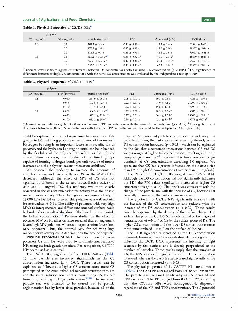
Physical Properties of NPs. The natural mucoadhesivepolymers CS and DS were used to formulate mucoadhesiveNPs using the ionic gelation method. For comparison, CS/TPPNPs were used as a control.The CS/DS NPs ranged in size from 110 to 360 nm (Table
1). The particle size increased significantly as the CSconcentration increased (p < 0.05). These results can beexplained as follows: at a higher CS concentration, more CSparticipated in the cross-linked gel network structure with DSand the stirrer solution was more viscous during CS/DS NPformation, resulting in large particle sizes.20,21 The increasedparticle size was assumed to be caused not by particleagglomeration but by larger sized particles, because all of the
prepared NPs revealed particle size distribution with only onepeak. In addition, the particle size decreased significantly as theDS concentration increased (p < 0.05), which can be explainedby the fact that electrostatic interactions between CS and DSwere stronger at higher DS concentrations, resulting in a morecompact gel structure.22 However, this force was no longerdominant at CS concentrations exceeding 1.0 mg/mL. Wespeculate that CS has a greater influence on the particle sizethan DS at high CS concentrations (greater than 1.0 mg/mL).The PDIs of the CS/DS NPs ranged from 0.26 to 0.44.
Although the DS concentration did not significantly influencethe PDI, the PDI values significantly increased at higher CSconcentrations (p < 0.05). This result was consistent with thechange of the particle size with the increase of CS, because PDIgenerally increases as the particle size increases.The ζ potential of CS/DS NPs significantly increased with
the increase of the CS concentration and reduced with theincrease of the DS concentration (p < 0.05). These resultscould be explained by the density of the surface charge. Thesurface charge of the CS/DS NP is determined by the degree ofneutralization of −NH3
+ of CS by the sulfate group of DS. Thehigher CS concentration and the lower DS concentration meanmore unneutralized −NH3
+ on the surface of the NP.The DCR significantly increased as the DS concentration
increased; however, the CS concentration did not significantlyinfluence the DCR. DCR represents the intensity of lightscattered by the particles and is directly proportional to thenumber of particles. These results imply that the number ofCS/DS NPs increased significantly as the DS concentrationincreased, whereas the particle size increased significantly as theCS concentration increased (p < 0.05).The physical properties of the CS/TPP NPs are shown in
Table 2. The CS/TPP NPs ranged from 180 to 590 nm in size.The particle size increased significantly as CS increased andTPP decreased. The PDI ranged from 0.22 to 0.27, indicatingthat the CS/TPP NPs were homogeneously dispersed,regardless of the CS and TPP concentrations. The ζ potential
Table 1. Physical Properties of CS/DS NPsa
polymer
CS (mg/mL) DS (mg/mL) particle size (nm) PDI ζ potential (mV) DCR (kcps)
0.5 0.1 204.2 ± 3.3 a 0.30 ± 0.03 a 57.2 ± 1.4 a 25581 ± 1662 b0.2 179.2 ± 2.6 b 0.27 ± 0.01 a 52.9 ± 2.0 b 58297 ± 4044 a0.3 114.1 ± 0.5 c 0.26 ± 0.01 a 41.3 ± 1.8 c 69023 ± 4825 a
1.0 0.1 355.2 ± 30.4 a*b 0.38 ± 0.02 a* 70.0 ± 2.3 a* 28659 ± 1849 b0.2 355.8 ± 20.8 a* 0.42 ± 0.01 a* 66.1 ± 1.7 b* 55694 ± 3417 b0.3 342.3 ± 16.8 a* 0.44 ± 0.03 a* 62.4 ± 1.2 c* 87529 ± 5814 a
aDifferent letters indicate significant differences between DS concentrations with the same CS concentration (p < 0.05). bThe significance ofdifferences between multiple CS concentrations with the same DS concentration was evaluated by the independent t test (p < 0.05).
Table 2. Physical Properties of CS/TPP NPsa
polymer
CS (mg/mL) TPP (mg/mL) particle size (nm) PDI ζ potential (mV) DCR (kcps)
0.5 0.050 287.9 ± 28.2 a 0.25 ± 0.02 a 59.3 ± 2.8 a 7024 ± 1200 c0.075 192.8 ± 32.4 b 0.22 ± 0.01 a 57.9 ± 4.1 a 21295 ± 3406 b0.100 184.7 ± 7.6 b 0.22 ± 0.01 a 49.8 ± 1.3 b 37098 ± 4849 a
1.0 0.050 586.2 ± 8.9 a*b 0.29 ± 0.02 a 70.3 ± 2.6 a* 5303 ± 130 c0.075 517.9 ± 21.8 b* 0.27 ± 0.01 a 66.5 ± 1.5 b* 15000 ± 1690 b*0.100 483.2 ± 38.4 b* 0.28 ± 0.01 a 65.5 ± 1.4 b* 18271 ± 647 a*
aDifferent letters indicate significant differences between TPP concentrations with the same CS concentration (p < 0.05). bThe significance ofdifferences between multiple CS concentrations with the same TPP concentration was evaluated by the independent t test (p < 0.05).
Journal of Agricultural and Food Chemistry Article
DOI: 10.1021/acs.jafc.6b00849J. Agric. Food Chem. 2016, 64, 5384−5388
5386
of CS/TPP NPs significantly increased as the CS concentrationincreased and the TPP concentration decreased (p < 0.05). TheDCR increased significantly as the TPP concentrationincreased, indicating that the number of CS/TPP NPsincreased as TPP increased (p < 0.05).In Vitro Mucoadhesive Activity of NPs. To investigate
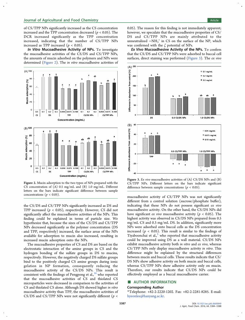
the mucoadhesive activities of the CS/DS and CS/TPP NPs,the amounts of mucin adsorbed on the polymers and NPs weredetermined (Figure 2). The in vitro mucoadhesive activities of
the CS/DS and CS/TPP NPs significantly increased as DS andTPP increased (p < 0.05), respectively. However, CS did notsignificantly affect the mucoadhesive activities of the NPs. Thisfinding could be explained in terms of particle size. Wehypothesize that, because the sizes of the CS/DS and CS/TPPNPs decreased significantly as the polymer concentration (DSand TPP, respectively) increased, the surface areas of the NPsavailable for adsorption to mucin also increased, resulting inincreased mucin adsorption onto the NPs.The mucoadhesive properties of CS and DS are based on the
electrostatic interaction of the amine groups in CS and thehydrogen bonding of the sulfate groups in DS to mucins,respectively. However, the negatively charged DS sulfate groupsbind to the positively charged CS amine groups during ionicgelation in NP formation, consequently reducing themucoadhesive activity of the CS/DS NPs. This result isconsistent with the findings of Pengpong et al.,16 who reportedthat the mucoadhesive activities of CS and thiolated CSmicroparticles were decreased in comparison to the activities ofCS and thiolated CS alone. Although DS showed higher in vitromucoadhesive activity than TPP, the mucoadhesive activities ofCS/DS and CS/TPP NPs were not significantly different (p <
0.05). The reason for this finding is not immediately apparent;however, we speculate that the mucoadhesive properties of CS/DS and CS/TPP NPs are mainly attributed to theunneutralized −NH3
+ in CS on the surface of the NP, whichwas confirmed with the ζ potential of NPs.
Ex Vivo Mucoadhesive Activity of the NPs. To confirmthat the CS/DS and CS/TPP NPs were adsorbed to buccal cellsurfaces, direct staining was performed (Figure 3). The ex vivo
mucoadhesive activity of CS/TPP NPs was not significantlydifferent from a control solution (sucrose/phosphate buffer),indicating that these NPs do not possess significant ex vivomucoadhesive activity. On the other hand, the CS/DS NPs didhave significant ex vivo mucoadhesive activity (p < 0.05). Thehighest activity was observed in CS/DS NPs prepared from 0.5mg/mL CS and 0.3 mg/mL DS. In addition, significantly moreNPs were adsorbed onto buccal cells as the DS concentrationincreased (p < 0.05). This result is similar to the findings ofTiyaboonchai et al.,5 who reported that mucoadhesive activitycould be improved using DS as a wall material. CS/DS NPsexhibit mucoadhesive activity both in vitro and ex vivo, whereasCS/TPP NPs only display mucoadhesive activity in vitro. Thisdifference might be explained by the structural differencesbetween mucin and buccal cells. These results indicate that CS/DS NPs show adhesive activity on both mucin and buccal cells,whereas CS/TPP NPs show adhesive activity only on mucin.Therefore, our results indicate that CS/DS NPs could beeffectively employed as a buccal mucoadhesive carrier.
■ AUTHOR INFORMATIONCorresponding Author*Telephone: +82-2-2220-1202. Fax: +82-2-2281-8285. E-mail:[email protected].
Figure 2. Mucin adsorption to the two types of NPs prepared with theCS concentration of (A) 0.5 mg/mL and (B) 1.0 mg/mL. Differentletters on the bars indicate significant difference between sampleconcentrations (p < 0.05).
Figure 3. Ex vivo mucoadhesive activities of (A) CS/DS NPs and (B)CS/TPP NPs. Different letters on the bars indicate significantdifference between sample concentrations (p < 0.05).
Journal of Agricultural and Food Chemistry Article
DOI: 10.1021/acs.jafc.6b00849J. Agric. Food Chem. 2016, 64, 5384−5388
5387
FundingThis research was supported by Basic Science ResearchProgram through the National Research Foundation of Korea(NRF) funded by the Ministry of Science, ICT & FuturePlanning (No. 2014M3A7B4051898).
NotesThe authors declare no competing financial interest.
■ REFERENCES(1) Cevher, E.; Taha, M. A. M.; Orlu, M.; Araman, A. Evaluation ofmechanical and mucoadhesive properties of clomiphene citrate gelformulations containing carbomers and their thiolated derivatives.Drug Delivery 2008, 15, 57−67.(2) Smart, J. D. The basics and underlying mechanisms ofmucoadhesion. Adv. Drug Delivery Rev. 2005, 57, 1556−1568.(3) Chowdary, K. P. R.; Srinivasa Rao, Y. Mucoadhesive micro-spheres for controlled drug delivery. Biol. Pharm. Bull. 2004, 27,1717−1724.(4) Salamat-Miller, N.; Chittchang, M.; Johnston, T. P. The use ofmucoadhesive polymers in buccal drug delivery. Adv. Drug DeliveryRev. 2005, 57, 1666−1691.(5) Tiyaboonchai, W.; Rodleang, I.; Ounaroon, A. Mucoadhesivepolyethylenimine−dextran sulfate nanoparticles containing Punicagranatum peel extract as a novel sustained-release antimicrobial.Pharm. Dev. Technol. 2015, 20, 426−432.(6) Mauludin, R.; Muller, R. H.; Keck, C. M. Kinetic solubility anddissolution velocity of rutin nanocrystals. Eur. J. Pharm. Sci. 2009, 36,502−510.(7) Rashidi, L.; Khosravi-Darani, K. The applications of nano-technology in food industry. Crit. Rev. Food Sci. Nutr. 2011, 51, 723−730.(8) Eouani, C.; Piccerelle, P.; Prinderre, P.; Bourret, E.; Joachim, J.In-vitro comparative study of buccal mucoadhesive performance ofdifferent polymeric films. Eur. J. Pharm. Biopharm. 2001, 52, 45−55.(9) Yu, T.; Andrews, P. G.; Jones, S. D. Mucoadhesion andcharacterization of mucoadhesive properties. In Mucosal Delivery ofBiopharmaceuticals: Biology, Challenges and Strategies; das Neves, J.,Sarmento, B., Eds.; Springer US: Boston, MA, 2014; pp 35−58, DOI:10.1007/978-1-4614-9524-6_2.(10) Bae, I. Y.; Lee, H. I.; Ko, A.; Lee, H. G. Substituting whole grainflour for wheat flour: Impact on cake quality and glycemic index. FoodSci. Biotechnol. 2013, 22, 1−7.(11) Muller, R. H.; Jacobs, C.; Kayser, O. Nanosuspensions asparticulate drug formulations in therapy: Rationale for developmentand what we can expect for the future. Adv. Drug Delivery Rev. 2001,47, 3−19.(12) Sosnik, A.; das Neves, J.; Sarmento, B. Mucoadhesive polymersin the design of nano-drug delivery systems for administration by non-parenteral routes: A review. Prog. Polym. Sci. 2014, 39, 2030−2075.(13) Anitha, A.; Deepagan, V. G.; Divya Rani, V. V.; Menon, D.; Nair,S. V.; Jayakumar, R. Preparation, characterization, in vitro drug releaseand biological studies of curcumin loaded dextran sulphate−chitosannanoparticles. Carbohydr. Polym. 2011, 84, 1158−1164.(14) Cho, Y.; Shi, R.; Ben Borgens, R. Chitosan nanoparticle-basedneuronal membrane sealing and neuroprotection following acrolein-induced cell injury. J. Biol. Eng. 2010, 4, 2.(15) Shao, Y.; Yang, L.; Han, H.-K. TPGS-chitosome as an effectiveoral delivery system for improving the bioavailability of CoenzymeQ10. Eur. J. Pharm. Biopharm. 2015, 89, 339−346.(16) Pengpong, T.; Sangvanich, P.; Sirilertmukul, K.; Muangsin, N.Design, synthesis and in vitro evaluation of mucoadhesive p-coumarate-thiolated-chitosan as a hydrophobic drug carriers. Eur. J.Pharm. Biopharm. 2014, 86, 487−497.(17) Kockisch, S.; Rees, G. D.; Young, S. A.; Tsibouklis, J.; Smart, J.D. A direct-staining method to evaluate the mucoadhesion of polymersfrom aqueous dispersion. J. Controlled Release 2001, 77, 1−6.
(18) Patel, D.; Smith, A. W.; Grist, N.; Barnett, P.; Smart, J. D. An invitro mucosal model predictive of bioadhesive agents in the oral cavity.J. Controlled Release 1999, 61, 175−183.(19) Gu, J. M.; Robinson, J. R.; Leung, S. H. Binding of acrylicpolymers to mucin/epithelial surfaces: Structure−property relation-ships. Crit. Rev. Ther. Drug Carrier Syst. 1988, 5, 21−67.(20) Vandenberg, G. W.; Drolet, C.; Scott, S. L.; de la Noue, J.Factors affecting protein release from alginate−chitosan coacervatemicrocapsules during production and gastric/intestinal simulation. J.Controlled Release 2001, 77, 297−307.(21) Alsarra, I. A.; Neau, S. H.; Howard, M. A. Effects of preparativeparameters on the properties of chitosan hydrogel beads containingCandida rugosa lipase. Biomaterials 2004, 25, 2645−2655.(22) Chen, Y.; Mohanraj, V.; Parkin, J. Chitosan−dextran sulfatenanoparticles for delivery of an anti-angiogenesis peptide. Lett. Pept.Sci. 2003, 10, 621−629.
Journal of Agricultural and Food Chemistry Article
DOI: 10.1021/acs.jafc.6b00849J. Agric. Food Chem. 2016, 64, 5384−5388
5388