Astrovirus Infection in Hospitalized Infants with Severe ... · Astrovirus Infection in...
9
Astrovirus Infection in Hospitalized Infants with Severe Combined Immunodeficiency after Allogeneic Hematopoietic Stem Cell Transplantation Werner Wunderli 1,2 * . , Astrid Meerbach 1. , Tayfun Guengoer 3. , Christoph Berger 3 , Oliver Greiner 3 , Rosmarie Caduff 4 , Alexandra Trkola 1 , Walter Bossart 1 , Daniel Gerlach 5 , Manuel Schibler 2 , Samuel Cordey 2,6 , Thomas Alexander McKee 7 , Sandra Van Belle 2 , Laurent Kaiser 2 , Caroline Tapparel 2 * 1 Division of Clinical Virology, University of Zurich, Zurich, Switzerland, 2 Laboratory of Virology, Division of Infectious Diseases and Division of Laboratory Medicine, University of Geneva Hospitals, Geneva, Switzerland, 3 Division of Immunology and Bone Marrow Transplantation, University Children’s Hospital, Zurich, Switzerland, 4 Division of Pathology, University of Zurich Hospitals, Zurich, Switzerland, 5 Department of Genetic Medicine and Development and Swiss Institute of Bioinformatics, University of Geneva Medical School, Geneva, Switzerland, 6 Swiss National Reference Centre for Emerging Viruses (CRIVE), University of Geneva Hospitals, Geneva, Switzerland, 7 Division of Pathology, University of Geneva Hospitals, Geneva, Switzerland Abstract Infants with severe primary combined immunodeficiency (SCID) and children post-allogeneic hematopoietic stem cell transplantation (HSCT) are extremely susceptible to unusual infections. The lack of generic tools to detect disease-causing viruses among more than 200 potential human viral pathogens represents a major challenge to clinicians and virologists. We investigated retrospectively the causes of a fatal disseminated viral infection with meningoencephalitis in an infant with gamma C-SCID and of chronic gastroenteritis in 2 other infants admitted for HSCT during the same time period. Analysis was undertaken by combining cell culture, electron microscopy and sequence-independent single primer amplification (SISPA) techniques. Caco-2 cells inoculated with fecal samples developed a cytopathic effect and non-enveloped viral particles in infected cells were detected by electron microscopy. SISPA led to the identification of astrovirus as the pathogen. Both sequencing of the capsid gene and the pattern of infection suggested nosocomial transmission from a chronically excreting index case to 2 other patients leading to fatal infection in 1 and to transient disease in the others. Virus-specific, real-time reverse transcription polymerase chain reaction was then performed on different stored samples to assess the extent of infection. Infection was associated with viremia in 2 cases and contributed to death in 1. At autopsy, viral RNA was detected in the brain and different other organs, while immunochemistry confirmed infection of gastrointestinal tissues. This report illustrates the usefulness of the combined use of classical virology procedures and modern molecular tools for the diagnosis of unexpected infections. It illustrates that astrovirus has the potential to cause severe disseminated lethal infection in highly immunocompromised pediatric patients. Citation: Wunderli W, Meerbach A, Guengoer T, Berger C, Greiner O, et al. (2011) Astrovirus Infection in Hospitalized Infants with Severe Combined Immunodeficiency after Allogeneic Hematopoietic Stem Cell Transplantation. PLoS ONE 6(11): e27483. doi:10.1371/journal.pone.0027483 Editor: Cheryl A. Stoddart, University of California San Francisco, United States of America Received June 5, 2011; Accepted October 17, 2011; Published November 11, 2011 Copyright: ß 2011 Wunderli et al. This is an open-access article distributed under the terms of the Creative Commons Attribution License, which permits unrestricted use, distribution, and reproduction in any medium, provided the original author and source are credited. Funding: The study was supported by SNF funding awarded to Caroline Tapparel (FNS 310030-127159) and Laurent Kaiser (32003B_127160). The funders had no role in study design, data collection and analysis, decision to publish, or preparation of the manuscript. Competing Interests: The authors have declared that no competing interests exist. * E-mail: [email protected] (CT); [email protected] (WW) . These authors contributed equally to this work. Introduction Infants with severe combined immunodeficiency (SCID) and children after allogeneic hematopoietic stem cell transplantation (HSCT) are exceptionally susceptible to viral infections and viral reactivations. The lack of functional cytotoxic T- and NK-cells prior to and for a certain time after HSCT opens the door to infections by unexpected pathogens either community acquired or nosocomial. Viral infections, including those that commonly cause self-limited childhood diseases, can lead to protracted infections with chronic viral shedding, but also to disseminated disease with infection of organs rarely affected in immunocompetent hosts [1]. When highly immunocompromised infants present prolonged illness despite broad spectrum antimicrobial therapy, extended microbiological investigations should be considered. Routine viral screening is limited to the most frequent groups of viruses including herpes-, hepatitis-, respiratory-, adeno-, polyoma- and selected gastrointes- tinal viruses. However, the number of different viruses potentially pathogenic in humans is estimated to be more than 200 [2]. Therefore, when standard investigations remain negative despite clinical suspicion for viral disease, screening has to be extended and depends on the availability of in-house assays. Under certain circumstances research techniques should be considered. Unfortu- nately, the clinical features presented by transplanted infants or patients with SCID are not always typical and often misleading. Generic molecular tools, such as microarrays [3], ultra-deep sequencing [4], sequence-independent single primer amplification (SISPA) [5], virus discovery based on cDNA-amplified fragment length polymorphism [6], or any other similar procedures, offer potentially attractive alternatives although the sensitivity is limited. PLoS ONE | www.plosone.org 1 November 2011 | Volume 6 | Issue 11 | e27483
Transcript of Astrovirus Infection in Hospitalized Infants with Severe ... · Astrovirus Infection in...
Astrovirus Infection in Hospitalized Infants with SevereCombined Immunodeficiency after AllogeneicHematopoietic Stem Cell TransplantationWerner Wunderli1,2*., Astrid Meerbach1., Tayfun Guengoer3., Christoph Berger3, Oliver Greiner3,
Rosmarie Caduff4, Alexandra Trkola1, Walter Bossart1, Daniel Gerlach5, Manuel Schibler2, Samuel
Cordey2,6, Thomas Alexander McKee7, Sandra Van Belle2, Laurent Kaiser2, Caroline Tapparel2*
1 Division of Clinical Virology, University of Zurich, Zurich, Switzerland, 2 Laboratory of Virology, Division of Infectious Diseases and Division of Laboratory Medicine,
University of Geneva Hospitals, Geneva, Switzerland, 3 Division of Immunology and Bone Marrow Transplantation, University Children’s Hospital, Zurich, Switzerland,
4 Division of Pathology, University of Zurich Hospitals, Zurich, Switzerland, 5 Department of Genetic Medicine and Development and Swiss Institute of Bioinformatics,
University of Geneva Medical School, Geneva, Switzerland, 6 Swiss National Reference Centre for Emerging Viruses (CRIVE), University of Geneva Hospitals, Geneva,
Switzerland, 7 Division of Pathology, University of Geneva Hospitals, Geneva, Switzerland
Abstract
Infants with severe primary combined immunodeficiency (SCID) and children post-allogeneic hematopoietic stem celltransplantation (HSCT) are extremely susceptible to unusual infections. The lack of generic tools to detect disease-causingviruses among more than 200 potential human viral pathogens represents a major challenge to clinicians and virologists.We investigated retrospectively the causes of a fatal disseminated viral infection with meningoencephalitis in an infant withgamma C-SCID and of chronic gastroenteritis in 2 other infants admitted for HSCT during the same time period. Analysiswas undertaken by combining cell culture, electron microscopy and sequence-independent single primer amplification(SISPA) techniques. Caco-2 cells inoculated with fecal samples developed a cytopathic effect and non-enveloped viralparticles in infected cells were detected by electron microscopy. SISPA led to the identification of astrovirus as thepathogen. Both sequencing of the capsid gene and the pattern of infection suggested nosocomial transmission from achronically excreting index case to 2 other patients leading to fatal infection in 1 and to transient disease in the others.Virus-specific, real-time reverse transcription polymerase chain reaction was then performed on different stored samples toassess the extent of infection. Infection was associated with viremia in 2 cases and contributed to death in 1. At autopsy,viral RNA was detected in the brain and different other organs, while immunochemistry confirmed infection ofgastrointestinal tissues. This report illustrates the usefulness of the combined use of classical virology procedures andmodern molecular tools for the diagnosis of unexpected infections. It illustrates that astrovirus has the potential to causesevere disseminated lethal infection in highly immunocompromised pediatric patients.
Citation: Wunderli W, Meerbach A, Guengoer T, Berger C, Greiner O, et al. (2011) Astrovirus Infection in Hospitalized Infants with Severe CombinedImmunodeficiency after Allogeneic Hematopoietic Stem Cell Transplantation. PLoS ONE 6(11): e27483. doi:10.1371/journal.pone.0027483
Editor: Cheryl A. Stoddart, University of California San Francisco, United States of America
Received June 5, 2011; Accepted October 17, 2011; Published November 11, 2011
Copyright: � 2011 Wunderli et al. This is an open-access article distributed under the terms of the Creative Commons Attribution License, which permitsunrestricted use, distribution, and reproduction in any medium, provided the original author and source are credited.
Funding: The study was supported by SNF funding awarded to Caroline Tapparel (FNS 310030-127159) and Laurent Kaiser (32003B_127160). The funders had norole in study design, data collection and analysis, decision to publish, or preparation of the manuscript.
Competing Interests: The authors have declared that no competing interests exist.
* E-mail: [email protected] (CT); [email protected] (WW)
. These authors contributed equally to this work.
Introduction
Infants with severe combined immunodeficiency (SCID) and
children after allogeneic hematopoietic stem cell transplantation
(HSCT) are exceptionally susceptible to viral infections and viral
reactivations. The lack of functional cytotoxic T- and NK-cells prior
to and for a certain time after HSCT opens the door to infections by
unexpected pathogens either community acquired or nosocomial.
Viral infections, including those that commonly cause self-limited
childhood diseases, can lead to protracted infections with chronic
viral shedding, but also to disseminated disease with infection of
organs rarely affected in immunocompetent hosts [1]. When highly
immunocompromised infants present prolonged illness despite
broad spectrum antimicrobial therapy, extended microbiological
investigations should be considered. Routine viral screening is
limited to the most frequent groups of viruses including herpes-,
hepatitis-, respiratory-, adeno-, polyoma- and selected gastrointes-
tinal viruses. However, the number of different viruses potentially
pathogenic in humans is estimated to be more than 200 [2].
Therefore, when standard investigations remain negative despite
clinical suspicion for viral disease, screening has to be extended and
depends on the availability of in-house assays. Under certain
circumstances research techniques should be considered. Unfortu-
nately, the clinical features presented by transplanted infants or
patients with SCID are not always typical and often misleading.
Generic molecular tools, such as microarrays [3], ultra-deep
sequencing [4], sequence-independent single primer amplification
(SISPA) [5], virus discovery based on cDNA-amplified fragment
length polymorphism [6], or any other similar procedures, offer
potentially attractive alternatives although the sensitivity is limited.
PLoS ONE | www.plosone.org 1 November 2011 | Volume 6 | Issue 11 | e27483

We describe here the retrospective analysis of a cluster of 3
infants prior to or after allogeneic HCST infected with an initially
unrecognized enteric virus. It was first detected by cell culture of
fecal specimens and identified as an astrovirus using a modified
SISPA protocol. Subsequent screening with a specific real-time
reverse transcription polymerase chain reaction (RT-PCR) assay
of different patients from the same time period revealed a cluster of
3 cases that remained undetected by standard investigation. In 1
fatal case, the infection involved multiple organs, including the
central nervous system. Viral genome sequencing revealed that all
cases were infected with the same astrovirus type 4 strain.
Materials and Methods
Cultivation of astrovirus, preparation for electronmicroscopy (EM), and immunofluorescence (IF)
Caco-2 cells (ATCC # HTB 37) were cultivated in M199
medium containing 10% fetal calf serum, glutamine and a mixture
of penicillin/streptomycin. Confluent cells in culture tubes were
used for the inoculation of specimens. Stool samples were
suspended in PBS (10% v/v) and centrifuged at 2000 g for
20 min. The supernatant was then removed and penicillin,
streptomycin, and fungizone were added. Culture tubes were
inoculated with 200 ml of the suspension in 2 ml culture medium
without the addition of trypsin (Tables S1,S2,S3). Cultures were
screened periodically for cytophathic changes for up 10 days and
compared with a non-infected cell-culture control. If rounding up
of cells started to appear (usually after 24–72 h), they were scraped
off and used to prepare cytospin slides for IF staining. After drying
and fixation of the slides, cells were incubated with monoclonal
antibodies directed against enteroviruses (pan-enterovirus blend,
Chemicon International). Of note, the antigen specificity of these
antibodies is not specified by the company and potential cross-
reaction with hepatitis A, reovirus 3, and some rhinovirus and
astrovirus strains is indicated in the data sheet. Some samples
(Tables S1,S2,S3) were analyzed retrospectively by IF after Caco-2
cell infection with the pan-enterovirus blend kit and, in parallel,
with anti-astrovirus-specific monoclonal antibody (Argene 11-301)
diluted 1/100 in PBS---1% BSA before addition of the anti-mouse
IgG AB/FITC (Light Diagnostics).
For EM, Caco-2 cells were infected with a 10% stool suspension
of a positive sample from patient 1 (sample no.12869; Table S1).
After adsorption for 2 h, the medium was changed and cells were
further incubated at 37uC with 5% CO2 for 2 days. Cells were
removed by trypsination and centrifuged at 900 g for 5 min. The
supernatant was removed and the cell pellet was fixed in
glutaraldehyde. After dehydration, the cell pellet was embedded
in epoxy resin and thin cuts were processed for standard EM
investigation. This procedure was performed by the Central EM
Service Unit of the University of Zurich.
Screening of patient material for the presence ofinfectious agents
Written informed consent was obtained from the children’s
parents and covered all investigations on patient samples. The
study was approved by the institutional ethics committee of the
University of Zurich Childrens Hospital, Zurich, Switzerland.
Standard histopathology was done on 5 mm-formalin-fixed,
paraffin-embedded tissue sections stained with hematoxylin-eosin,
Giemsa, and periodic acid Schiff (PAS). Additional staining
(according to Brown-Brenn, Ziehl-Neelsen and Grocott) as well
as immunohistochemistry for toxoplasmosis, herpes simplex virus
(HSV), varicella zoster virus (VZV), cytomegalovirus (CMV),
human herpes virus 8, parvovirus, and adenovirus (ADV) were
conducted, including PCR for mycobacterium tuberculosis-complex
and atypical nontuberculous mycobacteria.
Clinical specimens were screened for the presence of the
following viruses by real-time PCR or real-time RT-PCR as
previously described: influenza-, respiratory syncytial-, parainflu-
enza-, rhino-, entero-, metapneumo-, corona-, boca-, ADV,
CMV, Epstein-Barr -, HSV 1 and 2, noro-, parvovirus B19, and
VZV [7–14]. Only results from tests requested by the clinicians for
the detection of viruses are summarized in Tables S1,S2,S3. Other
tests were performed when the retrospective analysis on stored
samples was done to exclude other infections. Screening for
rotavirus was done by a latex agglutination kit (Orion Diagnostics,
Finland).
Identification of astrovirus and retrospective screening ofstored specimen
Astrovirus cloning and sequencing were performed on inocu-
lated infected Caco-2 cell supernatant (Table S1). For SISPA [5],
RNA was extracted with TRIzol (Invitrogen) from 200 ul of
infected Caco-2 cell supernatant and eluted in 20 ul of water. 9 ul
of RNA were reverse transcribed with primer FR20RV-N
(59GCCGGAGCTCTGCAGATATNNNNNN39) with Super-
script II (Invitrogen) according to the manufacturer’s instructions.
The cDNA was then treated for 1h with 2.5 units of Klenow (New
England Biolabs) before inactivation (10 min at 75uC). The
resulting double-stranded DNA was then PCR-amplified with
Taq Hifi polymerase (Invitrogen) with primer FR20RV
(59GCCGGAGCTCTGCAGATAT39) according to the manu-
facturer’s instructions. Amplification products were separated by
electrophoresis on agarose gel and fragments (0.6–2.5 kb) were
extracted with the QIAquick Gel Extraction kit (QIAGEN).
Purified products were cloned using the TOPO TA cloning kit
(Invitrogen). Minipreps were prepared and clones with the largest
inserts were selected for sequencing with the ABI Prism 3130XL
DNA Sequencer (Applied Biosystems). Chromatograms were
imported for proofreading and assembly with the Geneious Pro
5.0.3 software (Biomatters Ltd). Blast analysis was done with
http://blast.ncbi.nlm.nih.gov/Blast.cgi. Specific forward and
reverse primers (astro4 FwdA: 59CGTGCATCCTCCTTAAT-
CC39, astro4 FwdB: 59ATCTTGAATCACTCCATGGG39, as-
tro4 RevA: 59GAAGCATTATCATTTGTGTTTGTTAA39 and
Astro4 RevB: 59CGGCCATTGTTATTGACC39) were then
designed to amplify and sequence the related regions of additional
specimens collected from patients 1, 2 and 3 (Tables S1,S2,S3).
Resulting sequences (GenBank accession # HQ396880 to
HQ396890) were aligned with human astrovirus serotypes 1 to
8, as well as bat astrovirus sequences and cut to 543 nt. Alignments
were constructed using MUSCLE [15] with default parameters.
Multiple FastA was converted into PHYLIP format with the
EMBOSS program seqret [16]. Trees were built with PhyML [17]
using the GTR model, BIONJ for the initial tree, and optimized
tree topology and branch lengths. The transition/transversion
ratio was set to 4, and relative substitution rate categories were set
to 16. The gamma shape parameter alpha was set to 1. The
proportions of invariant sites were estimated from the data.
Astrovirus real-time RT-PCR was adapted from Logan et al
[18] with 600nM of Hast.fwd, 900nM of HastV.rev, and 150nM
Hast.Vpro (59-FAM-CAACTCAGGAAACARG-MGB) and run
under previously described experimental conditions [13] to
retrospectively screen clinical samples and paraffin-embedded
tissues. RNA was extracted with Easymag (Biomerieux) for clinical
specimens, and with the Absolutely RNA(R) FFPE Kit (Strata-
gene) for paraffin-embedded autopsy samples. As an internal
control, 10 ml of standardized canine distemper virus (CDV) of
Disseminated Astrovirus Infection
PLoS ONE | www.plosone.org 2 November 2011 | Volume 6 | Issue 11 | e27483

known concentration were added to each sample before extraction
to monitor for the RNA isolation and amplification procedures.
Immunohistochemistry for astrovirus detection was performed
on 3–5 mm autopsy tissue sections. The primary mouse anti-
astrovirus monoclonal antibody (Argene 11-301) and the second-
ary antibody (Envision Flex, Dako, K 8010) were used at 1:50 and
undiluted, respectively. An irrelevant isotype IgG1 diluted 1:10
was used as a negative control on the gut biopsy that was then
revealed by the same secondary antibody. Anti-keratin II (Dako,
M 3515) and anti-Glial Fibrillary Acidic Protein (GFAP) (Dako, Z
0334) antibodies were used as positive controls for small intestine
and brain tissue staining, respectively. A senior pathologist,
blinded to any viral real-time RT-PCR results, reviewed all slides.
Results
Identification of the infecting virusThree infants (patients 1-3) suffering from severe congenital
immunodeficiency were admitted for allogeneic HSCT at the
University of Zurich Children’s Hospital during the same time
period (Figure 1). Upon presentation of gastrointestinal symptoms
with or without fever during hospitalization, body fluids and
secretions were screened for the detection of the causative agent.
None of these investigations allowed precise identification of the
underlying disease (results not shown). However, a stool sample
from patient 1 inoculated on Caco-2 cells showed an unexplained
cytopathic effect (Tables S1) and prompted a closer investigation
of routine samples from this ward. Stool samples from patients 1
and 2 gave a reproducible cytopathic effect, which consisted in
rounding up and subsequent detachment of cells. A positive signal
by IF was observed with the pan-enterovirus blend antibody
(Chemicon International). Enteroviral real-time RT-PCR was
negative (Tables S1,S2,S3) in all samples. According to the
manufacturer’s instructions, this antibody is known to cross-react
with other viruses, such as hepatitis A, reovirus 3, astro- and
rhinovirus. Cells from a positive culture from a stool sample from
patient 1 were embedded and prepared for EM. Aggregates of
typical spherical structures from non-enveloped viruses with
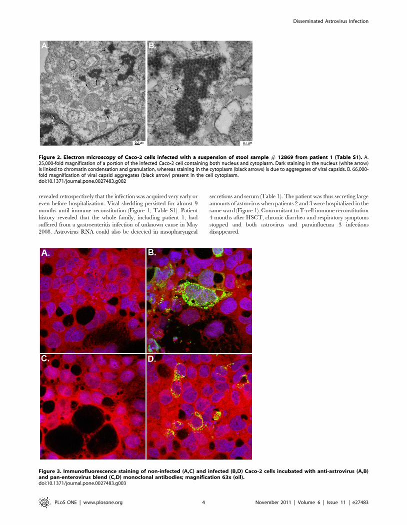
icosahedral capsids of approximately 30 nm diameter (Figures 2A
and 2B) were found in the cytoplasm of the infected cells.
Application of the SISPA method allowed the identification of an
astrovirus. 5 of 13 clones sequenced thanks to this method
represented astroviral sequences (1 clone covered nt 2540 to 2948
of the reference astrovirus 4 genome [GenBank DQ344027]); the
second, nt 3140 to 3795; and the 3 remaining clones overlapped
over nt 4101 to 5094. The 8 remaining clones contained human
sequences. IF performed with a specific anti-astrovirus monoclonal
antibody retrospectively confirmed the cross-reactivity of the pan-
enterovirus blend antibody with astrovirus-infected cells (Figure 3).
To investigate when and how the astrovirus infection was
introduced, stored, frozen clinical samples of a total of 5 suspected
cases were retrospectively screened. This was primarily done since
patient 1 who had a T-B+NK-(gamma-C) SCID was hospitalized
5 months earlier than the deceased patient with chronic diarrhea
and suspected viral disease. In fact, besides patient 1, 2 other
patients were identified positive for astrovirus. Since patient 1 had
stayed on the ward for a cumulative period of 9 months
with ongoing gastrointestinal symptoms until he became T-cell
engrafted, he was suspected to be the index case responsible for the
other infections (Figure 1). The first sample, which retrospectively
tested positive in patient 1, was from 11 July 2008 (Table S1), a
few months before patients 2 and 3 were hospitalized (Tables S2
and S3).
Clinical description of the 3 casesPatient 1 (index case). A 7-month-old boy was hospitalized
in a reduced general condition with frequent stools, chronic cough,
and fever. Following pneumocystis jirovecii pneumonia, he was
diagnosed with an X-linked (gamma-C deficient) T-B+NK- form
of SCID. At the age of 10 months (September 2008), he
underwent allogeneic unrelated HLA-identical HSCT after
pretreatment with rabbit anti-T-cell antibody (ATG) to prevent
graft-versus host disease (GvHD), but without chemotherapeutic
conditioning since he was critically ill. Despite laminar-airflow
conditions, regular administration of intravenous immunoglobulins
(virus inactivated by cold ethanol fractionation, solvent/detergent
treatment, pH 4 inactivation, and ultra filtration; 0,4 g/kg every 2
weeks), and the strict use of masks and hand hygiene antisepsis
measures, he developed a protracted respiratory parainfluenza virus
type 3 infection, as well as a transient rhinovirus infection (Table
S1), and was treated with oral ribavirin. At week 4 post-
transplantation, persistent diarrhea increased and prompted a
stool culture that revealed a positive cytopathic effect and was the
index specimen used for astrovirus identification. Gastrointestinal
symptoms deteriorated with bloody diarrhea without evidence for
GvHD. Stool specimens collected before and after transplantation
Figure 1. Timeline for the detection of astrovirus in samples of the 3 patients. Sample screening listed in Tables S1,S2,S3 is summarized asfollows. Grey circles indicate positive detection of astrovirus in any type of samples (stools, serum, plasma, nasopharyngeal swabs, and vesicle swabs)by real-time RT-PCR and/or cell culture confirmed by immunofluorescence. Black diamonds indicate negative astrovirus detection by real-time RT-PCR. Black star represents the end of hospitalization. T: bone marrow transplantation; {: death.doi:10.1371/journal.pone.0027483.g001
Disseminated Astrovirus Infection
PLoS ONE | www.plosone.org 3 November 2011 | Volume 6 | Issue 11 | e27483
revealed retrospectively that the infection was acquired very early or
even before hospitalization. Viral shedding persisted for almost 9
months until immune reconstitution (Figure 1; Table S1). Patient
history revealed that the whole family, including patient 1, had
suffered from a gastroenteritis infection of unknown cause in May
2008. Astrovirus RNA could also be detected in nasopharyngeal
secretions and serum (Table 1). The patient was thus secreting large
amounts of astrovirus when patients 2 and 3 were hospitalized in the
same ward (Figure 1). Concomitant to T-cell immune reconstitution
4 months after HSCT, chronic diarrhea and respiratory symptoms
stopped and both astrovirus and parainfluenza 3 infections
disappeared.
Figure 2. Electron microscopy of Caco-2 cells infected with a suspension of stool sample # 12869 from patient 1 (Table S1). A.25,000-fold magnification of a portion of the infected Caco-2 cell containing both nucleus and cytoplasm. Dark staining in the nucleus (white arrow)is linked to chromatin condensation and granulation, whereas staining in the cytoplasm (black arrows) is due to aggregates of viral capsids. B. 66,000-fold magnification of viral capsid aggregates (black arrow) present in the cell cytoplasm.doi:10.1371/journal.pone.0027483.g002
Figure 3. Immunofluorescence staining of non-infected (A,C) and infected (B,D) Caco-2 cells incubated with anti-astrovirus (A,B)and pan-enterovirus blend (C,D) monoclonal antibodies; magnification 63x (oil).doi:10.1371/journal.pone.0027483.g003
Disseminated Astrovirus Infection
PLoS ONE | www.plosone.org 4 November 2011 | Volume 6 | Issue 11 | e27483

Patient 2. A male infant and first-degree cousin of patient 1
was screened immediately after birth for SCID and was found to
be also gamma C-deficient. Regular intravenous immunoglobulin
infusions and cotrimoxazole and itraconazole prophylaxis were
initiated and the patient remained at home until transplantation.
An unrelated matched donor was identified and he received
HSCT at the age of 3 months (5 December 2008). Since he had no
clinical or laboratory hints for viral or other opportunistic infection
prior to transplantation, he received conditioning with intravenous
busulfan with therapeutic drug monitoring and rabbit ATG.
Respiratory and intestinal virus screening cultures were negative
prior to HSCT. Two days after infusion of the bone marrow, he
developed high grade fever that remained persistent and of
unknown origin, despite extensive microbiological and radiological
investigations and broad spectrum antibiotic and antifungal
treatment. Information from the donor center revealed that the
donor had not suffered from fever or gastrointestinal disease
prior to or following bone marrow sampling, thus excluding
transmission by the graft itself. Cultures from two stool samples
taken on 11 and 15 December revealed the same cytopathic effect
observed in patient 1. However, no virus could be detected by
standard screening procedures applied at that time, which did not
include screening by the astrovirus-specific real-time RT-PCR
(Table S2). Multiorgan dysfunction developed, including hepatic
dysfunction and pulmonary infiltrates with respiratory distress and
seizures, which required patient admission to the intensive care
unit. Blood culture and other usual bacterial, mycobacterial, and
fungal screening remained negative. Molecular screening for
respiratory viruses in nasopharyngeal swabs was negative (Table
S2) and none of the viruses tested could be detected in plasma.
Cerebrospinal fluid real-time RT-PCR was negative for
enterovirus. Cerebral magnetic resonance imaging revealed signs
suggesting a severe meningoencephalitis and the patient died on
day 17 post-transplantation. At autopsy, the heart, lungs, liver,
bone marrow, spleen, and testicles showed a severe necrotizing
inflammatory process. Neuropathological investigations revealed
distinct inflammatory leptomeningeal and ventricular infiltrates
consisting mainly of macrophages and granulocytes (Figure S1). In
addition, fresh necrosis was detected in the hippocampus, basal
ganglia, brain stem, and cerebellum. No infectious agent was
found by special staining and immunohistochemistry. Retrospec-
tive screening of specimens by culture and/or real-time RT-PCR
revealed that the first astrovirus-positive sample was collected at
day 13 post-hospitalization (Figure 1; Table S2). Subsequent stool,
plasma. and vesicle swab specimens revealed positive (Table 1).
RNA was extracted from paraffin-embedded autopsy samples and
tested by real-time RT-PCR. The small intestine was clearly
astrovirus-positive (consistent with high viral loads [CT values of
22; Table S2]; bone marrow and brain were also positive, but with
a lower viral load (CT around 30); kidney, spleen, lung and heart
tissues were weakly positive (CT values over 36), whereas the liver
was negative (Table 1; Table S2). Immunohistochemistry was
performed on the same autopsy tissue after a preliminary valida-
tion on paraffin-embedded, astrovirus-infected Caco-2 cells (data
not shown). The small intestine autopsy tissue section stained
positive (Figure 4), but no signals were detected in the brain and
bone marrow tissues although an anti-GFAP control antibody
resulted in the expected positive signals (data not shown). Positive
staining likely correlates with high viral load.
Patient 3. A 13-month-old boy with a profound T cell
deficiency and hypogammaglobulinemia (presumed ZAP 70
defect) had suffered from several episodes of bacterial infections
and pneumocystis jirovecii pneumonia. At the age of 16 months
(January 2009), he underwent an allogeneic HSCT from an
unrelated donor after reduced intensity conditioning. After 1 week,
the patient developed gastrointestinal symptoms with recurrent
diarrhea, nausea and vomiting without clinical evidence of GvHD.
Fecal and respiratory specimens revealed retrospectively to be
astrovirus-positive with the first positive sample detected 28 days
post-hospitalization (Table 1; Figure 1; Table S3). No blood
sample was available and viremia could not be documented.
Gastrointestinal symptoms improved simultaneous to emerging T
cells and moderate reduction of immunosuppression. The patient
was discharged on day 70 in good general condition.
Astrovirus transmissionIt is very likely that patient 1 was the index case for the following
reasons. 1) In May 2008, the patient’s family reported an episode of
virally transmitted gastrointestinal disease where patient 1 was also
infected. We could not document this hypothesis as his severe
immunodeficiency had not yet been detected at hospital admission
and, therefore, no stool samples had been analyzed from the patient
and his family. As he suffered from respiratory symptoms related to
pneumocystis jirovecii pneumonia, his concomitant diarrhea was not
further investigated. However, due to the absence of T- and NK-
cells in gamma SCID, a successful elimination of his acquired viral
gastrointestinal infection is not probable. 2) Stool samples were only
tested by standard cell culture at the beginning of hospitalization.
Because no cytopathic effect was observed, no IF test was done and
samples were eliminated after 2 months. For this reason, no real-
time RT-PCR could be done. The first strong positive sample was
on day 23 (11 July 2008; Table S1) of hospitalization. Virus
secretion in patient 1 could be documented on different samples
until 9 March 2009. 3) He had been secreting large amounts of
astrovirus (CT values: 15.3 and 12.23, respectively) when patients 2
and 3 were hospitalized in the same ward and was the very likely
source of contamination for the 2 other cases (Figure 1).
Table 1. Detection of astrovirus in different samples from the3 patients.
Duration (in days) of astrovirusidentification in different samplesfrom the 3 patients
Type of specimen Patient 1 Patient 2 Patient 3
Stool 302 4 82
Serum 1 - -
Plasma - 8 -
Nasopharyngeal swab 71 - 1
Pharyngeal swab 167 - 1
Vesicle swab - 1 -
Brain - PM -
Heart - PM * -
Lung - PM * -
Spleen - PM * -
Bone marrow - PM -
Kidney - PM * -
Small intestine - PM -
Duration of hospitalization 180 17 70
-, not tested or negative; PM, post mortem autopsy tissue tested positive (CTvalues,36); PM*, post mortem autopsy tissue tested weakly positive (CT values. 36). See Tables S1,S2,S3 for detailed results.doi:10.1371/journal.pone.0027483.t001
Disseminated Astrovirus Infection
PLoS ONE | www.plosone.org 5 November 2011 | Volume 6 | Issue 11 | e27483

When the 3 cases were hospitalized, standard precautions for
hospitalized (according to US Centers for Disease Prevention and
Control guidelines) and immunocompromised patients in the bone
marrow transplantation (BMT) unit (e.g. care in single-patient,
positive-pressure, laminar airflow boxes, introduction of disinfect-
ed material only, the wear of coats and masks for staff and parents
entering the box) were applied. However, subsequent investigation
by the infection control team revealed gaps and negligence in
some aspects of standard precautions, such as handling and
delivery of meals, hand hygiene of healthcare workers, and
insufficient information provided to parents. This is of particular
importance as the families of 2 patients were related. In addition,
the BMT unit was overcrowded both inside and outside the boxes,
thus preventing efficient environmental cleaning. In addition,
disinfection studies showed that the products used for standard
BMT procedures in our ward were insufficient in completely
inactivating enteroviruses (data not shown).
Stool specimens from the 3 patients were sequenced over 543 nt
of the capsid gene. A phylogenetic tree revealed that the patients
were infected with a unique astrovirus type 4 strain (Figure 5).
Furthermore, samples collected from patient 1 96 days post-
hospitalization had acquired a non synonymous change also
present in patients 2 and 3 (Table 2). As expected for protracted
RNA virus infections, viral populations sequenced from patient 1
between days 145 (sample no. 11196) and 250 (sample no. 1898)
post-hospitalization (the period where patients 2 and 3 were
infected) evolved. The exact sequence composition at the time of
transmission is not known, but the sequences of sample no. 12428
(patient 2) and 1391 (patient 3) are both present in the quasispecies
populations observed in sample no. 1898 (patient 1).
Discussion
We describe 3 young children with SCID in a pediatric BMT
unit who developed protracted astrovirus infection with dissem-
inated disease, which was finally fatal in one infant. Viral infection
was initially suspected, but routine diagnostic molecular investi-
gations remained negative. Generic viral discovery tools [5,19]
were then applied and led to the identification of astrovirus.
Subsequent screening by specific real-time RT-PCR confirmed
the presence of astrovirus RNA in multiple body sites in at least 2
of the 3 infected cases. Retrospective screening revealed that
patient 1 was already infected before transplantation, possibly even
before hospitalization, and secreting large amounts of virus when
patients 2 and 3 were hospitalized in the same ward. We cannot
completely exclude that patients 2 and 3 were infected from
another source, such as external visitors, but this seems very
unlikely due to the strict isolation measures applied to HSCT
recipients. The most probable scenario is that patient 1 was the
source of the infection to cases 2 and 3 and the virus was
transmitted by healthcare staff or the children’s parents due to
their close contact outside the laminar airflow cabins.
The hypothesis of nosocomial infection is further reinforced by
retrospective screening of samples, which revealed that astrovirus
was continuously detectable in patient 1 until immune reconsti-
tution (Figure 1; Table S1), and capsid sequence analysis of
different positive samples that confirmed the presence of a unique
astrovirus type 4 strain. One unlikely hypothesis is that cases 2 and
3 were infected in the community before hospitalization with low-
level replicating viruses that were not detected upon hospital
admission. Of note, investigation of the donor history of patients 1
and 2 showed no signs of diarrhea one month before donation.
Following the suspicion of nosocomial infection, preventive
measures were taken. The hand hygiene antisepsis agent was
replaced by a more virucidal product, the BMT unit was cleaned
room-by-room with highly virucidal surface disinfectant, and the
admission of visitors to these children was restricted to their
parents only.
This sequence of events illustrates the numerous challenges
facing clinicians, clinical virologists, and microbiologists caring for
highly immunocompromised infants. Astrovirus real-time RT-
PCR could have provided an early and simple diagnosis for these
patients. However, diarrhea observed in our index case could have
also been caused by many other viruses and a large panel of real-
time assays targeting all possible viral agents would have been
necessary. Due to the diversity of potentially involved viral agents
in humans [2], as well as emerging new viruses or variants, the
spectrum covered by such a panel would always be an issue. In the
present investigation, classical cell culture in combination with
modern generic molecular detection tools provided the diagnosis.
Traditional diagnosis by cell culture has technical limitations, such
as low sensitivity and existence of non-cultivable viruses, and has
been progressively abandoned. In this case, cell culture was the
first indication for the presence of a virus. This strongly supports
the notion that it is advantageous to preserve classical viral culture
in centers caring for immunocompromised hosts. Of note, negative
staining EM from stool suspensions or from a supernatant from
positive cell culture could also have given very fast, first indications
of the presence of an unexpected virus. However this technique is
not available as a routine tool in our institution. Nevertheless,
microarray-based assays [3], mass spectrometry [20], ultra-deep
sequencing [4], and other alternative advanced technologies will
likely change the diagnostic landscape once validated for clinical
use.
Astrovirus is a non-enveloped, positive-strand RNA virus that
infects mammalian as well as avian hosts [21]. The virus is
Figure 4. Immunohistochemistry of the small intestine autopsy tissue sections of patient 2. Immunohistochemistry with an anti-astrovirus antibody performed on non-infected (A) and infected (B) regions of the small intestine autopsy tissue sections.doi:10.1371/journal.pone.0027483.g004
Disseminated Astrovirus Infection
PLoS ONE | www.plosone.org 6 November 2011 | Volume 6 | Issue 11 | e27483

Figure 5. Phylogenetic analysis of the transmitted astrovirus strains. A. A maximum-likelihood-based phylogenetic tree was computedusing capsid gene sequences (corresponding to nt 4325 to 4867 of the astrovirus 4 complete genome [GenBank DQ344027] or nt 1 to 543 of thecapsid gene) obtained from 8 samples collected from patient 1, one from patient 2, and two from patient 3 (Table 2). Corresponding sequences fromrepresentatives of the 8 human astrovirus serotypes (HAstV) and the divergent VMLB1 human astrovirus (HastVMLB1) were included to show acomparison of evolutionary distances and relationships. Bat astrovirus (BatAstV, in light gray) was used as an outgroup. All virus strains are shownwith the corresponding GenBank accession number. The numbers of substitutions per site are indicated below the tree. Because of long branches,the outgroup BatAstV, as well as the strain HAstVMLB1, are shown with annotated branch lengths. B. Blow-up of a sub-tree from panel A marked withan asterisk showing relatively short branch lengths compared to the full tree.doi:10.1371/journal.pone.0027483.g005
Table 2. Genetic variability of the astrovirus genome in the 3 investigated cases.
Sample N6
Days post hospitalisationof patient 1 Patient Material Nucleotide (amino acid)
*4411 *4678 *4752 *4762 *4802
7125 24 1 Stool culture T(S) G(G) G(R) T(S) A(I)
8136 56 1 stool T(S) G(G) G(R) T(S) A(I)
8280 62 1 stool T(S) G(G) G(R) T(S) A(I)
9442 96 1 stool T(S) G(G) G(R) T(S) R(I/V)
9972 110 1 stool T(S) G(G) G(R) T(S) G(V)
10477 124 1 stool T(S) G(G) G(R) T(S) G(V)
11196 145 1 stool T(S) R (G) G(R) T(S) G(V))
1898 250 1 Stool culture Y(S) G(G) R (R/K) T(S) R(I/V)
12428 #180 2 Stool culture C (S) G(G) G(R) T(S) G(V)
1391 &237 3 stool C (S) G(G) G(R) T(S) G(V)
2619 272 3 stool Y (S) R (G) G(R) Y (S) G(V)
The astrovirus genome was sequenced over a 651 nt portion (corresponding to nt 4216 to 4867 of the astrovirus 4 complete genome (GenBank DQ344027) covering nt1 to 543 of the capsid gene). Sequences are available in GenBank under accession # HQ396880 to HQ396890.*Positions relative to astrovirus 4 genome (GenBank DQ344027). Capital letters outside brackets indicate nucleotides (A,T,G,C). Brackets indicate amino acids at theirrespective position. Synonymous changes are indicated in italic and non synonymous changes are indicated in bold.#4 days after detection of 1st astrovirus positive sample in patient 2.&time of detection of 1st astrovirus positive sample in patient 3.doi:10.1371/journal.pone.0027483.t002
Disseminated Astrovirus Infection
PLoS ONE | www.plosone.org 7 November 2011 | Volume 6 | Issue 11 | e27483

transmitted mainly by the oro-fecal route, circulates worldwide,
and is the cause of outbreaks in the community and healthcare
facilities [22]. After rotavirus, it is one of the most frequent causes
of viral gastroenteritis in young children [23,24]. The infection is
characterized by a gastrointestinal disease with nausea, vomiting,
diarrhoea, and sometimes fever, mostly self-limited to a few days.
The inflammatory response of the gastrointestinal mucosa is often
minimal [1], but there is a possible association with necrotizing
enterocolitis in newborns [25].
Atypical clinical presentations, such as prolonged viral shedding,
have been described in immunocompromised hosts [1,26] and this
was observed in all 3 patients reported in this study. We detected
also astrovirus RNA in unexpected body sites, such as the
respiratory tract, blood, bone marrow, skin, and brain. Recently, a
similar investigation in a child with primary immunodeficiency
and progressive encephalitis proved the ability of astrovirus to
infect the brain and likely cause progressive encephalitis [4]. The
authors of this study could not identify astrovirus in other sites,
probably due to the retrospective nature of their investigation and
a limited access to specimens. By demonstrating RNA in multiple
sites, our investigation supports their observation that astrovirus
can reach other organs, including the central nervous system. The
ability of astrovirus to cause viremia is probably due to high and
protracted replication in the gastrointestinal tract. Prolonged
shedding in immunocompromised hosts has been described for
other gastrointestinal RNA viruses, such as rotavirus [27], ADV
[28], and norovirus [29,30], as well as respiratory viruses [31], and
highlights the need for an efficient immune response to prevent
dissemination and to clear these usually self-limited infections. In
our cases, prolonged shedding and deficient environmental
disinfection procedures could have offered opportunities for
inter-individual transmission and has to be considered as the most
likely cause of the observed outbreak.
In conclusion, astrovirus needs to be considered as a potential
cause of protracted disease and disseminated infection in severely
immunocompromised infants with primary immunodeficiency
prior to and after allogeneic HSCT.
Supporting Information
Table S1 Virological and immunological results ob-tained in samples from patient 1 (index case).
(DOC)
Table S2 Virological and immunological results obtained in
samples from patient 2.
(DOC)
Table S3 Virological and immunological results ob-tained in samples from patient 3.
(DOC)
Figure S1 Histopathology of autopsy material of patient2. 1. Overview of hippocampus with necroses (arrow) and
meningo-ventriculo encephalitis (H&E x 10). 2. Destructive
ventriculitis consisting mainly of macrophages and granulocytes
(H&E x 200). 3. Meningoencephalitis consisting of macrophages,
lymphocytes, plasma cells and a few granulocytes (H&E x 50).
(TIF)
Acknowledgments
We thank Rosemary Sudan for editorial assistance, Patricia Gindre and
Johannes Alexander Lobrinus (Geneva) for help with immunohistochem-
istry, Chantal Gaille for sequencing, Ursula Luthi for electron microscopy,
and Pascal Cherpillod from the Swiss National Centre for Emerging Viral
Infections (CRIVE) in Geneva.
Author Contributions
Conceived and designed the experiments: CT LK AT WW. Performed the
experiments: SVB AM RC TAM WB MS SC. Analyzed the data: CT LK
WW AT DG TAM TG CB OG WB. Contributed reagents/materials/
analysis tools: WW AM RC WB TAM LK CT. Wrote the paper: WW CT
LK. Corrected the manuscript: CB TG AT WB MS SC TAM.
References
1. Sebire NJ, Malone M, Shah N, Anderson G, Gaspar HB, et al. (2004) Pathology
of astrovirus associated diarrhoea in a paediatric bone marrow transplant
recipient. J Clin Pathol 57: 1001–1003.
2. Taylor LH, Latham SM, Woolhouse ME (2001) Risk factors for human disease
emergence. Philos Trans R Soc Lond B Biol Sci 356: 983–989.
3. Victoria JG, Wang C, Jones MS, Jaing C, McLoughlin K, et al. (2010) Viralnucleic acids in live-attenuated vaccines: detection of minority variants and an
adventitious virus. J Virol 84: 6033–6040.
4. Quan PL, Wagner TA, Briese T, Torgerson TR, Hornig M, et al. (2010)
Astrovirus encephalitis in boy with X-linked agammaglobulinemia. Emerg InfectDis 16: 918–925.
5. Allander T, Tammi MT, Eriksson M, Bjerkner A, Tiveljung-Lindell A, et al.(2005) Cloning of a human parvovirus by molecular screening of respiratory
tract samples. Proc Natl Acad Sci U S A 102: 12891–12896.
6. de Vries M, Pyrc K, Berkhout R, Vermeulen-Oost W, Dijkman R, et al. (2008)
Human parechovirus type 1, 3, 4, 5, and 6 detection in picornavirus cultures.J Clin Microbiol 46: 759–762.
7. Berger C, Day P, Meier G, Zingg W, Bossart W, et al. (2001) Dynamics ofEpstein-Barr virus DNA levels in serum during EBV-associated disease. J Med
Virol 64: 505–512.
8. Heim A, Ebnet C, Harste G, Pring-Akerblom P (2003) Rapid and quantitative
detection of human adenovirus DNA by real-time PCR. J Med Virol 70: 228–239.
9. Hohne M, Schreier E (2004) Detection and characterization of norovirusoutbreaks in Germany: application of a one-tube RT-PCR using a fluorogenic
real-time detection system. J Med Virol 72: 312–319.
10. Kronenberg A, Schupbach R, Schuknecht B, Bossart W, Weber R, et al. (2002)
Multifocal vasculopathy due to Varicella-Zoster Virus (VZV): serial analysis ofVZV DNA and intrathecal synthesis of VZV antibody in cerebrospinal fluid.
Clin Infect Dis 35: 330–333.
11. Pevenstein SR, Williams RK, McChesney D, Mont EK, Smialek JE, et al. (1999)
Quantitation of latent varicella-zoster virus and herpes simplex virus genomes in
human trigeminal ganglia. J Virol 73: 10514–10518.
12. Soccal PM, Aubert JD, Bridevaux PO, Garbino J, Thomas Y, et al. (2010)
Upper and lower respiratory tract viral infections and acute graft rejection inlung transplant recipients. Clin Infect Dis 51: 163–170.
13. Tapparel C, Cordey S, Van Belle S, Turin L, Lee WM, et al. (2009) New
molecular detection tools adapted to emerging rhinoviruses and enteroviruses.J Clin Microbiol 47: 1742–1749.
14. Yun Z, Lewensohn-Fuchs I, Ljungman P, Vahlne A (2000) Real-time
monitoring of cytomegalovirus infections after stem cell transplantation usingthe TaqMan polymerase chain reaction assays. Transplantation 69: 1733–1736.
15. Edgar RC (2004) MUSCLE: multiple sequence alignment with high accuracyand high throughput. Nucleic Acids Res 32: 1792–1797.
16. Rice P, Longden I, Bleasby A (2000) EMBOSS: the European Molecular
Biology Open Software Suite. Trends Genet 16: 276–277.
17. Guindon S, Gascuel O (2003) A simple, fast, and accurate algorithm to estimate
large phylogenies by maximum likelihood. Syst Biol 52: 696–704.
18. Logan C, O’Leary JJ, O’Sullivan N (2007) Real-time reverse transcription PCRdetection of norovirus, sapovirus and astrovirus as causative agents of acute viral
gastroenteritis. J Virol Methods 146: 36–44.
19. Tapparel C, Junier T, Gerlach D, Van-Belle S, Turin L, et al. (2009) New
respiratory enterovirus and recombinant rhinoviruses among circulating
picornaviruses. Emerg Infect Dis 15: 719–726.
20. Sampath R, Russell KL, Massire C, Eshoo MW, Harpin V, et al. (2007) Global
surveillance of emerging Influenza virus genotypes by mass spectrometry. PLoS
One 2: e489.
21. Finkbeiner SR, Kirkwood CD, Wang D (2008) Complete genome sequence of a
highly divergent astrovirus isolated from a child with acute diarrhea. Virol J 5: 117.
22. Cunliffe NA, Booth JA, Elliot C, Lowe SJ, Sopwith W, et al. (2010) Healthcare-
associated viral gastroenteritis among children in a large pediatric hospital,
United Kingdom. Emerg Infect Dis 16: 55–62.
23. Glass RI, Noel J, Mitchell D, Herrmann JE, Blacklow NR, et al. (1996) The
changing epidemiology of astrovirus-associated gastroenteritis: a review. Arch
Virol Suppl 12: 287–300.
Disseminated Astrovirus Infection
PLoS ONE | www.plosone.org 8 November 2011 | Volume 6 | Issue 11 | e27483

24. Walter JE, Mitchell DK (2003) Astrovirus infection in children. Curr Opin Infect
Dis 16: 247–253.25. Bagci S, Eis-Hubinger AM, Franz AR, Bierbaum G, Heep A, et al. (2008)
Detection of astrovirus in premature infants with necrotizing enterocolitis.
Pediatr Infect Dis J 27: 347–350.26. Cubitt WD, Mitchell DK, Carter MJ, Willcocks MM, Holzel H (1999)
Application of electronmicroscopy, enzyme immunoassay, and RT-PCR tomonitor an outbreak of astrovirus type 1 in a paediatric bone marrow transplant
unit. J Med Virol 57: 313–321.
27. Liakopoulou E, Mutton K, Carrington D, Robinson S, Steward CG, et al. (2005)Rotavirus as a significant cause of prolonged diarrhoeal illness and morbidity
following allogeneic bone marrow transplantation. Bone Marrow Transplant 36:691–694.
28. de Mezerville MH, Tellier R, Richardson S, Hebert D, Doyle J, et al. (2006)
Adenoviral infections in pediatric transplant recipients: a hospital-based study.
Pediatr Infect Dis J 25: 815–818.
29. Kaufman SS, Chatterjee NK, Fuschino ME, Morse DL, Morotti RA, et al.
(2005) Characteristics of human calicivirus enteritis in intestinal transplant
recipients. J Pediatr Gastroenterol Nutr 40: 328–333.
30. Schorn R, Hohne M, Meerbach A, Bossart W, Wuthrich RP, et al. (2010)
Chronic norovirus infection after kidney transplantation: molecular evidence for
immune-driven viral evolution. Clin Infect Dis 51: 307–314.
31. Kaiser L, Aubert JD, Pache JC, Deffernez C, Rochat T, et al. (2006) Chronic
rhinoviral infection in lung transplant recipients. Am J Respir Crit Care Med
174: 1392–1399.
Disseminated Astrovirus Infection
PLoS ONE | www.plosone.org 9 November 2011 | Volume 6 | Issue 11 | e27483