Architecture and mechanism of the central gear in an...
14
rsif.royalsocietypublishing.org Review Cite this article: Egli M. 2017 Architecture and mechanism of the central gear in an ancient molecular timer. J. R. Soc. Interface 14: 20161065. http://dx.doi.org/10.1098/rsif.2016.1065 Received: 31 December 2016 Accepted: 27 February 2017 Subject Category: Life Sciences – Chemistry interface Subject Areas: biochemistry, biophysics, bioinformatics Keywords: ATPase, circadian clock, kinase, phosphorylation, subunit exchange, X-ray crystallography Author for correspondence: Martin Egli e-mail: [email protected] Architecture and mechanism of the central gear in an ancient molecular timer Martin Egli Department of Biochemistry, Vanderbilt University, Nashville, TN 37232, USA ME, 0000-0003-4145-356X Molecular clocks are the product of natural selection in organisms from bacteria to human and their appearance early in evolution such as in the prokaryotic cyanobacterium Synechococcus elongatus suggests that these timers served a crucial role in genetic fitness. Thus, a clock allows cyanobac- teria relying on photosynthesis and nitrogen fixation to temporally space the two processes and avoid exposure of nitrogenase carrying out fixation to high levels of oxygen produced during photosynthesis. Fascinating proper- ties of molecular clocks are the long time constant, their precision and temperature compensation. Although these are hallmarks of all circadian oscillators, the actual cogs and gears that control clocks vary widely between organisms, indicating that circadian timers evolved convergently multiple times, owing to the selective pressure of an environment with a daily light/dark cycle. In S. elongatus, the three proteins KaiA, KaiB and KaiC in the presence of ATP constitute a so-called post-translational oscillator (PTO). The KaiABC PTO can be reconstituted in an Eppendorf tube and keeps time in a temperature-compensated manner. The ease by which the KaiABC clock can be studied in vitro has made it the best-investigated mol- ecular clock system. Over the last decade, structures of all three Kai proteins and some of their complexes have emerged and mechanistic aspects have been analysed in considerable detail. This review focuses on the central gear of the S. elongatus clock and only enzyme among the three proteins: KaiC. Our determination of the three-dimensional structure of KaiC early in the quest for a better understanding of the inner workings of the cyanobacterial timer revealed its unusual architecture and conformational differences and unique features of the two RecA-like domains constituting KaiC. The structure also pinpointed phosphorylation sites and differential interactions with ATP molecules at subunit interfaces, and helped guide experiments to ferret out mechanistic aspects of the ATPase, auto-phosphorylation and auto- dephosphorylation reactions catalysed by the homo-hexamer. Comparisons between the structure of KaiC and those of nanomachines such as F1-ATPase and CaMKII also exposed shared architectural features (KaiC/ ATPase), mechanistic principles (KaiC/CaMKII) and phenomena, such as subunit exchange between hexameric particles critical for function (clock synchronization, KaiABC; memory-storage, CaMKII). 1. Introduction ‘Each thing is of like form from everlasting and comes round again in its cycle’ (Marcus Aurelius, second century A.D.). The emperor’s words spoken almost 2 millennia ago attest to the timeless fascination with the nature of time, and recurring patterns such as day and night or the seasons. Keeping track of time and correctly anticipating daybreak and nightfall is of such crucial impor- tance that organisms from simple cyanobacteria to humans all rely on precise molecular timers. Circadian clocks set the pace for a multitude of processes that include gene expression, cell division, development, metabolism and behaviour [1]. Circadian rhythms are characterized by three hallmarks indepen- dent of organism [2]: (i) endogenous oscillations with a period of approximately 24 h that persist under constant conditions, i.e. light or dark and temperature, & 2017 The Author(s) Published by the Royal Society. All rights reserved. on May 3, 2017 http://rsif.royalsocietypublishing.org/ Downloaded from
Transcript of Architecture and mechanism of the central gear in an...

on May 3, 2017http://rsif.royalsocietypublishing.org/Downloaded from
rsif.royalsocietypublishing.org
Review
Cite this article: Egli M. 2017 Architecture
and mechanism of the central gear in an
ancient molecular timer. J. R. Soc. Interface 14:
20161065.
http://dx.doi.org/10.1098/rsif.2016.1065
Received: 31 December 2016
Accepted: 27 February 2017
Subject Category:Life Sciences – Chemistry interface
Subject Areas:biochemistry, biophysics, bioinformatics
Keywords:ATPase, circadian clock, kinase,
phosphorylation, subunit exchange, X-ray
crystallography
Author for correspondence:Martin Egli
e-mail: [email protected]
& 2017 The Author(s) Published by the Royal Society. All rights reserved.
Architecture and mechanism of thecentral gear in an ancient molecular timer
Martin Egli
Department of Biochemistry, Vanderbilt University, Nashville, TN 37232, USA
ME, 0000-0003-4145-356X
Molecular clocks are the product of natural selection in organisms from
bacteria to human and their appearance early in evolution such as in the
prokaryotic cyanobacterium Synechococcus elongatus suggests that these
timers served a crucial role in genetic fitness. Thus, a clock allows cyanobac-
teria relying on photosynthesis and nitrogen fixation to temporally space the
two processes and avoid exposure of nitrogenase carrying out fixation to
high levels of oxygen produced during photosynthesis. Fascinating proper-
ties of molecular clocks are the long time constant, their precision and
temperature compensation. Although these are hallmarks of all circadian
oscillators, the actual cogs and gears that control clocks vary widely between
organisms, indicating that circadian timers evolved convergently multiple
times, owing to the selective pressure of an environment with a daily
light/dark cycle. In S. elongatus, the three proteins KaiA, KaiB and KaiC
in the presence of ATP constitute a so-called post-translational oscillator
(PTO). The KaiABC PTO can be reconstituted in an Eppendorf tube and
keeps time in a temperature-compensated manner. The ease by which the
KaiABC clock can be studied in vitro has made it the best-investigated mol-
ecular clock system. Over the last decade, structures of all three Kai proteins
and some of their complexes have emerged and mechanistic aspects have
been analysed in considerable detail. This review focuses on the central gear
of the S. elongatus clock and only enzyme among the three proteins: KaiC.
Our determination of the three-dimensional structure of KaiC early in the
quest for a better understanding of the inner workings of the cyanobacterial
timer revealed its unusual architecture and conformational differences and
unique features of the two RecA-like domains constituting KaiC. The structure
also pinpointed phosphorylation sites and differential interactions with
ATP molecules at subunit interfaces, and helped guide experiments to ferret
out mechanistic aspects of the ATPase, auto-phosphorylation and auto-
dephosphorylation reactions catalysed by the homo-hexamer. Comparisons
between the structure of KaiC and those of nanomachines such as
F1-ATPase and CaMKII also exposed shared architectural features (KaiC/
ATPase), mechanistic principles (KaiC/CaMKII) and phenomena, such as
subunit exchange between hexameric particles critical for function (clock
synchronization, KaiABC; memory-storage, CaMKII).
1. Introduction‘Each thing is of like form from everlasting and comes round again in its cycle’
(Marcus Aurelius, second century A.D.). The emperor’s words spoken almost
2 millennia ago attest to the timeless fascination with the nature of time, and
recurring patterns such as day and night or the seasons. Keeping track of
time and correctly anticipating daybreak and nightfall is of such crucial impor-
tance that organisms from simple cyanobacteria to humans all rely on precise
molecular timers. Circadian clocks set the pace for a multitude of processes
that include gene expression, cell division, development, metabolism and
behaviour [1]. Circadian rhythms are characterized by three hallmarks indepen-
dent of organism [2]: (i) endogenous oscillations with a period of approximately
24 h that persist under constant conditions, i.e. light or dark and temperature,

rsif.royalsocietypublishing.orgJ.R.Soc.Interface
14:20161065
2
on May 3, 2017http://rsif.royalsocietypublishing.org/Downloaded from
(ii) temperature compensation, i.e. the period is only
minimally affected by changes in temperature, and
(iii) entrainment of the endogenous timer by exogenous,
environmental cues, i.e. the light/dark cycle. The adaptive
importance of circadian programmes was rigorously tested
in cyanobacteria: competition experiments between Synecho-coccus elongatus strains showed that those with clocks that
resonated with their light/dark environment outcompeted
other strains, even if not set at 24 h [3]. Further, strains with
any type of functional clock outcompeted clock-disrupted
strains, although not under constant conditions [4]. Overall,
such experiments demonstrated that a precise clock system
enhances reproductive fitness.
Although all circadian oscillators share the above hall-
marks, the molecules that make up the timers vary greatly
among the lower and higher organisms. The KaiABC clock
in S. elongatus is composed of the KaiA, KaiB and KaiC pro-
teins and emerged some 3.5 billion years ago [5,6], but other
organisms whose clocks have been studied extensively, from
Neurospora crassa and Arabidopsis thaliana to Drosophila melano-gaster and Homo sapiens differ from the cyanobacterial
oscillator in their molecular make-up. For recent reviews of
the molecular clock components in diverse organisms,
mechanistic, functional and structural aspects as well as
emerging shared features and unique attributes, see [7–14].
Research into the inner workings of the KaiABC cyanobacter-
ial clock shattered the assumption that a transcription/
translation-based feedback loop (TTFL) is at the heart of all
biological timers [7,15]. Initially, it was discovered that circa-
dian rhythms of KaiC phosphorylation persisted in the
absence of transcription and translation [16]. Subsequent
investigations established that mixing the KaiA, KaiB and
KaiC proteins in an Eppendorf tube in the presence of ATP
constitutes a temperature-compensated circadian oscillator,
a so-called post-translational oscillator (PTO) [17]. The
cyanobacterial timer thus comprises a PTO—the master
clock—coupled to a TTFL that is required for robust oscil-
lations [1,18,19]. The discovery of the KaiABC PTO in
S. elongatus has led to a renewed interest in examining the
experimental evidence for the existence of PTOs or non-tran-
scriptional oscillators in eukaryotic clocks ([7] and references
therein). A PTO may have proved more resilient in maintain-
ing robust rhythms despite noise arising from cell division,
changes in metabolic rate and environmental stress. The
reduced susceptibility to external factors may have served
as a powerful evolutionary driving force for convergent
clock mechanisms in diverse organisms [20]. In cyanobac-
teria, the clock orchestrates gene expression and, although it
is not clear whether metabolism is regulated in a similar
fashion, changes in metabolism apparently serve the entrain-
ment of the clock [1]. In that context it is fascinating to
contemplate the possibility that temperature compensation,
a shared property of all biological timers, merely constitutes
a subset of metabolic compensation.
Among the three Kai proteins that constitute the S. elonga-tus PTO, only KaiC has enzymatic activity [21,22]. Actually,
KaiC exhibits multiple activities—kinase [17], ATPase [23]
and phosphotransferase/ATP synthase [24,25]—and its
(auto-) phosphorylation and (auto-) dephosphorylation pro-
ceed with a period of approximately 24 h [16,17], thus
setting the daily cycle. KaiC features N- and C-terminal
domains (KaiCI and KaiCII, respectively) that display high
sequence similarity (gene duplication) [26] (figure 1), and
create a double ring upon formation of KaiC homo-hexamer
[27–29]. Importantly, function, three-dimensional structure
and dynamics of the CI and CII domains and rings differ
[30]. Thus, both act as ATPases, but only CII possesses
kinase activity [31–34], whereby phosphates from phos-
phorylated threonine and serine residues are returned to
ADP to produce ATP, therefore rendering KaiC an ATP
synthase [24]. The CI ring acts as a platform or hub and tigh-
ter inter-domain contacts compared with CII are at the origin
of the higher stability of the CI ring [35]. KaiA binding to the
CII C-termini stimulates KaiC phosphorylation [36–40] and
KaiB sequesters KaiA in a KaiABC ternary complex during
the dephosphorylation phase [41]. Binding by KaiB coincides
with KaiC subunit shuffling, a process that is crucial for the
robustness of the oscillation [42–44]. Multiple activities
make this enzyme a fascinating target for mechanistic studies
and the observation that KaiC together with KaiA and KaiB
constitute a fully functional and temperature-compensated
circadian clock in vitro have advanced the KaiABC
nano-machine to the best studied molecular timer [1,7,45].
Circadian rhythms in cyanobacteria are pervasive from
molecules to cells. In addition to the aforementioned
rhythmic oscillation between the KaiC hypo- and hyper-
phosphorylated states and the above circadian assembly
and disassembly of binary and ternary Kai protein com-
plexes, the concentrations of the three Kai proteins as well
as those of the KaiAC, KaiBC and KaiABC complexes wax
and wane in a circadian fashion [44]. Bioluminescence
assays using a bacterial luciferase reporter demonstrated
genome-wide rhythmic promoter activity that is not dis-
rupted by cell division and persists in daughter cells
without a change in phase (reviewed in [29]). Additional cel-
lular phenomena that exhibit a circadian rhythm are
chromosomal compaction and expansion, and chromosomal
topology as evidenced by assaying supercoiling of an
endogenous plasmid [29].
This review focuses on KaiC, the central cog of the cyano-
bacterial clock, and some of its fascinating properties such as
multiple enzymatic activities. Functionally, structurally and
mechanistically, KaiC is the most interesting component of
the three-protein PTO of S. elongatus, a recognition that
prompted us to target it for initial three-dimensional struc-
ture determination by X-ray crystallography some 15 years
ago. Structures of the full-length enzyme indeed provided
insight into the order of phosphorylation events at the inter-
faces between KaiCII subunits [28,31,33,34], alternative
phosphorylation sites besides the primary threonine 432
(T432) and secondary serine 431 (S431), and the absence of
kinase activity in the KaiCI half [46]. Recent crystal structures
of the KaiCI ring provided a better understanding of the slow
ATPase activity in the N-terminal half [47]. Structural data
also confirmed that KaiC exhibits not only sequence simi-
larities to other proteins such as helicases (DnaB) and
recombinases (RecA) [26], but that the architectures of these
proteins show close resemblance, at least at the subunit
level of single-ring hexameric assemblies. By comparison,
the similar CI/CII and F1-ATPase active-site organizations
revealed by structural overlays [28], are not evident from
sequence alignments, but are consistent with the mechanism
of KaiC dephosphorylation, i.e. the transfer of phosphates on
threonine and serine back to ADP with concomitant synthesis
of ATP in CII [24]. Other unexpected structural and mechan-
istic similarities (double ring structure, kinase and

1 50292
100341
150383
198432
247480
b9
b7
b6
b4
b3b2b1
b5
b10 b11b8
a7 a8 a9
a6
a4a3
a1
a2
a5
251519
252
51293
101342
151384
199433
248481
Figure 1. Sequence alignment between the KaiCI (upper line) and KaiCII (lower line) halves. Secondary structure elements observed in the crystal structure aredepicted above the CI and CII sequences. Selected motifs and residues are highlighted in colour: Walker motif A (‘P-loop’, GXXXXGKT, blue), imperfect, truncated,Walker motif B (RXXXD, cyan), catalytic carboxylate glutamic acid (yellow), phosphorylation sites (CII, red) and equivalent residues in CI ( pink), arginine finger(orange) and arginine linker (CI, purple), and CI – CII linker (green). Residues contacting ATPs are marked by a black dot and threonines and serines within8 A from gP are starred. Residues E487 to I497 in the longer C-terminal region of CII are referred to as A-loop (binding of KaiA), and residues Q115 to D122in CI are referred to as B-loop (binding of KaiB).
rsif.royalsocietypublishing.orgJ.R.Soc.Interface
14:20161065
3
on May 3, 2017http://rsif.royalsocietypublishing.org/Downloaded from
phosphotransferase activity, and subunit exchange) also exist
between the KaiC clock protein and the Ca2þ/calmodulin-
dependent kinase II holoenzyme (CaMKII) that is involved
in memory formation [48].
2. Differences in the sequences and architecturesof the KaiCI and CII domains
Amino acid sequence comparisons established an evolution-
ary relationship between the RecA/Rad51/DCMI family of
ATP-dependent recombinases in archaea, bacteria and
eukaryota, the DnaB replication fork helicase in bacteria
and the Sms and KaiC families [26]. KaiC arose as a result
of the duplication of a gene that codes for approximately
250-amino acid ATP- and DNA-binding domain. However,
although this typically more than 500 amino acids long
protein composed of two RecA-like domains and joined in
a head-to-tail fashion is present in cyanobacteria (e.g. S. elon-gatus), the Archeoglobus and Pyrococcus archaea, and
Methanobacterium, it is not found in Aeropyrum and Methano-coccus. Besides the double-domain KaiCs, there are many
archaeal single-domain homologues and the KaiC protein
in the bacterium Thermotoga maritima (hyperthermophile) is
also of the single-domain type. Thus, it was suggested that
the single-domain protein was laterally transferred from the
bacteria to the archaea, followed by duplication and fusion
[26]. Sequence alignment of the KaiCI and KaiCII domains
from S. elongatus reveals similarities and clear differences
(figure 1). The two halves display some 20% identity and
30% similarity, and these numbers are similar for the proteins
from Thermosynechococcus elongatus, Synechocystis and
Anabaena. As expected, the KaiC domains feature a number
of sequence motifs and residues that are hallmarks of
ATPases, such as the Walker A and B motifs, catalytic
glutamate and the Arg finger that commonly contacts the
ATP g-phosphate at the active site. The N- and C-terminal
regions in the two domains differ significantly: at the
N-terminus, the CII domain exceeds in length the CI one
by a short stretch; as well the former domain features a
more than 30-residue long C-terminal tail. KaiC C-terminal
tails exhibit relatively high sequence conservation among
cyanobacterial strains and a noteworthy characteristic is the
presence of four or more acidic residues [40].
The likely absence of an N-terminal helix like those
present in DnaB or RecA was thought to preclude hexameri-
zation by KaiC and the head-to-tail fusion of two RecA-like
domains expected to perhaps promote a functional dimer
[26]. However, our crystal structure revealed that KaiC does
form homo-hexamers [28]. In fact, prior to the determination
of the structure, negative-stain electron micrographs pro-
vided unequivocal proof that KaiC molecules assemble into
hexamers (figure 2a). Once crystals of S. elongatus KaiC
became available [49], the self-rotation function calculated
based on diffraction data to better than 3 A resolution was
consistent with the presence of non-crystallographic sixfold
symmetry (figure 2b). Indeed, once the structure was
phased using a single Ta6Br122þ site, the experimental electron
density map showed two hexameric rings composed of KaiC
subunit domains that were slightly twisted relative to each
other (figure 2c). The initial structural model contained
about 490 amino acids per subunit, from ca residue 10–500,
whereby the two domains are nearly linearly arranged
(figure 3a). Translating KaiCI by 42 A and rotating it

12.5025.00
37.50
50.00
62.50
75.00
87.50
100.00 100.00
c*
c*
87.50
75.00
62.50
50.00
37.5
0
25.0
012.5
0
112.50
125.00
137.5
0
150.
00
162.
50
175.
00
175.00
162.50
150.00
137.50
125.00
112.50
y y
12.5025.00
37.50
50.00
62.50
75.00
87.50
100.00 100.00
87.50
75.00
62.50
50.00
37.5
0
25.0
012.5
0
112.50
125.00
137.5
0
150.
00
162.
50
175.
00
175.00
162.50
150.00
137.50
125.00
112.50
y y
(b) (i)
(ii)
(a)
(c)
Figure 2. Synechococcus elongatus KaiC forms a hexamer. (a) Negative-stain EM micrograph [27]. The magnified image in the inset shows hexameric rings. (b) Self-rotation function in space group P212121: k ¼ 1808 section, revealing non-crystallographic dyads every 308 (i); k ¼ 608 section, revealing non-crystallographicsixfold rotation axes that are slightly tilted relative to the crystallographic z-axis (ii). (c) Solvent flattened experimental electron density map at 2.8 A resolution.
rsif.royalsocietypublishing.orgJ.R.Soc.Interface
14:20161065
4
on May 3, 2017http://rsif.royalsocietypublishing.org/Downloaded from
clockwise by 158 around the axis of translation superimposes
it roughly onto KaiCII. The structural overlay of the two
domains results in an RMSD of approximately 2.5 A,
whereby loop regions exhibit a somewhat less optimal fit
than a helices and b strands (figure 3b). The 13-residue
linker region connects the b11(CI) and b1(CII) strands and
the most striking deviation concerns the C-terminal ends of
the two domains. Unlike the KaiCI domain that ends in
two short antiparallel b strands (b10/11), KaiCII continues
in an S-shaped turn and the last 20 residues then project
away from the surface. In the original structure, these tails
were not traced or traceable in the electron density [28], but
in a subsequent paper we reported a more complete KaiC
model with complete C-terminal tails in two subunits [37]
(figure 4a). Not surprisingly the tails exhibit considerable
flexibility, but as a result of packing interactions in the
KaiC crystal lattice that bring CI and CII surfaces of opposite
electrostatic polarizations into close vicinity [28,37], some of
the C-terminal regions become more ordered. Thus, in the
KaiC crystals, these tails adopt a random coil conformation
or are intrinsically disordered. However, in the structure of
a complex between full-length KaiA dimer and C-terminal
KaiCII peptides determined more recently, KaiC tails adopt
an a-helical conformation and form a coiled-coil motif with
KaiA helices [40].
ATP molecules are wedged between subunits of KaiC
hexamers (the crystal structure contains the slowly hydrolys-
ing ATPgS analogue [49]), and the structure disclosed some
C
C
N
N
N CN
C
C
N
KaiCII
KaiCI
D241 (474)
R226 (459)
T53 (295)P-loop (a2)
K52 (294)
T50 (292)
R218 (451)
K224(457)
K232(465)
H230(463)
S89(330)
KaiCI (a, b )/KaiCII (a , b )
KaiCII (a, b )/RecA (a , b )
b9
b8
b7
b6
b5
b4
b2
b1
b11
Kai
CI–
Kai
CII
link
er
(res
idue
s 24
8–26
0)
a4
a1b3
b10
a2
a3
a6
a9a7
a8
a5
turn after b9
(a) (b)
(c) (d)
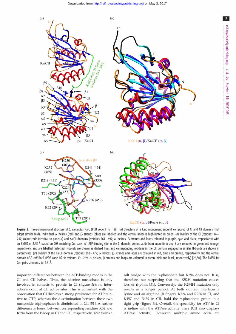
Figure 3. Three-dimensional structure of S. elongatus KaiC (PDB code 1TF7) [28]. (a) Structure of a KaiC monomeric subunit composed of CI and CII domains thatadopt similar folds. Individual a helices (red) and b strands (blue) are labelled and the central linker is highlighted in green. (b) Overlay of the CI (residues 14 –247; colour code identical to panel a) and KaiCII domains (residues 261 – 497; a helices, b strands and loops coloured in purple, cyan and black, respectively) withan RMSD of 2.45 A based on 208 matching Ca pairs. (c) ATP-binding site in the CI domain. Amino acids from subunits A and B are coloured in green and orange,respectively, and are labelled. Selected H-bonds are shown as dashed lines and corresponding residues in the CII domain engaged in similar H-bonds are shown inparentheses. (d ) Overlay of the KaiCII domain (residues 262 – 477; a helices, b strands and loops are coloured in red, blue and orange, respectively) and the centraldomain of E. coli RecA (PDB code 1G19; residues 39 – 269; a helices, b strands and loops are coloured in green, pink and black, respectively) [26,50]. The RMSD forCa pairs amounts to 1.3 A.
rsif.royalsocietypublishing.orgJ.R.Soc.Interface
14:20161065
5
on May 3, 2017http://rsif.royalsocietypublishing.org/Downloaded from
important differences between the ATP-binding modes in the
CI and CII halves. Thus, the adenine nucleobase is only
involved in contacts to protein in CI (figure 3c); no inter-
actions occur at CII active sites. This is consistent with the
observation that CI displays a strong preference for ATP rela-
tive to GTP, whereas the discrimination between these two
nucleoside triphosphates is diminished in CII [51]. A further
difference is found between corresponding residues K52 and
K294 from the P-loop in CI and CII, respectively. K52 forms a
salt bridge with the g-phosphate but K294 does not. It is,
therefore, not surprising that the K52H mutation causes
loss of rhythm [51]. Conversely, the K294H mutation only
results in a longer period. At both domain interfaces a
lysine and an arginine (R finger), K224 and R226 in CI, and
K457 and R459 in CII, hold the g-phosphate group in a
tight grip (figure 3c). Overall, the specificity for ATP in CI
is in-line with the ATPase activity there (CII also displays
ATPase activity). However, multiple amino acids are

KaiCII
KaiCI
90°
C
B
A
F
E
D
(c)
(b)(a)
Figure 4. Structure of the KaiC homo-hexamer (PDB code 3DVL) [28,37]. (a) View from the side with the N-terminal CI and C-terminal CII domains from individualsubunits coloured differently, and assuming the shape of a double doughnut. CI – CII linkers across the constricted waist are light green, C-terminal peptide tails arecyan and ATP molecules are shown in space filling mode, with carbon, oxygen, nitrogen and phosphorus atoms coloured in black, red, blue and orange, respectively.(b) The KaiC hexamer rotated by 908 around the horizontal axis and viewed from the CI side, exposing the central channel. Subunits are labelled A – F in a clockwisemanner. (c) Stereo diagram depicting the superimposed CI (residues 19 – 247; coloured in gold) and CII rings (residues 261 – 497; coloured in lilac).
rsif.royalsocietypublishing.orgJ.R.Soc.Interface
14:20161065
6
on May 3, 2017http://rsif.royalsocietypublishing.org/Downloaded from
contacting the g-phosphate group both at CI and CII subunit
interfaces, and subtle differences between the interactions
do not allow a rationalization as to why CI is exclusively an
ATPase, whereas CII exhibits both ATPase and kinase
activities. Both core domains, as predicted by sequence com-
parisons, adopt a fold that exhibits close similarity to the
structure of Escherichia coli RecA [50] (figure 3d ).
3. Three-dimensional structure and dynamics ofthe KaiCI and CII rings
The KaiC hexamer resembles a double doughnut with a
diameter and height of approximately 100 A. The view
along the non-crystallographic sixfold rotation axis shows a
central channel that is somewhat wider at the CI end
(figure 4). The CI–CII linker that comprises residues 248–
260 runs on the outer surface and across the constricted
waist region, and C-terminal tails (residues 497–519) pro-
trude from the dome-shaped surface of the CII ring.
Whereas CI and CII core domains of about 245 residues
adopt similar folds, the C-terminal extension breaks the
more or less symmetrical stack formed by the hexameric
rings. Binding sites of ATP molecules between subunits are
located closer to the upper (C-terminal) surface of rings, i.e.
ATPs bound in the CI half lie close to the central waist and
those bound in the CII half lie close to the dome-shaped sur-
face that is marked by clefts harbouring the ATPs [28]. The
lower (N-terminal) and upper (C-terminal) surfaces of rings
exhibit opposing electrostatic surface potentials (ESPs).
Thus, the N-terminal surface of the double doughnut is
strongly negatively polarized, but the C-terminal surface
(bottom and top, respectively, in figure 4a) displays a largely
positive ESP [28,30]. Analysis of the ESPs at subunit inter-
faces reveals the strongly hydrophilic nature of the
interaction between individual KaiC molecules. Similarly,
the central pore is rich in arginine, histidine and glutamate.
However, KaiC shows only weak affinity for DNA [27] and
no helicase activity has been established to date for the cen-
tral cog of the KaiABC clock. Consistent with these
observations, structural overlays of KaiC and helicase hexam-
ers reveal only a suboptimal fit and misaligned ATP-binding
sites at interfaces [28]. This is in contrast with superimposi-
tions of monomeric domains, e.g. KaiCII and RecA [50]
(figure 3d ), that reveal an excellent match.
Several motifs in the CI and CII domains mediate contacts
among subunits besides the extensive interactions at subunit
interfaces that harbour ATP molecules. Among them is the
arginine linker (residues R215, R216, R217 and R218,
figure 1) in CI that does not just mediate contacts between

rsif.royalsocietypublishing.orgJ.R.Soc.Interface
14:20161065
7
on May 3, 2017http://rsif.royalsocietypublishing.org/Downloaded from
CI domains from adjacent subunits but also reaches across the
CI–CII waist region [52], and the A-loop (residues E487–I497,
figures 1 and 3a) near the CII C-terminal end [38,39]. Another
motif in the CII half is constituted by D417(A) and H429(B)
and D427(B) from neighbouring subunits (A, B, C, etc.) that
form a circle around the central channel, whereby the histidine
side chain is sandwiched between the D417 carboxylate
moiety and the D427 main chain keto group [24]. Importantly,
mutation of some of the residues that are part of the above
motifs results in drastically distorted clock periods or arrhyth-
mic behaviour. Examples of the latter are the R216A and
R217A [50], D427A [24] and E487A [38] mutants. Proper func-
tioning of the central cog of the cyanobacterial clock is
dependent on allosteric effects among subunits and distorting
the latter by mutation of key residues severely impairs or
destroys the oscillator. In addition to non-covalent lateral,
inter-subunit CI–CI and CII–CII and intra-subunit CI–CII
interactions, the CI–CII linker that joins the two domains is
crucial for clock function. Severing the linker abolishes rhyth-
micity and therefore the clock cannot be reconstituted from
separate CI and CII rings [35]. The CI domain from S. elongatusKaiC expressed separately forms a stable hexamer but the CII
domain does not [53]. The crystal structure of the KaiCI hex-
amer has recently been published and the ATPase
mechanism and activity of the isolated CI half can be assayed
[47]. However, this has not been possible for the CII half,
although the CII domain expressed separately still exhibits
auto-kinase activity [53].
A superimposition of the KaiCI and CII hexamers reveals
two regions where their conformations obviously differ from
each other (figure 4c). The A-loops surround the channel at
the C-terminal end of the double doughnut in a crown-like
fashion and the 20-residue CII tails then protrude from the
dome-shaped surface. The tails serve the interaction with
KaiA and the activation of the kinase in the CII half
[36,37,40], and this entire 35 amino acids long stretch is miss-
ing in the CI half. At the other end, the loop between b5
and a5 in CII is much tighter than in CI as a consequence of
a deletion (figure 1). The more extended loop in CI (B-loop)
is readily recognizable at the bottom of figure 4c and has
been implicated in KaiB binding [30,54,55]. Molecular
dynamics simulations of KaiC hexamers (including residues
14–497 in all subunits) demonstrated the drastically enhanced
mobility of the A- and B-loop motifs located at the top and
bottom, respectively, of the double doughnut [39]. The crystal
structure thus unveiled unique conformational properties of
the two regions that obviously deviate also in terms of the
KaiCI and CII primary sequences. These regions turn out to
be of critical importance for mediating Kai protein–protein
interactions and clock mechanism and also exhibit distinct
dynamic properties, such that altering the latter via mutation
affects clock period and/or temperature compensation.
4. Functional divergence between KaiCI and CIIand why CII acts as a kinase but not CI
The observation that the KaiCI half has ATPase activity and that
KaiCII is both an ATPase and a kinase adds to the fascination
with the cyanobacterial PTO and prompted us to take a closer
look at the reasons for water being the only nucleophile in one
case, whereas threonine and serine act as nucleophiles in the
other [56]. KaiCII is a Ser/Thr kinase and it turns out that the
identity of the phosphorylation sites is very important for
proper working of the oscillator, but more about that in the
next chapter. The initial structure of KaiC found that T432 in
all six subunits was phosphorylated and that S431 was phos-
phorylated in four of them [28]. Although no order of the
phosphorylation events could be deduced based on this obser-
vation, it was apparent that Og of T432 was situated closer to
the g-phosphate group of ATP than Og of S431 on average,
and it seemed therefore reasonable to view T432 as the primary
phosphorylation site [31]. A mass-spectrometric analysis of pro-
teolytic fragments of KaiC confirmed the T432 and S431
phosphorylation sites [32]. Phosphorylation occurs across the
subunit interface such that the g-phosphate of ATP bound to
subunit A is transferred to threonine and serine from subunit
B (figure 5b). Subsequent work revealed a strict order of phos-
phorylation and dephosphorylation: TS! pTS! pTpS!TpS! TS [32,33]. An interesting difference in the environments
of the T432 and S431 residues is that the latter is positioned in the
vicinity of T426 across a tight turn. When S431 is phosphory-
lated, the g-hydroxyl group of T426 engages in an H-bond to
the phosphate group. An initial round of mutations showed
that the T432A, S431A and T426A mutants were all arrhythmic
in vitro [31]. A more in-depth investigation using the T426A,
T426N and T426E mutants along with others demonstrated
that alterations at residue 426 abolished circadian rhythms of
luminescence in vivo [57]. The finding that the T426N mutant
was arrhythmic supports the conclusion that it is not sufficient
for the side chain of residue 426 to be able to engage in an H-
bond (as seen in the crystal structure of T426N-KaiC [46]).
Rather, it has to be phosphorylatable in order for 426 to fulfil
its proper regulatory role. Indeed, we discovered that T426 car-
ried a phosphate group in a single subunit and in four subunits
in the crystal structures of the S431A and T432E/S431A KaiC
mutants, respectively [46]. For an overview of phosphorylation
sites and number of phosphates in CII based on crystal struc-
tures of mutant enzymes, see [30]. What is clear is that a full
understanding of the complex mechanistic aspects of the KaiC
auto-kinase activity and its regulation requires one to look
beyond the two main sites and the strict order, starting from
the hypo- to the hyper- and back to the hypo-phosphorylated
state, over the daily cycle.
Alignment of the CI and CII sequences shows that the CI
residues that correspond to T432, S431 and T426 in CII are
E198, E197 and A192 (figure 1). Neither glutamate nor ala-
nine can be phosphorylated; examination of the separate CI
half by polyacrylamide gel electrophoresis (SDS-PAGE) and
inspection of the electron density in the CI region did not
reveal phosphorylation. It appears that nature deliberately
precluded kinase activity in KaiCI by mimicking pT432 and
pS431 with glutamates in CI and T426 with a non-phosphor-
ylatable alanine (figure 5). We investigated the possibility of
phosphorylation in a KaiCI triple mutant E198T/E197S/
A192T (TST-KaiCI) using the T. elongatus system, but did
not detect kinase activity [52]. Closer examination of the
superimposed CI and CII active sites reveals subtle devi-
ations in the loop region leading up to the a9 helix that
harbours the phosphorylation sites in CII. Thus, a slight
kink at G195 changes the register of the following V196,
E197, E198 and F199 in CI relative to I430, S431, T432 and
I433 in CII. Instead of the correspondence between E197/
E198 and S431/T432, as suggested by the sequence align-
ment, the three-dimensional structural alignment shows
that E198 maps to S431 and F199 maps to T432. As a result,
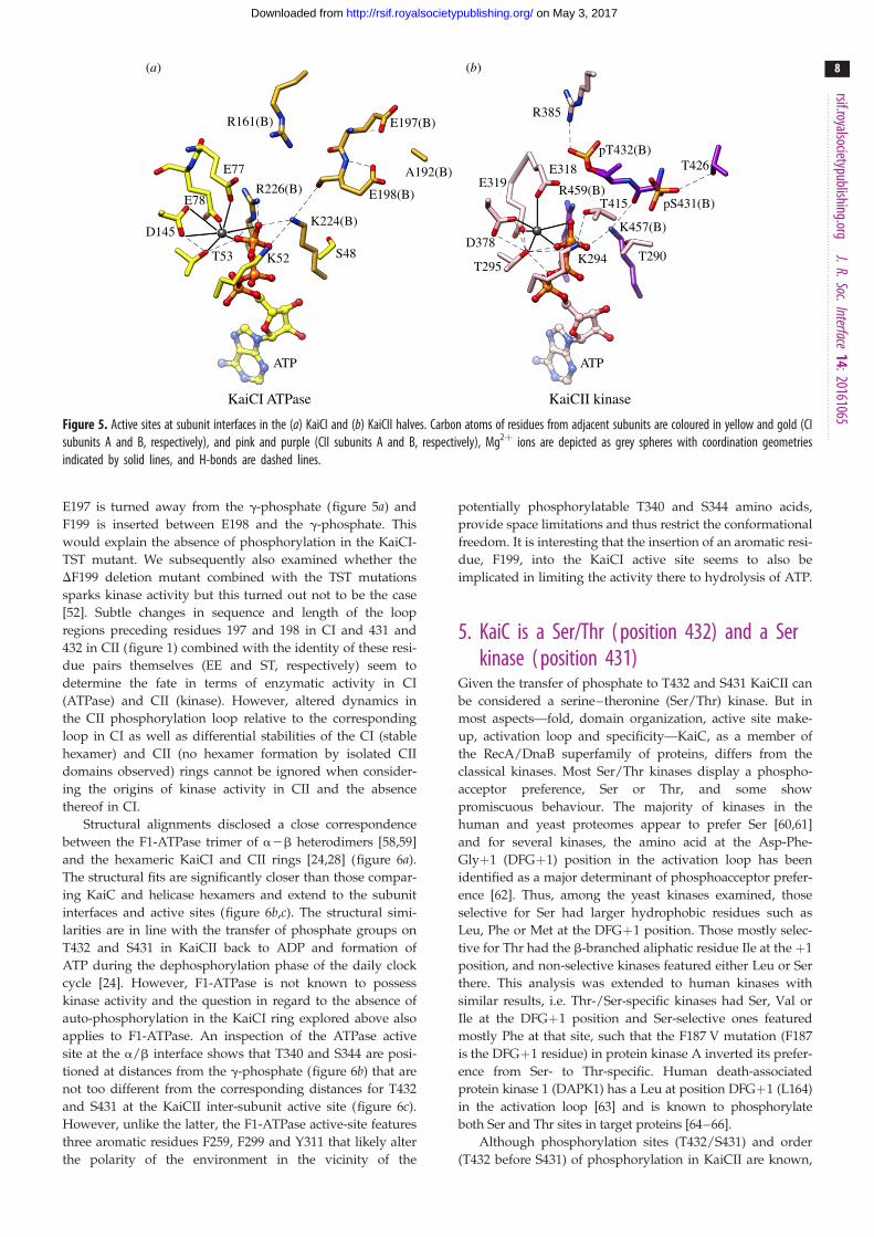
R161(B)
E77
E78
D145
T53 K52
R226(B)
K224(B)
S48T295
D378
E319E318
R459(B)
K457(B)
K294 T290
pS431(B)
T426pT432(B)
R385
T415
ATP ATP
KaiCI ATPase KaiCII kinase
E198(B)
A192(B)
E197(B)
(a) (b)
Figure 5. Active sites at subunit interfaces in the (a) KaiCI and (b) KaiCII halves. Carbon atoms of residues from adjacent subunits are coloured in yellow and gold (CIsubunits A and B, respectively), and pink and purple (CII subunits A and B, respectively), Mg2þ ions are depicted as grey spheres with coordination geometriesindicated by solid lines, and H-bonds are dashed lines.
rsif.royalsocietypublishing.orgJ.R.Soc.Interface
14:20161065
8
on May 3, 2017http://rsif.royalsocietypublishing.org/Downloaded from
E197 is turned away from the g-phosphate (figure 5a) and
F199 is inserted between E198 and the g-phosphate. This
would explain the absence of phosphorylation in the KaiCI-
TST mutant. We subsequently also examined whether the
DF199 deletion mutant combined with the TST mutations
sparks kinase activity but this turned out not to be the case
[52]. Subtle changes in sequence and length of the loop
regions preceding residues 197 and 198 in CI and 431 and
432 in CII (figure 1) combined with the identity of these resi-
due pairs themselves (EE and ST, respectively) seem to
determine the fate in terms of enzymatic activity in CI
(ATPase) and CII (kinase). However, altered dynamics in
the CII phosphorylation loop relative to the corresponding
loop in CI as well as differential stabilities of the CI (stable
hexamer) and CII (no hexamer formation by isolated CII
domains observed) rings cannot be ignored when consider-
ing the origins of kinase activity in CII and the absence
thereof in CI.
Structural alignments disclosed a close correspondence
between the F1-ATPase trimer of a2b heterodimers [58,59]
and the hexameric KaiCI and CII rings [24,28] (figure 6a).
The structural fits are significantly closer than those compar-
ing KaiC and helicase hexamers and extend to the subunit
interfaces and active sites (figure 6b,c). The structural simi-
larities are in line with the transfer of phosphate groups on
T432 and S431 in KaiCII back to ADP and formation of
ATP during the dephosphorylation phase of the daily clock
cycle [24]. However, F1-ATPase is not known to possess
kinase activity and the question in regard to the absence of
auto-phosphorylation in the KaiCI ring explored above also
applies to F1-ATPase. An inspection of the ATPase active
site at the a/b interface shows that T340 and S344 are posi-
tioned at distances from the g-phosphate (figure 6b) that are
not too different from the corresponding distances for T432
and S431 at the KaiCII inter-subunit active site (figure 6c).
However, unlike the latter, the F1-ATPase active-site features
three aromatic residues F259, F299 and Y311 that likely alter
the polarity of the environment in the vicinity of the
potentially phosphorylatable T340 and S344 amino acids,
provide space limitations and thus restrict the conformational
freedom. It is interesting that the insertion of an aromatic resi-
due, F199, into the KaiCI active site seems to also be
implicated in limiting the activity there to hydrolysis of ATP.
5. KaiC is a Ser/Thr ( position 432) and a Serkinase ( position 431)
Given the transfer of phosphate to T432 and S431 KaiCII can
be considered a serine–theronine (Ser/Thr) kinase. But in
most aspects—fold, domain organization, active site make-
up, activation loop and specificity—KaiC, as a member of
the RecA/DnaB superfamily of proteins, differs from the
classical kinases. Most Ser/Thr kinases display a phospho-
acceptor preference, Ser or Thr, and some show
promiscuous behaviour. The majority of kinases in the
human and yeast proteomes appear to prefer Ser [60,61]
and for several kinases, the amino acid at the Asp-Phe-
Glyþ1 (DFGþ1) position in the activation loop has been
identified as a major determinant of phosphoacceptor prefer-
ence [62]. Thus, among the yeast kinases examined, those
selective for Ser had larger hydrophobic residues such as
Leu, Phe or Met at the DFGþ1 position. Those mostly selec-
tive for Thr had the b-branched aliphatic residue Ile at the þ1
position, and non-selective kinases featured either Leu or Ser
there. This analysis was extended to human kinases with
similar results, i.e. Thr-/Ser-specific kinases had Ser, Val or
Ile at the DFGþ1 position and Ser-selective ones featured
mostly Phe at that site, such that the F187 V mutation (F187
is the DFGþ1 residue) in protein kinase A inverted its prefer-
ence from Ser- to Thr-specific. Human death-associated
protein kinase 1 (DAPK1) has a Leu at position DFGþ1 (L164)
in the activation loop [63] and is known to phosphorylate
both Ser and Thr sites in target proteins [64–66].
Although phosphorylation sites (T432/S431) and order
(T432 before S431) of phosphorylation in KaiCII are known,

E192f
T163f
R373b
K162f
F1-ATPase (a, b )3 / (KaiCII)6 F1-ATPase, B:F subunit interface KaiCII, A:F subunit interface
10.0Å
7.8Å
8.2ÅY311f
F259f
Y337b
F299b
R260f
R229f E319f
D378f
S379f
R385f
S381fT415fK294f
H429a
T426a
R393a
E318fP-T432a
P-S431a
T295f
R459a
K457a
D256f
R189fE188f
S344b
T340b
PbPb
Pg(Pg)5.7Å
(a) (b) (c)
Figure 6. KaiC and F1-ATPase form similar ring-shaped complexes and active sites at subunit interfaces. (a) Overlay of the KaiC hexamer (red; CII ring only) and thea3b3 ring from F1-ATPase (PDB code 1BMF) [58]. The a and b subunits of F1-ATPase are coloured cyan and yellow, respectively. (b) ATP-binding site at thea2b heterodimer interface in F1-ATPase, taken from the crystal structure of the bovine enzyme inhibited by ADP and beryllium fluoride (PDB code 1W0 J) [59].Carbon atoms of amino acid side chains in a chain B of F1-ATPase are cyan, and carbon atoms of side chains in b chain F of F1-ATPase are coloured yellow andMg2þ is a pink sphere with coordinated water molecules depicted as smaller red spheres. (c) ATP-binding site at the subunit interface in KaiCII, taken from thecrystal structure of the full-length enzyme from S. elongatus (PDB code 3DVL) [28,37]. Carbon atoms of CII subunit A are grey and carbon atoms of CII subunit F arepink, and Mg2þ is a cyan sphere. Only b- and g-phosphates of ATP are depicted, whereby the latter is mimicked by BF3 (green) in the structure of F1-ATPase.Carbon atoms of catalytic glutamates (E188 in F1-ATPase and E318 in KaiCII) are highlighted with magenta carbon atoms, and the water nucleophile H-bonded toE188 (F1-ATPase) is shown as a red sphere of slightly larger radius.
rsif.royalsocietypublishing.orgJ.R.Soc.Interface
14:20161065
9
on May 3, 2017http://rsif.royalsocietypublishing.org/Downloaded from
the phosphoacceptor preference of the auto-kinase remained
unknown. We found in crystal structures that mutation of
either T432 (T432S/S431), S431 (T432/S431T), or both
(T432S/S431T), resulted in changes in the phosphorylation
levels compared with wild-type KaiC (table 1). All three
mutants were arrhythmic (T Mori, Y Xu, CH Johnson, 2015),
indicating that tampering with phosphorylation sites results
in an identity crisis and abolishes clock function, despite the
fact that KaiC can phosphorylate Thr or Ser at position 432
(but apparently only Ser and not Thr at position 431;
table 1). Altering the identity of the secondary phosphoryl-
ation site T426 to Ser while retaining T432 and S431
abolishes phosphorylation of the latter according to the crystal-
lographic analysis of this KaiC mutant (table 1) and triggers an
extremely long period. Once again, these data suggest a crucial
role of residue 426 in KaiC conformation and clock regulation.
6. Origins of the ‘slowness’ of the KaiCI ATPaseOne of the most fascinating and difficult to explain properties
of molecular clocks is the long-time constant of the period.
Recent crystal structures of the KaiCI hexamer alone led to
the identification of two possible origins of the slowness of
the ATPase [47]. One was a suboptimal (not in-line with respect
to the scissile Pg-O3b bond) orientation of the water molecule
initiating hydrolysis of ATP. The other relates to the cis/transisomerization of the peptide bond between D145 and S146,
i.e. between a predominantly cis conformation in the pre-
hydrolysis state and a trans conformation in the post-hydrolysis
state (see figure 5a for the location of D145). The switch from cisto trans is accompanied by a repositioning of the a6-a7/a8
helices (see figure 3a for the location of these helices in
KaiCI), and modified interactions with ATP at the adjacent
subunit interface. Interestingly, the conformational changes
in the helix–loop–helix region seen in KaiCI as a result of
ATP hydrolysis are very similar to the deviating positions
between a6-a7/a8 helices of KaiCI and KaiCII in the
superimposed rings. In figure 4c, the different orientations of
a8 helices in one subunit are visible near the bottom exit of
the central channel, adjacent to the B-loop. Thus, it is feasible
that the ATPases in CI and CII were trapped in different
states in the crystal structure of full-length KaiC. The actions
of the two enzymes in the upper and lower rings are coupled
and ATPase activity in CII is required for auto-dephosphoryla-
tion [47]. Crystals of full-length KaiC were grown in the
presence of ATPgS [28,49], a slowly hydrolysing analogue of
ATP, and ATPase activity is therefore not prevented. However,
the structures of the separate CI half of KaiC and mutants
thereof in the presence of ADP or ATP were determined at res-
olutions of around or better than 2 A [47]. Conversely, the
resolutions of crystal structures of full-length KaiC are typi-
cally closer to 3 A. The highest resolution for a KaiC
structure we obtained so far was for the A422S mutant protein
(2.6 A resolution; Egli lab, 2013). However, even at this resol-
ution, the quality of the electron density does not permit one
to distinguish between the cis and trans conformations of a
peptide bond and both isomers of the D145-S146 dimer can
be built into the density. Moreover, a careful inspection of
the MD simulation data generated for KaiC (with subunits
comprising 14–497 or 14–487 residues [39]), did not reveal
any transition from the preferred trans to the cis conformation.
This is perhaps not surprising given the steep energy barrier of
14–16 kcal mol21 that needs to be overcome for cis/transisomerization to occur [47].
7. Comparison between CaMKII and KaiCLooking beyond one’s own field of investigation or particular
subject of interest can be challenging and confusing, but ever
so often an initially strange territory presents familiar views
and reveals common principles. The structural similarity
between KaiC and F1-ATPase despite very different
sequences, distinct functions and cellular locations offers
a case in point. The comparison between KaiABC, a

Table 1. Phosphorylation sites and levels in KaiC mutant enzymes.
residue T432 S431 T432 T431 S432 S431 S432 T431 T432 S431 S426
phosphorylated 6 4 6 — 6 6 6 — 6 — —
non-phosphorylated — 2 — 6 — — — 6 — 6 6
rsif.royalsocietypublishing.orgJ.R.Soc.Interface
14:20161065
10
on May 3, 2017http://rsif.royalsocietypublishing.org/Downloaded from
nano-machine in cyanobacteria that keeps track of time
(figure 7a), and human Ca2þ/calmodulin-dependent kinase
II (CaMKII, figure 7b), a dodecameric complex of multi-
domain subunits that is involved in synaptic and behavioural
memory [67] also shows that the two share quite a few traits.
For one, both use atypical folds for kinase function—oligo-
meric assemblies that allow cooperative effects, fluctuations
in the interactions at subunit interfaces and trans-phosphoryl-
ation. Both KaiC and CaMKII catalyse auto-phosphorylation
and the dephosphorylation reactions proceed through a
reversal of auto-phosphorylation with synthesis of ATP
[24,68]. The use of ATPgS for structural and functional
assays demonstrated in both cases that the analogue does
not affect the forward reaction but that phosphorothioate-
threonine and -serine can no longer be dephosphorylated.
CaMKII forms hexamers of dimers, with the hub domains
on the inside, establishing the core, and the kinase domains
on the outside (figure 7b). In the double ring composed of
hub domains, the vertical dimer is more stable than the equa-
torial one. This is not unlike the architecture of the KaiC
hexamer, although CI and CII in the clock enzyme form a
covalently linked and serially arranged ‘dimer’ that arises
from a gene duplication. However, the KaiCI ring can be con-
sidered a hub because separate CI domains form a stable
hexamer; conversely separate CII kinase domains do not hex-
amerize. Trans-phosphorylation in KaiC involves the two
primary sites, T432 and S431, and a secondary site, T426,
and KaiA acts as an activator. In CaMKII, the initial phos-
phorylation concerns T286 in the regulatory domain and
requires activation by Ca2þ/calmodulin; subsequently, two
further threonines, T305 and T306, undergo phosphorylation
[47] (figure 7b). The activated form of CaMKII shows spon-
taneous subunit exchange as binding of a peptide portion
to the hub as a result of phosphorylation causes subunits of
the hub ring to fall apart. Also the deletion of the kinase
domain results in subunit exchange [48]. Eventually, fluctu-
ations in the lateral interfaces between vertical hub dimers
open up a gap and this allows capture of a free dimer unit
and expansion from dodecameric CaMKII to a tetradecameric
form (figure 7b). KaiC subunit exchange is also a key feature
of the daily cycle in the KaiABC circadian clock. KaiC mol-
ecules alone swap subunits as well and as in the case of
CaMKII, experiments with fluorescently labelled complexes
were used to establish this principle [44]. However, in the
full S. elongatus PTO, subunit exchange sets in during the
dephosphorylation phase when KaiB is associated with
KaiC (figure 7a). Unlike in the case of CaMKII, the KaiC hex-
amer does not expand to a heptameric particle upon subunit
exchange; it seems that monomeric units join pentameric
complexes that have released a subunit. That both CaMKII
and KaiABC rely on subunit exchange is possibly the most
remarkable shared property of the two systems. Mathemat-
ical models demonstrated that prevention of subunit
exchange in the clock system leads to rapid attenuation of
the amplitude of the oscillation and loss of function [44,45].
Phase-dependent exchange of subunits allows individual
KaiC hexamers to maintain similar levels of phosphorylation
and thus provides a mechanism of clock synchronization. In
CaMKII that is a likely candidate for a memory-storage
molecule, subunit exchange allows passing on an activated
state from an activated complex to an unactivated one.
In this fashion, a level of activation can be maintained in a
CaMKII holoenzyme even when some subunits are degraded
and certainly long after the initial activation signal has
disappeared [48].
8. ConclusionOne of the key points, I tried to convey in this review is the
crucial role structural insights have played for a better
understanding of the KaiABC clock. Apart from obvious
observations such as fold, active-site configuration (i.e. ATP
binding) and phosphorylation sites, the KaiC structure
revealed upfront important differences between the KaiCI
and KaiCII rings that would, however, not necessarily have
been obvious to the observer in terms of their meaning.
Thus, a simple superimposition between the two hexamers
exposes three regions of notable conformational and/or
dynamic variance: the KaiCII A-loop and tail, the KaiCI
B-loop, and misaligned a8 helices in CI and CII (figure 4).
These three regions turn out to play key roles in clock
protein–protein interactions (A-/B-loop), regulation (A-/B-
loop) and ATP processing (a8 and preceding loop and
helix). An examination of the differences in the primary
sequences of the CI and CII halves would also have led one
to the CII-terminal A-loop and tail as well as the CI B-loop
regions, but there is no arguing as to which approach—
sequence alignment versus three-dimensional structure and
dynamics—is more informative.
Although we have gained a much better understanding of
the KaiA–KaiC and KaiB–KaiC interactions over the last
decade, much more remains to be learnt in terms of the acti-
vation of KaiC phosphorylation by KaiA (single molecule
pulling experiments could tell us more about the forces
involved and cooperative effects among subunits) and the
precise role(s) played by KaiB in the PTO and in linking the
PTO to the TTFL. There also exists a void in terms of getting
a better grip on the molecular underpinnings of and specific
involvement of individual amino acids and regulatory motifs
in temperature compensation. Not only are the reactions cat-
alysed by KaiC subjects to temperature compensation, but the
entire PTO and, therefore, the various protein–protein inter-
actions that take place over the daily cycle also have to be
temperature-compensated. As far as KaiC is concerned, a
detailed mutational analysis combined with in vitro or

conformationreverts
KaiCphosphorylation
KaiCdephosphorylation
T286 phosphorylation
dodecameric CaMKII
subunit exchange
tetradecameric CaMKII
docking of regulatory
segment onto hub
release ofdimeric hub
subunit
capture of a free
vertical dimer unit
other kinase domainsremoved in drawing
other kinase domainsremoved in drawing
CaM release andT305/T306
phosphorylation
conformationalchange and
dephosphorylation
Ca2+/CaM binding
hub cavity
KaiA
KaiA
KaiCmonomers KaiB
KaiA KaiB
KaiB
KaiA bd
c
a
aA
KaiA
subunit exchange
(a)
(b)
Figure 7. Subunit exchange in a hexameric molecular complex that measures time, the cyanobacterial KaiABC circadian clock, and in a dodecameric molecularcomplex that is critical for memory formation, human CaMKII. (a) Diagram depicting the daily clock cycle controlled by KaiA, KaiB and KaiC and featuring phos-phorylation and dephosphorylation of the KaiC homo-hexamer [44,45]. Cyan and blue dumbbell shapes represent two conformational states of KaiC subunits/monomers in the phosphorylated and non-phosphorylated forms, respectively. Phosphate groups at the T432 and S431 sites of KaiC are red dots. A subpopulationof KaiC hexamers associates with and dissociates from KaiA and KaiB during the 24-h cycle. Double-headed arrows in the centre indicate subunit exchange betweenKaiC hexamers. Rates of subunit exchange vary among KaiC states: a solid line indicates a high rate and a dashed line indicates a low rate. (b) Diagram depicting apotential mechanism of subunit exchange in CaMKII [48]. The sequence starts with an auto-inhibited dodecamer CaMKII holoenzyme in a compact conformation.The hub domain is grey, the kinase domain is blue, the regulatory segment is yellow, the CaM footprint is red, and upper and lower hub domains of one verticaldimer are coloured blue and purple, respectively. In the subsequent drawings, only the kinase of a single subunit is shown. CaMKII is auto-phosphorylated at T286 inthe regulatory element. After CaM dissociates, T305 and T306 are phosphorylated if T286 is already phosphorylated. The regulatory segment is then released and canbind the available b-sheet of its own hub domain. As a result of binding by the regulatory domain, the lateral association between hub domain dimers is wea-kened, opening a crack between adjacent hub domains and causing release of a domain dimer from the dodecameric holoenzyme. Owing to fluctuations in theconformation of the auto-inhibited form of the CaMKII holoenzyme lateral interactions are weakened, thus allowing re-entry of the domain dimer into a dodecamerwith formation of a CaMKII tetradecamer. The panels are adapted versions of illustrations published in [45] (KaiABC) and [48] (CaMKII).
rsif.royalsocietypublishing.orgJ.R.Soc.Interface
14:20161065
11
on May 3, 2017http://rsif.royalsocietypublishing.org/Downloaded from

rsif.royalsocietypublishing.orgJ.R.Soc.Interface
14:20161065
12
on May 3, 2017http://rsif.royalsocietypublishing.org/Downloaded from
in vivo measurements of temperature-dependent changes in
the period as a consequence of mutation could pinpoint key
residues and key regions in this regard.
Finally, the emergence of common principles in molecular
machines that keep track of time and play a role in memory
formation is particularly exciting. Subunit exchange has
been studied to some degree in the KaiABC clock and in
greater structural and mechanistic detail in CaMKII. In the
former, more remains to be learnt about how KaiB affects
subunit exchange. There is some evidence from cryo-EM
and negative-stain EM in combination with gold labelling
that KaiB monomers congregate at some point in the daily
cycle (KaiB–KaiC complex) along the KaiCII rim [69,70],
thereby potentially covering ATP-binding clefts and serving
KaiA sequestration and initiation of the dephosphorylation
stage. However, in CaMKII subunit exchange is triggered
by destabilization of lateral interactions between hub
domains. The KaiCI ring constitutes the hub in KaiC and
destabilizing interactions among CI domains would be
expected to be the best way to induce subunit exchange
and degradation of the KaiABC particle. There is good sup-
port for KaiB binding to KaiCI and interactions between
KaiB and KaiCI in the monomeric state [55]. However, it is
worth mentioning that the above data in support of the CII
binding mode by KaiB were obtained using wild-type pro-
teins, whereas the studies supporting a CI binding mode
used B and CI proteins that both contained mutations
(reviewed in [30]). Neither the structures of complexes com-
posed of monomeric species nor those of KaiCI hexamers
in different states (e.g. with ADP or ATP bound) can
enlighten us as to the changes at the CI/CII interface in the
KaiC homo-hexamer. Moreover, crystal structures of KaiC
have all trapped the hexamer in the hyper- or near hyper-
phosphorylated state, thereby limiting our understanding of
the detailed conformational changes and cooperative effects
that a hexamer undergoes as it transitions over the daily
cycle from the hypo- to the hyper- and back to the hypo-
phosphorylated state before coming apart. Thus, many
more questions exist regarding the detailed mechanism of
the KaiABC cyanobacterial circadian clock.
Competing interests. The author declares that he has no competinginterests.
Funding. Supported by the US NIH and Vanderbilt University.
References
1. Johnson CH, Egli M. 2014 Metabolic compensationand circadian resilience in prokaryotic cyanobacteria.Annu. Rev. Biochem. 83, 221 – 247. (doi:10.1146/annurev-biochem-060713-035632)
2. Dunlap JC, Loros JJ, DeCoursey PJ (eds) 2004Chronobiology: biological timekeeping. Sunderland,MA: Sinauer.
3. Ouyang Y, Andersson CR, Kondo T, Golden SS,Johnson CH. 1998 Resonating circadian clocksenhance fitness in cyanobacteria. Proc. Natl Acad.Sci. USA 95, 8660 – 8664. (doi:10.1073/pnas.95.15.8660)
4. Woelfle MA, Ouyang Y, Phanvijhitsiri K, Johnson CH.2004 The adaptive value of circadian clocks: anexperimental assessment in cyanobacteria. Curr. Biol.14, 1481 – 1486. (doi:10.1016/j.cub.2004.08.023)
5. Ishiura M, Kutsuna S, Aoki S, Iwasaki H, AnderssonCR, Tanabe A, Golden SS, Johnson CH, Kondo T.1998 Expression of a gene cluster kaiABC as acircadian feedback process in cyanobacteria. Science281, 1519 – 1523. (doi:10.1126/science.281.5382.1519)
6. Dvornyk V, Vinogradova O, Nevo E. 2003 Origin andevolution of circadian clock genes in prokaryotes.Proc. Natl Acad. Sci. USA 100, 2495 – 2500. (doi:10.1073/pnas.0130099100)
7. Egli M, Johnson CH. 2015 Biochemistry that timesthe day. Biochemistry 54, 104 – 109. (doi:10.1021/bi5014968)
8. Sancar A et al. 2015 Circadian clock, cancer, andchemotherapy. Biochemistry 54, 110 – 123. (doi:10.1021/bi5007354)
9. Kojima S, Green CB. 2015 Circadian genomics reveala role for post-transcriptional regulation inmammals. Biochemistry 54, 124 – 133. (doi:10.1021/bi500707c)
10. Gustafson CL, Partch CL. 2015 Emerging models forthe molecular basis of mammalian circadian timing.Biochemistry 54, 134 – 149. (doi:10.1021/bi500731f )
11. Cha J, Zhou M, Liu Y. 2015 Mechanism of theNeurospora circadian clock, a FREQUENCY-centricview. Biochemistry 54, 150 – 156. (doi:10.1021/bi5005624)
12. Shim JS, Imaizumi T. 2015 Circadian clock andphotoperiodic response in Arabidopsis: fromseasonal flowering to redox homeostasis.Biochemistry 54, 157 – 170. (doi:10.1021/bi500922q)
13. Noordally ZB, Millar AJ. 2015 Clocks in algae.Biochemistry 54, 171 – 183. (doi:10.1021/bi501089x)
14. Hoyle NP, O’Neill JS. 2015 Oxidation – reductioncycles of peroxiredoxin proteins andnontranscriptional aspects of timekeeping.Biochemistry 54, 184 – 193. (doi:10.1021/bi5008386)
15. Egli M, Johnson CH. 2013 A circadian clocknanomachine that runs without transcription ortranslation. Curr. Opin. Neurobiol. 23, 732 – 740.(doi:10.1016/j.conb.2013.02.012)
16. Tomita J, Nakajima M, Kondo T, Iwasaki H. 2005Circadian rhythm of KaiC phosphorylation withouttranscription – translation feedback. Science 307,251 – 254. (doi:10.1126/science.1102540)
17. Nakajima M, Imai K, Ito H, Nishiwaki T, MurayamaY, Iwasaki H, Oyama T, Kondo T. 2005 Reconstitutionof circadian oscillation of cyanobacterial KaiCphosphorylation in vitro. Science 308, 414 – 415.(doi:10.1126/science.1108451)
18. Qin X, Byrne M, Yu X, Mori T, Johnson CH. 2010Coupling of a core post-translational pacemaker to a
slave transcription/translation feedback loop in acircadian system. PLoS Biol. 8, e1000394. (doi:10.1371/journal.pbio.1000394)
19. Teng SW, Mukherji S, Moffitt JR, de Buyl S, O’SheaEK. 2013 Robust circadian oscillations in growingcyanobacteria require transcriptional feedback.Science 340, 737 – 740. (doi:10.1126/science.1230996)
20. Johnson CH. 2010 Circadian clocks and cell division:what’s the pacemaker? Cell Cycle 9, 3864 – 3873.(doi:10.4161/cc.9.19.13205)
21. Williams SB, Vakonakis I, Golden SS, LiWang AC.2002 Structure and function from the circadianclock protein KaiA of Synechococcus elongatus: apotential clock input mechanism. Proc. Natl Acad.Sci. USA 99, 15 357 – 15 362. (doi:10.1073/pnas.232517099)
22. Xu Y, Mori T, Johnson CH. 2003 Cyanobacterialcircadian clockwork: roles of KaiA, KaiB, and thekaiBC promoter in regulating KaiC. EMBO J. 22,2117 – 2126. (doi:10.1093/emboj/cdg168)
23. Terauchi K, Kitayama Y, Nishiwaki T, Miwa K,Murayama Y, Oyama T, Kondo T. 2007 ATPaseactivity of KaiC determines the basic timing forcircadian clock of cyanobacteria. Proc. Natl Acad. Sci.USA 104, 16 377 – 16 381. (doi:10.1073/pnas.0706292104)
24. Egli M, Mori T, Pattanayek R, Xu Y, Qin X, JohnsonCH. 2012 Dephosphorylation of the core clockprotein KaiC in the cyanobacterial KaiABC circadianoscillator proceeds via an ATP synthase mechanism.Biochemistry 51, 1547 – 1558. (doi:10.1021/bi201525n)
25. Nishiwaki T, Kondo T. 2012 Circadianautodephosphorylation of cyanobacterial clockprotein KaiC occurs via formation of ATP as

rsif.royalsocietypublishing.orgJ.R.Soc.Interface
14:20161065
13
on May 3, 2017http://rsif.royalsocietypublishing.org/Downloaded from
intermediate. J. Biol. Chem. 287, 18 030 – 18 035.(doi:10.1074/jbc.M112.350660)
26. Leipe DD, Aravind L, Grishin NV, Koonin EV. 2000The bacterial replicative helicase DnaB evolved froma RecA duplication. Genome Res. 10, 5 – 16.
27. Mori T, Saveliev SV, Xu Y, Stafford WF, Cox MM,Inman RB, Johnson CH. 2002 Circadian clock proteinKaiC forms ATP-dependent hexameric rings andbinds DNA. Proc. Natl Acad. Sci. USA 99, 17 203 –17 208. (doi:10.1073/pnas.262578499)
28. Pattanayek R, Wang J, Mori T, Xu Y, Johnson CH,Egli M. 2004 Visualizing a circadian clock protein:crystal structure of KaiC and functional insights. Mol.Cell 15, 375 – 388. (doi:10.1016/j.molcel.2004.07.013)
29. Johnson CH, Egli M, Stewart PL. 2008 Structuralinsights into a circadian oscillator. Science 322,697 – 701. (doi:10.1126/science.1150451)
30. Egli M. 2014 Intricate protein – protein interactionsin the cyanobacterial circadian clock. J. Biol. Chem.289, 21 267 – 21 275. (doi:10.1074/jbc.R114.579607)
31. Xu Y, Mori T, Pattanayek R, Pattanayek S, Egli M,Johnson CH. 2004 Identification of keyphosphorylation sites in the circadian clock proteinKaiC by crystallographic and mutagenetic analyses.Proc. Natl Acad. Sci. USA 101, 13 933 – 13 938.(doi:10.1073/pnas.0404768101)
32. Nishiwaki T et al. 2004 Role of KaiC phosphorylationin the circadian clock system of Synechococcuselongatus PCC 7942. Proc. Natl Acad. Sci. USA 101,13 927 – 13 932. (doi:10.1073/pnas.0403906101)
33. Nishiwaki T, Satomi Y, Kitayama Y, Terauchi K,Kiyohara R, Takao T, Kondo T. 2007 A sequentialprogram of dual phosphorylation of KaiC as a basisfor circadian rhythm in cyanobacteria. EMBO J. 26,4029 – 4037. (doi:10.1038/sj.emboj.7601832)
34. Rust MJ, Markson JS, Lane WS, Fisher DS, O’SheaEK. 2007 Ordered phosphorylation governsoscillation of a three-protein circadian clock. Science318, 809 – 812. (doi:10.1126/science.1148596)
35. Hayashi F, Iwase R, Uzumaki T, Ishiura M. 2006Hexamerization by the N-terminal domain andintersubunit phosphorylation by the C-terminaldomain of cyanobacterial circadian clock proteinKaiC. Biochem. Biophys. Res. Commun. 348,864 – 872. (doi:10.1016/j.bbrc.2006.07.143)
36. Vakonakis I, LiWang AC. 2004 Structure of theC-terminal domain of the clock protein KaiA incomplex with a KaiC-derived peptide: implicationsfor KaiC regulation. Proc. Natl Acad. Sci. USA 101,10 925 – 10 930. (doi:10.1073/pnas.0403037101)
37. Pattanayek R, Williams DR, Pattanayek S, Xu Y, MoriT, Johnson CH, Stewart PL, Egli M. 2006 Analysis ofKaiA – KaiC protein interactions in the cyano-bacterial circadian clock using hybrid structuralmethods. EMBO J. 25, 2017 – 2028. (doi:10.1038/sj.emboj.7601086)
38. Kim YI, Dong G, Carruthers CWJr, Golden SS,LiWang A. 2008 The day/night switch in KaiC, acentral oscillator component of the circadian clock ofcyanobacteria. Proc. Natl Acad. Sci. USA 105,12 825 – 12 830. (doi:10.1073/pnas.0800526105)
39. Egli M, Pattanayek R, Sheehan JH, Xu Y, Mori T,Smith JA, Johnson CH. 2013 Loop – loop interactionsregulate KaiA-stimulated KaiC phosphorylation inthe cyanobacterial KaiABC circadian clock.Biochemistry 52, 1208 – 1220. (doi:10.1021/bi301691a)
40. Pattanayek R, Egli M. 2015 Protein – proteininteractions in the cyanobacterial circadian clock:structure of KaiA dimer in complex with C-terminalKaiC peptides at 2.8 Angstrom resolution.Biochemistry 54, 4575 – 4578. (doi:10.1021/acs.biochem.5b00694)
41. Brettschneider C, Rose RJ, Hertel S, Axmann IM,Heck AJ, Kollmann M. 2010 A sequestrationfeedback determines dynamics and temperatureentrainment of the KaiABC circadian clock. Mol. Syst.Biol. 6, 389. (doi:10.1038/msb.2010.44)
42. Kageyama H, Nishiwaki T, Nakajima M, Iwasaki H,Oyama T, Kondo T. 2006 Cyanobacterial circadianpacemaker: Kai protein complex dynamics in theKaiC phosphorylation cycle in vitro. Mol. Cell 23,161 – 171. (doi:10.1016/j.molcel.2006.05.039)
43. Ito H, Kageyama H, Mutsuda M, Nakajima M,Oyama T, Kondo T. 2007 Autonomoussynchronization of the circadian KaiCphosphorylation rhythm. Nat. Struct. Mol. Biol. 14,1084 – 1088. (doi:10.1038/nsmb1312)
44. Mori T, Williams DR, Byrne MO, Qin X, Egli M,Mchaourab HS, Stewart PL, Johnson CH. 2007Elucidating the ticking of an in vitro circadianclockwork. PLoS Biol. 5, e93. (doi:10.1371/journal.pbio.0050093)
45. Johnson CH, Stewart PL, Egli M. 2011 Thecyanobacterial circadian system: frombiophysics to bioevolution. Annu. Rev. Biophys. 40,143 – 167. (doi:10.1146/annurev-biophys-042910-155317)
46. Pattanayek R, Mori T, Xu Y, Pattanayek S, JohnsonCH, Egli M. 2009 Structures of KaiC circadian clockmutant proteins: a new phosphorylation site atT426 and mechanisms of kinase, ATPase andphosphatase. PLoS ONE 4, e7529. (doi:10.1371/journal.pone.0007529)
47. Abe J et al. 2015 Atomic-scale origins of slownessin the cyanobacterial circadian clock. Science 349,312 – 316. (doi:10.1126/science.1261040)
48. Stratton M, Lee I, Bhattacharyya M, Christensen SM,Chao LH, Schulman H, Groves JT, Kuriyan J. 2014Activation-triggered subunit exchange betweenCaMKII holoenzymes facilitates the spread of kinaseactivity. eLife 3, e01610. (doi:10.7554/elife.02490)
49. Egli M. 2015 Structural and biophysical methods toanalyze clock function and mechanism. In Methodsin enzymology, vol. 551 (ed. A Seghal), pp.223 – 266. Oxford, UK: Elsevier.
50. Story RM, Weber IT, Steitz TA. 1992 The structure ofthe E. coli recA protein monomer and polymer.Nature 355, 318 – 325. (doi:10.1038/355318a0)
51. Nishiwaki T, Iwasaki H, Ishiura M, Kondo T. 2000Nucleotide binding and autophosphorylation of theclock protein KaiC as a circadian timing process ofcyanobacteria. Proc. Natl Acad. Sci. USA 97, 495 –499. (doi:10.1073/pnas.97.1.495)
52. Pattanayek R, Xu Y, Lamichhane A, Johnson CH, EgliM. 2014 An arginine tetrad as mediator of input-dependent and -independent ATPases in the clockprotein KaiC. Acta Cryst. D 70, 1375 – 1390. (doi:10.1107/S1399004714003228)
53. Pattanayek R, Williams DR, Pattanayek S, Mori T,Johnson CH, Stewart PL, Egli M. 2008 Structuralmodel of the circadian clock KaiB – KaiC complexand mechanism for modulation of KaiCphosphorylation. EMBO J. 27, 1767 – 1778. (doi:10.1038/emboj.2008.104)
54. Tseng R, Chang Y-G, Bravo I, Latham R, ChaudharyA, Kuo N-W, LiWang A. 2014 Cooperative KaiA –KaiB – KaiC interactions affect KaiB/SasA competitionin the circadian clock of cyanobacteria. J. Mol. Biol.426, 389 – 402. (doi:10.1016/j.jmb.2013.09.040)
55. Chang Y-G et al. 2015 A protein fold switch joinsthe circadian oscillator to clock output incyanobacteria. Science 349, 324 – 328. (doi:10.1126/science.1260031)
56. Koshland DEJr. 1994 The key – lock theory and theinduced fit theory. Angew. Chem. Int. Ed. Engl. 33,2375 – 2378. (doi:10.1002/anie.199423751)
57. Xu Y, Mori T, Qin X, Yan H, Egli M, Johnson CH.2009 Intramolecular regulation of phosphorylationstatus of the circadian clock protein KaiC. PLoS ONE4, e7509. (doi:10.1371/journal.pone.0007509)
58. Abrahams JP, Leslie AGW, Lutter R, Walker JE. 1994Structure at 2.8 A resolution of F1 ATPase frombovine heart mitochondria. Nature 370, 621 – 628.(doi:10.1038/370621a0)
59. Kagawa R, Montgomery G, Braig K, Walker JE, LeslieAGW. 2004 The structure of bovine F1-ATPaseinhibited by ADP and beryllium fluoride.EMBO J. 23, 2734 – 2744. (doi:10.1038/sj.emboj.7600293)
60. Hornbeck PV, Kornhauser JM, Tkachev S, Zhang B,Skrzypek E, Murray B, Latham V, Sullivan M. 2012PhosphoSitePlus: a comprehensive resource forinvestigating the structure and function ofexperimentally determined post-translationalmodifications in man and mouse. Nucleic Acids Res.40(Database issue), D261 – D270. (doi:10.1093/nar/gkr1122)
61. Sadowski I et al. 2013 The PhosphoGRIDSaccharomyces cerevisiae protein phosphorylationsite database: version 2.0 update. Database (Oxford)2013, pbat026. (doi:10.1093/database/bat026)
62. Chen C et al. 2014 Identification of a majordeterminant for serine-threonine kinasephosphoacceptor specificity. Mol. Cell 53, 140 – 147.(doi:10.1016/j.molcel.2013.11.013)
63. Tereshko V, Teplova M, Brunzelle J, Watterson DM,Egli M. 2001 Crystal structures of the catalyticdomain of human protein kinase associated withapoptosis and tumor suppression. Nat. Struct. Biol.8, 899 – 907. (doi:10.1038/nsb1001-899)
64. Shani G, Marash L, Gozuacik D, Bialik S, TeitelbaumL, Shohat G, Kimchi A. 2004 Death-associatedprotein kinase phosphorylates ZIP kinase, forming aunique kinase hierarchy to activate its cell deathfunctions. Mol. Cell. Biol. 24, 8611 – 8626. (doi:10.1128/MCB.24.19.8611-8626.2004)

rsif.royalsocietypublishing.org
14
on May 3, 2017http://rsif.royalsocietypublishing.org/Downloaded from
65. Zalckvar E, Berissi H, Mizrachy L, Idelchuk Y, Koren I,Eisenstein M, Sabanay H, Pinkas-Kramarski R,Kimchi A. 2009 DAP-kinase-mediatedphosphorylation on the BH3 domain of beclin 1promotes dissociation of beclin 1 from Bcl-XL andinduction of autophagy. EMBO Rep. 10, 285 – 292.(doi:10.1038/embor.2008.246)
66. Lee TH et al. 2011 Death-associated protein kinase 1phosphorylates Pin1 and inhibits its prolylisomerase activity and cellular function. Mol.
Cell 42, 147 – 159. (doi:10.1016/j.molcel.2011.03.005)
67. Lisman J, Schulman H, Cline H. 2002 The molecularbasis of CaMKII function in synaptic and behaviouralmemory. Nat. Rev. Neurosci. 3, 175 – 190. (doi:10.1038/nrn753)
68. Kim SA, Hudmon A, Volmer AM, Waxham MN. 2001CaM-Kinase II dephosphorylates Thr286 by a reversal ofthe autophosphorylation reaction. Biochem. Biophys. Res.Commun. 282, 773 – 780. (doi:10.1006/bbrc.2001.4651)
69. Villarreal SA, Pattanayek R, Williams DR, Mori T, QinX, Johnson CH, Egli M, Stewart PL. 2013 CryoEMand molecular dynamics of the circadian KaiB – KaiCcomplex indicates KaiB monomers interact with KaiCand block ATP binding clefts. J. Mol. Biol. 425,3311 – 3324. (doi:10.1016/j.jmb.2013.06.018)
70. Pattanayek R, Yadagirib KK, Ohi MD, Egli M. 2013Nature of KaiB – KaiC binding in the cyanobacterialcircadian oscillator. Cell Cycle 12, 810 – 817. (doi:10.4161/cc.23757)
J.
R.Soc.Interface14:20161065