Applications of Radiation Candace Davison Brenden Heidrich Penn State University Michael Erdman PSU...
62
Applications of Radiation Candace Davison Brenden Heidrich Penn State University Michael Erdman PSU Milton S. Hershey Medical Center Mary Lou Dunzik-Gougar, Ph.D. Idaho State University and Idaho National Laborator with special thanks to
-
Upload
erik-baker -
Category
Documents
-
view
218 -
download
0
Transcript of Applications of Radiation Candace Davison Brenden Heidrich Penn State University Michael Erdman PSU...

Applications of Radiation
Candace Davison
Brenden Heidrich
Penn State University
Michael Erdman
PSU Milton S. Hershey Medical Center
Mary Lou Dunzik-Gougar, Ph.D.Idaho State University and Idaho National Laboratory
with special thanks to

Overview
• General applications by radiation type• Radiography - process• Medical Research• Medical Applications• Space

Alpha Radiation
• Highly ionizing• Removes Static Charge
Static ChargeAlpha Particle

Uses of Alpha Radiation
• Pacemakers (Older models)
• Airplanes
• Copy Machines
•Smoke Detectors
•Space exploration

Beta Radiation
• Small electron particle• More penetrating than alpha
e-

Beta radiation is used in thickness gauging
The thicker the material the less radiation will pass through the material.

Gauging is used to
• Measure and control thickness of paper, plastic, and aluminum.
• Measure the amount of glue placed on a postage stamp
• Measure the amount of air whipped into ice cream.
• Measure the density of the road during construction.

Back Scattering
Detector

Gamma Radiation
A penetrating wave

Uses for Gamma Radiation
Food irradiation Sterilization of medical equipment Creation of different varieties of flowers Inspect bridges, vessel welds and Statue Of
Liberty
What were original uses of mysterious rays?
• Becquerel’s discovery• Roentgen x-ray of wife’s hand• Marie Curie – WWI – x-ray unit

Early X-ray
http://www.uihealthcare.com/depts/medmuseum/galleryexhibits/colle
ctingfrompast/xray/xray.html

Radiographs
• Radiograph - radiation energy passes through object
• Autoradiograph - use
radiation from object itself
X-ray
Photo-Film

Radiography• Let’s explore two different methods of
using radiation to capture images– X—rays– Neutrons
• The next graph shows attenuation of the radiation vs. atomic number. The shading on the right shows how much radiation is blocked – black indicates completely blocked.

(0.025 eV)

Comparing Different Materials
• Cadmium ( Cd )
• Lead ( Pb )
• Polyethylene [ (CH3)n ]

CODE Box at Penn StateStudent Project to Demonstrate X-Ray/Neutron Radiography
Was originally in cardboard shoe box, but was replaced by more durable aluminum.

C O D E
Cadmium = red
Lead = white

X-Ray Image

C O
P S U
D E
Cadmium = red
Lead = white
Polyethylene = gray

Neutron Radiograph

Hydrogen Fuel Cell Imaging
Fuel Cell research conducted at RSEC

Hydrogen Fuel Cell Imaging
Water Calibration Wedge

Hydrogen Fuel Cell Imaging

Hydrogen Fuel Cell Imaging

Clinical Uses of
Radioactive Materials

Understanding the Replication Process of the HIV Retrovirus
• DNA sequencing, using 35S and 32P, is used to investigate the process by which new viruses “bud” or form from host cells

5000 Premature Infants Die Annually from Respiratory Distress/SIDS
• The infant lacks a protein which produces a surfactant in the lung alveoli
• Without the surfactant, there is too much surface tension – the lung is too weak to expand. A respirator is needed.
• 32P-research identified the missing protein• Gene therapy may one day be available

Benefits fromRadioisotope Research
The Penn State Artificial Heart
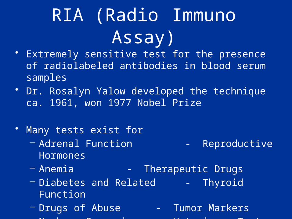
RIA (Radio Immuno Assay)• Extremely sensitive test for the presence of radiolabeled
antibodies in blood serum samples• Dr. Rosalyn Yalow developed the technique ca. 1961, won
1977 Nobel Prize
• Many tests exist for– Adrenal Function - Reproductive Hormones– Anemia - Therapeutic Drugs– Diabetes and Related - Thyroid Function– Drugs of Abuse - Tumor Markers– Newborn Screening - Veterinary Tests

RIA Kit
• A standard test kit includes reagents, antigens, and a minute amount of radioactivity
• One kit can be used to test 100 to 500 patient serum samples

14C Test for Helicobacter pylori• H. pylori is often implicated in Gastric Reflux Disease• If present, a specific antibiotic can be prescribed to eliminate it• The use of radioactive 14C provides a simple and sure test

137Cs Blood Irradiator • Delivers 2500 rads to blood products
• Reduces potential for Graft-vs-Host Disease
• Essential for bone marrow transplants

Diagnostic RadiologyModalities
• X-Ray Radiography – Angiography– Mammography– Fluoroscopy– Cardiac Catheterization– CT (Computed Tomography)

Heart Image• Gated study of radiolabeled cardiac muscle• Allows visualization of heart tissue viability

Bone Scan with 99mTc-HDP• Active bone surface is
labeled• Note “hot spots” and
kidneys

Nuc Med & Radiographic Images Compared• Metabolic hotspots highlighted
– possibly cancerous• X-ray image shows break,
but no metabolic information

The New(er) Kid on the Block: PET Positron Emission Tomography
• 18FDG Images of a normal vs. an epileptic brain• Rapidly growing in popularity for tumor imaging

• p+ à n0 + e+ + n• Positron escapes the nucleus• Two oppositely directed photons result from the
annihilation of the positron with an electron
Positron Decay and Coincidence Photon Detection
PET Scanner Coincidence Detectors
Negatron-

18FDG
• Fluorodeoxyglucose
• Most commonly used PET radiocompound
• A glucose analog, useful for– Differentiating malignant from benign tumors– Differentiating scar from viable myocardial
tissue– Brain function studies

Cerebral Glucose Metabolism
• Brain tumor diagnosed• MRI scan suspicious for low-grade astrocytoma• PET/CT scan shows large hypo-metabolic area in left posterior
temporal lobe Siemens Clinical Solutions, www.medical.siemens.com

• Neurological studies– Epilepsy– Alzheimers– Parkinson’s Disease– Addictions
• Cancer imaging and localization– In demand by Oncologists
• Cardiology studies
Other PET Applications

The ‘Historical’ Problem in Modern Radiology
• Images obtained from Nuclear Medicine were obtained on a computer platform different from those obtained from CT, and also from MRI, Ultrasound, etc.
• Thus, images could not be easily overlaid
• A common software was needed to make best use of the information from each modality

Radiation Doses from Medical X-rays
• Medical Radiation (Effective Whole Body Dose Equivalent)– Chest X-ray: 8 mrem (0.08 mSv)– Head CT scan: 111 mrem (1.11 mSv)– Barium Enema: 406 mrem (4.06 mSv)– Extremity X-ray: 1 mrem (0.01 mSv)
Source: NCRP Report 100

Radiation Doses and Dose LimitsFlight from Los Angeles to London 5 mrem
Annual public dose limit 100 mrem
Annual natural background 300 mrem
Fetal dose limit 500 mrem
Barium enema 870 mrem
Annual radiation worker dose limit 5,000 mrem
Heart catheterization 45,000 mrem
Life saving exposure (NCRP-116) 50,000 mrem
Mild acute radiation syndrome 200,000 mrem
LD50/60 for humans (bone marrow dose) 350,000 mrem
Radiation therapy (localized & fractionated) 6,000,000 mrem
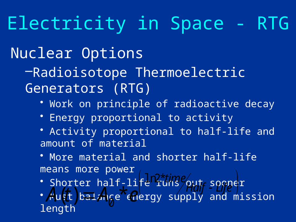
Electricity in Space - RTG
Nuclear Options–Radioisotope Thermoelectric Generators (RTG)
• Work on principle of radioactive decay• Energy proportional to activity• Activity proportional to half-life and amount of material• More material and shorter half-life means more power• Shorter half-life runs out sooner• Must balance energy supply and mission length
LifeHalftime
eAtA*2ln
0 *)(

RTGs in Space - TheoryWork on the thermoelectric principle also
known as the ‘Seebeck Effect’
~10% efficiency
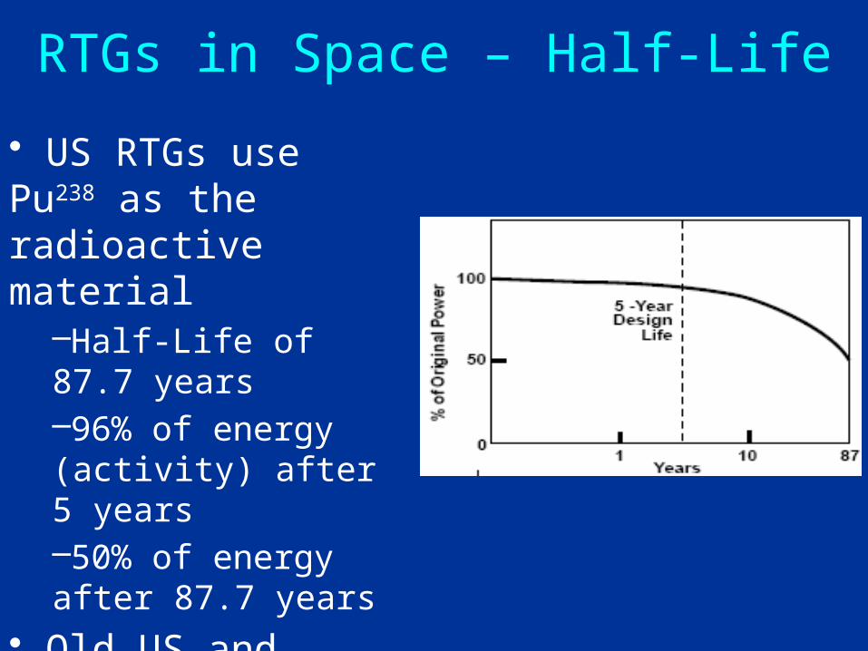
RTGs in Space – Half-Life
• US RTGs use Pu238 as the radioactive material
–Half-Life of 87.7 years–96% of energy (activity) after 5 years–50% of energy after 87.7 years
• Old US and Russian RTGs used Po210
–Half-Life of 138 days

RTGs in Space - Radiation
• 238Puand 210Po are alpha (α) emitters• Alpha radiation cannot penetrate very far
–Stopped by a sheet of paper or 10cm of air–Turns into heat in the RTG material–Very little radiation gets out of the shielding–Not ‘weapons grade’ material–Ceramic form that is very heat and impact
resistant

RTGs in Space - History1959: Atomic Energy Commission members show President Eisenhower the new ‘nuclear battery’ for use in US satellites

RTGs in Space - History
• Original RTG in space was for a US Navy navigation satellite– 1961 SNAP-3 unit (Space Nuclear Auxiliary
Power)– 2.7 watts of electrical power– Lasted for 15 years
• RTGs were used in 25 other missions from 1961 to 2005 from military satellites to the Apollo missions

RTGs in Space - History
•1972: Pioneer 10 & 11 launched to explore the outer planets
– Both survived high radiation around Jupiter– Both crafts left solar system after mission performed and continued to send data for 17 years– Still in contact with crafts

RTGs in Space - History
•1977: Voyager 1&2 launched to explore the outer planets
– Transmitted high speed data and first high-quality pictures– Both crafts left solar system after mission was performed and continue to send data

RTGs in Space - History
•1990: Ulysses launched to explore top and bottom of sun
– Mission extended after initial successes– First mission to explore solar system outside the ‘disk’ of the planets– Can you see the RTG?

RTGs in Space - History
•1989: Galileo launched to explore Jupiter and her moons
–Took the long-way to Jupiter; by Venus and Earth twice–Required long-lived power supply to make the 4-year flight–Operated for 14 years–Can you see the RTG?

RTGs in Space - History
• Galileo also needed heat for its long mission• 120 - 1watt Radioactive Heater Units (RHU) placed all over the spacecraft• Safety design similar to RTGs

RTGs in Space - History
•1997 -Cassini Mission to Saturn and moons
– 3 General Purpose Heat Source RTGs (current generation)– 4-year mission once the craft gets to Saturn.– Great results coming back from craft– Recently discovered new moon of Saturn

RTGs in Space - History•January 2006•New Horizons Mission
–Pluto & Charon–Kuiper Belt Objects

RTGs in Space - History
•Viking Landers- 1975– Used RTGs for power– 6 Years on Nuclear Power
•Mars Pathfinder-1997– Rover used RHUs for heat– 3 months on solar power

THANK YOU

MRI:Radio Waves & Magnetic Fields

MRI – Magnetic Resonance Imaging• Utilizes magnetic fields and RF (radio-frequency) energy
to gain information via Nuclear Magnetic Resonance• No ionizing radiation is used in this process