
Antitumoral Effects of Calcitriol in Basal Cell...
11
Molecular Medicine in Practice Antitumoral Effects of Calcitriol in Basal Cell Carcinomas Involve Inhibition of Hedgehog Signaling and Induction of Vitamin D Receptor Signaling and Differentiation Anja Uhmann 1 , Hannah Niemann 1 ,B er enice Lammering 1 , Cornelia Henkel 1 , Ina Heß 1 , Frauke Nitzki 1 , Anne Fritsch 1 , Nicole Pr€ ufer 1 , Albert Rosenberger 2 , Christian Dullin 3 , Anke Schraepler 7 , Julia Reifenberger 8 , Stefan Schweyer 4 , Torsten Pietsch 9 , Frank Strutz 5 , Walter Schulz-Schaeffer 6 , and Heidi Hahn 1 Abstract Activation of the Hedgehog (Hh)-signaling pathway due to deficiency in the Hh receptor Patched1 (Ptch) is the pivotal defect leading to formation of basal cell carcinoma (BCC). Recent reports provided evidence of Ptch-dependent secretion of vitamin D 3 -related compound, which functions as an endogenous inhibitor of Hh signaling by repressing the activity of the signal transduction partner of Ptch, Smoothened (Smo). This suggests that Ptch-deficient tumor cells are devoid of this substance, which in turn results in activation of Hh- signaling. Here, we show that the application of the physiologically active form of vitamin D 3, calcitriol, inhibits proliferation and growth of BCC of Ptch mutant mice in vitro and in vivo. This is accompanied by the activation of the vitamin D receptor (Vdr) and induction of BCC differentiation. In addition, calcitriol inhibits Hh signaling at the level of Smo in a Vdr-independent manner. The concomitant antiproliferative effects on BCC growth are stronger than those of the Hh-specific inhibitor cyclopamine, even though the latter more efficiently inhibits Hh signaling. Taken together, we show that exogenous supply of calcitriol controls the activity of 2 independent pathways, Hh and Vdr signaling, which are relevant to tumorigenesis and tumor treatment. These data suggest that calcitriol could be a therapeutic option in the treatment of BCC, the most common tumor in humans. Mol Cancer Ther; 10(11); 2179–88. Ó2011 AACR. Introduction The Hedgehog (Hh)-signaling pathway regulates cell differentiation, organ patterning, and cell proliferation (1). In the absence of a ligand, the activity of the Hh-pathway is inhibited due to an interaction between the Hh receptor Patched1 (Ptch) and its partner Smoothened (Smo). Bind- ing of Hh to Ptch, inactivating Ptch mutations, or activating Smo mutations may suspend this inhibition, resulting in the transcription of target genes including Gli1 (1). Thus aberrant (e.g., mutation driven) Hh signaling results in tumor formation (2). One prominent example is basal cell carcinoma (BCC), the most frequent tumor entity in humans, with active Hh-signaling due to mutations in Ptch. Today, the inhibition of the Hh pathway is considered to be a promising strategy in the treatment of these tumors. Thus specific Smo inhibitors such as cyclopamine and GDC0449 have been tested in several small, nonrando- mized clinical trials (2). GDC0449 was recently shown to elicit antitumoral effects in 18 of 33 patients with locally advanced or metastatic BCC (3). It remains to be eluci- dated if these promising results will be confirmed in a prospective, randomized, and controlled study and if Hh targeting alone will be sufficient. In this context, a recent report describes resistance to GDC0449 due to a therapy- associated Smo mutation (4). As with other tumors, ther- apy of those associated with abnormal Hh signaling may require targeting of additional signaling pathways. Vitamin D 3 and its derivatives (e.g., EB1089; Paricalci- triol) are known to have antitumoral effects on different cancer types (5) including squamous cell carcinoma (6, 7) or on hyperproliferative skin diseases such as psoriasis (8, 9). These effects comprise G 0 –G 1 arrest, cellular differen- tiation, induction of apoptosis, and modulation of inflam- mation or of different signaling pathways in tumor cells, as well as inhibiting tumor angiogenesis (5). Until now, the antitumoral effects of vitamin D 3 were explained by bind- ing of the biologically active form of vitamin D 3 , calcitriol (1a,25-dihydroxy vitamin D 3 ), to the vitamin D receptor (Vdr) and the subsequent regulation of Vdr-bound genes. Authors' Affiliations: 1 Institute of Human Genetics; Departments of 2 Genetic Epidemiology, 3 Diagnostic Radiology, 4 Pathology, 5 Nephrology and Rheumatology, and 6 Neuropathology; 7 Central Animal Facility, Uni- versity of Goettingen, Goettingen; 8 Department of Dermatology, University of Duesseldorf, Duesseldorf; and 9 Department of Neuropathology, Uni- versity of Bonn, Bonn, Germany Corresponding Author: Anja Uhmann, Institute of Human Genetics, University of Goettingen, Heinrich-D€ uker-Weg 12, 37073 Goettingen, Germany. Phone: 49-551-3914100; Fax: 49-551-396580; E-mail: [email protected] doi: 10.1158/1535-7163.MCT-11-0422 Ó2011 American Association for Cancer Research. Molecular Cancer Therapeutics www.aacrjournals.org 2179 on July 29, 2018. © 2011 American Association for Cancer Research. mct.aacrjournals.org Downloaded from Published OnlineFirst August 30, 2011; DOI: 10.1158/1535-7163.MCT-11-0422
Transcript of Antitumoral Effects of Calcitriol in Basal Cell...

Molecular Medicine in Practice
Antitumoral Effects of Calcitriol in Basal Cell CarcinomasInvolve Inhibition of Hedgehog Signaling and Inductionof Vitamin D Receptor Signaling and Differentiation
Anja Uhmann1, Hannah Niemann1, B�er�enice Lammering1, Cornelia Henkel1, Ina Heß1, Frauke Nitzki1,Anne Fritsch1, Nicole Pr€ufer1, Albert Rosenberger2, Christian Dullin3, Anke Schraepler7,Julia Reifenberger8, Stefan Schweyer4, Torsten Pietsch9, Frank Strutz5,Walter Schulz-Schaeffer6, and Heidi Hahn1
AbstractActivation of the Hedgehog (Hh)-signaling pathway due to deficiency in the Hh receptor Patched1 (Ptch) is
the pivotal defect leading to formation of basal cell carcinoma (BCC). Recent reports provided evidence of
Ptch-dependent secretion of vitaminD3-related compound, which functions as an endogenous inhibitor of Hh
signaling by repressing the activity of the signal transduction partner of Ptch, Smoothened (Smo). This
suggests that Ptch-deficient tumor cells are devoid of this substance, which in turn results in activation of Hh-
signaling. Here, we show that the application of the physiologically active form of vitamin D3, calcitriol,
inhibits proliferation and growth of BCC of Ptch mutant mice in vitro and in vivo. This is accompanied by the
activation of the vitamin D receptor (Vdr) and induction of BCC differentiation. In addition, calcitriol inhibits
Hh signaling at the level of Smo in a Vdr-independent manner. The concomitant antiproliferative effects on
BCC growth are stronger than those of the Hh-specific inhibitor cyclopamine, even though the latter more
efficiently inhibits Hh signaling. Taken together, we show that exogenous supply of calcitriol controls the
activity of 2 independent pathways, Hh and Vdr signaling, which are relevant to tumorigenesis and tumor
treatment. These data suggest that calcitriol could be a therapeutic option in the treatment of BCC, the most
common tumor in humans. Mol Cancer Ther; 10(11); 2179–88. �2011 AACR.
Introduction
The Hedgehog (Hh)-signaling pathway regulates celldifferentiation, organ patterning, and cell proliferation (1).In the absence of a ligand, the activity of theHh-pathway isinhibited due to an interaction between the Hh receptorPatched1 (Ptch) and its partner Smoothened (Smo). Bind-ing ofHh toPtch, inactivatingPtchmutations, or activatingSmo mutations may suspend this inhibition, resulting inthe transcription of target genes including Gli1 (1). Thusaberrant (e.g., mutation driven) Hh signaling results intumor formation (2). One prominent example is basal cellcarcinoma (BCC), the most frequent tumor entity inhumans,withactiveHh-signalingdue tomutations inPtch.
Today, the inhibitionof theHhpathway is considered tobe a promising strategy in the treatment of these tumors.Thus specific Smo inhibitors such as cyclopamine andGDC0449 have been tested in several small, nonrando-mized clinical trials (2). GDC0449 was recently shown toelicit antitumoral effects in 18 of 33 patients with locallyadvanced or metastatic BCC (3). It remains to be eluci-dated if these promising results will be confirmed in aprospective, randomized, and controlled study and if Hhtargeting alone will be sufficient. In this context, a recentreport describes resistance to GDC0449 due to a therapy-associated Smo mutation (4). As with other tumors, ther-apy of those associated with abnormal Hh signaling mayrequire targeting of additional signaling pathways.
Vitamin D3 and its derivatives (e.g., EB1089; Paricalci-triol) are known to have antitumoral effects on differentcancer types (5) including squamous cell carcinoma (6, 7)or on hyperproliferative skin diseases such as psoriasis (8,9). These effects comprise G0–G1 arrest, cellular differen-tiation, induction of apoptosis, andmodulation of inflam-mation or of different signaling pathways in tumor cells,aswell as inhibiting tumorangiogenesis (5).Until now, theantitumoral effects of vitamin D3were explained by bind-ing of the biologically active form of vitamin D3, calcitriol(1a,25-dihydroxy vitamin D3), to the vitamin D receptor(Vdr) and the subsequent regulation of Vdr-bound genes.
Authors' Affiliations: 1Institute of Human Genetics; Departments of2Genetic Epidemiology, 3Diagnostic Radiology, 4Pathology, 5Nephrologyand Rheumatology, and 6Neuropathology; 7Central Animal Facility, Uni-versity of Goettingen, Goettingen; 8Department of Dermatology, Universityof Duesseldorf, Duesseldorf; and 9Department of Neuropathology, Uni-versity of Bonn, Bonn, Germany
Corresponding Author: Anja Uhmann, Institute of Human Genetics,University of Goettingen, Heinrich-D€uker-Weg 12, 37073 Goettingen,Germany. Phone: 49-551-3914100; Fax: 49-551-396580; E-mail:[email protected]
doi: 10.1158/1535-7163.MCT-11-0422
�2011 American Association for Cancer Research.
MolecularCancer
Therapeutics
www.aacrjournals.org 2179
on July 29, 2018. © 2011 American Association for Cancer Research. mct.aacrjournals.org Downloaded from
Published OnlineFirst August 30, 2011; DOI: 10.1158/1535-7163.MCT-11-0422

Calcitriol is produced from vitamin D3 by 2 hydrox-ylation steps in the liver and kidney, respectively, andto a lesser extent in other organs and in tumor cells (5).Through binding to Vdr, calcitriol regulates the tran-scription of Vdr target genes (5). This so-called genomiccalcitriol/Vdr signaling regulates a variety of physio-logic processes including cellular differentiation, espe-cially in the skin (10, 11), proliferation, and apoptosis,and can be monitored by measuring transcription of thecalcitriol metabolizing enzyme 24-hydroxylase (5). Inaddition, calcitriol elicits rapid, so-called nongenomic(i.e., transcription independent) effects such as calciuminflux (5).
Recent data suggest a cross-talk between vitamin D3
and Hh signaling. Through medium transfer experi-ments Bijlsma and colleagues provided first evidencefor a Ptch-dependent secretion of vitamin D3 com-pounds. They also showed that vitamin D3 inhibitsthe Hh-pathway at the level of Smo (12). This suggeststhat, in addition to or instead of a direct protein–proteininteraction, Ptch may repress Smo via secretion of avitamin D3 derivative (12).
The possibility of a Ptch-dependent secretion of avitamin D3 derivative with Smo-inhibitory propertiesopens new perspectives for therapies of tumors that arisedue to mutations in Ptch. One would expect that inacti-vation of Ptch results in a disrupted secretion of thisderivative. This should result in a potential deprivationof the vitamin D3 derivative, and in lack of Smo inhibi-tion. If this hypothesis is true, the concerted action ofactive Hh and inactive Vdr signaling may be the drivingforce leading to enhanced cell proliferation, compro-mised differentiation, and ultimately to tumor formation.It follows that it should be possible to revert or at least toimpede these processes by administration of the respec-tive vitamin D3 derivative.
Here, we investigated the effect of the biologicallyactive vitamin D3 derivative calcitriol on Vdr- and Hh-signaling, growth, apoptosis, and differentiation of Ptch-deficient BCC cells in vitro and in vivo using the Ptchflox/flox
Rosa26CreERT2þ/� (Ptchflox/flox ERT2þ/�) mouse model forBCC (13). In addition, we compared its effects with thoseof cyclopamine and sought to unravel the molecularmechanisms underlying the calcitriol-mediated effectson Hh signaling.
Materials and Methods
CompoundsCalcitriol (Sigma-Aldrich) and cyclopamine (Toronto
Research Chemicals Inc.) were dissolved in ethanol(EtOH). Final concentrations for in vitro experimentsare indicated in the respective experiments and corre-spond to those normally used in cell culture (5, 14, 15). Forin vivo use, calcitriol was diluted individually for eachanimal in 20 mL EtOH/1,200 mL sterile sunflower oil(Sigma-Aldrich) to obtain a final concentration of 40 or100 ng/kg in 50 mL.
Animals and treatment of tumor-bearing Ptchflox/flox
ERT2þ/� mice with calcitriolConditional Ptchflox/flox ERT2þ/� mice were random-
ized into 2 groups and BCC was induced in all animalsby intramuscular injection of 100 mg tamoxifen as de-scribed (13, 16). Tumors of this animal model lack theexpression of wt Ptch alleles and are therefore deficientin Ptch (13, 17). Starting points of the daily intraperito-neal treatment with 100 ng/kg calcitriol or vehicle wereday 0 or 60 after BCC induction. Treatment of eachcohort was conducted until day 90 after BCC induction.For RNA isolation and histologic examinations, skinsamples were collected from tails 24 hours after the lastcalcitriol injection.
Mice were fed with calcium- and phosphate-reducedand vitamin D3-free food (ssniff Spezialdiaeten, E15312-14; ref. 18) 1 week before and during the injectionperiod.
All animals were treated and housed in accordancewith the German animal protection law.
Measurement of tumor sizeBCC size was measured on hematoxylin and eosin
(H&E)-stained sections using the area calculation toolof the software CellF (Olympus Soft Imaging SolutionsGmbH).
Analysis of calcium blood serum valuesAmounts of 100 mL of blood were collected from the
retroorbital plexus. Serum calcium concentrations weremeasured using an O-cresolphthaleine–based assay(cobas, Roche Diagnostics GmbH).
Histopathology and immunohistochemistryBCC and normal skin from the tail were embedded in
paraffin for histologic analyses or were used for isolationof total RNA. The identity of BCC was confirmed byexamination of H&E-stained sections. Paraffin sectionswere stained using an anti-Ki67- and anti-active caspase3-antibody as described (19).
Cell lines and primary cell culture of BCCThe fibroblast cell line Ptchflox/floxERT2þ/� was estab-
lished fromdermis of aPtchflox/floxERT2þ/�mouse.Ptch�/�
cells are stable Ptch-deficient cells derived from tamox-ifen-treated Ptchflox/floxERT2þ/� fibroblasts. Smo�/� andVdr�/� fibroblasts have been described in Ma and collea-gues (20) and Sun and colleagues (21), respectively. Allfibroblast cell lines were maintained in Dulbecco’s Modi-fied Eagle Medium (DMEM), 10% fetal calf serum, and1% penicillin/streptomycine (PS).
Themurine BCC cell line ASZ001 was established fromUV-induced BCC of Ptchneo12/þ mice and maintained asdescribed (22). All cell lines have been tested and au-thenticated before using by genotyping PCRs on genomicDNA as described (13, 20, 21).
Skin punches were isolated from BCC-bearingPtchflox/floxERT2þ/� mice 30, 40, and 60 days after tumor
Uhmann et al.
Mol Cancer Ther; 10(11) November 2011 Molecular Cancer Therapeutics2180
on July 29, 2018. © 2011 American Association for Cancer Research. mct.aacrjournals.org Downloaded from
Published OnlineFirst August 30, 2011; DOI: 10.1158/1535-7163.MCT-11-0422
initiation by tamoxifen. Punches were maintained for10 days in culture as reported (16).
Cell culture experimentsFor gene expression analysis or 5-bromo-20-deoxyuri-
dine (BrdU) incorporation and caspase assays, 100,000 or4,000 cells/well were seeded in 6-well and 96-well plates,respectively. For short interfering RNA (siRNA)-mediat-ed knockdown of Vdr expression, Ptchflox/flox ERT2þ/�
cells were transfected with Vdr-specific or control siRNAas described below. After 24 hours, the cells were washedand incubated for additional 48 hours with mediumsupplemented with calcitriol, cyclopamine, or EtOH asindicated in the respective experiments.Cell proliferation wasmeasured after BrdU-pulsing for
the last 22 hours using a Cell Proliferation BrdU ELISA(Roche Diagnostics GmbH).Activity of caspase 3 and 7 was measured using the
Caspase-Glo 3/7 Assay (Promega) and a microplatereader (Biotek instruments Inc.) according the man-ufacturer’s instructions.Shh-N–conditioned medium (Shh-N-CM) or respec-
tive control medium were obtained from HEK293-Shh(HEK293-Shh express one of the 3 mammalian Hh pro-teins Sonic hedgehog (Shh)) or HEK293 cells, respective-ly, as described (14).Knockdown ofVdr expression in Ptch�/� and Ptchflox/flox
ERT2þ/� cells was achieved by using a Vdr-specificsiRNA(50-CAGGCGGAGCATGAAGCGCAA-30). Scram-bled siRNA (AllStars negative, Qiagen) was used as con-trol siRNA. 75 ng siRNA and 4.5 mL HiPerFect (Qiagen)were mixed with 100 mL DMEM and incubated for 10minutes. Simultaneously, 15,000 cells/well were seededin DMEM/10% fetal calf serum/2% penicillin/strepto-mycine in 24-well plates. Subsequently, the siRNA/HiPerFect-mix was added to the cells. After 24 hours,the cells were incubated with the respective media (asindicated in the experiments) supplemented with vehicleor 10 nmol/L calcitriol. After additional 48 hours, the cellswere collected and used for subsequent experiments.For activation of Hh-signaling pathway in Smo�/�
fibroblasts the cells were transfected with a plasmid-expressing hSMO (15, 23) using RotiFect following themanufacturer’s instructions. After 6 hours, the cells wereincubated with media containing 10 nmol/L calcitriol, 5mmol/L cyclopamine, or EtOH for 48 hours.Data shown are representative for at least 3 indepen-
dent experiments each conducted in triplicate.
Reverse transcription PCR and quantitative real-time PCR analysesTotal RNA was extracted using TriReagent (Sigma-
Aldrich). For skin samples, the RNeasy fibrous tissueminikit (Qiagen) was used. Synthesis of cDNA and primercombinations for amplification of 18S rRNA, Gli1 andKeratin10 (K10) transcripts used for quantitative real-timePCR (qRT-PCR) were described previously (16, 19). Prim-er pairs used for qRT-PCR analysis of Gli2, Vdr, Cyp24a1,
and transglutaminase 1 (Tgm1) were 50-GCAAGGTCAA-GACTGAGGCTGA-30/50-GCTGCTCCTGTGTGTCATA-CTCCTT-30; 50-AGAACATGTGCTGCTCATGGC-30/50-TCATCTTGGCGTAGAGCTGGTGGCT-30; 50-GTGTGG-CAAGCGCACACGCT-30/50-CCGTGACAGCAGCGTA-CAGT-30; and 50-GCAGTGGTGTAAATGCAGCTGG-30/50-ATGAGGAGCTCAAGGGCAATGC-30, respectively.
Amplification of 18S rRNAwas done as an endogenouscontrol for the normalization of target gene expression.The amount of target and endogenous reference wasdetermined using the relative standard curve method.Each sample was measured in triplicates. Graphs repre-sent the mean value of all measurements.
Glycerol-3-phosphate dehydrogenase (mGapd), wild-type (wt) Ptch transcripts (derived from the nonrecom-bined Ptchflox locus) and Ptchdel transcripts weredetected by RT-PCR as described in Zibat and collea-gues (13, 24).
Protein extraction and Western blotPtch�/� fibroblasts were transfected with Vdr-specific
siRNA or control siRNA as described. After 72 hours, thecells were harvested. Nuclear fractions of the transfectedcells and of Vdr�/� fibroblasts were isolated using the kitNE-PER Nuclear and Cytoplasmatic Extraction Reagents(Thermo Scientific) according to the manufacturer’sinstructions. Protein concentrations were measured byPierce BCA Protein Assay Kit (Thermo Scientific) and16.5 mg of each protein sample of the nuclear fractionwere analyzed by Western blot using a rabbit anti-Vdrantibody (1:100; clone 9A7; Abcam) and a mouse anti-heat shock 70 kDa protein 8 (HSC-70; 1:10,000; SantaCruz) in combination with horseradish peroxidase-con-jugated goat anti-rat IgG (1:10,000; Thermo Scientific) andrabbit antimouse IgG (1:5,000; GE Healthcare), respec-tively. Signals were detected by using ECLþ reagent (GEHealthcare).
StatisticsMann–Whitney U testing was done to determine the
significance of the results.
Results
Calcitriol activates the Vdr pathway, suppressesHh signaling and inhibits proliferation of culturedPtch mutant BCC cells
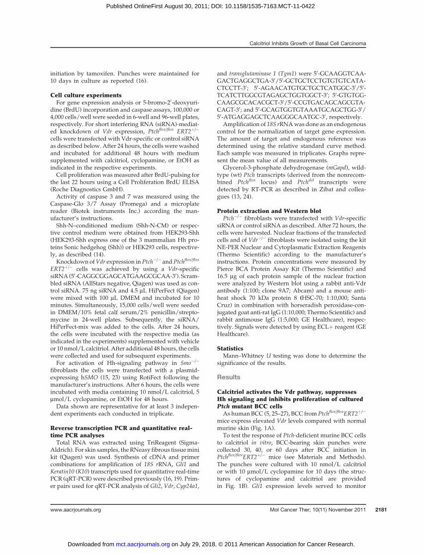
As human BCC (5, 25–27), BCC from Ptchflox/floxERT2þ/�
mice express elevated Vdr levels compared with normalmurine skin (Fig. 1A).
To test the response of Ptch-deficient murine BCC cellsto calcitriol in vitro, BCC-bearing skin punches werecollected 30, 40, or 60 days after BCC initiation inPtchflox/floxERT2þ/� mice (see Materials and Methods).The punches were cultured with 10 nmol/L calcitriolor with 10 mmol/L cyclopamine for 10 days (the struc-tures of cyclopamine and calcitriol are providedin Fig. 1B). Gli1 expression levels served to monitor
Calcitriol Inhibits Growth of Basal Cell Carcinoma
www.aacrjournals.org Mol Cancer Ther; 10(11) November 2011 2181
on July 29, 2018. © 2011 American Association for Cancer Research. mct.aacrjournals.org Downloaded from
Published OnlineFirst August 30, 2011; DOI: 10.1158/1535-7163.MCT-11-0422
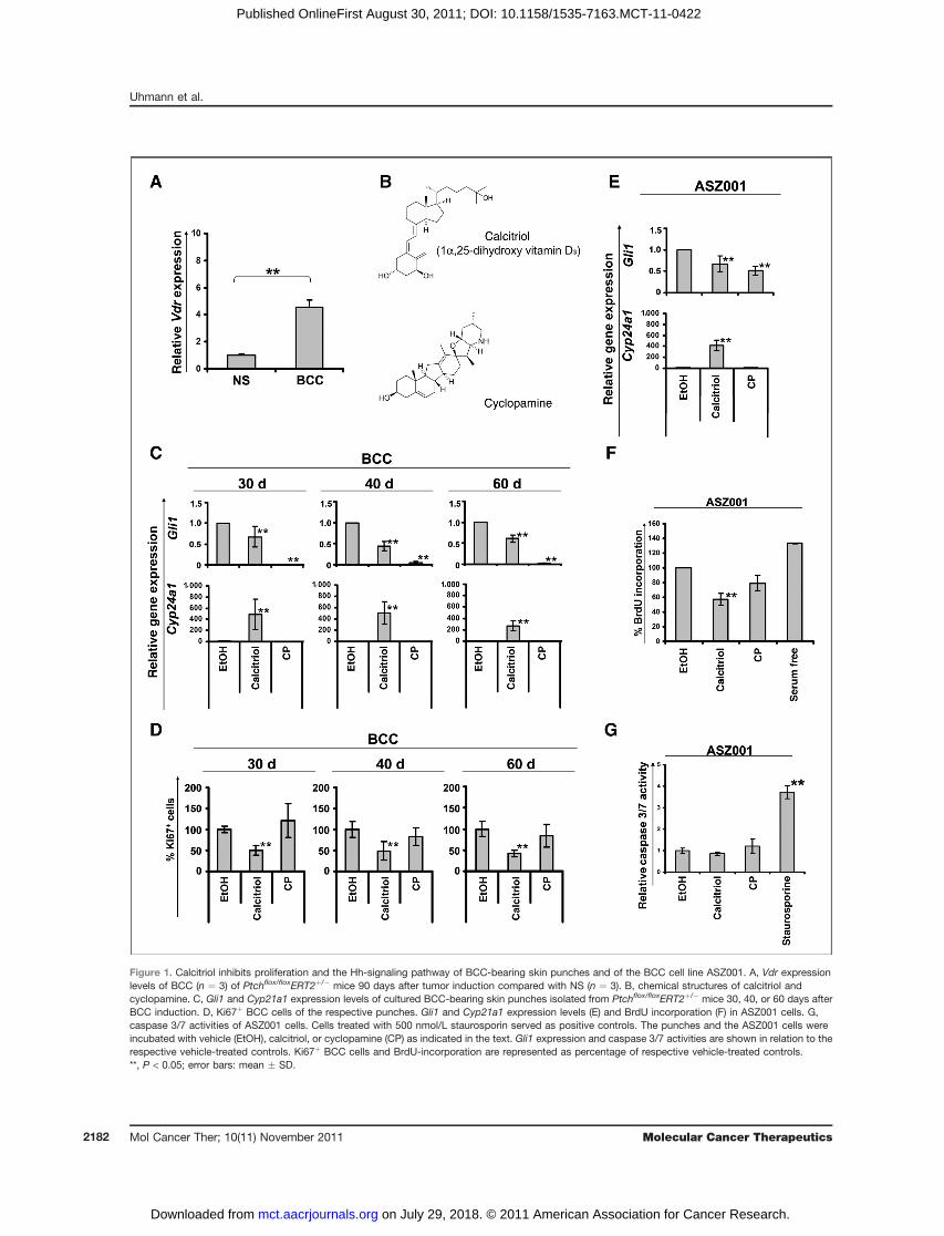
Figure 1. Calcitriol inhibits proliferation and the Hh-signaling pathway of BCC-bearing skin punches and of the BCC cell line ASZ001. A, Vdr expressionlevels of BCC (n ¼ 3) of Ptchflox/floxERT2þ/� mice 90 days after tumor induction compared with NS (n ¼ 3). B, chemical structures of calcitriol andcyclopamine. C, Gli1 and Cyp21a1 expression levels of cultured BCC-bearing skin punches isolated from Ptchflox/floxERT2þ/� mice 30, 40, or 60 days afterBCC induction. D, Ki67þ BCC cells of the respective punches. Gli1 and Cyp21a1 expression levels (E) and BrdU incorporation (F) in ASZ001 cells. G,caspase 3/7 activities of ASZ001 cells. Cells treated with 500 nmol/L staurosporin served as positive controls. The punches and the ASZ001 cells wereincubated with vehicle (EtOH), calcitriol, or cyclopamine (CP) as indicated in the text. Gli1 expression and caspase 3/7 activities are shown in relation to therespective vehicle-treated controls. Ki67þ BCC cells and BrdU-incorporation are represented as percentage of respective vehicle-treated controls.**, P < 0.05; error bars: mean � SD.
Uhmann et al.
Mol Cancer Ther; 10(11) November 2011 Molecular Cancer Therapeutics2182
on July 29, 2018. © 2011 American Association for Cancer Research. mct.aacrjournals.org Downloaded from
Published OnlineFirst August 30, 2011; DOI: 10.1158/1535-7163.MCT-11-0422
Hh-signaling activity. Cyp24a1 (24-hydroxylase) tran-scripts were measured to estimate activation of Vdr.Calcitriol led to a significant induction of Cyp24a1
transcription (Fig. 1C), which was consistent with thepresence of Vdr in BCC (Fig 1A). In addition, calcitrioldecreased Gli1 expression, thus indicating an inhibitionof the Hh-signaling pathway. As expected, cyclopaminedid not induce Cyp24a1 transcription but repressed Gli1expression. The repressive effect was more pronouncedthan that achieved with calcitriol (Fig. 1C).Next, we assessed the antiproliferative effects of calci-
triol. As revealed by anti-Ki67 antibody staining, calcitirolinhibited proliferation of tumor cells in BCC-bearing skinpunches (Fig. 1D). Most interestingly, the antiprolifera-tive effect of calcitriol was more pronounced than thatachieved with the Hh-specific inhibitor cyclopamine(Fig. 1D).Similar results were obtained when the BCC-derived
cell line ASZ001 was incubated with 10 nmol/L calcitriolor with 10 mmol/L cyclopamine for 48 hours. As shown inFig. 1E both substances efficiently inhibited Hh signaling.However, only calcitriol significantly inhibited BrdU-incorporation in these cells (Fig. 1F). Inhibition of prolif-eration apparently was not accompanied by an increasein apoptosis, as calcitriol had no effect on caspase 3/7activity (Fig. 1G).Together, these results show that calcitriol efficiently
inhibits the Hh-signaling pathway and activates Vdr-signaling in BCC. In contrast to cyclopamine, onlycalcitriol mediated antiproliferative effects in BCC,even though cyclopamine more efficiently inhibits Hh-signaling.
Calcitriol treatment of Ptchmutant mice inhibits theHh-signaling pathway and growth of BCC andstimulates BCC differentiationNext the in vivo antitumoral effects of calcitriol were
tested in the Ptchflox/floxERT2þ/� mouse model for BCC. Inthis model, where all mice develop full-blown BCC90 days after activation of ERT2 recombinase by tamox-ifen (13), treatment can be commenced at a specified timeafter tumor induction and at a defined age of the animals.Preliminary studies, in which mice were treated daily
with 40 ng/kg calcitriol starting at the day of tamoxifeninjection for 90 subsequent days (days 0–90), did notresult in an induction of Vdr signaling or differentiation(data not shown). Therefore, we increased the dailycalcitriol dose to 100 ng/kg. Calcitriol treatment wasstarted either immediately (day 0) or 60 days afterBCC initiation (n60–90d ¼ 6; n0–90d ¼ 4). Vehicle-treatedanimals (nvehicle ¼ 17) served as controls. The treatmentwas continued until day 90, when all mice were sacrificed(Fig. 2A).Calcitriol therapy led to a significant increase in serum
calcium concentrations (Fig. 2A) without causing weightloss, hypercalcemia-driven kidney damage or signs ofnephrocalcinosis (data not shown). This indicates that thetreatment induced calcitriol-specific systemic effects
without causing toxicity. Tumor areas on H&E-stainedskin sections in mice treated on days 0 to 90, but not ondays 60 to 90, were significantly decreased when com-pared with the vehicle-treated group (Fig. 2B and C).Furthermore, calcitriol inhibited tumor cell proliferationas measured by Ki67þ cells in tumors treated on days 0 to90 (Fig. 2D). This was accompanied by a significantdecreased expression of Hh-pathway target genes Gli1and Gli2 (28–30) in BCC treated for days 0 to 90 (Fig. 2E).Consistent with the results from our in vitro studies nosignificant increase of caspase 3 positive BCC cells wereobserved (data not shown).Moreover, calcitriol treatmentresulted in a substantial activation of Vdr signaling asrevealed by an enhanced expression of the immediateVdr-target gene Cyp24a1 (Fig. 2E). Finally, a significantlyincreased expression of the keratinocyte differentiationmarkers and Vdr-target genes Tgm1 and K10 weredetected (Fig. 2F). Whereas both differentiation markerscan be induced by active Vdr signaling, K10 expressionalso depends on Hh-signaling activity (10, 29, 31–34).
In summary, our data show that calcitriol significantlyinhibits proliferation and induces cellular differentiationof Ptch-associated BCC in vivo. Moreover, this response isaccompanied by activation of Vdr- and inactivation of theHh-signaling pathways.
Calcitriol inhibits Hh signaling downstream of Ptchbut upstream of Gli1
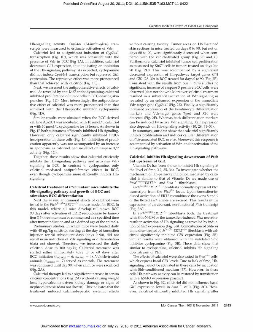
Vitamin D3 has been shown to inhibit Hh signaling atthe level of Smo (12, 35, 36). To investigate whether themechanism of Hh-pathway inhibition mediated by calci-triol is similar to that of Vitamin D3 we made use ofPtchflox/floxERT2þ/� and Smo�/� fibroblasts.
Ptchflox/floxERT2þ/� fibroblasts normally express wt Ptchtranscripts from the Ptchflox locus. Upon tamoxifen-in-duced activation of ERT2 recombinase the exons 8 and 9of the floxed Ptch alleles are excised. This results in theexpression of an aberrant, nonfunctional Ptch transcript(Fig. 3A).
In Ptchflox/floxERT2þ/� fibroblasts both, the treatmentwith Shh-N-CM or the tamoxifen-induced Ptch mutationresult in activation of Hh signaling as revealed by induc-tion of Gli1 expression (Fig. 3B). Coincubation of Shh- ortamoxifen-treated Ptchflox/floxERT2þ/� fibroblasts with cal-citriol significantly inhibited Gli1 expression (Fig. 3B).Similar results were obtained with the validated Smoinhibitor cyclopamine (Fig. 3B). These data show thatsimilar to cyclopamine, calcitriol inhibits Hh signalingdownstream of Ptch.
The effects of calcitriol were also tested in Smo�/� cells,which express basal Gli1 levels. Due to lack of Smo, Hh-signaling cannot be activated in these cells by incubationwith Shh-conditioned medium (37). However, in thesecells Hh-pathway activity can be restored by transfectionwith a hSMO expression plasmid.
As shown in Fig. 3C, calcitriol did not influence basalGli1 expression levels in Smo�/� cells (Fig. 3C). How-ever, calcitriol efficiently inhibited Hh signaling after
Calcitriol Inhibits Growth of Basal Cell Carcinoma
www.aacrjournals.org Mol Cancer Ther; 10(11) November 2011 2183
on July 29, 2018. © 2011 American Association for Cancer Research. mct.aacrjournals.org Downloaded from
Published OnlineFirst August 30, 2011; DOI: 10.1158/1535-7163.MCT-11-0422
restoration of Hh-pathway activity upon transfectionwith hSMO. These results are similar to those obtainedwith cyclopamine (Fig. 3C) and show that calcitriolnormally inhibits Gli1 expression at the level of Smo.
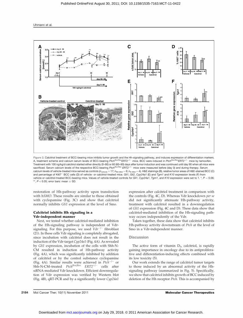
Calcitriol inhibits Hh signaling in aVdr-independent manner
Next, we tested whether calcitriol-mediated inhibitionof the Hh-signaling pathway is independent of Vdr-signaling. For this purpose, we used Vdr�/� fibroblast(21). In these cells Vdr signaling is completely abrogated,since incubation with calcitriol does not result in theinduction of the Vdr-targetCyp24a1 (Fig. 4A). As revealedby Gli1 expression, incubation of the cells with Shh-N-CM resulted in induction of Hh-pathway activity(Fig. 4A), which was significantly inhibited by additionof calcitriol or by the control substance cyclopamine(Fig. 4A). Similar results were achieved in Ptch�/� orShh-N-CM–treated Ptchflox/flox ERT2þ/� cells aftersiRNA-mediated Vdr knockdown. Efficient downregula-tion of Vdr expression was verified by Western blot(Fig. 4B), qRT-PCR and by a significantly lower Cyp24a1
expression after calcitriol treatment in comparison withthe controls (Fig. 4C, D). Whereas Vdr knockdown per sedid not significantly attenuate Hh-pathway activity,treatment with calcitriol resulted in a downregulationof Gli1 expression (Fig. 4C and D). These data show thatcalcitriol-mediated inhibition of the Hh-signaling path-way occurs independently of the Vdr.
Taken together, these data show that calcitriol inhibitsHh-pathway activity downstream of Ptch at the level ofSmo in a Vdr-independent manner.
Discussion
The active form of vitamin D3, calcitriol, is rapidlygaining importance in oncology due to its antiprolifera-tive and differentiation-inducing effects combined withits low toxicity (5).
Our work extends the range of calcitriol tumor targetsto those induced by an abnormal activity of the Hh-signaling pathway (summarized in Fig. 5). Specifically,we show that calcitriol inhibits growth of BCC induced bydeletion of the Hh receptor Ptch. This is accompanied by
Figure 2. Calcitriol treatment of BCC-bearing mice inhibits tumor growth and the Hh-signaling pathway, and induces expression of differentiation markers.A, treatment scheme and calcium serum levels of BCC-bearing Ptchflox/floxERT2þ/� mice. BCC were induced in Ptchflox/floxERT2þ/� mice by tamoxifen.Treatment with 100 ng/kg/d calcitriol started either directly (0–90) or 60 (60–90) days after tumor induction and was continued until day 90 when all mice weresacrificed. Serum calcium levels of the respective BCC-bearing Ptchflox/flox ERT2þ/� mice were measured before (day 0) and during therapy. Serumcalcium levels of vehicle-treated mice served as controls (nvehicle¼ 17; n60–90d¼ 6; n0–90d¼ 4). H&E stainings (B), relative tumor areas of H&E-stained BCC (C)and percentage of Ki67þ BCC cells (D) of vehicle- or calcitriol-treated mice. Gli1, Gli2, Cyp24a1 (E) and Tgm1 and K10 expression levels (F) fromvehicle or calcitriol-treated BCC-bearing mice. Values of vehicle-treated controls for Gli1, Cyp24a1, Tgm1, and K10 expression were set to 1. *, P ¼ 0.06;**, P < 0.05; error bars: mean � SD.
Uhmann et al.
Mol Cancer Ther; 10(11) November 2011 Molecular Cancer Therapeutics2184
on July 29, 2018. © 2011 American Association for Cancer Research. mct.aacrjournals.org Downloaded from
Published OnlineFirst August 30, 2011; DOI: 10.1158/1535-7163.MCT-11-0422

the expression of relevant cell differentiation markers.Our data suggest that calcitriol could be a valuablesupplement or even alternative to the established treat-ments of BCC, the most common tumor in humans,associated with aberrant Hh-pathway activity.
As assessed by reduced Gli1 transcription, calcitriolinhibits canonical Hh signaling independently of Vdrsignaling and downstream of Ptch (Fig. 5). An obviousmolecular target of this Vdr-independent effect of calci-triol is Smo, because Smo-deficient cells (unlike thosereconstituted with Smo or Ptch-deficient ones) show nodecreasedGli1 transcription in response to this substance.A similar observation has beenmade for the inactive formof calcitriol, vitamin D3 (12). Nevertheless, whether calci-triol directly binds to Smo should be addressed in futurestudies.
Besides inhibition of Hh-signaling pathway, calcitriolinhibits proliferation, and increases the expression ofskin differentiation marker in BCC. The latter effects arealso induced by calcitriol in skin of patients with hyper-proliferative skin diseases such as psoriasis (6–9, 34, 38).Calcitriol treatment specifically results in inhibition ofproliferation of psoriatic skin and induction of differ-entiation of keratinocytes (9, 39, 40). Similarly to BCC,psoriatic skin also expresses high levels of the Hh-targetgene Gli1 (41). These facts raise the question whethercalcitriol mediates its antiproliferative effects and dif-ferentiation stimuli via activation of Vdr signaling orrather via inhibition of the Hh-signaling pathway. Aclue might come from our present study: Our in vitrostudy shows that calcitriol has a significantly strongerantiproliferative effect on BCC than the pure Smo in-hibitor cyclopamine, even though the latter substanceinhibits Hh signaling more efficiently (Fig. 1D and F).Together with the fact that both calcitriol (presentstudy) and cyclopamine (42) inhibit BCC proliferationin vivo, it is possible that calcitriol exerts its antiproli-ferative effects via both signaling pathways (e.g., byinhibition or activation of Hh or Vdr signaling, respec-tively). On the other hand, differentiation of calcitriol-treated BCC is probably unrelated to inhibition of thecanonical Hh-signaling pathway, because cylopaminenever has been reported to induce the expression ofkeratinocyte differentiation markers in BCC. Morelikely, calcitriol induces BCC differentiation via Vdrsignaling, which is supported by the increased expres-sion of the Vdr-target genes Cyp24a1 and Tgm1(Fig. 2F).
Antiproliferative and Hh-signaling inhibitory proper-ties have also been described for the inactive form ofcalcitriol, vitamin D3, on murine BCC in vitro and in vivo(35). In contrast to calcitriol, vitamin D3 did not affect
Figure 3. Calcitriol inhibits the Hh-signaling pathway downstream of Ptchbut upstream of Gli1. A, Ptch ablation in tamoxifen-treated Ptchflox/flox
ERT2þ/� fibroblasts after incubation with 10 mmol/L tamoxifen for 48hours. Wt Ptch transcripts (derived from the nonrecombined Ptchflox locus)and Ptchdel transcripts were detected by PCR using the primer pairmPtc11/mPtc7R as described in Zibat and colleagues (13, 24). cDNAderived from 12.5 days old wild-type mouse embryos (E12.5 Ptchþ/þ) wasused as a control. Gli1 and Cyp24a1 expression levels of Ptchflox/flox
ERT2þ/� (B) and Smo�/� (C) fibroblasts after treatment with vehicle (EtOH),calcitriol, or cyclopamine (CP). Ptchflox/floxERT2þ/� fibroblasts were
cultured in either Shh-N-CM or control medium (CoM) with or without 10mmol/L tamoxifen as indicated. Smo�/� fibroblasts were either transfectedwith a hSMO expressing plasmid or with empty vector as indicated.**, P < 0.05; error bars, mean � SD; ntc, no template control.
Calcitriol Inhibits Growth of Basal Cell Carcinoma
www.aacrjournals.org Mol Cancer Ther; 10(11) November 2011 2185
on July 29, 2018. © 2011 American Association for Cancer Research. mct.aacrjournals.org Downloaded from
Published OnlineFirst August 30, 2011; DOI: 10.1158/1535-7163.MCT-11-0422

differentiation in BCC, although it was topically appliedat high concentrations. Supposedly the treatment periodusing vitamin D3 (i.e., 30 days)may have been too short toinduce Vdr signaling and thus a differentiation response.
This suggestion is based on a comparison with our studyin which a 30 days calcitriol application also had nosignificant effects on these processes (see BCC treatedfor days 60 to 90, Fig. 2B and E).
Figure 4. Calcitriol inhibits the Hh-signaling pathway in a Vdr-independent manner. A, Gli and Cyp24a1 expression of Vdr�/� fibroblasts after treatment withvehicle (EtOH), calcitriol, or cyclopamine (CP) and Shh-N-CM or control medium (CoM). B, analyses of the Vdr protein level of nuclear extracts of si-Vdr or si-control transfected Ptch�/� and Vdr�/� fibroblasts by Western blot. Detection of HSC-70 protein served as control. C and D, Gli1, Cyp24a1, and Vdrexpression of Ptch�/� and Shh-stimulated Ptchflox/floxERT2þ/� fibroblasts after si-Vdr or si-control transfection and calcitriol treatment. Gli1 expression ofPtch�/� fibroblasts and Vdr expression of Ptch�/� and Ptchflox/flox ERT2þ/� cells are shown in relation to the respective vehicle-treated control. **, P < 0.05;error bars: mean � SD.
Figure 5.Model for the dual function of calcitriol in Ptch-associated BCC. Normally Ptch inhibits its signaling partner Smo, thereby regulating the activity of theHh-signaling pathway (normal cell). Mutations of Ptch lead to a constitutive activation of the Gli transcription factors, which resulted in cellproliferation and tumor formation (BCC cell). The known Smo-inhibitor cyclopamine inhibits Hh-pathway in the Ptch-mutant cells and thus Hh-relatedprocesses involved in tumor growth (cyclopamine-treated BCC cell). In contrast, calcitriol inhibits Hh-pathway activity and additionally activates Vdr signaling(calcitriol-treated BCC cell). Consequently, calcitriol not only inhibits tumor-relevant processes mediated by Hh signaling, but also induces antiproliferativeeffects and differentiation processes via the Vdr-signaling pathway.
Uhmann et al.
Mol Cancer Ther; 10(11) November 2011 Molecular Cancer Therapeutics2186
on July 29, 2018. © 2011 American Association for Cancer Research. mct.aacrjournals.org Downloaded from
Published OnlineFirst August 30, 2011; DOI: 10.1158/1535-7163.MCT-11-0422

Finally, a new model of tumorigenesis driven by Ptch-deficiency may emerge from our study. According to arecent work, Ptch might function as an efflux pump forvitamin D3-related compounds with Hh-inhibitory prop-erties (12). A deficiency of this compound due to Ptchinactivation would pathologically activate Hh-pathwayand reduce Vdr signaling. Vice versa application of thiscompound should result in inhibition ofHh-pathway andactivation of Vdr signaling (Fig. 5). Whether this vitaminD3-related compound is calcitriol remains to be analyzedin the future.Taken together the application of calcitriol holds pro-
mises as an effective anticancer drug in the treatment ofBCC. Due to its dual effects on both Vdr and Hh signal-ing, it may be superior to substances that solely target theHh-signaling pathway. Calcitriol treatment may also besuperior to application of vitamin D3, which has to bemetabolized before activating Vdr signaling. The benefitsof topical application of calcitriol in treatment of BCChave to be tested in the future.
Disclosure of Potential Conflicts of Interest
No potential conflicts of interest were disclosed.
Acknowledgments
We thank Stefan Wolf and Susan Peter for excellent animal care. Wealso thank J. Taipale, University of Helsinki, Finland for provision ofSmo�/� fibroblasts, E. Epstein, UCSF, for the BCC cell line ASZ001, R.Toftgard, Karolinska Institute, Huddinge, Sweden for hSMO plasmid,Steven Johnsen, University of G€ottingen, Germany for proofreading andfor comments on the manuscript, and Leszek Wojnoswki, University ofMainz, Germany for comments on the manuscript.
Grant Support
This work was supported by grants of the Deutsche ForschungsgemeinschaftUH 228/2-1 and UH228/2-2 to A. Uhmann and HA 2197/5-1 FOR942 to H. Hahn.
The costs of publication of this article were defrayed in part by the paymentof page charges. This article must therefore be hereby marked advertisement inaccordance with 18 U.S.C. Section 1734 solely to indicate this fact.
Received June 8, 2011; revised August 3, 2011; accepted August 24, 2011;published OnlineFirst August 30, 2011.
References1. Hooper JE, Scott MP. Communicating with Hedgehogs. Nat Rev Mol
Cell Biol 2005;6:306–17.2. Yang L, Xie G, Fan Q, Xie J. Activation of the hedgehog-signaling
pathway in human cancer and the clinical implications. Oncogene2009;29:469–81.
3. Von Hoff DD, LoRusso PM, Rudin CM, Reddy JC, Yauch RL, Tibes R,et al. Inhibition of the hedgehog pathway in advanced basal-cellcarcinoma. N Engl J Med 2009;361:1164–72.
4. Yauch RL, Dijkgraaf GJ, Alicke B, Januario T, Ahn CP, Holcomb T,et al. Smoothened mutation confers resistance to a Hedgehog path-way inhibitor in medulloblastoma. Science 2009;326:572–4.
5. Deeb KK, Trump DL, Johnson CS. Vitamin D signalling pathways incancer: potential for anticancer therapeutics. Nat Rev Cancer2007;7:684–700.
6. Hershberger PA, Modzelewski RA, Shurin ZR, Rueger RM, Trump DL,Johnson CS. 1,25-Dihydroxycholecalciferol (1,25-D3) inhibits thegrowth of squamous cell carcinoma and down-modulates p21(Waf1/Cip1) in vitro and in vivo. Cancer Res 1999;59:2644–9.
7. Majewski S, Skopinska M, Bollag W, Jablonska S. Combination ofisotretinoin and calcitriol for precancerous and cancerous skinlesions. Lancet 1994;344:1510–1.
8. Stewart DG, Lewis HM. Vitamin D analogues and psoriasis. J ClinPharm Ther 1996;21:143–8.
9. vandeKerkhofPC.AnupdateonvitaminD3analogues in the treatmentof psoriasis. Skin Pharmacol Appl Skin Physiol 1998;11:2–10.
10. Bikle DD. Vitamin D and the skin. J Bone Miner Metab 2010;28:117–30.
11. Haussler MR, Whitfield GK, Haussler CA, Hsieh JC, Thompson PD,Selznick SH, et al. The nuclear vitamin D receptor: biological andmolecular regulatory properties revealed. J Bone Miner Res 1998;13:325–49.
12. Bijlsma MF, Spek CA, Zivkovic D, van de Water S, Rezaee F, Peppe-lenbosch MP. Repression of smoothened by patched-dependent(pro-)vitamin D3 secretion. PLoS Biol 2006;4:e232.
13. Zibat A, Uhmann A, Nitzki F, Wijgerde M, Frommhold A, Heller T, et al.Time-point and dosage of gene inactivation determine the tumorspectrum in conditional Ptch knockouts. Carcinogenesis 2009;30:918–26.
14. Chen JK, Taipale J, Young KE, Maiti T, Beachy PA. Small moleculemodulation of Smoothened activity. Proc Natl Acad Sci U S A 2002;99:14071–6.
15. Taipale J, Chen JK, CooperMK,Wang B,Mann RK,Milenkovic L, et al.Effects of oncogenic mutations in Smoothened and Patched can bereversed by cyclopamine. Nature 2000;406:1005–9.
16. Nitzki F, Zibat A, K€onig S, Wijgerde M, Rosenberger A, Brembeck FH,et al. Tumor stroma-derivedWnt5a induces differentiation of basal cellcarcinoma of Ptch mutant mice via CaMKII. Cancer Res 2010;70:2739–48.
17. Calzada-Wack J, Kappler R, Schnitzbauer U, Richter T, Nathrath M,RosemannM, et al. Unbalanced overexpression of the mutant allele inmurine Patched mutants. Carcinogenesis 2002;23:727–34.
18. Banach-Petrosky W, Ouyang X, Gao H, Nader K, Ji Y, Suh N, et al.Vitamin D inhibits the formation of prostatic intraepithelial neoplasia inNkx3.1;Pten mutant mice. Clin Cancer Res 2006;12:5895–901.
19. Ecke I, Rosenberger A, Obenauer S, Dullin C, Aberger F, Kimmina S,et al. Cyclopamine treatment of full-blown Hh/Ptch-associated RMSpartially inhibits Hh/Ptch signaling, but not tumor growth. Mol Carci-nog 2008;47:361–72.
20. Ma Y, Erkner A, Gong R, Yao S, Taipale J, Basler K, et al. Hedgehog-mediated patterning of the mammalian embryo requires transporter-like function of dispatched. Cell 2002;111:63–75.
21. Sun J, Kong J, Duan Y, Szeto FL, Liao A, Madara JL, et al. IncreasedNF-kappaB activity in fibroblasts lacking the vitamin D receptor. Am JPhysiol Endocrinol Metab 2006;291:E315–22.
22. Xie J, AszterbaumM, Zhang X, Bonifas JM, Zachary C, Epstein E, et al.A role of PDGFRalpha in basal cell carcinoma proliferation. Proc NatlAcad Sci U S A 2001;98:9255–9.
23. Xie J, Murone M, Luoh SM, Ryan A, Gu Q, Zhang C, et al. ActivatingSmoothened mutations in sporadic basal-cell carcinoma. Nature1998;391:90–2.
24. Uhmann A, Dittmann K, Nitzki F, Dressel R, Koleva M, Frommhold A,et al. The Hedgehog receptor Patched controls lymphoid lineagecommitment. Blood 2007;110:1814–23.
25. Mitschele T, Diesel B, Friedrich M, Meineke V, Maas RM, Gartner BC,et al. Analysis of the vitamin D system in basal cell carcinomas (BCCs).Lab Invest 2004;84:693–702.
26. Kamradt J, Rafi L, Mitschele T, Meineke V, Gartner BC, Wolfgang T,et al. Analysis of the vitamin D system in cutaneous malignancies.Recent Results Cancer Res 2003;164:259–69.
27. Reichrath J, Kamradt J, Zhu XH, Kong XF, Tilgen W, Holick MF.Analysis of 1,25-dihydroxyvitamin D(3) receptors (VDR) in basal cellcarcinomas. Am J Pathol 1999;155:583–9.
Calcitriol Inhibits Growth of Basal Cell Carcinoma
www.aacrjournals.org Mol Cancer Ther; 10(11) November 2011 2187
on July 29, 2018. © 2011 American Association for Cancer Research. mct.aacrjournals.org Downloaded from
Published OnlineFirst August 30, 2011; DOI: 10.1158/1535-7163.MCT-11-0422

28. Ikram MS, Neill GW, Regl G, Eichberger T, Frischauf AM, Aberger F,et al. GLI2 is expressed in normal human epidermis and BCC andinduces GLI1 expression by binding to its promoter. J Invest Dermatol2004;122:1503–9.
29. Regl G, Kasper M, Schnidar H, Eichberger T, Neill GW, IkramMS, et al.The zinc-finger transcription factor GLI2 antagonizes contact inhibi-tion and differentiation of human epidermal cells. Oncogene2004;23:1263–74.
30. Regl G, Neill GW, Eichberger T, Kasper M, Ikram MS, Koller J, et al.Human GLI2 and GLI1 are part of a positive feedback mechanism inbasal cell carcinoma. Oncogene 2002;21:5529–39.
31. Stark HJ, Breitkreutz D, Limat A, Ryle CM, Roop D, Leigh I, et al.Keratins 1 and 10 or homologues as regular constituents of inner rootsheath and cuticle cells in the human hair follicle. Eur J Cell Biol1990;52:359–72.
32. Beck B, Lehen'kyi V, Roudbaraki M, Flourakis M, Charveron M,Bordat P, et al. TRPC channels determine human keratinocytedifferentiation: new insight into basal cell carcinoma. Cell Calcium2008;43:492–505.
33. Lee SC, Ikai K, Ando Y, Imamura S. Effects of 1 alpha, 25-dihydroxyvitamin D3 on the transglutaminase activity of trans-formed mouse epidermal cells in culture. J Dermatol 1989;16:7–11.
34. Bikle DD. The vitamin D receptor: a tumor suppressor in skin. DiscovMed 2011;11:7–17.
35. Tang JY, Xiao TZ, Oda Y, Chang KS, Shpall E, Wu A, et al. Vitamin d3inhibits hedgehog signaling and proliferation in murine Basal cellcarcinomas. Cancer Prev Res (Phila) 2011;4:744–51.
36. Bruggemann LW, Queiroz KC, Zamani K, van Straaten A, Spek CA,Bijlsma MF. Assessing the efficacy of the hedgehog pathway inhibitorvitamin D3 in a murine xenograft model for pancreatic cancer. CancerBiol Ther 2010;10:79–88.
37. Varjosalo M, Li SP, Taipale J. Divergence of hedgehog signal trans-duction mechanism between Drosophila and mammals. Dev Cell2006;10:177–86.
38. Bikle DD. Vitamin D and skin cancer. J Nutr 2004;134;3472–8S.39. Bikle DD. 1,25(OH)2D3-modulated calcium induced keratinocyte dif-
ferentiation. J Investig Dermatol Symp Proc 1996;1:22–7.40. Gottlieb AB. Therapeutic options in the treatment of psoriasis and
atopic dermatitis. J Am Acad Dermatol 2005;53:S3–16.41. Endo H, Momota Y, Oikawa A, Shinkai H. Psoriatic skin expresses the
transcription factor Gli1: possible contribution of decreased neurofi-bromin expression. Br J Dermatol 2006;154:619–23.
42. Tas S, Avci O. Rapid clearance of psoriatic skin lesions induced bytopical cyclopamine. A preliminary proof of concept study. Derma-tology 2004;209:126–31.
Uhmann et al.
Mol Cancer Ther; 10(11) November 2011 Molecular Cancer Therapeutics2188
on July 29, 2018. © 2011 American Association for Cancer Research. mct.aacrjournals.org Downloaded from
Published OnlineFirst August 30, 2011; DOI: 10.1158/1535-7163.MCT-11-0422

2011;10:2179-2188. Published OnlineFirst August 30, 2011.Mol Cancer Ther Anja Uhmann, Hannah Niemann, Bérénice Lammering, et al. Receptor Signaling and DifferentiationInhibition of Hedgehog Signaling and Induction of Vitamin D Antitumoral Effects of Calcitriol in Basal Cell Carcinomas Involve
Updated version
10.1158/1535-7163.MCT-11-0422doi:
Access the most recent version of this article at:
Cited articles
http://mct.aacrjournals.org/content/10/11/2179.full#ref-list-1
This article cites 42 articles, 8 of which you can access for free at:
Citing articles
http://mct.aacrjournals.org/content/10/11/2179.full#related-urls
This article has been cited by 3 HighWire-hosted articles. Access the articles at:
E-mail alerts related to this article or journal.Sign up to receive free email-alerts
Subscriptions
Reprints and
To order reprints of this article or to subscribe to the journal, contact the AACR Publications Department at
Permissions
Rightslink site. Click on "Request Permissions" which will take you to the Copyright Clearance Center's (CCC)
.http://mct.aacrjournals.org/content/10/11/2179To request permission to re-use all or part of this article, use this link
on July 29, 2018. © 2011 American Association for Cancer Research. mct.aacrjournals.org Downloaded from
Published OnlineFirst August 30, 2011; DOI: 10.1158/1535-7163.MCT-11-0422


![Vitamin D in PdPregnancy and Infancy · 1,25-dihydroxy vitamin D = calcitriol [1,25(OH)2D] assessment (hormonal form) ↓ Calcitriol binds to vitamin D receptor (VDR) to regulate](https://static.fdocuments.in/doc/165x107/5ffb3ce26517d830b10f5d09/vitamin-d-in-pdpregnancy-and-125-dihydroxy-vitamin-d-calcitriol-125oh2d.jpg)

