Dr. Paul MacKoul MD: Learn About Adenomyosis and Minimally Invasive Adenomyosis Surgery at CIGC
1
Genital system-2
Adenomyosis
Endometrial glandular hyperplasia
Endometrial adenocarcinoma
Residua post abortum
Hydatidiform mole
Choriocarcinoma
Endometriosis and Adenomyosis
Endometriosis is the term used to describe the presence of endometrial glands or stroma in abnormal locations outside the uterus.
It occurs in the following sites, in descending order of frequency:
(1) ovaries;
(2) uterine ligaments;
(3) rectovaginal septum;
(4) pouch of Douglas
(5) pelvic peritoneum;
(6) large and small bowel
(7) vagina, vulva, or appendix.
(8) laparotomy scars;
Adenomyosis (earlier term „internal endometriosis”) refers to the
presence of endometrial glands and stroma within the myometrium of the
uterus
Epidemiology of endometriosis
►Affects 5-10% of the female population Presentation: Up to 20% are asymptomatic
► 40-60% in women with dysmenorrhoea
► 20-30% in women with subfertility
► Global survey data indicate that 62% of women with endometriosis are under age 30 at onset of symptoms
► References Mounsey AL, Wilgus A, Slawson DC. Diagnosis and management of endometriosis: Am Fam Phys 2006;74:594–600.
Endometriosis – etiology ?
1. Transplantation theory (most widely accepted): menstrual backflow through the fallopian tubes and implantation
2. Vascular or lymphatic dissemination
3. Metaplasia theory: peritoneum may differentiate intoendometrial tissue
4. iatrogenic (cesarian cut)
5. Induction theory: endometrial-derived substancesinduces endometrial differentiation at ectopic sites
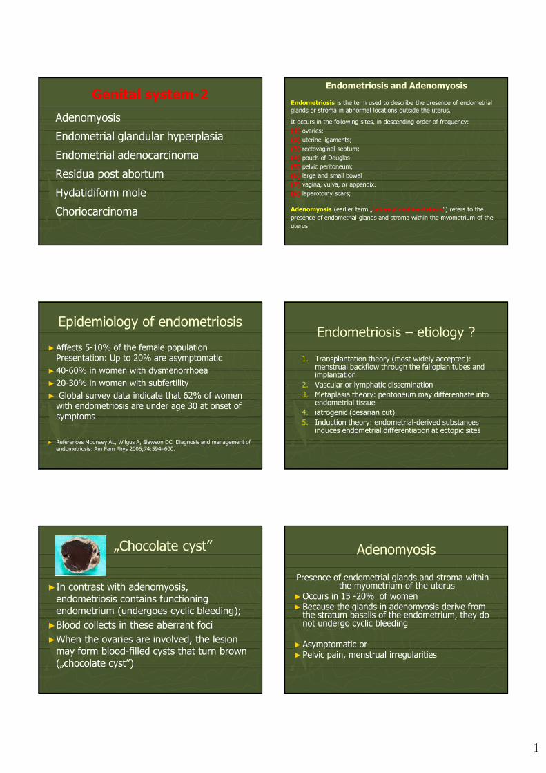
„Chocolate cyst”
►In contrast with adenomyosis, endometriosis contains functioning endometrium (undergoes cyclic bleeding);
►Blood collects in these aberrant foci
►When the ovaries are involved, the lesion may form blood-filled cysts that turn brown („chocolate cyst”)
Adenomyosis
Presence of endometrial glands and stroma within the myometrium of the uterus
►Occurs in 15 -20% of women►Because the glands in adenomyosis derive from
the stratum basalis of the endometrium, they do not undergo cyclic bleeding
►Asymptomatic or► Pelvic pain, menstrual irregularities
2
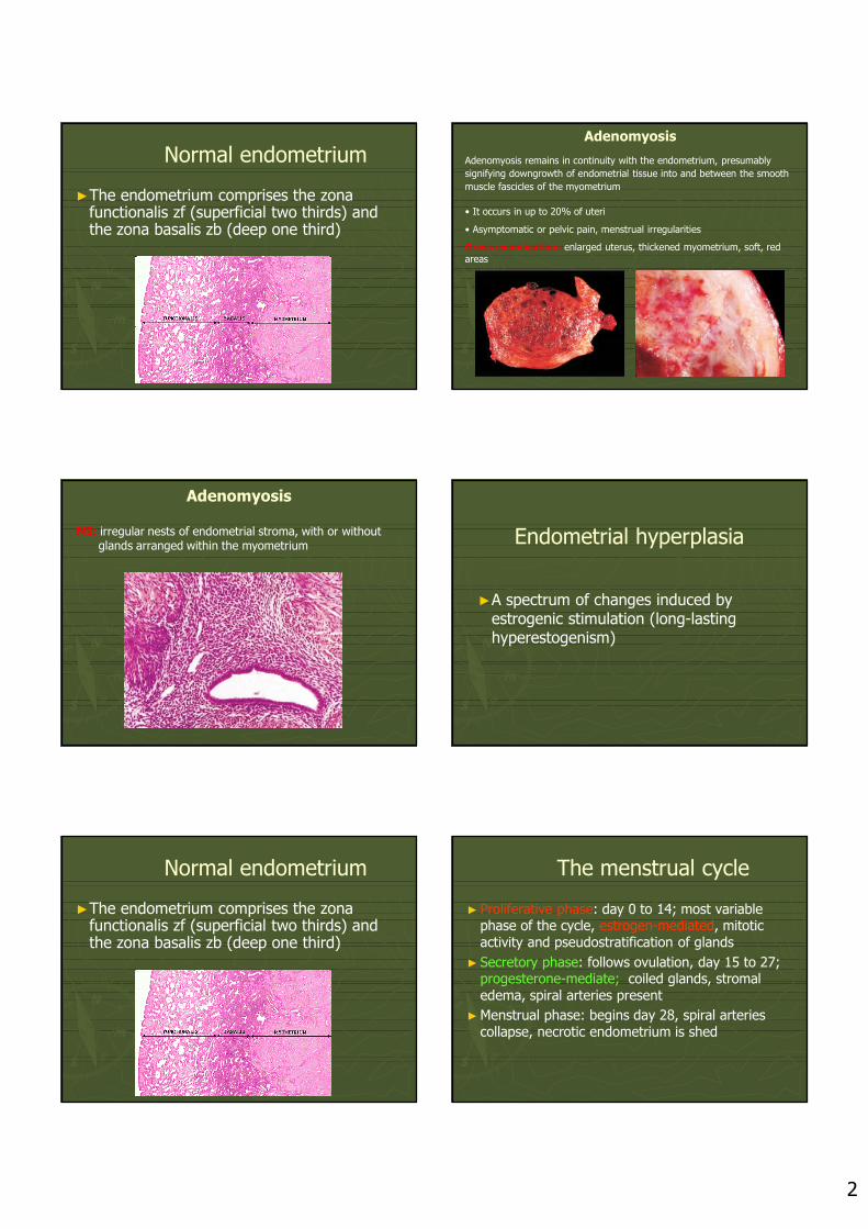
Normal endometrium
►The endometrium comprises the zona functionalis zf (superficial two thirds) and the zona basalis zb (deep one third)
Adenomyosis
Adenomyosis remains in continuity with the endometrium, presumably
signifying downgrowth of endometrial tissue into and between the smooth
muscle fascicles of the myometrium
• It occurs in up to 20% of uteri
• Asymptomatic or pelvic pain, menstrual irregularities
Gross examination: enlarged uterus, thickened myometrium, soft, red areas
Adenomyosis
MI: irregular nests of endometrial stroma, with or without glands arranged within the myometrium Endometrial hyperplasia
►A spectrum of changes induced by estrogenic stimulation (long-lasting hyperestogenism)
Normal endometrium
►The endometrium comprises the zona functionalis zf (superficial two thirds) and the zona basalis zb (deep one third)
The menstrual cycle
► Proliferative phase: day 0 to 14; most variable phase of the cycle, estrogen-mediated, mitotic activity and pseudostratification of glands
► Secretory phase: follows ovulation, day 15 to 27;progesterone-mediate; coiled glands, stromal edema, spiral arteries present
►Menstrual phase: begins day 28, spiral arteries collapse, necrotic endometrium is shed
3
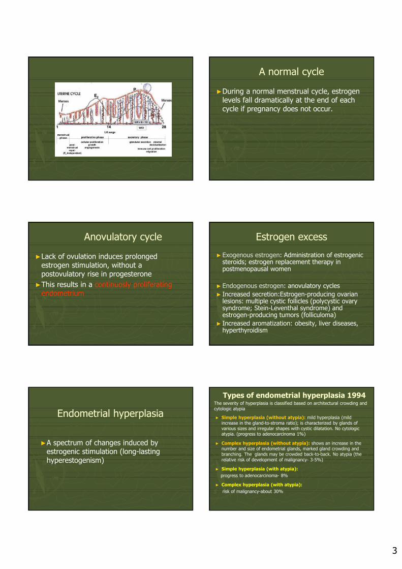
A normal cycle
►During a normal menstrual cycle, estrogen levels fall dramatically at the end of each cycle if pregnancy does not occur.
Anovulatory cycle
►Lack of ovulation induces prolonged estrogen stimulation, without a postovulatory rise in progesterone
►This results in a continuosly proliferating endometrium
Estrogen excess
► Exogenous estrogen: Administration of estrogenic steroids; estrogen replacement therapy in postmenopausal women
► Endogenous estrogen: anovulatory cycles
► Increased secretion:Estrogen-producing ovarian lesions: multiple cystic follicles (polycystic ovary syndrome; Stein-Leventhal syndrome) and estrogen-producing tumors (folliculoma)
► Increased aromatization: obesity, liver diseases, hyperthyroidism
Endometrial hyperplasia
►A spectrum of changes induced by estrogenic stimulation (long-lasting hyperestogenism)
Types of endometrial hyperplasia 1994The severity of hyperplasia is classified based on architectural crowding and cytologic atypia
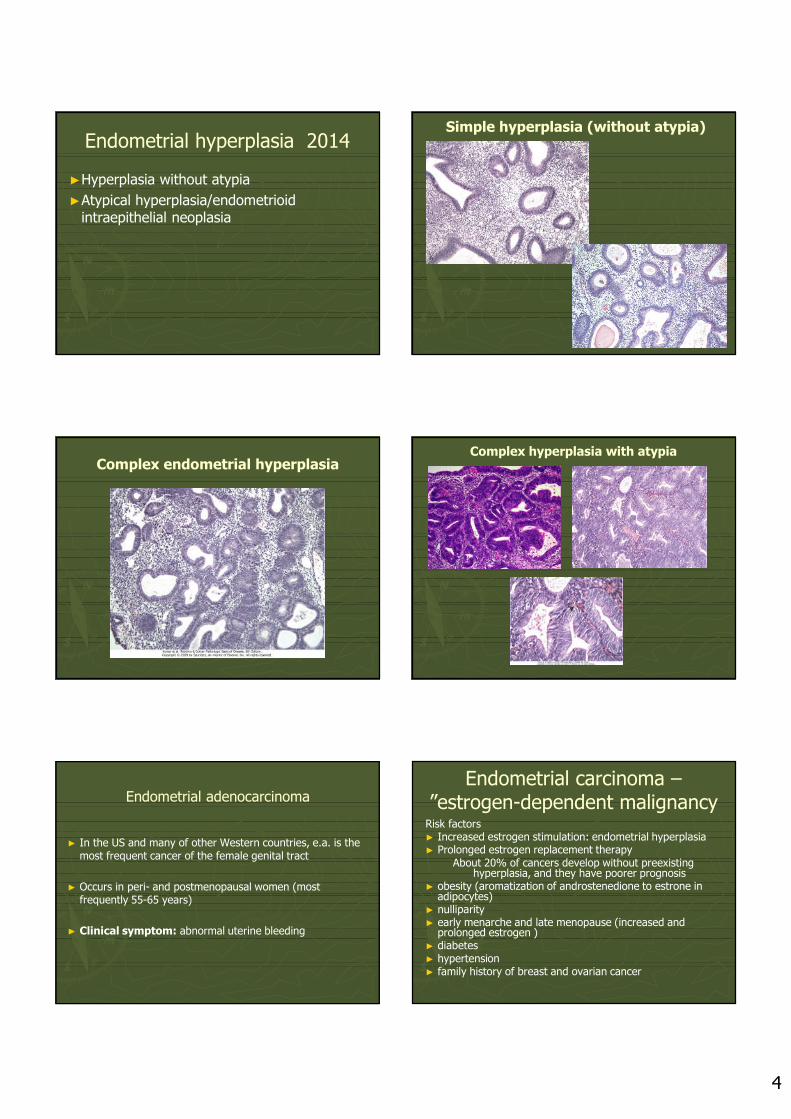
► Simple hyperplasia (without atypia): mild hyperplasia (mild increase in the gland-to-stroma ratio); is characterized by glands of various sizes and irregular shapes with cystic dilatation. No cytologic atypia. (progress to adenocarcinoma 1%)
► Complex hyperplasia (without atypia): shows an increase in the number and size of endometrial glands, marked gland crowding andbranching. The glands may be crowded back-to-back. No atypia (the relative risk of development of malignancy- 3-5%)
► Simple hyperplasia (with atypia):
progress to adenocarcinoma- 8%
► Complex hyperplasia (with atypia):
risk of malignancy-about 30%
4
Endometrial hyperplasia 2014
►Hyperplasia without atypia
►Atypical hyperplasia/endometrioidintraepithelial neoplasia
Simple hyperplasia (without atypia)
Complex endometrial hyperplasiaComplex hyperplasia with atypia
Endometrial adenocarcinoma
► In the US and many of other Western countries, e.a. is the most frequent cancer of the female genital tract
► Occurs in peri- and postmenopausal women (most frequently 55-65 years)
► Clinical symptom: abnormal uterine bleeding
Endometrial carcinoma –”estrogen-dependent malignancy
Risk factors► Increased estrogen stimulation: endometrial hyperplasia► Prolonged estrogen replacement therapy
About 20% of cancers develop without preexisting hyperplasia, and they have poorer prognosis
► obesity (aromatization of androstenedione to estrone in adipocytes)
► nulliparity► early menarche and late menopause (increased and
prolonged estrogen )► diabetes► hypertension► family history of breast and ovarian cancer
5
Histologic forms of endometrial carcinoma1. endometrioid (accounts for 60% of all endometrial
carcinomas)
► resemble normal endometrium but
► glands contain atypical cells with pleomorphic nuclei and abnormal mitotic figures
Graded according to the International Federation of Gynecology and Obstetric system: worsening grade corresponding to areas of SOLID growth
► G1 well differentiated >95% of glands (low-grade)
► G2 moderately differentiated >50% of glands (low-grade)
► G3 poorly differentiated (high-grade)
Endometrial adenocarcinomaNot graded:2. serous adenocarcinoma► epithelium similar to that found in the
fallopian tube► particularly aggressive3. clear cell adenocarcinoma► cells are of müllerian origin, large, with
cytoplasmic glycogen► tumor tends to occur in older women, with
no history of estrogen replacement► behaves as poorly differentiated cancer
(Müllerian ducts – uterus and tubes)
WHO classification of the tumors of the uterine corpus 2014
► Precursors: hyperplasiawithout atypia; atypicalhyperplasia
► Endometrial carcinomas
Endometrioid ( with squamousdifferentiation, villoglandular, secretory)
Mucinous carcinoma
Serous carcinoma
Clear cell carcinoma
Neuroendocrinetumors (carcinoid, high-grade tumors)
Undifferentiatedcarcinoma
• Mesenchymal tumors
Leiomyoma
leiomyosarcomas
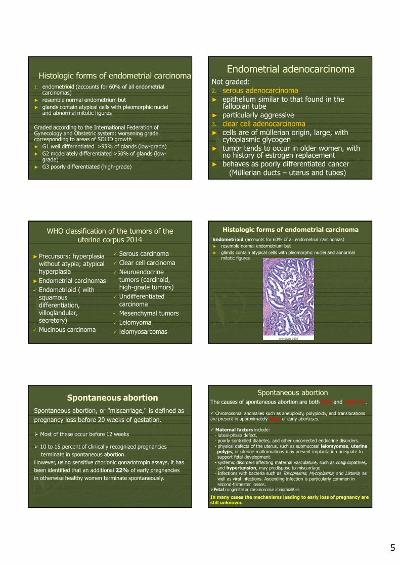
Histologic forms of endometrial carcinoma
Endometrioid (accounts for 60% of all endometrial carcinomas)
► resemble normal endometrium but
► glands contain atypical cells with pleomorphic nuclei and abnormal mitotic figures
Spontaneous abortion
Spontaneous abortion, or "miscarriage," is defined as
pregnancy loss before 20 weeks of gestation.
Most of these occur before 12 weeks
10 to 15 percent of clinically recognized pregnancies
terminate in spontaneous abortion.
However, using sensitive chorionic gonadotropin assays, it has
been identified that an additional 22% of early pregnancies
in otherwise healthy women terminate spontaneously.
Spontaneous abortionThe causes of spontaneous abortion are both fetal and maternal.
Chromosomal anomalies such as aneuploidy, polyploidy, and translocations are present in approximately 50% of early abortuses.
Maternal factors include: - luteal-phase defect, - poorly controlled diabetes, and other uncorrected endocrine disorders. - physical defects of the uterus, such as submucosal leiomyomas, uterine polyps, or uterine malformations may prevent implantation adequate to support fetal development.
- systemic disorders affecting maternal vasculature, such as coagulopathies, and hypertension, may predispose to miscarriage.
- Infections with bacteria such as Toxoplasma, Mycoplasma, and Listeria, as well as viral infections. Ascending infection is particularly common in second-trimester losses.
Fetal congenital or chromosomal abnormalities
In many cases the mechanisms leading to early loss of pregnancy are still unknown.
6
Early abortion - foetal partsResidua post abortum
Biopsy material shows:a) Placental villi normal sizeProtective epithelium (throphoblast) consist of:- superficial, multilayered syncytiotrophoblast;-Profound cytotrophoblast Its cuboid cells form one layerb) Decidua which consists of enlarged cells of endomertial stroma (swallen cells exhibit the epithelioid arrangenent)
First-trimester chorionic villi composed of delicate mesh of central stroma surrounded by two discrete layers of epithelium-the outer layer consisting of syncytiotrophoblast (two arrows) and the inner layer consisting of cytotrophoblast (arrow).
Gestational trophoblastic disease GTD
►GTD is a heterogenous group of gestational and neoplastic conditions arising from trophoblast, including molar gestations and trophoblastic tumors
Gestational trophoblastic disease
Traditionally, the gestational trophoblastic tumors have been divided into three overlapping morphologic categories: 1. hydatidiform mole2. invasive mole3. choriocarcinoma
They range in level of aggressiveness from the hydatidiform moles, most of which are benign, to the highly malignant choriocarcinomas.
All elaborate human chorionic gonadotropin (hCG), which can be detected in the circulating blood and urine at titers considerably higher than those found during normal pregnancy; the titers progressively rising from hydatidiform mole to invasive mole to choriocarcinoma.
Clinicians prefer the term gestational trophoblastic disease, because the response to therapy as judged by the hormone titers is significantly more important than any arbitrary anatomic segregation of one lesion from another.
Nonetheless, it is necessary to understand their individual characteristics to appreciate the spectrum of lesions.
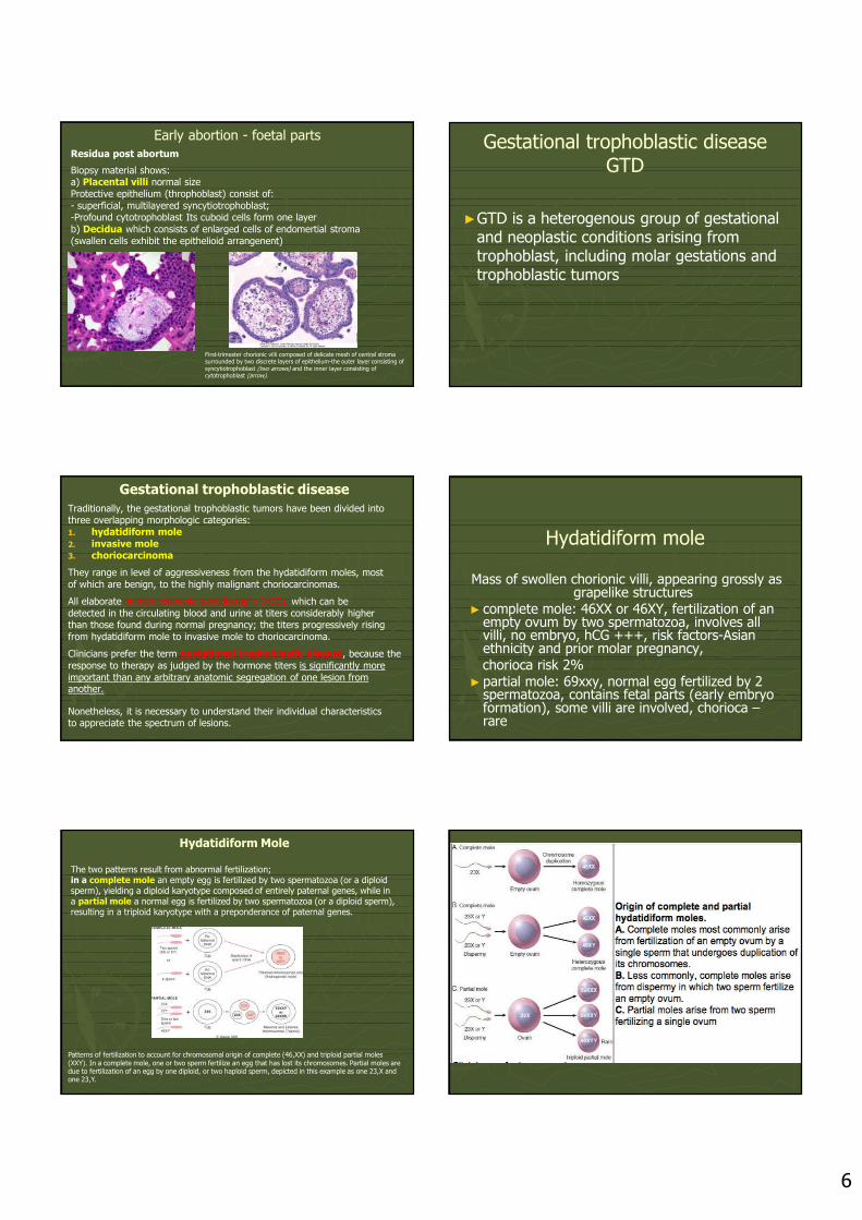
Hydatidiform mole
Mass of swollen chorionic villi, appearing grossly as grapelike structures
► complete mole: 46XX or 46XY, fertilization of an empty ovum by two spermatozoa, involves all villi, no embryo, hCG +++, risk factors-Asian ethnicity and prior molar pregnancy, chorioca risk 2%
► partial mole: 69xxy, normal egg fertilized by 2 spermatozoa, contains fetal parts (early embryo formation), some villi are involved, chorioca –rare
Hydatidiform Mole
The two patterns result from abnormal fertilization; in a complete mole an empty egg is fertilized by two spermatozoa (or a diploid sperm), yielding a diploid karyotype composed of entirely paternal genes, while in a partial mole a normal egg is fertilized by two spermatozoa (or a diploid sperm), resulting in a triploid karyotype with a preponderance of paternal genes.
Patterns of fertilization to account for chromosomal origin of complete (46,XX) and triploid partial moles (XXY). In a complete mole, one or two sperm fertilize an egg that has lost its chromosomes. Partial moles are due to fertilization of an egg by one diploid, or two haploid sperm, depicted in this example as one 23,X and one 23,Y.

7
Hydatidiform Mole
Two distinctive subtypes of moles have been characterized:
a. complete and b. partial moles.
The complete hydatidiform mole does not permit embryogenesis and therefore never contains fetal parts. All of the chorionic villi are abnormal, and the chorionic epithelial cells are diploid (46,XX or, uncommonly, 46,XY).
The partial hydatidiform mole is compatible with early embryo formation and therefore contains fetal parts, has some normal chorionic villi, and is almost always triploid (e.g., 69,XXY).
The typical hydatidiform mole is a voluminous mass of swollen, sometimes cystically dilated, chorionic villi, appearing grossly as grapelike structures.
Hydatidiform Mole
The incidence of complete hydatidiform moles is about 1 to 1.5 per 2000 pregnancies in the United States and other Western countries. For unknown reasons there is a much higher incidence in Asian countries.
Moles are most common before age 20 years and after age 40 years, and a history of the condition increases the risk in subsequent pregnancies.
Hydatidiform mole
Histology►Hydropic swelling of chorionic villi, no
vessels, proliferation of chorionic epithelium (cyto- and syncytiotrophoblast), sometimes present nucleated erythrocytes (partial)
Complete hydatidiform mole
Hydropic villi Invasive mole
►Mole that is invasive locally, with no metastatic potential
►complications: rupture of the uterus and haemorrhage
►spontaneous regress or
►chemotherapy
8
Choriocarcinoma
This very aggressive malignant tumor arises either from gestational chorionic epithelium or, less frequently, from totipotential cells within the gonads or elsewhere.
• Choriocarcinomas are rare in the Western hemisphere, and in the United States they occur in about 1 in 30,000 pregnancies.
• They are much more common in Asian and African countries, reaching a frequency of 1 in 2000 pregnancies.
• The risk is somewhat greater before age 20 and is significantly elevated after age 40.
• Approximately 50% of choriocarcinomas arise in complete hydatidiform moles; about 25% arise after an abortion
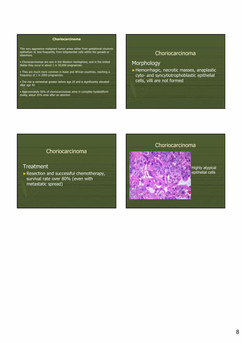
Choriocarcinoma
Morphology►Hemorrhagic, necrotic masses, anaplastic
cyto- and syncytiotrophoblastic epithelial cells, villi are not formed
Choriocarcinoma
Treatment►Resection and successful chemotherapy,
survival rate over 80% (even with metastatic spread)
Choriocarcinoma
Highly atypical epithelial cells