ANTEPARTUM HAEMORRHAGE. Definition: Bleeding from the genital tract after 22 weeks of pregnancy or...
23
ANTEPARTUM HAEMORRHAGE
-
Upload
jessie-rogers -
Category
Documents
-
view
225 -
download
1
Transcript of ANTEPARTUM HAEMORRHAGE. Definition: Bleeding from the genital tract after 22 weeks of pregnancy or...
ANTEPARTUM HAEMORRHAGE
Definition:
Bleeding from the genital tract after 22
weeks of pregnancy or during the 1st
stage of labor.
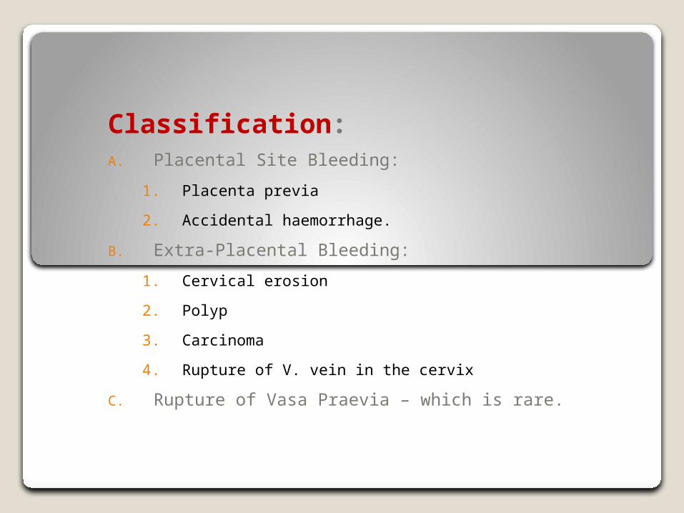
Classification:A. Placental Site Bleeding:
1. Placenta previa
2. Accidental haemorrhage.
B. Extra-Placental Bleeding:
1. Cervical erosion
2. Polyp
3. Carcinoma
4. Rupture of V. vein in the cervix
C. Rupture of Vasa Praevia – which is rare.
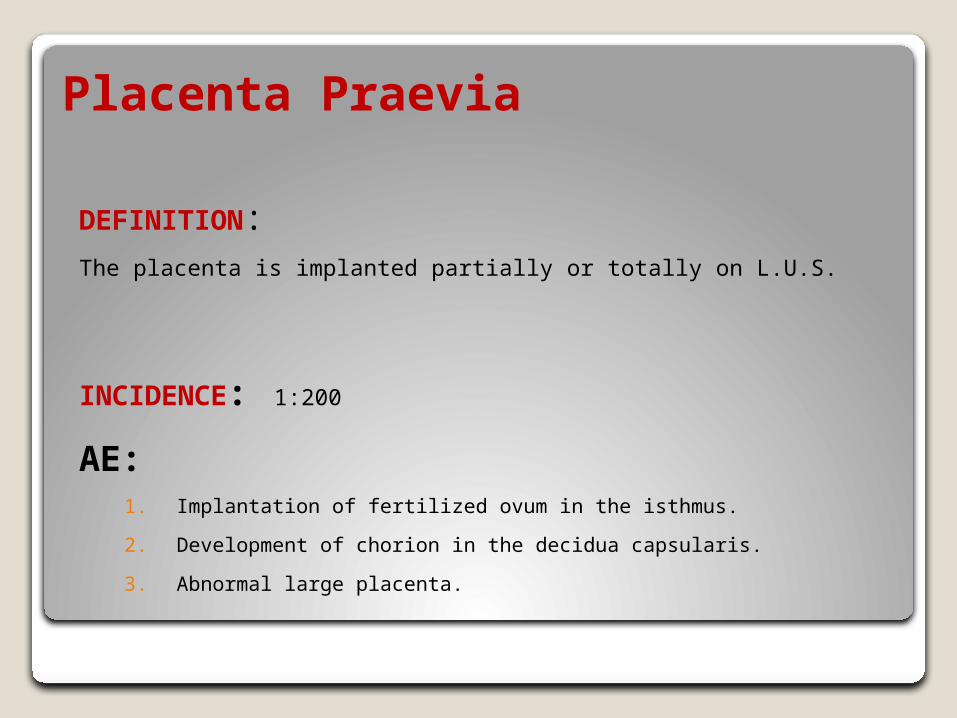
Placenta Praevia
DEFINITION:The placenta is implanted partially or totally on L.U.S.
INCIDENCE: 1:200
AE:1. Implantation of fertilized ovum in the isthmus.
2. Development of chorion in the decidua capsularis.
3. Abnormal large placenta.
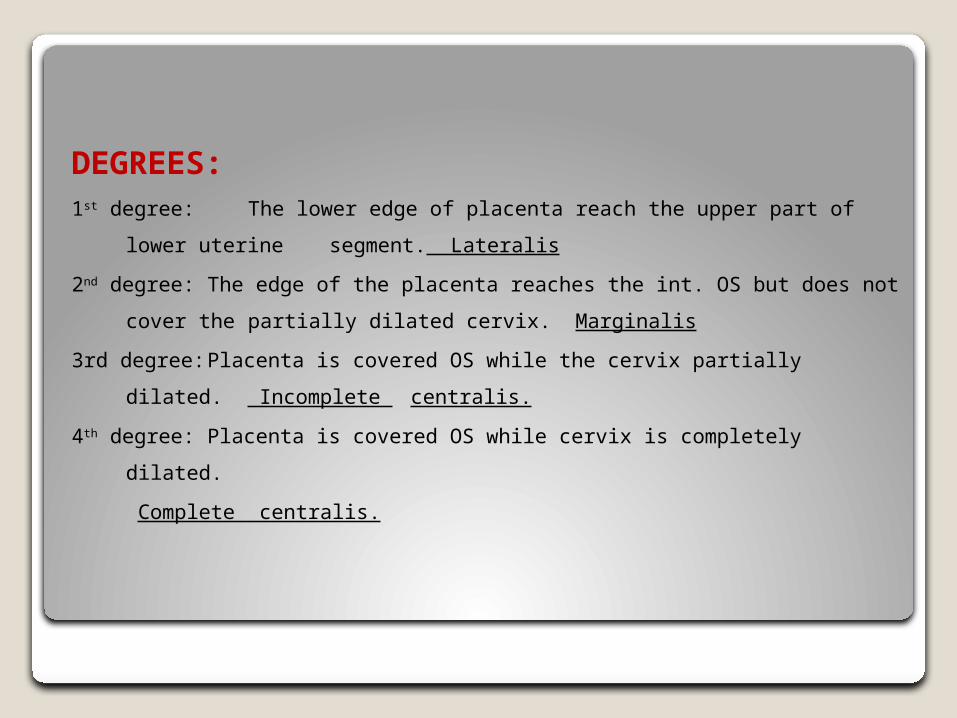
DEGREES:1st degree: The lower edge of placenta reach the upper part of lower
uterine segment. Lateralis
2nd degree: The edge of the placenta reaches the int. OS but does not
cover the partially dilated cervix. Marginalis
3rd degree: Placenta is covered OS while the cervix partially dilated.
Incomplete centralis.
4th degree: Placenta is covered OS while cervix is completely dilated.
Complete centralis.
MECHANISM OF BLEEDING:
During labour sheering of placenta. due to uterine contraction.
CLINICAL PICTURE:
1. Painless2. Causeless3. Recurrent bleeding after 28 weeks.
SIGNSGeneral examination
◦ Depend on severity of bleeding ± anaemia
Abdominal examination◦ Nothing characteristic◦ Maybe malpresentation – non engagement head
Uterus is not tender Supra pubic fullness
Vaginal examination◦ Should not be done except in theatre◦ Vaginal examination is done when active treatment is indicated
Active Treatment will be done:1. Fetal maturity2. During labor (patient in labour)3. Severe bleeding.
INVESTIGATION:
1. Ultrasound – accurate
2. Radiology – this is in the past
3. Soft tissue placentography◦ Amniography – radio opaque substance
◦ Detection of fetal head displacement
◦ Public angiography
◦ Cystography – inject sodium iodide 12.5.
4. IV radio active istope
5. Thermography
TREATMENT
A. At home: In case of emergency
1. Cervix generally – no PV
2. Sterile vulval pad
3. Morphine + transfer to hospital
B. At Hospital:
1. Antenatal record – all investigation should be done including coagulation profile
2. Blood transfusion if needed
3. Abdominal examination
C. Not in Labour:
1. Severe bleeding – Cesarean section.
2. Mild bleeding.
Expect ttt less than 37 weeks do U/S
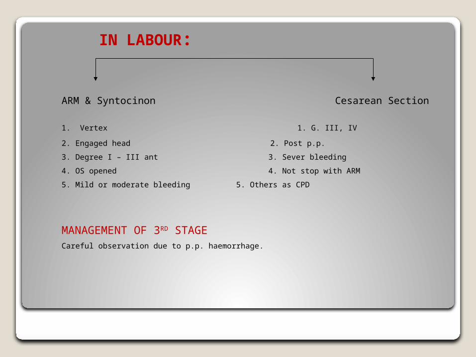
IN LABOUR:
ARM & Syntocinon Cesarean Section
1. Vertex 1. G. III, IV
2. Engaged head 2. Post p.p.
3. Degree I – III ant 3. Sever bleeding
4. OS opened 4. Not stop with ARM
5. Mild or moderate bleeding 5. Others as CPD
MANAGEMENT OF 3RD STAGECareful observation due to p.p. haemorrhage.
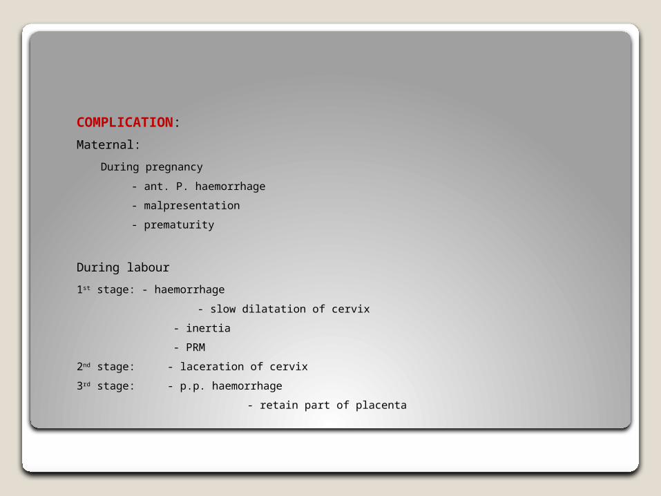
COMPLICATION:
Maternal:
During pregnancy
- ant. P. haemorrhage
- malpresentation
- prematurity
During labour
1st stage: - haemorrhage
- slow dilatation of cervix
- inertia
- PRM
2nd stage: - laceration of cervix
3rd stage: - p.p. haemorrhage
- retain part of placenta
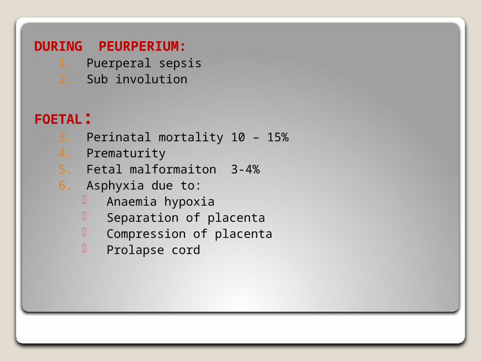
DURING PEURPERIUM:1. Puerperal sepsis2. Sub involution
FOETAL:3. Perinatal mortality 10 – 15%4. Prematurity5. Fetal malformaiton 3-4%6. Asphyxia due to:
Anaemia hypoxia Separation of placenta Compression of placenta Prolapse cord
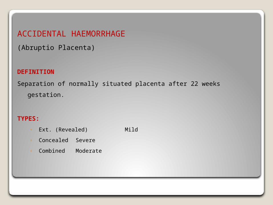
ACCIDENTAL HAEMORRHAGE
(Abruptio Placenta)
DEFINITION
Separation of normally situated placenta after 22 weeks gestation.
TYPES:
◦ Ext. (Revealed) Mild
◦ Concealed Severe
◦ Combined Moderate
INCIDENCE
1. About 1%
2. Severe form are seen more in primigravida
AE:
1. Toxaemia: 25 – 60%
◦ Spasm of vessel degenerage change.
◦ Rupture Hge
Toxaemia increased in patient with severe type
NON-TOXAEMIA: Trauma as ECV
Circumrellate placenta
Traction on cord
Sudden increased in IUP as R.M. in case of hydramnios
Vit. C, K, E, deficiency
Folate deficiency
Detachment of placenta after delivery of 1st twin.
PATHOLOGY:
All degree of placenta separation can occur from few millions to whole
placenta
Degeneration change in the decidual rupture haemorrhage in the decidual
basalis
A. 1. Decidual than splits
2. Leaving then layer adherent to myometrium
3. Decidual haematoma separation compression
B. Utero-placenta apoplexy (couviar’s ut)
C. Coagulation Defect
1. DIC low thromboplastin + fibrimolysin high FDP
2. Fibrinolysis
INVESTIGATION:
1. Urine analysis albumin PET
2. Fibrinogen
3. FDF Reveled
Clinical Picture
Symptoms:
4. Vaginal bleeding – dark, not severe
5. Tender abdomen
6. History of trauma
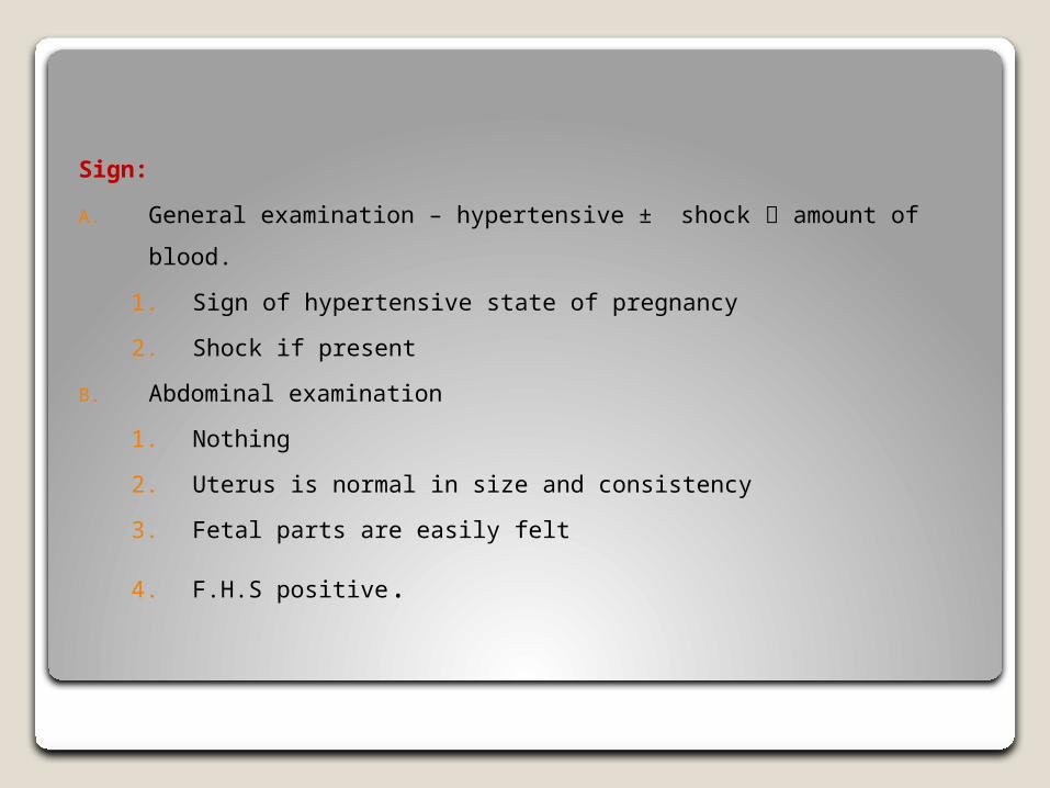
Sign:
A. General examination – hypertensive ± shock amount of blood.
1. Sign of hypertensive state of pregnancy
2. Shock if present
B. Abdominal examination
1. Nothing
2. Uterus is normal in size and consistency
3. Fetal parts are easily felt
4. F.H.S positive.
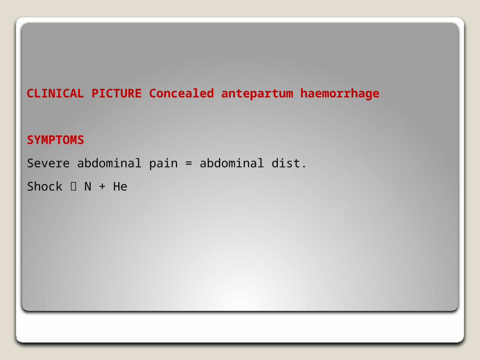
CLINICAL PICTURE Concealed antepartum haemorrhage
SYMPTOMS
Severe abdominal pain = abdominal dist.
Shock N + He
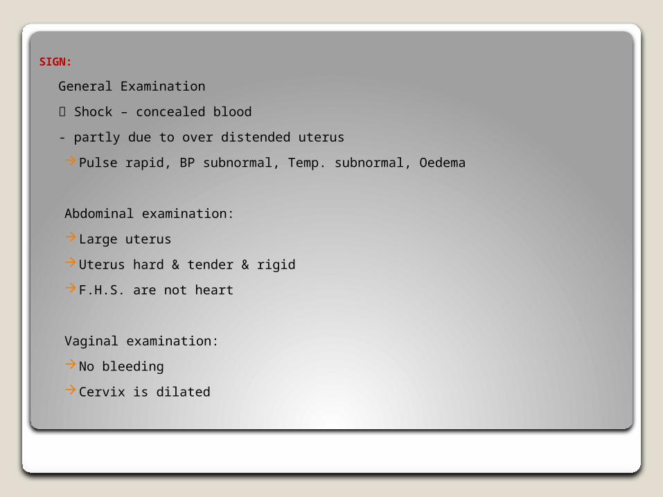
SIGN:
General Examination
Shock – concealed blood
- partly due to over distended uterus
Pulse rapid, BP subnormal, Temp. subnormal, Oedema
Abdominal examination:
Large uterus
Uterus hard & tender & rigid
F.H.S. are not heart
Vaginal examination:
No bleeding
Cervix is dilated
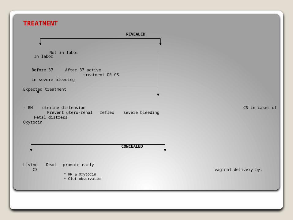
TREATMENT
REVEALED
Not in labor In labor
Before 37 After 37 active treatment OR CS in severe bleeding
Expected treatment
- RM uterine distension CS in cases of Prevent utero-renal reflex severe bleeding
Fetal distressOxytocin
CONCEALED
Living Dead – promote early CS vaginal delivery by:
* RM & Oxytocin* Clot observation
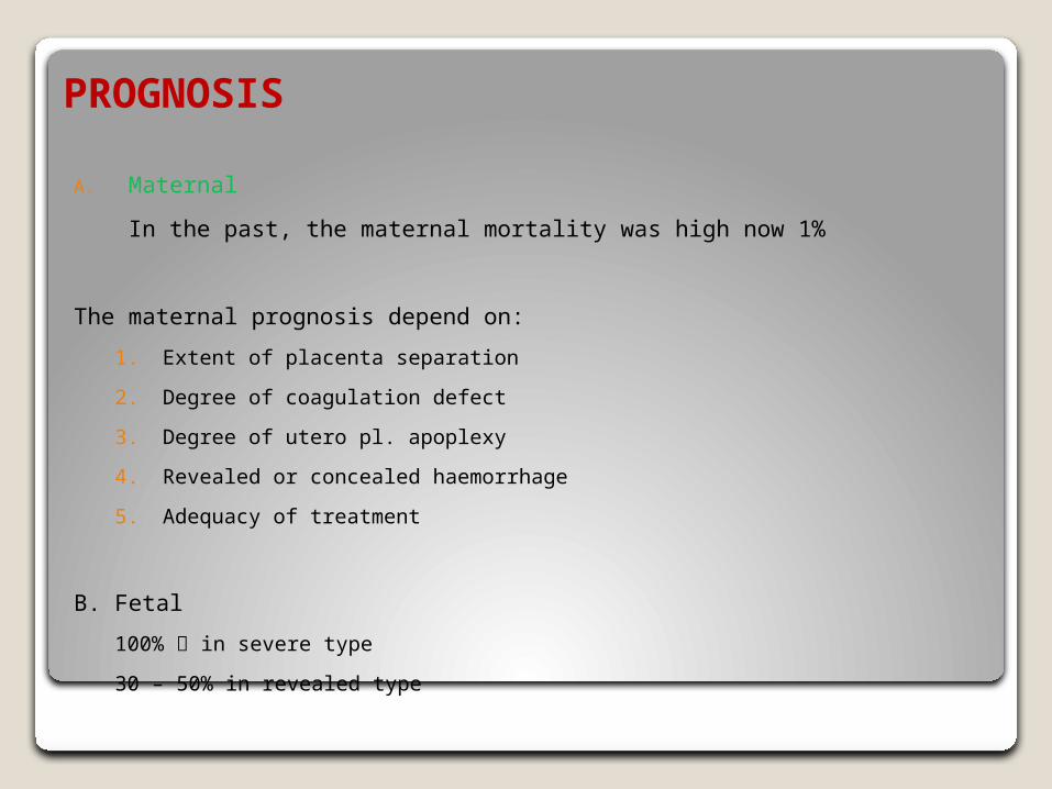
PROGNOSIS
A. Maternal
In the past, the maternal mortality was high now 1%
The maternal prognosis depend on:
1. Extent of placenta separation
2. Degree of coagulation defect
3. Degree of utero pl. apoplexy
4. Revealed or concealed haemorrhage
5. Adequacy of treatment
B. Fetal
100% in severe type
30 – 50% in revealed type

THANK YOU