Arteriographic Assessment Placental Vascularity Antepartum ... · of antepartum haemorrhage by...
3
6 April 1968 Arteriographic Assessment of Placental Vascularity after Antepartum Haemorrhage J. G. HILL,* M.B., F.R.C.S.GLASG., M.R.C.O.G.; F. J. BRUNTONt M.B., M.R.C.P., F.F.R. [WITH SPECIAL PLATE FACING PAGE 28] Brii. med. J., 1968, 2, 25-26 During the last decade there has been a gradual shift of emphasis in the management of antepartum haemorrhage. Placenta praevia, dangerous as it may be, presents fewer prob- lems of management since the adoption of conservative therapy and the increased use of caesarean section as advocated by Macafee (1945). Stallworthy (1951) declared that in the man- agement of placenta praevia our aim should be a maternal mortality of nil and a foetal mortality of less than 10%, and these figures have now been achieved in most modem depart- ments. However, in many cases of antepartum haemorrhage no cause is found for the bleeding, and the foetal wastage in this group of "unclassified haemorrhage" is still high. Different writers put the mortality in this group between 10 and 27%. The high mortality is presumably due to circulatory inadequacy of the placenta after separation of a portion of its surface. As bed rest is known to increase the uterine blood flow (Morris et al., 1956), there are grounds for detaining in hospital until delivery all patients who have had an antepartum haemorrhage irrespective of the situation of the placenta. Such a policy would be expected to increase the foetal salvage but has not been generally accepted, as it results in a large number of women being unnecessarily detained in hospital for many weeks. From the foetal mortality figures we can assume that in only a minority of cases is the placental circulation dangerously reduced. The problem is to recognize the cases at risk. If information could be obtained about the effective circula- tion of the placenta after the occurrence of haemorrhage it should not be difficult to pick out these cases-that is, those in which the placental vascularity is considerably diminished. Brink (1960) observed that arteriography is not only useful in locating the placenta but can also give an assessment of its vascularity. He suggested that it is possible to recognize a deficiency in sinusoids as an indication of inadequate placental circulation. Location of the placental site by arteriography was begun in Southampton in July 1965. In September of that year the following case was investigated and the outcome prompted us to think along the lines suggested by Brink. A primigravida aged 23 was admitted to hospital at 32 weeks' gestation after a slight, painless antepartum haemorrhage. Arterio- graphy was carried out and only two sinusoids were demonstrated. The small number of sinusoids was thought to be due to a technical fault, and as there was no radiographic evidence of placenta praevia the patient was allowed to go home. Two weeks later the foetal heart was heard but the uterus was noted to be small for dates. One week later, at the 35th week, she was admitted in labour and the foetal heart could not be heard. A macerated stillborn infant weighing 2 lb. 5 oz. (1,050 g.) was delivered and the placenta showed evidence of previous separation and old retroplacental haemorrhage. * Senior Registrar, Department of Obstetrics, Southampton General Hos- pital. Present address: Department of Obstetrics, St. George's Hospital, London S.W.1. t Consultant Radiologist, Southampton General HospitaL Present address: Department of Obstetrics, St. George's Hospital, London S.W.1. This case bore a remarkable similarity to the one described by Brink in which only a few sinusoids were shown and the foetus died in utero some weeks later. In both these cases it is assumed that the haemorrhage had separated so much of the placental surface that the circulation to the foetus was greatly reduced; with the continuation of the pregnancy this circula- tion became ultimately inadequate and the foetus died. It was decided, therefore, to continue investigating all case of antepartum haemorrhage by arteriography and to carry out a prospective survey of the placental vascularity. Material and Methods Fifty-eight patients were investigated by arteriography between September 1965 and February 1967. During this time the total number of deliveries in the area was 9,500. All patients with an antepartum haemorrhage before the 35th week, exclud- ing cases of abruptio placentae and premature labour, were examined by arteriography. The procedure was also carried out after the 35th week in four cases in which there was also a malpresentation. No other method of placentography was used. Arteriography was performed as early as the 27th week of pregnancy in three patients, and the average for the series was 311 weeks. The examination was not carried out until bleeding had ceased for at least four days. Patients diagnosed radiographically as having a placenta praevia were detained in hospital and managed in the usual manner. All the other patients were allowed- home and were not readmitted unless further obstetric complications arose. It was decided not to induce labour routinely at the 38th week in these cases as the significance of the radiographic assessment of the placental vascularity was not known. The pregnancy was allowed to continue until 42 weeks unless there was some other obstetric complication. In seven patients labour was induced before term. In three of these there was an associated pre- eclampsia, and in four there was further haemorrhage. Technique The procedure is carried out under sedation and local anaes- thesia. The catheterization is by the standard Seldinger method and is similar to that advocated by Sutton (1966). The Seldinger-Sutton needle, the fine guide wire (both size P.E. 160), but the larger polyethylene catheter (size P.E. 240) are used. The catheter, which has a tapered tip and at least two side holes, is introduced about 25 cm. so that it reaches just above the aortic bifurcation. The contrast used is Conray 420 (sodium isothalamate), 25 ml., and is injected forcibly by hand. A single film is taken two and a half seconds after the comple- tion of the injection. There is some variation of the flow rate, and occasionally the film has had to be repeated, usually after a longer time interval. A position S degrees from the true lateral has been found to be best for assessing the position of BRrnS MEICAL JOUML 25 on 11 May 2020 by guest. Protected by copyright. http://www.bmj.com/ Br Med J: first published as 10.1136/bmj.2.5596.25 on 6 April 1968. Downloaded from
Transcript of Arteriographic Assessment Placental Vascularity Antepartum ... · of antepartum haemorrhage by...

6 April 1968
Arteriographic Assessment of Placental Vascularity afterAntepartum Haemorrhage
J. G. HILL,* M.B., F.R.C.S.GLASG., M.R.C.O.G.; F. J. BRUNTONt M.B., M.R.C.P., F.F.R.
[WITH SPECIAL PLATE FACING PAGE 28]
Brii. med. J., 1968, 2, 25-26
During the last decade there has been a gradual shift ofemphasis in the management of antepartum haemorrhage.Placenta praevia, dangerous as it may be, presents fewer prob-lems of management since the adoption of conservative therapyand the increased use of caesarean section as advocated byMacafee (1945). Stallworthy (1951) declared that in the man-agement of placenta praevia our aim should be a maternalmortality of nil and a foetal mortality of less than 10%, andthese figures have now been achieved in most modem depart-ments.
However, in many cases of antepartum haemorrhage nocause is found for the bleeding, and the foetal wastage in thisgroup of "unclassified haemorrhage" is still high. Differentwriters put the mortality in this group between 10 and 27%.The high mortality is presumably due to circulatory inadequacyof the placenta after separation of a portion of its surface. Asbed rest is known to increase the uterine blood flow (Morriset al., 1956), there are grounds for detaining in hospital untildelivery all patients who have had an antepartum haemorrhageirrespective of the situation of the placenta. Such a policywould be expected to increase the foetal salvage but has notbeen generally accepted, as it results in a large number of womenbeing unnecessarily detained in hospital for many weeks. Fromthe foetal mortality figures we can assume that in only aminority of cases is the placental circulation dangerouslyreduced. The problem is to recognize the cases at risk.
If information could be obtained about the effective circula-tion of the placenta after the occurrence of haemorrhage itshould not be difficult to pick out these cases-that is, those inwhich the placental vascularity is considerably diminished.Brink (1960) observed that arteriography is not only useful inlocating the placenta but can also give an assessment of itsvascularity. He suggested that it is possible to recognize a
deficiency in sinusoids as an indication of inadequate placentalcirculation.
Location of the placental site by arteriography was begun inSouthampton in July 1965. In September of that year thefollowing case was investigated and the outcome prompted usto think along the lines suggested by Brink.A primigravida aged 23 was admitted to hospital at 32 weeks'
gestation after a slight, painless antepartum haemorrhage. Arterio-graphy was carried out and only two sinusoids were demonstrated.The small number of sinusoids was thought to be due to a technicalfault, and as there was no radiographic evidence of placenta praeviathe patient was allowed to go home. Two weeks later the foetalheart was heard but the uterus was noted to be small for dates.One week later, at the 35th week, she was admitted in labour andthe foetal heart could not be heard. A macerated stillborn infantweighing 2 lb. 5 oz. (1,050 g.) was delivered and the placentashowed evidence of previous separation and old retroplacentalhaemorrhage.
* Senior Registrar, Department of Obstetrics, Southampton General Hos-pital. Present address: Department of Obstetrics, St. George'sHospital, London S.W.1.
t Consultant Radiologist, Southampton General HospitaL Presentaddress: Department of Obstetrics, St. George's Hospital, LondonS.W.1.
This case bore a remarkable similarity to the one describedby Brink in which only a few sinusoids were shown and thefoetus died in utero some weeks later. In both these cases it isassumed that the haemorrhage had separated so much of theplacental surface that the circulation to the foetus was greatlyreduced; with the continuation of the pregnancy this circula-tion became ultimately inadequate and the foetus died.
It was decided, therefore, to continue investigating all caseof antepartum haemorrhage by arteriography and to carry outa prospective survey of the placental vascularity.
Material and Methods
Fifty-eight patients were investigated by arteriographybetween September 1965 and February 1967. During this timethe total number of deliveries in the area was 9,500. All patientswith an antepartum haemorrhage before the 35th week, exclud-ing cases of abruptio placentae and premature labour, wereexamined by arteriography. The procedure was also carried out
after the 35th week in four cases in which there was also a
malpresentation. No other method of placentography was used.Arteriography was performed as early as the 27th week ofpregnancy in three patients, and the average for the series was
311 weeks. The examination was not carried out until bleedinghad ceased for at least four days.
Patients diagnosed radiographically as having a placentapraevia were detained in hospital and managed in the usualmanner.
All the other patients were allowed- home and were notreadmitted unless further obstetric complications arose. It wasdecided not to induce labour routinely at the 38th week in thesecases as the significance of the radiographic assessment of theplacental vascularity was not known. The pregnancy wasallowed to continue until 42 weeks unless there was some otherobstetric complication. In seven patients labour was inducedbefore term. In three of these there was an associated pre-eclampsia, and in four there was further haemorrhage.
Technique
The procedure is carried out under sedation and local anaes-thesia. The catheterization is by the standard Seldinger methodand is similar to that advocated by Sutton (1966). TheSeldinger-Sutton needle, the fine guide wire (both size P.E. 160),but the larger polyethylene catheter (size P.E. 240) are used.The catheter, which has a tapered tip and at least two sideholes, is introduced about 25 cm. so that it reaches just abovethe aortic bifurcation. The contrast used is Conray 420(sodium isothalamate), 25 ml., and is injected forcibly by hand.A single film is taken two and a half seconds after the comple-tion of the injection. There is some variation of the flow rate,and occasionally the film has had to be repeated, usually aftera longer time interval. A position S degrees from the truelateral has been found to be best for assessing the position of
BRrnSMEICAL JOUML 25
on 11 May 2020 by guest. P
rotected by copyright.http://w
ww
.bmj.com
/B
r Med J: first published as 10.1136/bm
j.2.5596.25 on 6 April 1968. D
ownloaded from
26 6 April 1968 Placentography-Hill and Brunton NftDICAL JOU
the placenta (Special Plate, Fig. 1). Earlier in our investigationsome patients were examined in the anteroposterior and/oroblique position.
Cervical Marker.-With a Sims speculum a 50-mm. contra-ceptive cap is gently placed over the cervix. The metal springin the rim is thus in close proximity to the internal os (SpecialPlate, Fig. 1). We consider this form of marking superior toother methods and of great help in placental location.Complications.-The patients require reassurance, but tolerate
the examination extremely well. We have encountered noserious complications. After an uneventful placentogram, onepatient complained of a painful area on the outer aspect of thehip some days later, which necessitated readmission to hospital.The area was well away from the skin puncture and hersymptoms gradually regressed over two to three weeks.Film Interpretation.-If the lowermost sinus was within
10 cm. of the cervical marker the placenta was considered to bepraevia and these patients were kept in hospital. We have beenparticularly interested in the films showing the placentae in theupper segment, and in these we have studied the sinusoids care-fully. No attempt was made to grade the degree of filling, butfrom the appearance of the sinusoids the vascularity was con-sidered to be either adequate or deficient. Exact enumeration wasfound to be impracticable owing to the frequency with whichthe sinusoids appeared to be confluent. In practice little diffi-culty was experienced in deciding whether the filling of thesinusoids was adequate or not. A technical problem in assessingplacental vascularity has been the individual variation in circu-lation time. For this reason a second film has sometimes beenrequired, but with increasing experience this has become lessfrequent.
Results
From a total of 58 patients in the series there were 69 births.There were two stillbirths and no neonatal deaths, giving aperinatal mortality of 3.3 %. There were no maternal deaths.Of the 58 cases investigated the placenta was found to
encroach on the lower segment in 10. This study concerns theremaining 48. Forty-four of these patients had a well-vascularized placenta, the foetal mortality being nil. Theremaining four were thought to have a poorly vascularizedplacenta, and in this group there was one foetal death.A gravida-2 aged 27 had slight painless antepartum haemorrhage
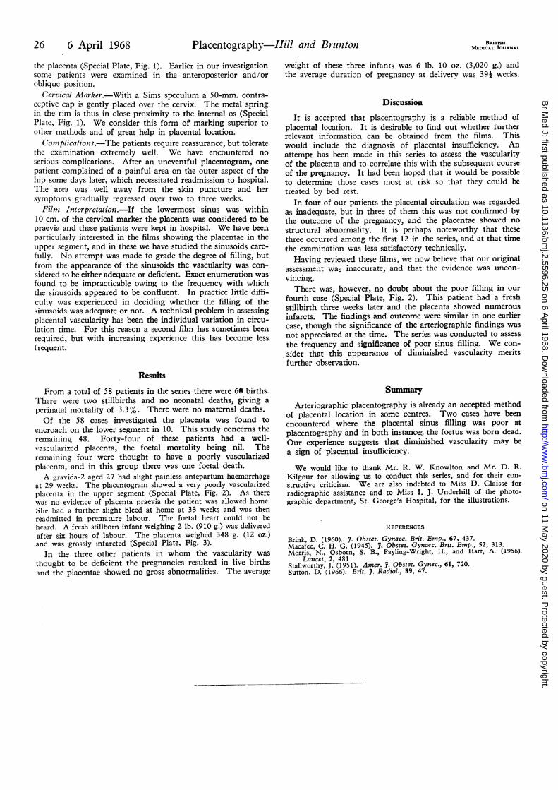
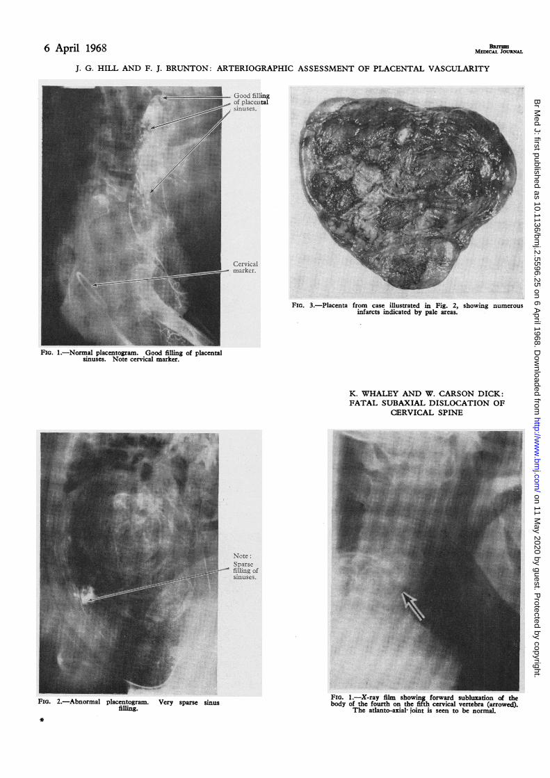
at 29 weeks. The placentogram showed a very poorly vascularizedplacenta in the upper segment (Special Plate, Fig. 2). As therewas no evidence of placenta praevia the patient was allowed home.She had a further slight bleed at home at 33 weeks and was thenreadmitted in premature labour. The foetal heart could not beheard. A fresh stillborn infant weighing 2 lb. (910 g.) was deliveredafter six hours of labour. The placenta weighed 348 g. (12 oz.)and was grossly infarcted (Special Plate, Fig. 3).
In the three other patients in whom the vascularity wasthought to be deficient the pregnancies resulted in live birthsand the placentae showed no gross abnormalities. The average
weight of these three infants was 6 lb. 10 oz. (3,020 g.) andthe average duration of pregnancy at delivery was 39i weeks.
Discussion
It is accepted that placentography is a reliable method ofplacental location. It is desirable to find out whether furtherrelevant information can be obtained from the films. Thiswould include the diagnosis of placental insufficiency. Anattempt has been made in this series to assess the vascularityof the placenta and to correlate this with the subsequent courseof the pregnancy. It had been hoped that it would be possibleto determine those cases most at risk so that they could betreated by bed rest.
In four of our patients the placental circulation was regardedas inadequate, but in three of them this was not confirmed bythe outcome of the pregnancy, and the placentae showed nostructural abnormality. It is perhaps noteworthy that thesethree occurred among the first 12 in the series, and at that timethe examination was less satisfactory technically.Having reviewed these films, we now believe that our original
assessment was inaccurate, and that the evidence was uncon-vincing.There was, however, no doubt about the poor filling in our
fourth case (Special Plate, Fig. 2). This patient had a freshstillbirth three weeks later and the placenta showed numerousinfarcts. The findings and outcome were similar in one earliercase, though the significance of the arteriographic findings wasnot appreciated at the time. The series was conducted to assessthe frequency and significance of poor sinus filling. We con-sider that this appearance of diminished vascularity meritsfurther observation.
Summary
Arteriographic placentography is already an accepted methodof placental location in some centres. Two cases have beenencountered where the placental sinus filling was poor atplacentography and in both instances the foetus was born dead.Our experience suggests that diminished vascularity may bea sign of placental insufficiency.
We would like to thank Mr. R. W. Knowlton and Mr. D. R.Kilgour for allowing us to conduct this series, and for their con-structive criticism. We are also indebted to Miss D. Claisse forradiographic assistance and to Miss I. J. Underhill of the photo-graphic department, St. George's Hospital, for the illustrations.
REFERENCES
Brink, D. (1960). 7. Obstet. Gynaec. Brit. Emp., 67, 437.Macafee, C. H. G. (1945). 7. Obstet. Gynaec. Brit. Emp., 52, 313.Morris, N., Osborn, S. B., Payling-Wright, H., and Hart, A. (1956).
Lancet, 2, 481Stallworthy, J. (1951). Amer. 7. Obstet. Gynec., 61, 720.Sutton, D. (1966). Brit. 7. Radiol., 39, 47.
on 11 May 2020 by guest. P
rotected by copyright.http://w
ww
.bmj.com
/B
r Med J: first published as 10.1136/bm
j.2.5596.25 on 6 April 1968. D
ownloaded from
6 April 1968 BRITIMIMEDICAL JOURNAL
J. G. HILL AND F. J. BRUNTON: ARTERIOGRAPHIC ASSESSMENT OF PLACENTAL VASCULARITY
________________ Good fillingof placentalsinuses.
.. .x~... .....
B.
:::.>..F
Cervical
ark~~~~~~~~mrer.
FIG. 1.-Normal placentogram. Good filling of placentalsinuses. Note cervical marker.
FIG. 3.-Placenta from case illustrated in Fig. 2, showing numerousinfarcts indicated by pale areas.
Note:Sparsefilling ofsinuses.
FIG. 2.-Abnormal placentogram. Very sparse sinusfilling.
*
K. WHALEY AND W. CARSON DICK:FATAL SUBAXIAL DISLOCATION OF
CERVICAL SPINE
FIG. 1.-X-ray film showing forward subluxation of thebody of the fourth on the fifth cervical vertebra (arrowed).
The atlanto-axial joint is seen to be normal.
on 11 May 2020 by guest. P
rotected by copyright.http://w
ww
.bmj.com
/B
r Med J: first published as 10.1136/bm
j.2.5596.25 on 6 April 1968. D
ownloaded from