Anatomy of facial nerve - Government Medical College … lectures/ENT/Anatomy of facial nerve...
37
Anatomy of facial nerve
Transcript of Anatomy of facial nerve - Government Medical College … lectures/ENT/Anatomy of facial nerve...

Anatomy of facial nerve

Embryology of the facial nerve
Weeks 0-4- 3rd wk : facioacoustic (acousticofacial) primordium - 4th wk : chorda tympani nerve exits rostrally and courses ventrally to the first pharyngeal pouch to enter the mandibular arch
Weeks 5-6- The greater superficial petrosal nerve (GSPN) is
appreciable- The chorda tympani nerve enters the mandibular arch
and terminates just proximal to the submandibular ganglion, near a branch of the trigeminal nerve
2

Week 7 :-The chorda tympani and lingual nerve unite proximal to the submandibular gland-The parotid gland begins to develop . The temporal, zygomatic, and upper buccal branches are superficial to the parotid primordium
Week 8 :-Beginning of the fallopian canal
Weeks 10-15 :-The vertical portion of the facial nerve begins in the middle ear, and its overall relationship to external and middle ear structures is far more anterior than in the adult.
3
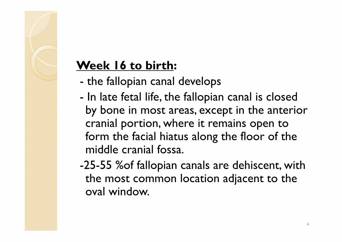
Week 16 to birth:- the fallopian canal develops- In late fetal life, the fallopian canal is closed by bone in most areas, except in the anterior cranial portion, where it remains open to form the facial hiatus along the floor of the middle cranial fossa.
-25-55 %of fallopian canals are dehiscent, with the most common location adjacent to the oval window.
4

The Anatomy Of Facial Nerve
Broadly divided into 3 partsIntracranial PortionIntratemporal PortionExtratemporal Portion
5


2 roots:◦ Motor root :moderate in size◦ Sensory root (Nervus intermedius of wrisberg)
- Very slender & lies posterior to motor root.
7

Facial nerve: sensory root
Special Visceral Efferent/Branchial Motor General Visceral Efferent/Parasympathetic General Sensory afferent Special Visceral Afferent/Taste
8

Special sensory afferent
•Origin : Unipolar neurons in geniculate ganglia
•Central : Nucleus of Tractus
Solitarius •Peripheral : Chorda
tympani nv & lingual nv to ant 2/3rd
tongue
9

General sensory afferent
Deep sensibility of face
10
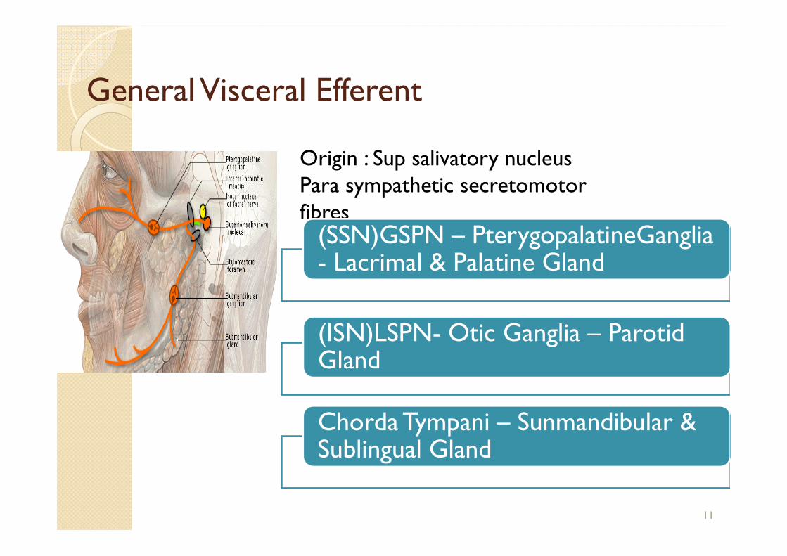
General Visceral Efferent
Origin : Sup salivatory nucleusPara sympathetic secretomotor fibres
(SSN)GSPN – PterygopalatineGanglia- Lacrimal & Palatine Gland
(ISN)LSPN- Otic Ganglia – Parotid Gland
Chorda Tympani – Sunmandibular & Sublingual Gland
11

Segment Location Length mm
Supranuclear Cerebral cortex Brain stem Brain stem to IAC 24
Meatal segment
IAM to fundus 5-12
Labyrinthine segment
Fundus of IAC to first genu
3-4
Tympanic segment
Geniculate ganglion to pyramidal eminence
8-11
Mastoid segment
Pyramidal process to stylomastoid foramen
10-14
Extratemporal segment
Stylomastoid foramen to pes anserinus
15-20
12


Intracranial Portion Of Facial nerve
14
•Facial nv & nerve of intermedius lie above & slightly ant to vestibulocochlear nv•Distance between exit & entrance in IAC :23-24 mm

15

Intratemporal portion of Facial nerve
fallopian canal (after Gabriel Fallopius).
Divided into 4 segments:◦ Meatal◦ Labyrinthine◦ Tympanic, horizontal◦ Mastoid, vertical
16

Meatal Segment (5-12 mm)
0.68mm
17


labyrinthine segment: Size -3-5 mm , 0.68 mm Lies beneath the middle cranial
fossa
Direction
Meninges
20

labyrinthine segment:
- meningeal cover
- narrow constriction (0.68mm)
- 132 deg bend
Slight constriction from vertical crest , thick periosteum
Only segment of the facial nerve that lacks anastomosing arterial cascades : embolic phenomena, low-flow states, or vascular compression
21

Geniculate ganglion:
Forming a acute angle of variable degree but usually not less than 75˚.
1st genu Cog The geniculate ganglion
is formed by the junction of the nervus intermedius and the facial nerve into a common trunk
22

Tympanic or horizontal segment(8-11 mm) :
Geniculate ganglion to the 2nd
genu Course : inclined inferiorly
forming a angle of <10˚
Dehiscent fallopian canal in 25-
55% of postmortem specimens.
oval window
23


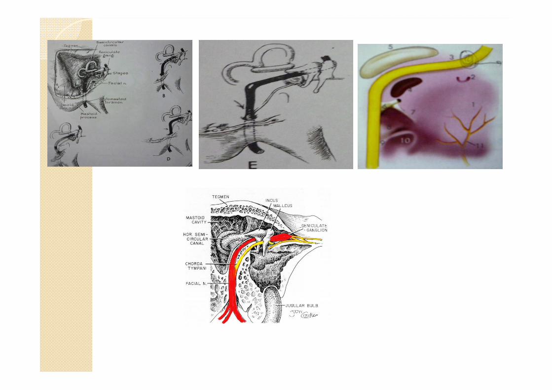


Mastoid (Vertical) segment of facial canal
15-20 mm Course Angulation 3 branches
28

Chorda tympani nerve Terminal branch of the
nervus intermedius Course
29

Exits the fallopian canal via the stylomastoid foramen. Stylomastoid foramen opens at base of petrosa between the
mastoid process and styloid. Once it exit the fallopian canal at the stylomastoid foramen, it
gives off several rami before it divides into its main branches
30

Branches of Facial Nerve1. Ansa of Haller (inconstant)2. Posterior auricular branch3. Stylohyoid branch4. Posterior belly of digastric branch5. Pes anserinus
Extratemporal Facial Nerve
31

Extratemporal Facial Nerve Runs anteriorly in
the substance of parotid gland, crosses the ECA & divides at the posterior border of ramus of mandible into 2 primary branches:
32
Sup : TemporozygomaticInf : Cervicofacial

After the main point of division, 5 major branches of the facial nerve exist:
Temporal (i.e., frontal), zygomatic, buccal, marginal mandibular, and cervical.
33

Tragal pointer Styloid process Posterior belly of digastric Peripheral branches Stylomastoid foramen Tympanomastoid suture Vaginomastoid angle Post auricular muscle branch
Surgical landmarks for the extratemporal facial nerve
34

Frontal branch Ramus mandibularis
(post facial vein ) Buccal branch
35

Vascular supply of the facial nerve
The cortical motor area : Rolandic branch
Pons : anterior inferior cerebellar artery (AICA)
Superficial petrosal artery
Posterior auricular artery
36

Thank U