Clinical anatomy of facial nerve and facial nerve palsy
38
Dr.Ramesh Parajuli,MS Chitwan Medical College Teaching Hospital,Bharatpur- 10,Chitwan, Nepal
-
Upload
ramesh-parajuli -
Category
Health & Medicine
-
view
104 -
download
3
Transcript of Clinical anatomy of facial nerve and facial nerve palsy
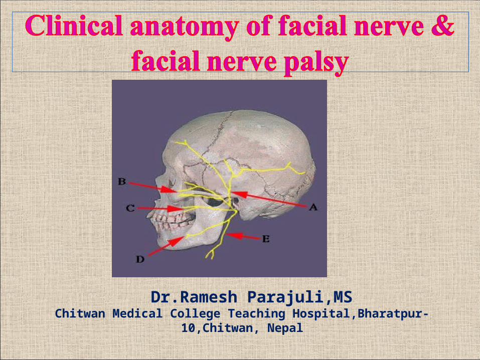
Dr.Ramesh Parajuli,MSChitwan Medical College Teaching Hospital,Bharatpur-10,Chitwan, Nepal

Anatomy of Facial NerveMixed nerve
1. Motor: supply to the facial muscles
2. Sensory: Sensory root (Nervus intermedius/Wrisberg) joins motor
root at fundus of Internal Auditory Canal(IAC)
-part of post-aural / concha / ext. auditory canal,supratonsillar
fossa
3. Secretomotor: lacrimal, submandibular, sublingual
4.Taste: anterior 2/3rd of tongue (via chorda tympani nerve)
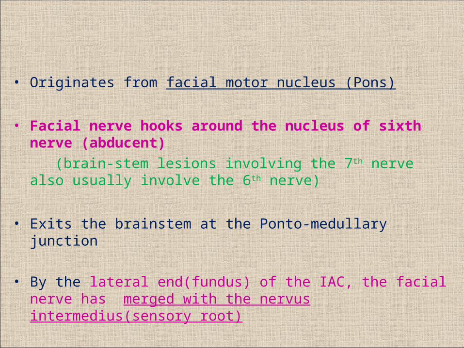
• Originates from facial motor nucleus (Pons)
• Facial nerve hooks around the nucleus of sixth nerve (abducent) (brain-stem lesions involving the 7th nerve also usually involve the 6th
nerve)
• Exits the brainstem at the Ponto-medullary junction
• By the lateral end(fundus) of the IAC, the facial nerve has merged with the nervus intermedius(sensory root)

Course of facial nerve


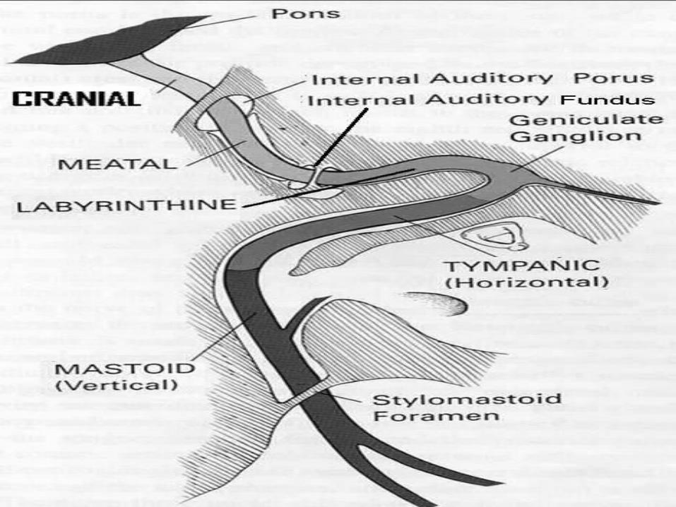
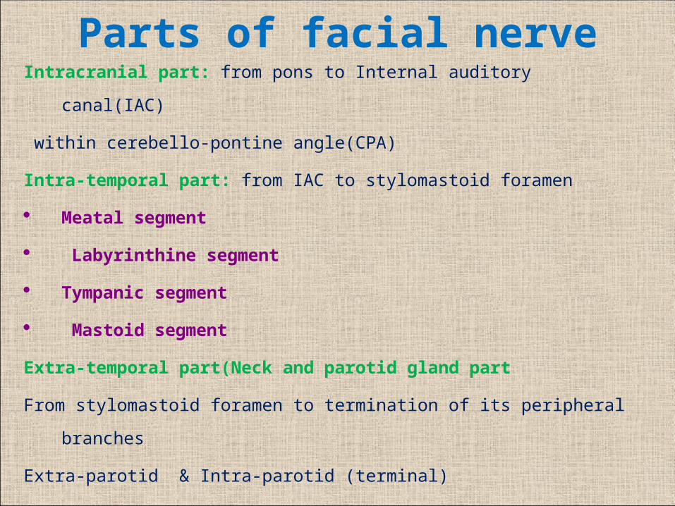
Parts of facial nerveIntracranial part: from pons to Internal auditory canal(IAC)
within cerebello-pontine angle(CPA)
Intra-temporal part: from IAC to stylomastoid foramen
Meatal segment
Labyrinthine segment
Tympanic segment
Mastoid segment
Extra-temporal part(Neck and parotid gland part
From stylomastoid foramen to termination of its peripheral branches
Extra-parotid & Intra-parotid (terminal)
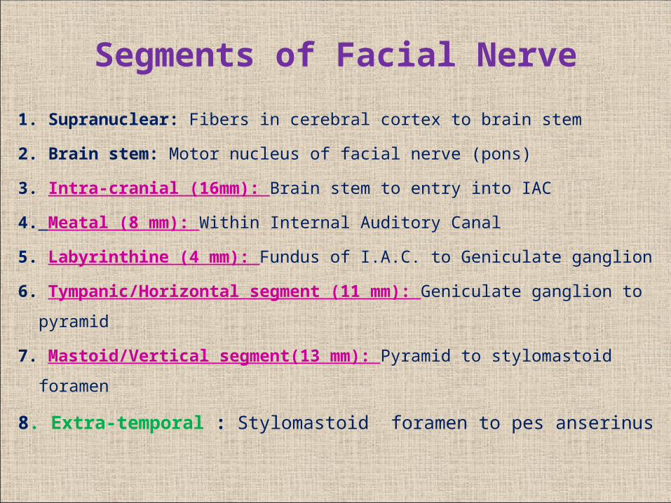
1. Supranuclear: Fibers in cerebral cortex to brain stem
2. Brain stem: Motor nucleus of facial nerve (pons)
3. Intra-cranial (16mm): Brain stem to entry into IAC
4. Meatal (8 mm): Within Internal Auditory Canal
5. Labyrinthine (4 mm): Fundus of I.A.C. to Geniculate ganglion
6. Tympanic/Horizontal segment (11 mm): Geniculate ganglion to pyramid
7. Mastoid/Vertical segment(13 mm): Pyramid to stylomastoid foramen
8. Extra-temporal : Stylomastoid foramen to pes anserinus
Segments of Facial Nerve
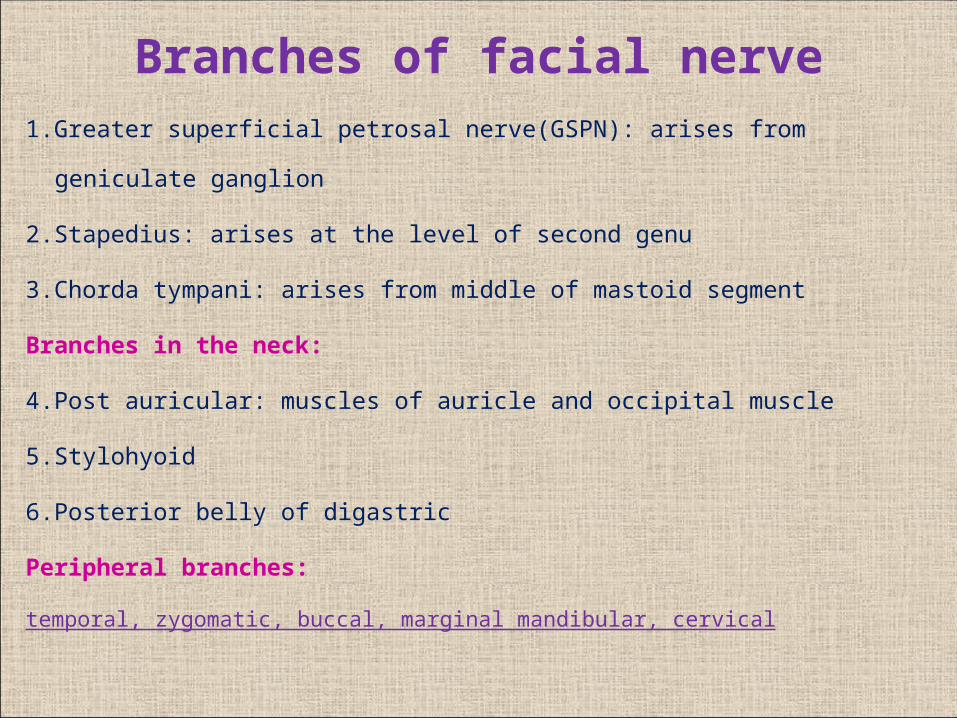
Branches of facial nerve
1.Greater superficial petrosal nerve(GSPN): arises from geniculate ganglion
2.Stapedius: arises at the level of second genu
3.Chorda tympani: arises from middle of mastoid segment
Branches in the neck:
4.Post auricular: muscles of auricle and occipital muscle
5.Stylohyoid
6.Posterior belly of digastric
Peripheral branches:
temporal, zygomatic, buccal, marginal mandibular, cervical

Intra-tympanic branches

Extra-tympanic branches
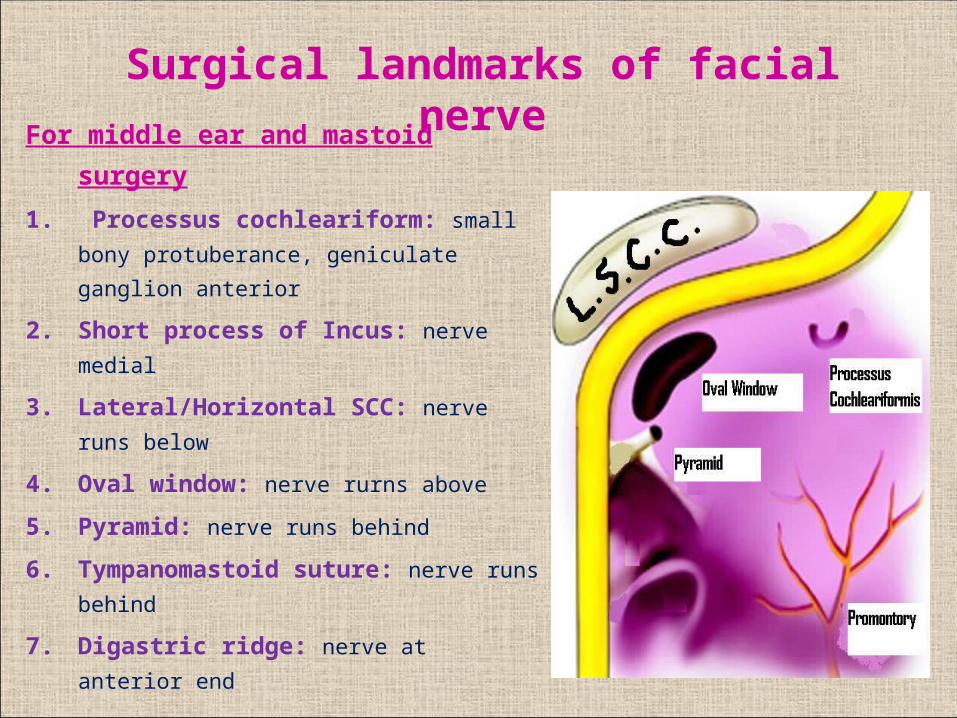
For middle ear and mastoid surgery
1. Processus cochleariform: small bony
protuberance, geniculate ganglion
anterior
2. Short process of Incus: nerve medial
3. Lateral/Horizontal SCC: nerve runs
below
4. Oval window: nerve rurns above
5. Pyramid: nerve runs behind
6. Tympanomastoid suture: nerve runs
behind
7. Digastric ridge: nerve at anterior end
Surgical landmarks of facial nerve

For parotid surgery:
1. Tympano-mastoid suture: 6-8 mm deep to this suture
2. Groove between mastoid & bony EAC: bisected by facial nerve
3. Tragal pointer: 1 cm antero-infero-medial is facial nerve
4. Styloid process: lateral lies facial nerve
5. Posterior belly of digastric: superior & parallel lies facial nerve

Lesions of Facial Nerve


1. Idiopathic: Bell’s palsy
Melkersson Rosenthal syndrome
2. Temporal bone trauma: Road traffic accident
3. Infection: C.S.O.M., Herpes Zoster oticus
Malignant otitis externa
4. Neoplasm: Parotid tumors, Acoustic Neuroma,
Glomus tumors, Malignancy of ear
5. Congenital: Moebius syndrome
6. Iatrogenic: Mastoidectomy, Parotid surgery
7. Metabolic: Diabetes mellitus, Hypertension
Etiology of Facial Nerve Palsy
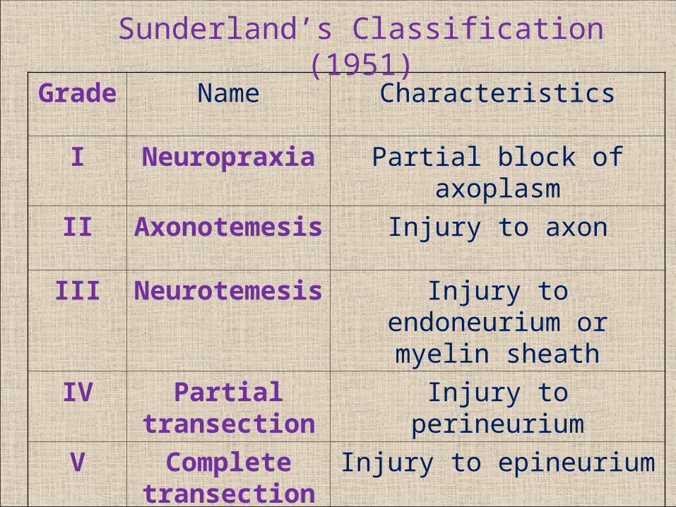
Grade Name Characteristics
I Neuropraxia Partial block of axoplasm
II Axonotemesis Injury to axon
III Neurotemesis Injury to endoneurium or myelin sheath
IV Partial transection
Injury to perineurium
V Complete transection
Injury to epineurium
Sunderland’s Classification (1951)

Grade Description Characteristics
I Normal Normal facial function
II Mild dysfunction Slight weakness seen only on close inspection
III Moderate dysfunction Obvious asymmetry; complete eye closure
IV Moderately severe dysfunction
Obvious asymmetry; incomplete eye closure
V Severe dysfunction Only minimal motion seen; asymmetry at rest
VI Total paralysis No movement
House Brackmann Classification (post-injury)

Diagnosis
• Topo-diagnostic Tests
• Electrical Tests
• Magnetic stimulation of intra-cranial facial nerve
• CT scan temporal bone: for progressive palsy
• MRI brain
• Surgical exploration

Topo-diagnostic tests
• Audiometry: cochlear nerve function
• Vestibulometry: vestibular function
• Schirmer’s test: Greater Superficial Petrosal Nerve
• Stapedial reflex test: Nerve to stapedius
• Electrogustometry: Chorda tympani
• Submandibular salivary flow: Chorda tympani
• Examination for terminal facial nerve branches
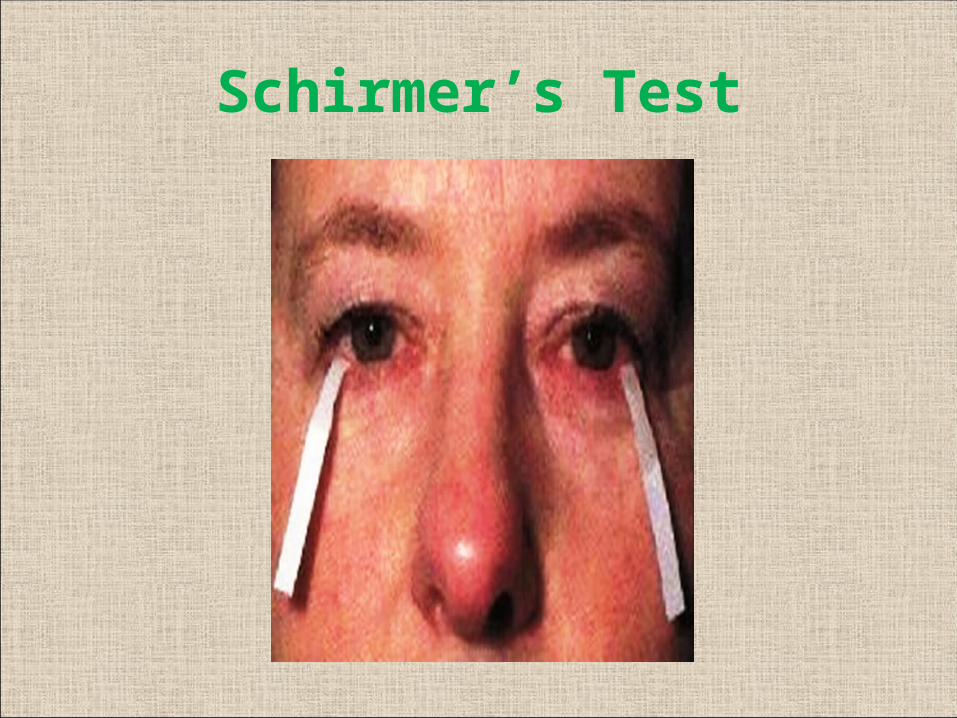
Schirmer’s Test

Stapedial Reflex

Electrogustometry Measures minimum amount of current required to
excite sensation of taste

Muscles supplied by terminal branches

Temporal branch
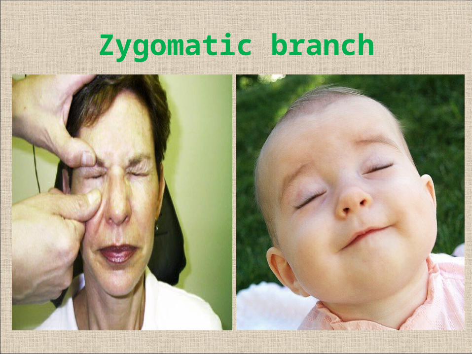
Zygomatic branch
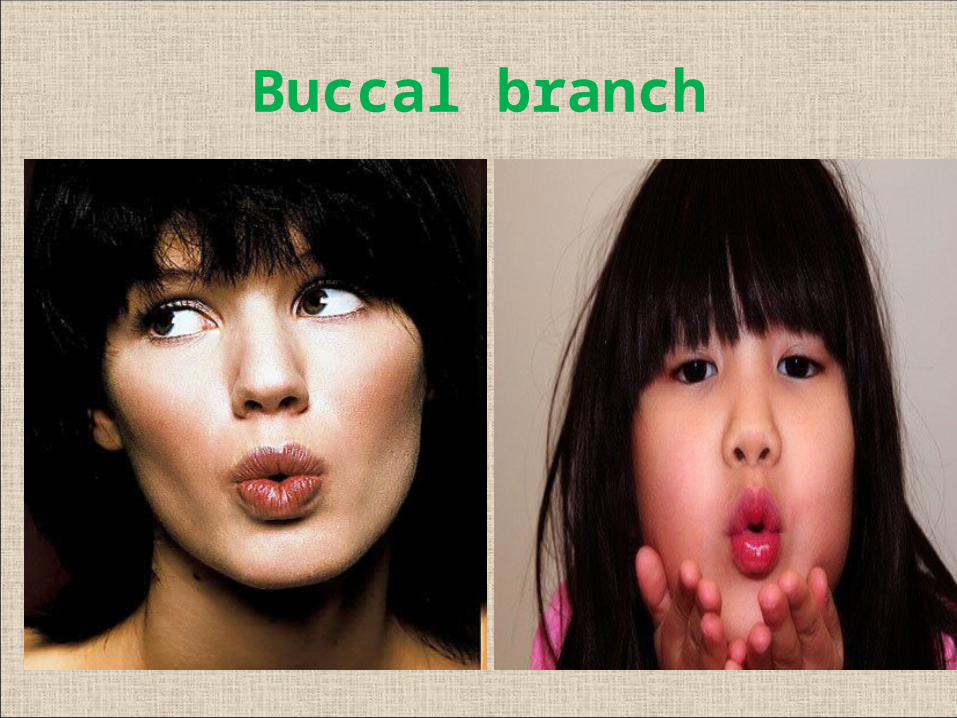
Buccal branch

Marginal mandibular
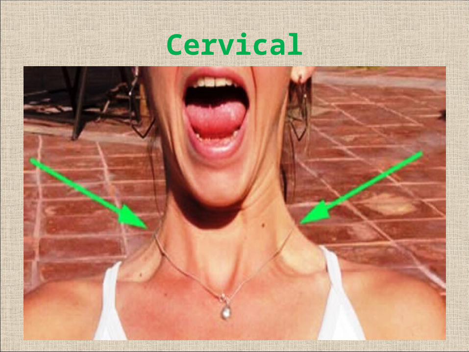
Cervical

• Nerve Excitability Test
• Maximal stimulation test
• Electro-neuronography
Electrical tests

Electromyography Responses
Polyphasic potentials Fibrillation potentials
Re-innervation of muscles Denervation of muscles

Bell’s Palsy
• Acute onset, idiopathic, unilateral, self-limiting, non-
progressive, peripheral facial nerve palsy
• 85% start recovering within 3 weeks
• Etiology:
1. Viral: Herpes simplex, Herpes Zoster
2. Ischemia of facial nerve: exposure to cold,
emotional stress, nerve compression
3. Hereditary 4. Autoimmune
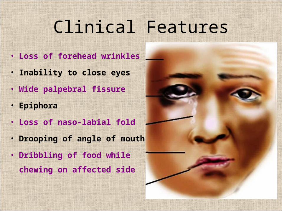
Clinical Features
• Loss of forehead wrinkles
• Inability to close eyes
• Wide palpebral fissure
• Epiphora
• Loss of naso-labial fold
• Drooping of angle of mouth
• Dribbling of food while chewing
on affected side
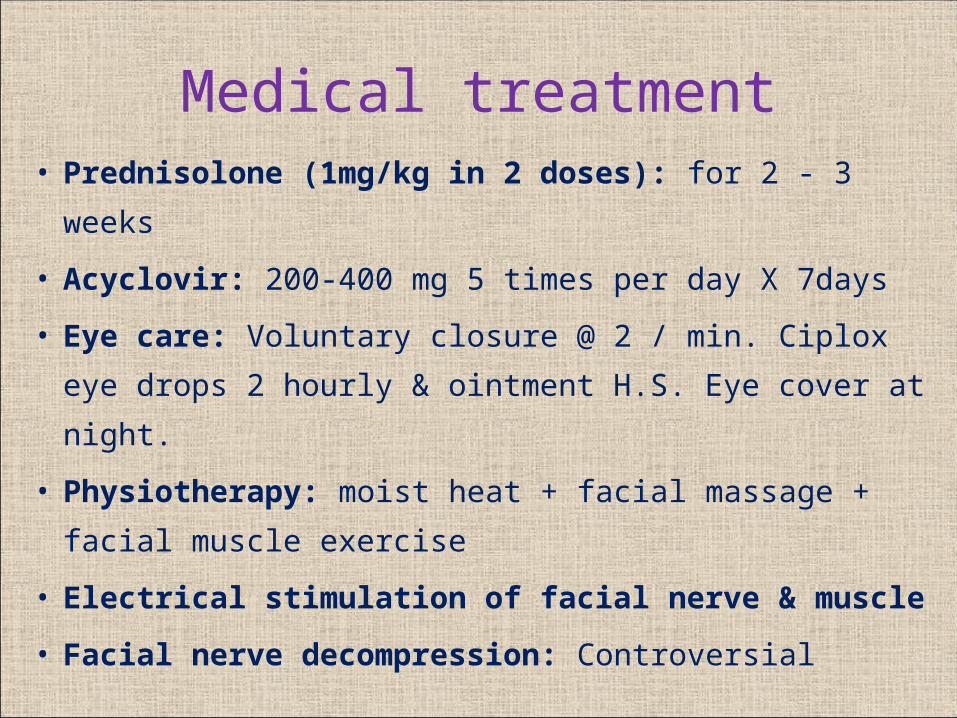
Medical treatment• Prednisolone (1mg/kg in 2 doses): for 2 - 3 weeks
• Acyclovir: 200-400 mg 5 times per day X 7days
• Eye care: Voluntary closure @ 2 / min. Ciplox eye drops 2
hourly & ointment H.S. Eye cover at night.
• Physiotherapy: moist heat + facial massage + facial
muscle exercise
• Electrical stimulation of facial nerve & muscle
• Facial nerve decompression: Controversial

Surgical Treatment for Facial Nerve Injury
A. Facial nerve decompression: till meatal foramen
B. Neurorrhaphy (Nerve repair)
1. Direct end to end anastomosis
2. Interposition Cable grafting: sural, greater auricular
C. Nerve Transposition: hypoglossal-facial
D. Muscle Transposition: temporalis, masseter
E. Micro-neuro-vascular muscle flaps
F. Static Procedures: eyelid implant, fascial sling

Thank you