Anatomía senos paranasales
30
Anatomía senos paranasales
-
Upload
fiorellalima -
Category
Health & Medicine
-
view
100 -
download
1
Transcript of Anatomía senos paranasales
Anatomía senos paranasales

Senos paranasales
Sociedad francesa de rinología

Senos paranasales
Levine-Sinus Surgery Endoscopic and Microscopic Approaches

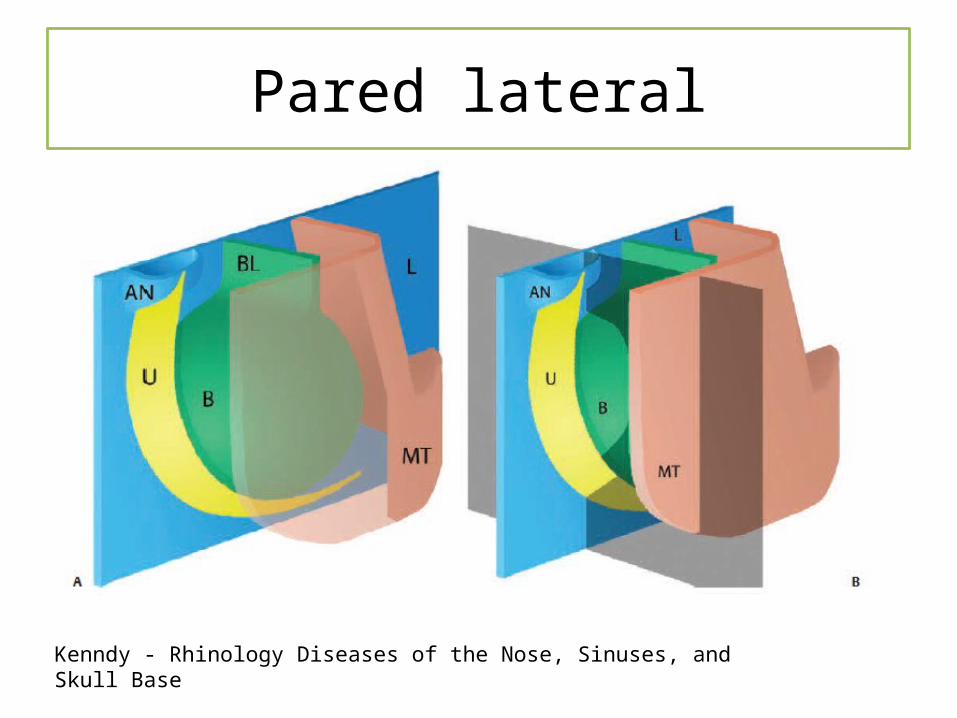
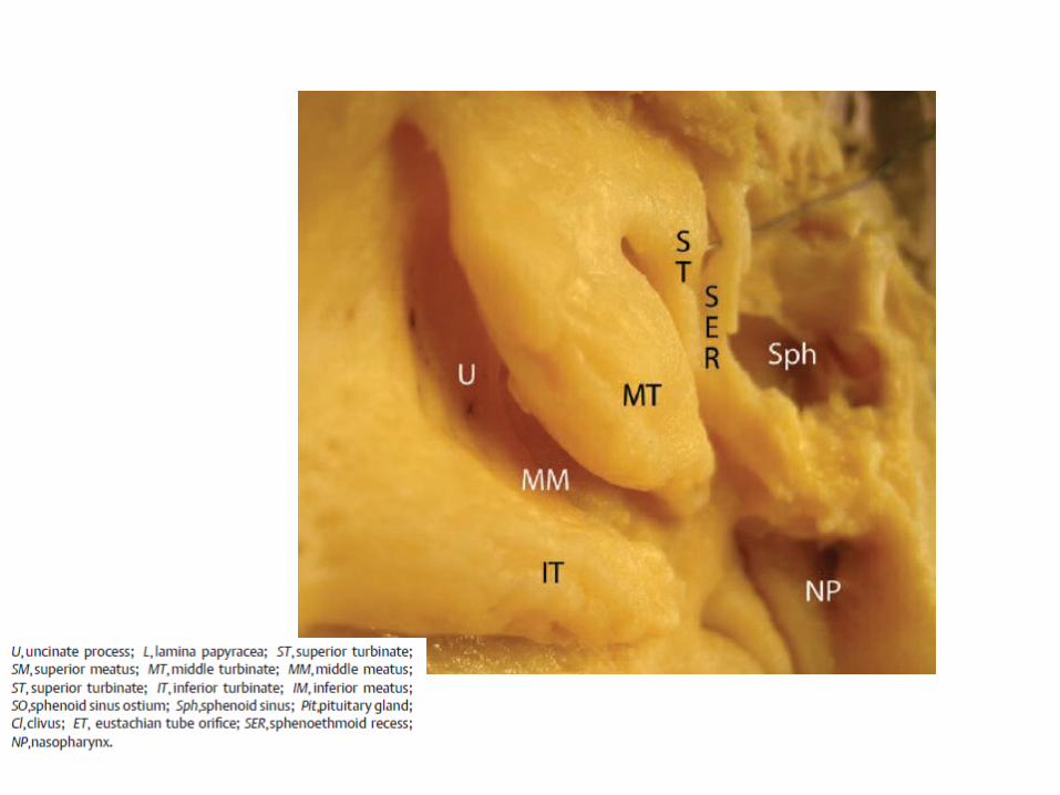
Pared lateral
Kenndy - Rhinology Diseases of the Nose, Sinuses, and Skull Base

Vista endoscópica
Pared lateral
Kenndy - Rhinology Diseases of the Nose, Sinuses, and Skull Base

Kenndy - Rhinology Diseases of the Nose, Sinuses, and Skull Base
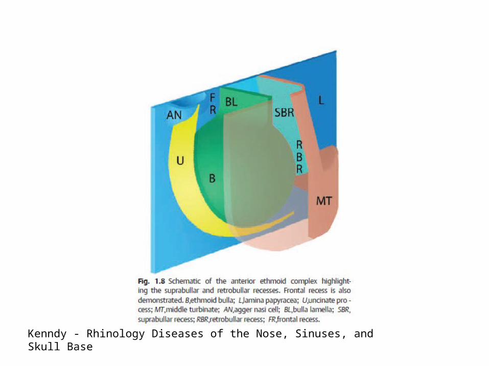
Kenndy - Rhinology Diseases of the Nose, Sinuses, and Skull Base

Kenndy - Rhinology Diseases of the Nose, Sinuses, and Skull Base
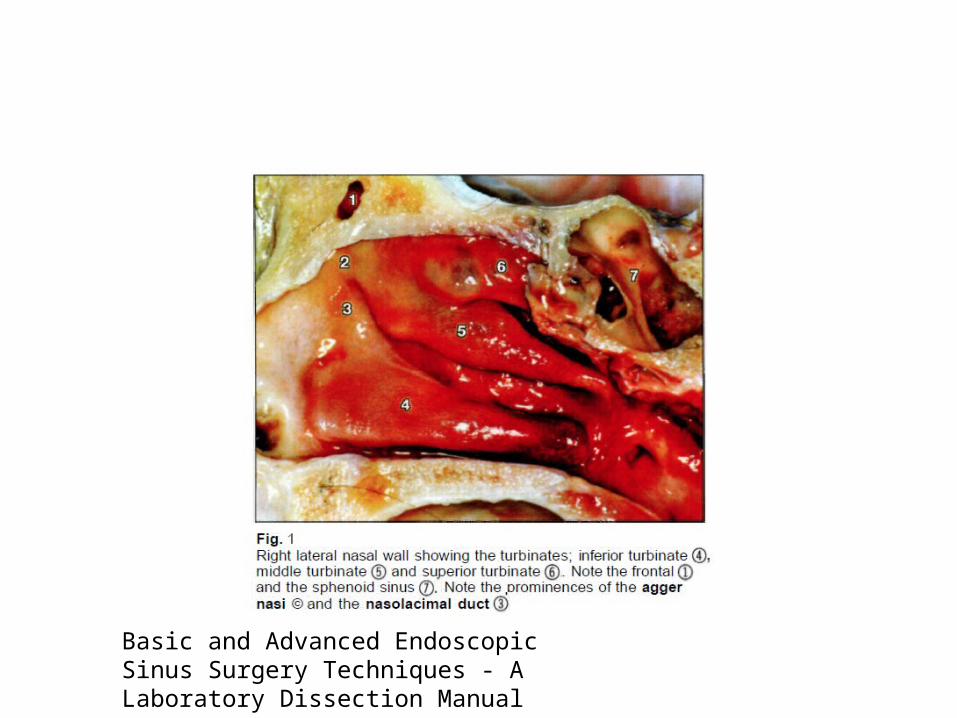
Basic and Advanced Endoscopic Sinus Surgery Techniques - A Laboratory Dissection Manual
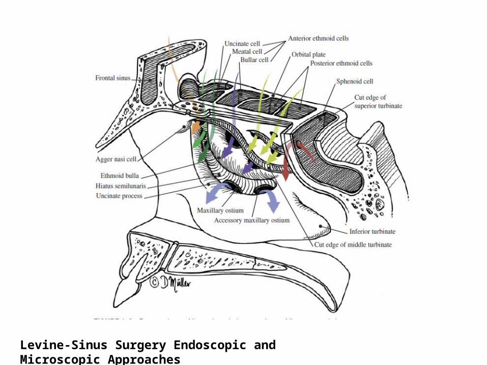
Levine-Sinus Surgery Endoscopic and Microscopic Approaches

Hiato semilunar superior
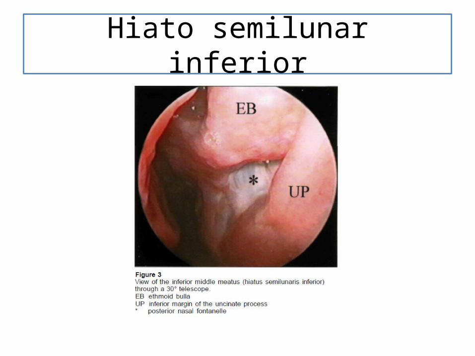
Hiato semilunar inferior


Normal sinonasal endoscopic surgical anatomy
Operative TechniquesinOtolaryngology(2014)25,144–148
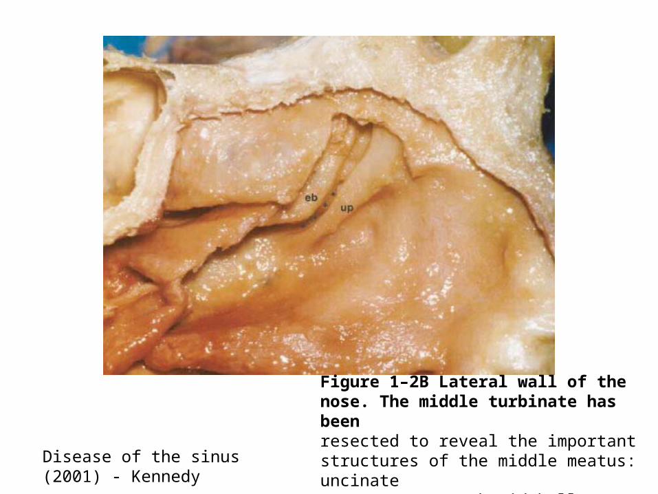
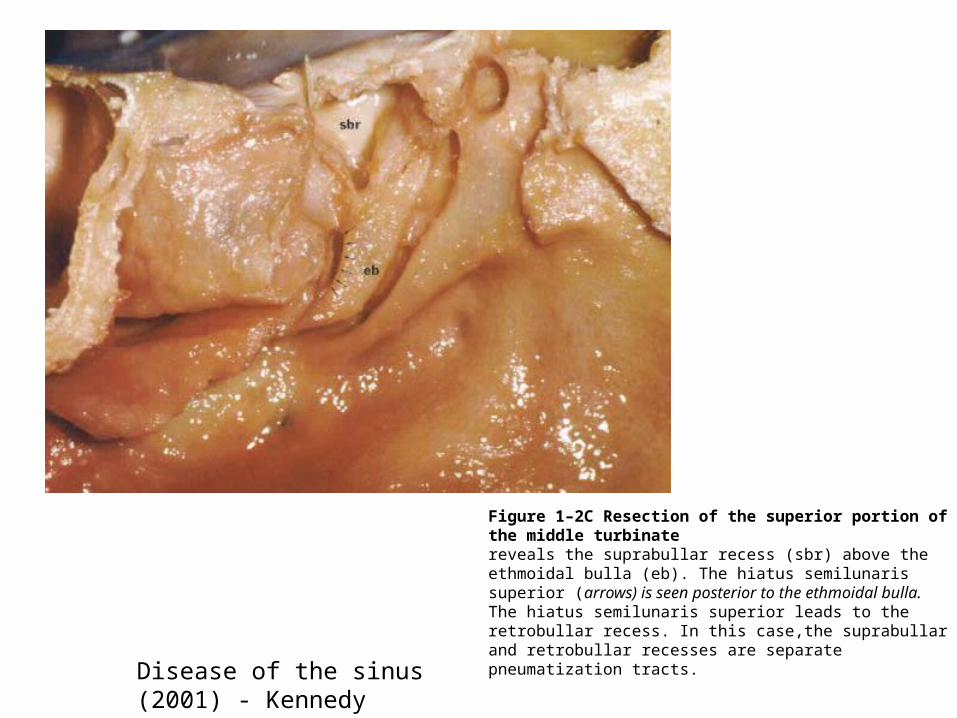
Disease of the sinus (2001) - Kennedy
Figure 1–2B Lateral wall of the nose. The middle turbinate has beenresected to reveal the important structures of the middle meatus: uncinateprocess (up), ethmoid bulla (eb), and the hiatus semilunaris (inferior) (*).
Disease of the sinus (2001) - Kennedy
Figure 1–2C Resection of the superior portion of the middle turbinatereveals the suprabullar recess (sbr) above the ethmoidal bulla (eb). The hiatus semilunaris superior (arrows) is seen posterior to the ethmoidal bulla.The hiatus semilunaris superior leads to the retrobullar recess. In this case,the suprabullar and retrobullar recesses are separate pneumatization tracts.

Ostium Maxilar
Figure 1–2E Following resection of the uncinate process, the naturalostium of the maxillary sinus is evident (o).

Lamelas basales
Levine-Sinus Surgery Endoscopic and Microscopic Approaches

Receso frontal

Celdas frontales

Esfenoidal

Vista endoscópica
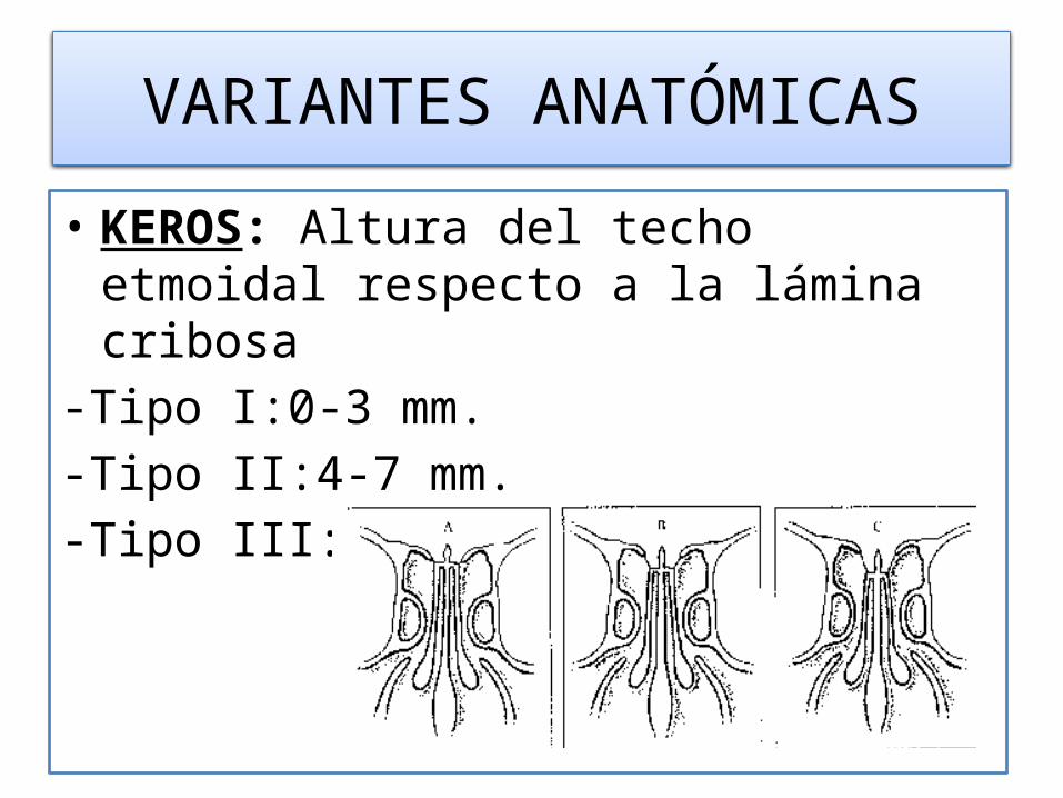
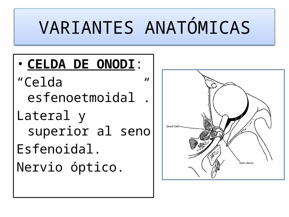
VARIANTES ANATÓMICAS
• KEROS: Altura del techo etmoidal respecto a la lámina cribosa
-Tipo I:0-3 mm.-Tipo II:4-7 mm.-Tipo III:8-16 mm.
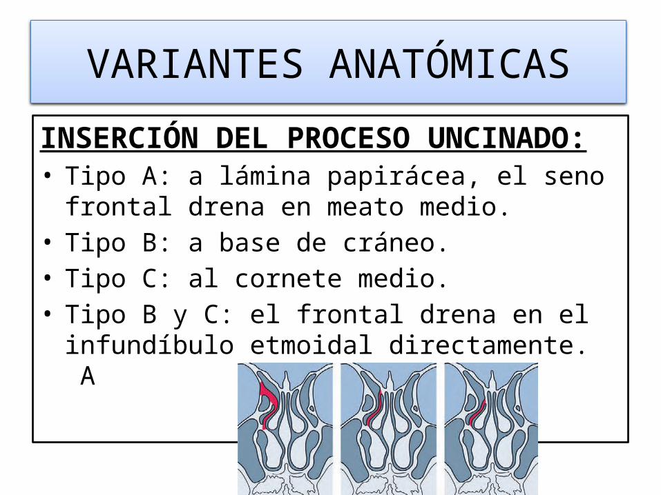
VARIANTES ANATÓMICAS
INSERCIÓN DEL PROCESO UNCINADO:• Tipo A: a lámina papirácea, el seno frontal drena en
meato medio.• Tipo B: a base de cráneo.• Tipo C: al cornete medio.• Tipo B y C: el frontal drena en el infundíbulo etmoidal
directamente. A B C
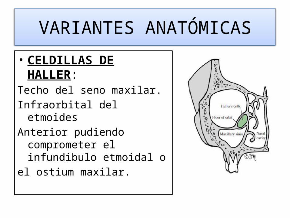
VARIANTES ANATÓMICAS
• CELDILLAS DE HALLER:Techo del seno maxilar. Infraorbital del etmoides Anterior pudiendo comprometer
el infundibulo etmoidal o el ostium maxilar.
VARIANTES ANATÓMICAS
• CELDA DE ONODI:“Celda esfenoetmoidal”.Lateral y superior al senoEsfenoidal.Nervio óptico.
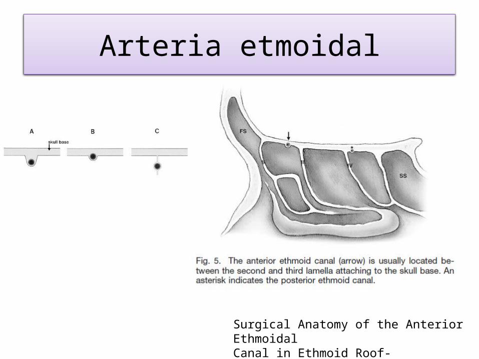
Arteria etmoidal
Surgical Anatomy of the Anterior EthmoidalCanal in Ethmoid Roof-laryngoscope-2011
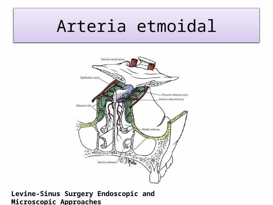
Arteria etmoidal
Levine-Sinus Surgery Endoscopic and Microscopic Approaches