
Anaplasma phagocytophilum MSP4 and HSP70 Proteins Are ...
16
ORIGINAL RESEARCH published: 05 July 2017 doi: 10.3389/fcimb.2017.00307 Frontiers in Cellular and Infection Microbiology | www.frontiersin.org 1 July 2017 | Volume 7 | Article 307 Edited by: Daniel E. Voth, University of Arkansas for Medical Sciences, United States Reviewed by: Edward Shaw, Oklahoma State University, United States Cornelius Joel Funk, John Brown University, United States *Correspondence: Alejandro Cabezas-Cruz [email protected] José de la Fuente [email protected] Received: 08 March 2017 Accepted: 20 June 2017 Published: 05 July 2017 Citation: Contreras M, Alberdi P, Mateos-Hernández L, Fernández de Mera IG, García-Pérez AL, Vancová M, Villar M, Ayllón N, Cabezas-Cruz A, Valdés JJ, Stuen S, Gortazar C and de la Fuente J (2017) Anaplasma phagocytophilum MSP4 and HSP70 Proteins Are Involved in Interactions with Host Cells during Pathogen Infection. Front. Cell. Infect. Microbiol. 7:307. doi: 10.3389/fcimb.2017.00307 Anaplasma phagocytophilum MSP4 and HSP70 Proteins Are Involved in Interactions with Host Cells during Pathogen Infection Marinela Contreras 1 , Pilar Alberdi 1 , Lourdes Mateos-Hernández 1 , Isabel G. Fernández de Mera 1 , Ana L. García-Pérez 2 , Marie Vancová 3, 4 , Margarita Villar 1 , Nieves Ayllón 1 , Alejandro Cabezas-Cruz 3, 4, 5 *, James J. Valdés 3, 6 , Snorre Stuen 7 , Christian Gortazar 1 and José de la Fuente 1, 8 * 1 SaBio, Instituto de Investigación en Recursos Cinegéticos, Consejo Superior de Investigaciones Científicas, CSIC-UCLM-JCCM, Ciudad Real, Spain, 2 Departamento de Sanidad Animal, Instituto Vasco de Investigación y Desarrollo Agrario (NEIKER), Derio, Spain, 3 Biology Centre, Czech Academy of Sciences, Institute of Parasitology, ˇ Ceské Bud ˇ ejovice, Czechia, 4 Faculty of Science, University of South Bohemia, ˇ Ceské Bud ˇ ejovice, Czechia, 5 UMR BIPAR, Animal Health Laboratory, INRA, ANSES, ENVA, Maisons Alfort, France, 6 Department of Virology, Veterinary Research Institute, Brno, Czechia, 7 Department of Production Animal Clinical Sciences, Norwegian University of Life Sciences, Sandnes, Norway, 8 Department of Veterinary Pathobiology, Center for Veterinary Health Sciences, Oklahoma State University, Stillwater, OK, United States Anaplasma phagocytophilum transmembrane and surface proteins play a role during infection and multiplication in host neutrophils and tick vector cells. Recently, A. phagocytophilum Major surface protein 4 (MSP4) and Heat shock protein 70 (HSP70) were shown to be localized on the bacterial membrane, with a possible role during pathogen infection in ticks. In this study, we hypothesized that A. phagocytophilum MSP4 and HSP70 have similar functions in tick-pathogen and host-pathogen interactions. To address this hypothesis, herein we characterized the role of these bacterial proteins in interaction and infection of vertebrate host cells. The results showed that A. phagocytophilum MSP4 and HSP70 are involved in host-pathogen interactions, with a role for HSP70 during pathogen infection. The analysis of the potential protective capacity of MSP4 and MSP4-HSP70 antigens in immunized sheep showed that MSP4- HSP70 was only partially protective against pathogen infection. This limited protection may be associated with several factors, including the recognition of non-protective epitopes by IgG in immunized lambs. Nevertheless, these antigens may be combined with other candidate protective antigens for the development of vaccines for the control of human and animal granulocytic anaplasmosis. Focusing on the characterization of host protective immune mechanisms and protein-protein interactions at the host-pathogen interface may lead to the discovery and design of new effective protective antigens. Keywords: anaplasmosis, immunology, HL60, tick, vaccine, sheep, Anaplasma phagocytophilum INTRODUCTION Anaplasma phagocytophilum (Rickettsiales: Anaplasmataceae) is an emerging tick-borne intracellular bacterial pathogen in many regions of the world, but vaccines are not available for prevention of transmission and infection in humans and animals (Dumler et al., 2001; Severo et al., 2013; Stuen et al., 2013, 2015; Bakken and Dumler, 2015). Anaplasma phagocytophilum causes
Transcript of Anaplasma phagocytophilum MSP4 and HSP70 Proteins Are ...

ORIGINAL RESEARCHpublished: 05 July 2017
doi: 10.3389/fcimb.2017.00307
Frontiers in Cellular and Infection Microbiology | www.frontiersin.org 1 July 2017 | Volume 7 | Article 307
Edited by:
Daniel E. Voth,
University of Arkansas for Medical
Sciences, United States
Reviewed by:
Edward Shaw,
Oklahoma State University,
United States
Cornelius Joel Funk,
John Brown University, United States
*Correspondence:
Alejandro Cabezas-Cruz
José de la Fuente
Received: 08 March 2017
Accepted: 20 June 2017
Published: 05 July 2017
Citation:
Contreras M, Alberdi P,
Mateos-Hernández L, Fernández de
Mera IG, García-Pérez AL,
Vancová M, Villar M, Ayllón N,
Cabezas-Cruz A, Valdés JJ, Stuen S,
Gortazar C and de la Fuente J (2017)
Anaplasma phagocytophilum MSP4
and HSP70 Proteins Are Involved in
Interactions with Host Cells during
Pathogen Infection.
Front. Cell. Infect. Microbiol. 7:307.
doi: 10.3389/fcimb.2017.00307
Anaplasma phagocytophilum MSP4and HSP70 Proteins Are Involved inInteractions with Host Cells duringPathogen Infection
Marinela Contreras 1, Pilar Alberdi 1, Lourdes Mateos-Hernández 1,
Isabel G. Fernández de Mera 1, Ana L. García-Pérez 2, Marie Vancová 3, 4, Margarita Villar 1,
Nieves Ayllón 1, Alejandro Cabezas-Cruz 3, 4, 5*, James J. Valdés 3, 6, Snorre Stuen 7,
Christian Gortazar 1 and José de la Fuente 1, 8*
1 SaBio, Instituto de Investigación en Recursos Cinegéticos, Consejo Superior de Investigaciones Científicas,
CSIC-UCLM-JCCM, Ciudad Real, Spain, 2Departamento de Sanidad Animal, Instituto Vasco de Investigación y Desarrollo
Agrario (NEIKER), Derio, Spain, 3 Biology Centre, Czech Academy of Sciences, Institute of Parasitology, Ceské Budejovice,
Czechia, 4 Faculty of Science, University of South Bohemia, Ceské Budejovice, Czechia, 5UMR BIPAR, Animal Health
Laboratory, INRA, ANSES, ENVA, Maisons Alfort, France, 6Department of Virology, Veterinary Research Institute, Brno,
Czechia, 7Department of Production Animal Clinical Sciences, Norwegian University of Life Sciences, Sandnes, Norway,8Department of Veterinary Pathobiology, Center for Veterinary Health Sciences, Oklahoma State University, Stillwater, OK,
United States
Anaplasma phagocytophilum transmembrane and surface proteins play a role during
infection and multiplication in host neutrophils and tick vector cells. Recently, A.
phagocytophilum Major surface protein 4 (MSP4) and Heat shock protein 70 (HSP70)
were shown to be localized on the bacterial membrane, with a possible role during
pathogen infection in ticks. In this study, we hypothesized that A. phagocytophilumMSP4
and HSP70 have similar functions in tick-pathogen and host-pathogen interactions. To
address this hypothesis, herein we characterized the role of these bacterial proteins
in interaction and infection of vertebrate host cells. The results showed that A.
phagocytophilum MSP4 and HSP70 are involved in host-pathogen interactions, with
a role for HSP70 during pathogen infection. The analysis of the potential protective
capacity of MSP4 and MSP4-HSP70 antigens in immunized sheep showed that MSP4-
HSP70 was only partially protective against pathogen infection. This limited protection
may be associated with several factors, including the recognition of non-protective
epitopes by IgG in immunized lambs. Nevertheless, these antigens may be combined
with other candidate protective antigens for the development of vaccines for the control of
human and animal granulocytic anaplasmosis. Focusing on the characterization of host
protective immune mechanisms and protein-protein interactions at the host-pathogen
interface may lead to the discovery and design of new effective protective antigens.
Keywords: anaplasmosis, immunology, HL60, tick, vaccine, sheep, Anaplasma phagocytophilum
INTRODUCTION
Anaplasma phagocytophilum (Rickettsiales: Anaplasmataceae) is an emerging tick-borneintracellular bacterial pathogen in many regions of the world, but vaccines are not available forprevention of transmission and infection in humans and animals (Dumler et al., 2001; Severo et al.,2013; Stuen et al., 2013, 2015; Bakken and Dumler, 2015). Anaplasma phagocytophilum causes

Contreras et al. Anaplasma-Host Interactions
human granulocytic anaplasmosis (HGA), which has emergedas a tick-borne disease of humans in the United States, Europeand Asia (Severo et al., 2013). In Europe, A. phagocytophilumis an established pathogen of small ruminants, most notablyin sheep, where it was first described as the etiologic agentof tick-borne fever (TBF; Gordon et al., 1932; Foggie, 1951;Dugat et al., 2015). Clinical presentation of A. phagocytophiluminfection has been also documented in goats, cattle, horses, dogs,cats, roe deer, and reindeer (Severo et al., 2013). Although, A.phagocytophilum is recognized as a threat for human and animalhealth in Europe and the United States, its pathogenic andepidemic potential is neglected in tropical regions of the world(Heyman et al., 2010; Dugat et al., 2015). Prophylactic uses oftetracycline together with acaricide applications for tick controlare the main measures to controlA. phagocytophilum infection inendemic areas (Woldehiwet, 2006; Stuen et al., 2015). However,these control measures raise concerns about their impact on theenvironment and human health, and the selection of resistantpathogens and ticks (Woldehiwet, 2006; Stuen et al., 2015).
Results using next generation sequencing technologies haveadvanced our understanding of the mechanisms by whichA. phagocytophilum infection affects gene expression, proteincontent and microbiota in the vertebrate host and tick vector(Ge and Rikihisa, 2006; Sukumaran et al., 2006; de la Fuenteet al., 2010, 2016a,b,c,d, 2017, Neelakanta et al., 2010; Rikihisa,2011; Severo et al., 2012; Ayllón et al., 2013, 2015; Hajdušeket al., 2013; Villar et al., 2015a; Cabezas-Cruz et al., 2016, 2017;Gulia-Nuss et al., 2016; Abraham et al., 2017; Mansfield et al.,2017). However, less information is available on the bacterialmolecules involved in pathogen infection and multiplication(Ge and Rikihisa, 2007; Huang et al., 2010; Lin et al., 2011;Troese et al., 2011; Mastronunzio et al., 2012; Oliva Chávezet al., 2015; Seidman et al., 2015; Villar et al., 2015b; Truchanet al., 2016). Definition of bacterial proteins involved in host-pathogen and vector-pathogen interactions may provide targetantigens for the development of vaccines and therapeutics thatinterfere with pathogen host infection and transmission by ticks(Gomes-Solecki, 2014; de la Fuente and Contreras, 2015).
Recently, Villar et al. (2015b) demonstrated that A.phagocytophilum activates a new mechanism associatedwith bacterial cell stress and membrane proteins to counteracttick cell response to infection and favor pathogen infection andmultiplication. Their results showed that A. phagocytophilumproteins, Major surface protein 4 (MSP4) and Heat shock protein70 (HSP70), are localized on the bacterial membrane wherethey interact with a possible role during pathogen infectionin ticks (Villar et al., 2015b). Furthermore, antibodies againstMSP4 and HSP70 inhibited pathogen infection of tick cells,supporting that these proteins are involved in tick-pathogeninteractions (Villar et al., 2015b). They proposed that theinhibitory effect of anti-MSP4 and anti-HSP70 antibodies couldbe the result of the antibodies blocking the interaction betweenbacterial ligands (e.g., MSP4) and tick receptors or an effect onproteins functionally important for bacterial infection and/ormultiplication in tick cells (e.g., HSP70 and those physicallyand/or functionally interacting with it; Villar et al., 2015b). Theresults of these experiments suggested that A. phagocytophilum
MSP4 and HSP70 proteins constitute candidate protectiveantigens to interfere with pathogen infection in the tick vector,Ixodes scapularis.
The characterization of the A. phagocytophilum proteomedemonstrated that chaperones, surface and stress responseproteins are among the most abundant proteins found in I.scapularis tick salivary glands (Mastronunzio et al., 2012). HSP70is a chaperone involved in protein folding and stress response(Johnson, 2012). This protein functions by protecting cells fromstress-induced lethal damage and under physiological growthconditions by acting as carriers for immunogenic peptides,assisting in protein export or mediating adherence to host cellsand may play an essential role during cell division (Scopio et al.,1994; Susin et al., 2006; Multhoff, 2007; Seydlová et al., 2012). Therole of MSPs such as MSP4 in adhesion to tick cells for bacterialinfection has been demonstrated in A. marginale (de la Fuenteet al., 2001) and A. phagocytophilum (Villar et al., 2015b).
Anaplasma phagocytophilum infects vertebrate hostneutrophils and various tick tissues, where it develops withinmembrane-bound inclusions in the cell cytoplasm (Severo et al.,2013). However, this pathogen has evolved common molecularmechanisms to establish infection in tick vectors and vertebratehosts that collectively mediate pathogen infection, development,persistence, and survival (de la Fuente et al., 2016a). Thesestrategies include, but are not limited to, remodeling of thecytoskeleton, inhibition of cell apoptosis, manipulation ofthe immune response, and the use of rickettsial proteins forinfection and manipulation of tick and host gene expression(Cabezas-Cruz et al., 2016; de la Fuente et al., 2016a).
Based on these results, we hypothesized that the use ofcommon strategies by A. phagocytophilum to establish infectionin ticks and vertebrate hosts resulted in similar functions forMSP4 and HSP70 proteins in host-pathogen and tick-pathogeninteractions. To address this hypothesis, in this study wecharacterized the role of these bacterial proteins in infection ofvertebrate host cells and their potential protective capacity inimmunized sheep. The results showed that A. phagocytophilumMSP4 and HSP70 are involved in host-pathogen interactionsduring pathogen infection, but were only partially protectiveagainst pathogen infection in sheep.
MATERIALS AND METHODS
Ethics StatementThe study was ethically approved by the local Animal Health andWelfare Authority (Diputación Foral de Alava) with referenceNo. 1820, 12th May 2015, following Spanish ethical guidelinesand animal welfare regulations (Real Decreto 53/2013).
Production of Recombinant Proteins andRabbit AntibodiesThe His-tag recombinant A. phagocytophilum human NY18isolate (Asanovich et al., 1997) proteins MSP4 (AFD54597) andHSP70 (KX891324) were produced in Escherichia coli BL21 cells(Champion pET101 Directional TOPO Expression kit, Carlsbad,CA, USA), after induction with IPTG and purified using theNi-NTA affinity column chromatography system (Qiagen Inc.,
Frontiers in Cellular and Infection Microbiology | www.frontiersin.org 2 July 2017 | Volume 7 | Article 307

Contreras et al. Anaplasma-Host Interactions
Valencia, CA, USA) as previously described (Villar et al., 2015b).Recombinant purified proteins showed purity higher than 85% oftotal proteins and were used to immunize rabbits to purify IgGsfrom preimmune and immunized animals (Montage AntibodyPurification Kit and Spin Columns with PROSEP-A Media,Millipore, Billerica, MA, USA) for Western blot and antibodyinhibition analyses as previously described (Villar et al., 2015b).
Surface Trypsin Digestion ofA. phagocytophilum from Infected HL60Human CellsThe A. phagocytophilum human NY18 isolate was propagatedin cultured HL60 human promyelocytic cells as previouslydescribed (de la Fuente et al., 2005). The A phagocytophilum-infected cells (∼1 × 10⁷ cells) were collected when 70–80% ofthe cells were infected as determined by detection of intracellularmorulae in stained cytospin cell smears. Host cell-free bacteriawere isolated from cell lysates after five passages through a 27-gauge syringe, followed by differential centrifugation in Percollgradients as previously described for A. marginale to separatebacteria from host cell debris (Lis et al., 2014). The pellet ofpurified A. phagocytophilum was resuspended in 200µl of SPGbuffer (0.25mM sucrose, 10mM sodium phosphate, 5mM L-glutamic acid, pH 7.2), and 5µl of sequencing-grade trypsin(Promega, Madison, WI, USA) was added to half of the cellreaction mixture. Bacteria were incubated at 37◦C for 30minand then centrifuged at 10,000 × g for 15min and resuspendedin Laemmli protein loading buffer, boiled for 5min and loadedonto a 12% SDS-PAGE and analyzed byWestern blot using rabbitantibodies against recombinant MSP4 and HSP70 proteins aspreviously described (Villar et al., 2015b).
Tertiary Models and Protein-ProteinDockingThe active A. phagocytophilum HSP70 and MSP4 proteins weremodeled using I-TASSER (Zhang, 2008), and the unbound (apo)-HSP70 protein was modeled using Robetta (Kim et al., 2004). Alltertiary models were optimized with the Schrödinger’s MaestroProtein Preparation Wizard (Li et al., 2007). All steric clasheswere resolved via minimization with the default settings inthe Schrodinger’s Maestro package. For the tertiary models,the Protein Preparation Wizard clusters at the highest degreeof hydrogen bonding in equilibrium were used. Monte Carloorientations were performed (100,000) for each cluster. Theoptimized structure is based on electrostatic and geometricscoring functions. The membrane positioning for MSP4 wascalculated by the OPM database (Lomize et al., 2006) andgenerated using the Desmond systems builder (Bowers et al.,2006) as part of the Schrodinger’s Maestro package. Theprotein-protein docking was assessed using the SwarmDockserver (Torchala et al., 2013) that incorporates flexible dockingby exploring in proximity to the Cartesian center of massof the target protein. Minimization steps are included forthe whole system. The poses are calculated based on themost energy favorable poses, minimized once again and sentto the user. We chose to analyze the top 10 poses since
these were highly energy favorable (−41 to −54 kcal/mol).The server also produces the residue contacts made betweenboth proteins. All structures were visualized and analyzedusing the Visual Molecular Dynamics (Humphrey et al.,1996).
Adhesion of Recombinant E. coli Strains toHL60 Human CellsThe adhesion of recombinant E. coli strains to HL60 humancells was characterized as previously reported (de la Fuenteet al., 2001; Villar et al., 2015b). Briefly, E. coli strainsproducingA. phagocytophilumMSP4, HSP70, andmutantHSP70with truncated peptide-binding domains that are involved inprotein-protein interactions (Villar et al., 2015b) recombinantproteins were grown and induced as described before. TheE. coli cells transformed with expression vector alone wereused as negative control. Cell densities were determined andadjusted to 108 cells per ml in Luria Broth (LB). One hundredmicroliters (107 bacteria) culture were added to 900µl of 106
cells per ml suspensions of HL60 human cells in LB. Humancells and bacteria were incubated for 30min at 37◦C withoccasional agitation. Cells were then collected by centrifugation,washed two times in PBS and resuspended in 100ml ofPBS. Elimination of unbound bacteria from human cells withbound bacteria was performed by Percoll (Sigma, St. Louis,MI, USA) gradient separation (de la Fuente et al., 2001). Theband containing human cells was removed with a pipette andwashed in PBS. The final cell pellet was lysed in 1ml ofsterile water and 5µl plated onto LB agar plates containing100µg of ampicillin per ml. Two replicates were done foreach experiment. Adhesive bacteria were quantitated as thenumber of colony forming units (CFU) recovered from each testand compared to the control values by Chi2 test (P = 0.001;N = 2).
Transmission Electron Microscopy (TEM)The HL60 human cells incubated as described above with E.coli strains producing A. phagocytophilum MSP4 and HSP70recombinant proteins or transformed with expression vectoralone as controls were pelleted and fixed in 2.5% glutaraldehydein 0.1M phosphate buffer for 1 h at room temperature. Fixedcells were washed three times in 0.1M phosphate buffer with4% glucose and embedded in 2% agar at 60◦C. Sampleswere post-fixed using 2% OsO4 for 2 h at room temperature,three times washed and dehydrated in a graded series ofacetone (30–100%) solutions for 15min at each step. Sampleswere infiltrated with 25, 50, and 75% solutions of Spi PonEpoxy resin (Structure Probe, Inc. Supplies, West Chester,PA, USA) diluted in anhydrous acetone for 1 h at each step.Samples were left in 100% resin overnight, transferred toembedding molds and polymerized at 60◦C for 48 h. Ultrathinsections were contrasted in ethanolic uranyl acetate and leadcitrate, carbon coated and observed in a JEOL 1010 TEM(JEOL Ltd., Akishima, Tokio, Japan) at an accelerating voltageof 80 kV. Images were captured using a Mega View IIIcamera (Olympus Soft Imaging Solutions GmbH, Münster,Germany).
Frontiers in Cellular and Infection Microbiology | www.frontiersin.org 3 July 2017 | Volume 7 | Article 307

Contreras et al. Anaplasma-Host Interactions
Prediction of B-cell EpitopesAnaplasma phagocytophilum MSP4 (AFD54597) and HSP70(AAC31306) amino acid sequences were aligned using MAFFTversion 7, applying a gap opening penalty of 3 (range 1–3,default 1.53; Katoh and Standley, 2013). Sequence homologywas calculated using Clustal Omega (Sievers et al., 2011).The linear B-cell epitopes on A. phagocytophilum MSP4and HSP70 proteins were predicted using the BepipredLinear Epitope Prediction tool (http://tools.immuneepitope.org/bcell/; Haste Andersen et al., 2006; Larsen et al., 2006;Ponomarenko and Bourne, 2007). Subsequently, to searchfor epitope homology, each predicted epitope within eachprotein was aligned with the full sequence of the otherprotein.
Antibody Inhibition AssayThe inhibitory effect of rabbit IgG antibodies against MSP4and HSP70 recombinant proteins on A. phagocytophilum humanNY18 (Asanovich et al., 1997) and sheep (Alberdi et al.,2015) isolates infection of HL60 human cells was conductedas described previously for tick cells (Villar et al., 2015b). Theinhibitory effect of IgG antibodies purified from MSP4 andMSP4-HSP70 immunized and control sheep at 0 and 94 dayspost-infection (dpi) on A. phagocytophilum human NY18 isolateinfection of HL60 human cells was conducted using the sameexperimental approach as for rabbit IgG. The IgGs were purifiedfrom sheep sera using the NAb Protein G spin kit (ThermoFisher Scientific, Waltham, MA, USA) following manufacturer’srecommendations. HL60 cells were pooled and used to seed 24-well plates for each assay. Each well received 1 × 10⁶ cells inRPMI 1,640 medium (Gibco, Thermo Fisher, Madrid, Spain) 48h prior to inoculation with A. phagocytophilum. Infected culturesfor inoculum were harvested when infection reached 80% andhost cells were mechanically disrupted with a syringe and 26-gauge needle. Rabbit or sheep purified IgGs (2.2–2.4mg/ml) weremixed with inoculum (1:1) for 60 min before being placed onthe cell monolayers. Each monolayer then received 100µl ofthe inoculum plus IgG mix and plates were incubated at 37◦Cfor 30min. The inoculum was removed from the wells and cellmonolayers washed three times with PBS. Complete medium (1ml) was added to each well and the plates were incubated at37◦C. The control for each trial included inoculum incubatedwith rabbit pre-immune IgG or control sheep IgG. Four replicateswere done for each treatment. After 7 days, cells from allwells were harvested, resuspended in 1ml PBS and frozen at−70◦C. Samples were thawed and solubilized with 1% Triton-X100 and processed for A. phagocytophilum detection by PCRafter DNA extraction using TriReagent (Sigma) according to themanufacturer’s recommendations. Anaplasma phagocytophiluminfection levels were determined by msp4 real-time PCRnormalizing against human β-actin as described previously(de la Fuente et al., 2005) but using oligonucleotide primersMSP4-L (5′-CCTTGGCTGCAGCACCACCTG-3′) and MSP4-R (5′-TGCTGTGGGTCGTGACGCG3′), with PCR conditionsof 5min at 95◦C and 35 cycles of 10 s at 95◦C, 30 s at 55◦Cand 30 s at 60◦C. Results were compared between treatments
by the Student’s t-test with unequal variance (P = 0.05;N = 4).
Protein Inhibition AssayHL60 cells were incubated with 4µM MSP4 and HSP70recombinant proteins or their combination in culture mediafor 1 h at 37◦C and 5% CO2 in a humidified atmosphere.For antigen combination, equal molar ratios of each protein,equivalent to one part of HSP70 and two parts of MSP4,were incubated at 4◦C on a rotator overnight. HL-60 cells(4 × 10⁵ cells/well) were fixed in 4% paraphormaldehyde(PFA) in PBS for 1 h at room temperature (RT), and thenincubated with a 6x-His epitope tag monoclonal antibody(3µg/ml mouse IgG1, Thermo Fisher 4A12E4) for 1 h atRT. After washing, cells were incubated with FITC-conjugatedgoat anti-mouse IgG (1/100, Sigma F2012) for 1 h at RT.Protein binding was assessed by flow cytometry using aFACScalibur R© Flow Cytometer, equipped with the CellQuestPro R© software (BD-Biosciences, San Jose, CA, USA) aspreviously described (Seidman et al., 2015; Hebert et al., 2017).Incubation with PBS was used as negative control. The viablecell population was gated according to forward scatter andside scatter parameters. To determine the effect of MSP4 andHSP70 recombinant proteins on A. phagocytophilum infection,HL60 cells were incubated with 4µM MSP4 and HSP70recombinant proteins or their combination for 1 h, after whichA. phagocytophilum human NY18 isolate bacteria purified asdescribed above were added and incubated with the host cells inthe continued presence of recombinant protein for 2 h. Unboundbacteria and proteins were removed and the infection wasallowed to proceed for 48 h. Then, cells were harvested andinfection levels determined by PCR and statistically analyzedas described above. The Rhipicephalus microplus Subolesinrecombinant protein (SUB; Merino et al., 2011) and PBSwere included as controls. Four replicates were done for eachtreatment.
Lamb Immunization and Infection withA. phagocytophilumThe recombinant MSP4 was formulated alone or combined withHSP70. Antigen combination was done as described above inSection Protein Inhibition Assay. Recombinant antigens or salinecontrol were adjuvated in Montanide ISA 50 V2 (Seppic, Paris,France; Merino et al., 2013). Nine 3-month old lambs of theLatxa breed (Basque Country, Spain) were selected from theexperimental sheep flock maintained at NEIKER and were keptindoor during the experiment. This flock has no known historyof ticks or tick-borne diseases. However, blood of lambs andtheir dams were analyzed prior to the start of the study to checktheir status for hemoparasites Theileria, Babesia, and Anaplasmaspp. as previously described (Hurtado et al., 2015). All animalswere negative for these hemoparasites. Three groups of 3 lambseach with similar live weight were formed. Lambs from eachgroup were injected subcutaneously in the loose skin of theaxilla (armpit) using a sterile syringe with removable needle 20G × 1′′ (9.0 × 25mm), and taking aseptic precautions. Lambswere immunized three times on days 55, 30, and 10 before
Frontiers in Cellular and Infection Microbiology | www.frontiersin.org 4 July 2017 | Volume 7 | Article 307

Contreras et al. Anaplasma-Host Interactions
experimental infection with 1ml doses of MSP4 (100µg/dose),MSP4-HSP70 (100µg of equal molar ratios of each protein/dose)or adjuvant/saline as control. The strain of A. phagocytophilumused for experimental infection originated from an infected lambin Norway, which suffered TBF but was negative to other tick-borne pathogens (Alberdi et al., 2015; Stuen et al., 2015). Theinoculum consisted of A. phagocytophilum infected heparinisedblood that had been stored at−70◦Cwith 10% dimethyl sulfoxide(DMSO). Once unfrozen, 1ml of infected blood containing 1.8× 10⁶ infected neutrophils per ml was intravenously inoculatedinto each experimental lamb through the jugular vein usingsterile winged infusion sets with needle 21 G × 3/4′′ (0.8 ×
19mm).
Sheep Samples and AnalysisWhole blood and serum samples were collected from the jugularvein of each lamb previous to each immunization, daily startingon infection day during 10 days, and at weekly intervals untilthe end of the experiment at 94 dpi (Supplementary Table 1).Rectal temperatures were taken daily until 10 dpi and then weeklyuntil 94 dpi (Supplementary Table 1). Lambs were also weighedperiodically (Supplementary Table 1). Hematological analysesincluding leukocyte and erythrocyte cell counts, leukocyte celldifferentiation (percent neutrophils, lymphocytes, monocytesand eosinophils), hemoglobin levels, hematocrit, mean cellvolume (MCV), and mean corpuscular hemoglobin (MCH)were performed with an electronic counter (Hemavet 950,Drew, USA) in blood samples collected in EDTA-containingtubes (Supplementary Table 1). Blood smears stained withGiemsa stain were examined to investigate the presence of A.phagocytophilum in blood cells (Supplementary Table 1). Atleast 100 neutrophils were counted and examined to calculatethe number of infected neutrophils per milliliter blood of eachlamb throughout the experiment. The differential percent ofA. phagocytophilum-infected neutrophils was calculated as thedifference between values at different dpi and values at 3 dpiwhen infected neutrophils were first detected (SupplementaryTable 1).
Analysis of the Antibody Response inLambsAn indirect ELISA test was performed to detect IgG antibodiesagainst MSP4 and HSP70 proteins in immunized and controllambs using serum samples collected before each immunizationand at 0, 7, 10, and 94 dpi. High absorption capacity polystyrenemicrotiter plates were coated with 100µl (0.01µg/µl solutionof purified recombinant MSP4 or HSP70 protein) per wellin carbonate-bicarbonate buffer (Sigma). After an overnightincubation at 4◦C, coated plates were blocked with 100µl/well ofblocking solution (5% skim milk in PBS). Serum samples or PBSas negative control were diluted (1:100, v/v) in blocking solutionand 100µl/well were added into duplicate wells of the antigen-coated plates. After an overnight incubation at 4◦C, the plateswere washed three times with a washing solution (PBS containing0.05% Tween 20). A donkey anti-sheep IgG-peroxidase conjugate(Sigma) was added (diluted 1:1000 in blocking solution) and
incubated at room temperature for 1 h. After three washeswith washing solution, 100µl/well of substrate solution (FastOPD; Sigma) was added. Finally, the reaction was stopped with50µl/well of 3N H2SO4 and the optical density (OD) wasmeasured in a spectrophotometer at 450 nm. Antibody titers wereexpressed as OD450nm (ODlambsera ODPBScontrol). The antigen-specific antibody response in immunized lambs was corroboratedby ELISA using pooled sera collected at 0 dpi, but incubating serawith A. phagocytophilum purified from infected HL60 humancells as described above in Section Antibody Inhibition Assay.Results from rectal temperature and hematological analyses,differential percent of A. phagocytophilum-infected neutrophilsand antibody titers were compared between immunized andcontrol groups by two-way ANOVA test (P = 0.05; N = 3).
Analysis of A. phagocytophilum DNALevels in LambsFor the analysis of A. phagocytophilum infection in lambsduring the trial, DNA was extracted from 200µl of bloodusing the QIAamp DNA Mini Kit (Qiagen, Hilden, Germany),including negative extraction controls every 9 samples. DNAwas stored at −20◦C until subsequent analysis. The presenceof Anaplasma spp. was firstly determined using a real-timePCR assay for the screening of piroplasms and Anaplasmaspp. (RTi-PCR1) that targets the 16S rRNA gene of Anaplasmaspp. and the 18S rRNA gene of piroplasms of the generaTheileria and Babesia (Hurtado et al., 2015). The assay alsoincludes an internal amplification control (IAC) to monitor forpossible inhibition of the PCR reaction. All samples positiveto Anaplasma spp. in the RTi-PCR1 were analyzed with amultiplex PCR assay that specifically amplifies the major surfaceprotein 2 (msp2) gene of A. phagocytophilum, and the msp4gene of Anaplasma ovis (RTi-PCR2). Sequences of primers andprobes, as well as details on cycling conditions were as reportedpreviously (Hurtado et al., 2015). Analyses were performedin 20µl volume reactions using an ABI PRISM 7500 FastSequence Detection System (Applied Biosystems, Foster City,CA, USA). For the quantitative analysis of A. phagocytophiluminfection levels, DNA was extracted from 200µl blood samplesusing Nucleospin 96 Blood (Machery-Nagel, Düren Germany).A quatitative real-time PCR was conducted on DNA samplesusing a Quantitect SYBR Green RT-PCR Kit and a Rotor GeneQ thermocycler (Qiagen, Inc. Valencia, CA, USA) followingmanufacturer’s recommendations. A dissociation curve was runat the end of the reaction to ensure that only one ampliconwas formed and that the amplicon denatured consistently inthe same temperature range for every sample (Ririe et al.,1997). The DNA levels were normalized against sheep aldolase Busing primers Ovi-ALDOB-F: CCCATCTTGCTATCCAGGAAand Ovi-ALDOB-R: TACAGCAGCCAGGACCTTCT, and thegenNorm method (ddCT method as implemented by Bio-RadiQ5 Standard Edition, Version 2.0; Livak and Schmittgen, 2001).Normalized Ct values were compared between immunized andcontrol groups by Student’s t-test with unequal variance (P =
0.05; N = 3).
Frontiers in Cellular and Infection Microbiology | www.frontiersin.org 5 July 2017 | Volume 7 | Article 307

Contreras et al. Anaplasma-Host Interactions
RESULTS
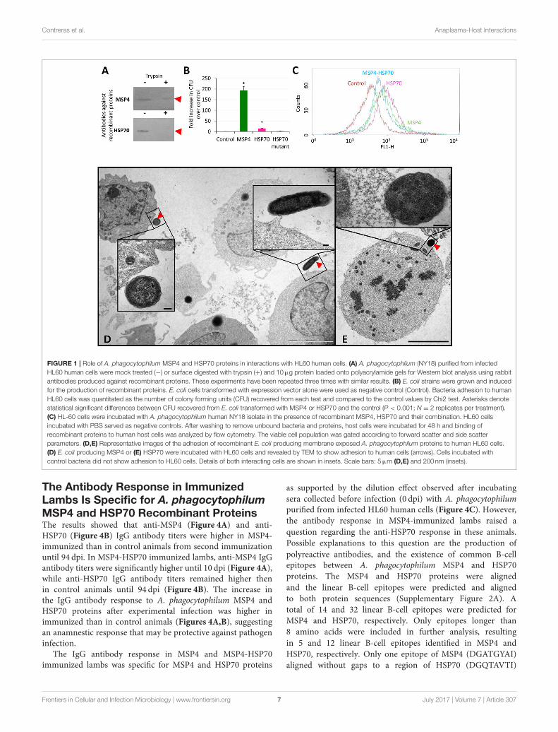
The A. phagocytophilum MSP4 and HSP70Proteins are Localized on the BacterialMembrane and Involved in PathogenInfection of HL60 Human CellsThe subcellular localization of MSP4 and HSP70 proteins wascharacterized in A. phagocytophilum purified from infectedHL60 human promyelocytic leukemia cells, mock treated orsurface digested with trypsin and loaded onto polyacrylamidegels for Western blot analysis using rabbit antibodies specificagainst recombinant proteins. The results showed that as atransmembrane protein, MSP4 was partially resistant to trypsindigestion in A. phagocytophilum from HL60 cells, while HSP70was extracellular and exposed to protease digestion (Figure 1A).
The apo and bound A. phagocytophilum HSP70 modelsshowed that the major structural difference between thetwo HSP70 tertiary structures is at the C-terminus (residues400–533), with a 40–90 Å α-carbon backbone root meansquare deviation (RMSD; Supplementary Figure 1A). The A.phagocytophilum MSP4 showed a 25◦ ± 1◦ tilt from themembrane with its N/C-terminus oriented toward the cytosol,and the β-sheets buried within the membrane with remainingβ-hairpin loops exposed extracellularly (Supplementary Figure1B). The models suggested a limited number of possible MSP4-HSP70 binding positions due to the membrane orientation ofMSP4 (Supplementary Figure 1C). The energy score of theoptimum MSP4-HSP70 bound state (Supplementary Figure 1C)was calculated at −46 kcal/mol. Furthermore, although theresidue map showed that the majority of protein-protein contactsare formed between the β-sheets of MSP4 buried within themembrane and the C-terminus of HSP70, several contactsbetween the β-hairpin loops of MSP4 and the N-terminus ofHSP70 are exposed extracellularly, and therefore these residuesmay act as markers for mutational studies and antibodytargeting (Supplementary Figure 1C). These models supportedthe interaction between A. phagocytophilum MSP4 and HSP70proteins when localized on the bacterial membrane.
The binding of HSP70 and MSP4 to HL60 human cellswas characterized using recombinant proteins and E. coliproducing surface-exposed A. phagocytophilum proteins. Theresults demonstrated that MSP4 and HSP70 are involved inbinding to human promyelocytic leukemia cells (Figures 1B,C).Furthermore, E. coli producing themutant HSP70 with truncatedpeptide-binding domains that are involved in protein-proteininteractions did not bind to human HL60 cells, thus supportingthe role of this protein in interactions with host cells. Theinteraction of recombinant E. coli producing A. phagocytophilumMSP4 (Figure 1D) and HSP70 (Figure 1E) proteins with HL60human cells was also characterized by electron microscopy incomparison with control E. coli cells to provide additionalevidence for the role of these proteins in the interaction withvertebrate host cells.
To provide additional support for the role of A.phagocytophilum MSP4 and HSP70 proteins in the interactionwith and infection of vertebrate host cells, recombinant proteins
and antibodies against these proteins were used to evaluate theireffect on pathogen infection of HL60 human cells. Anti-MSP4and anti-HSP70 or recombinant MSP4 and HSP70 proteinswere incubated with HL60 cells prior to infection with A.phagocytophilum. The results showed an inhibitory effect ofanti-MSP4 and anti-HSP70 antibodies on infection of humancells with A. phagocytophilum human NY18 (Figure 2A) andsheep (Figure 2B) isolates when compared to cells treated withpre-immune serum. Furthermore, incubation with HSP70 andMSP4-HSP70 but not MSP4 recombinant proteins inhibitedinfection of human cells with A. phagocytophilum human NY18isolate when compared to cells incubated with PBS or SUBcontrols (Figure 2C).
These results evidenced a role for MSP4 and HSP70 proteinsin A. phagocytophilum adhesion to vertebrate host cells, andsuggested a role for HSP70 during pathogen infection. Theseresults also suggested that these proteins might constitutecandidate protective antigens to prevent or control pathogeninfection.
Experimental Infection withA. phagocytophilum Correlates with TBF inLambsTo gain additional information on the role ofA. phagocytophilumMSP4 and HSP70 proteins in host-pathogen interactions,sheep that are natural hosts for this pathogen were selectedfor immunization with recombinant proteins followed byexperimental infection with A. phagocytophilum. Groups ofthree lambs each were immunized with recombinant MSP4,MSP4-HSP70 combination or adjuvant/saline control andinfected with a sheep isolate of A. phagocytophilum. Then,several parameters including rectal temperature, animal weight,hemoglobin content, and hematological variables were evaluatedin immunized and control A. phagocytophilum-infected lambsto correlate with TBF main clinical signs (SupplementaryTable 1).
The results showed signs of TBF in lambs infected with A.phagocytophilum. Evidence of A. phagocytophilum in neutrophilswas obtained for all animals (Supplementary Table 1). Fever wasevident in animals from all groups, primarily between 3 and 9 dpi(Figure 3A and Supplementary Table 1). Although immunizedlambs tend to gain more weight, differences with controlswere not significant (Supplementary Table 1). Control sheepshowed evidence of anemia at 4 dpi, and between 8 and 10 dpiwith all animals being anemic at 9 dpi, a result that correlatedwith low erythrocyte counts at 8–10 dpi (Figure 3B andSupplementary Table 1). The percent neutrophils in the leukocytepopulation increased in all animals after A. phagocytophiluminfection between 4 and 9 dpi (Figure 3C and SupplementaryTable 1). A severe neutropenia was evident in all animals after59 dpi and lasted until the end of the experiment at 94 dpi(Figure 3C and Supplementary Table 1). Although monocytelevels were within normal values throughout the experiment,an increase was observed in all animals after infection between2–10, 38–45, and 59–94 dpi (Figure 3D and SupplementaryTable 1).
Frontiers in Cellular and Infection Microbiology | www.frontiersin.org 6 July 2017 | Volume 7 | Article 307
Contreras et al. Anaplasma-Host Interactions
FIGURE 1 | Role of A. phagocytophilum MSP4 and HSP70 proteins in interactions with HL60 human cells. (A) A. phagocytophilum (NY18) purified from infected
HL60 human cells were mock treated (−) or surface digested with trypsin (+) and 10µg protein loaded onto polyacrylamide gels for Western blot analysis using rabbit
antibodies produced against recombinant proteins. These experiments have been repeated three times with similar results. (B) E. coli strains were grown and induced
for the production of recombinant proteins. E. coli cells transformed with expression vector alone were used as negative control (Control). Bacteria adhesion to human
HL60 cells was quantitated as the number of colony forming units (CFU) recovered from each test and compared to the control values by Chi2 test. Asterisks denote
statistical significant differences between CFU recovered from E. coli transformed with MSP4 or HSP70 and the control (P < 0.001; N = 2 replicates per treatment).
(C) HL-60 cells were incubated with A. phagocytophilum human NY18 isolate in the presence of recombinant MSP4, HSP70 and their combination. HL60 cells
incubated with PBS served as negative controls. After washing to remove unbound bacteria and proteins, host cells were incubated for 48 h and binding of
recombinant proteins to human host cells was analyzed by flow cytometry. The viable cell population was gated according to forward scatter and side scatter
parameters. (D,E) Representative images of the adhesion of recombinant E. coli producing membrane exposed A. phagocytophilum proteins to human HL60 cells.
(D) E. coli producing MSP4 or (E) HSP70 were incubated with HL60 cells and revealed by TEM to show adhesion to human cells (arrows). Cells incubated with
control bacteria did not show adhesion to HL60 cells. Details of both interacting cells are shown in insets. Scale bars: 5µm (D,E) and 200 nm (insets).
The Antibody Response in ImmunizedLambs Is Specific for A. phagocytophilumMSP4 and HSP70 Recombinant ProteinsThe results showed that anti-MSP4 (Figure 4A) and anti-HSP70 (Figure 4B) IgG antibody titers were higher in MSP4-immunized than in control animals from second immunizationuntil 94 dpi. In MSP4-HSP70 immunized lambs, anti-MSP4 IgGantibody titers were significantly higher until 10 dpi (Figure 4A),while anti-HSP70 IgG antibody titers remained higher thenin control animals until 94 dpi (Figure 4B). The increase inthe IgG antibody response to A. phagocytophilum MSP4 andHSP70 proteins after experimental infection was higher inimmunized than in control animals (Figures 4A,B), suggestingan anamnestic response that may be protective against pathogeninfection.
The IgG antibody response in MSP4 and MSP4-HSP70immunized lambs was specific for MSP4 and HSP70 proteins
as supported by the dilution effect observed after incubatingsera collected before infection (0 dpi) with A. phagocytophilumpurified from infected HL60 human cells (Figure 4C). However,the antibody response in MSP4-immunized lambs raised aquestion regarding the anti-HSP70 response in these animals.Possible explanations to this question are the production ofpolyreactive antibodies, and the existence of common B-cellepitopes between A. phagocytophilum MSP4 and HSP70proteins. The MSP4 and HSP70 proteins were alignedand the linear B-cell epitopes were predicted and alignedto both protein sequences (Supplementary Figure 2A). Atotal of 14 and 32 linear B-cell epitopes were predicted forMSP4 and HSP70, respectively. Only epitopes longer than8 amino acids were included in further analysis, resultingin 5 and 12 linear B-cell epitopes identified in MSP4 andHSP70, respectively. Only one epitope of MSP4 (DGATGYAI)aligned without gaps to a region of HSP70 (DGQTAVTI)
Frontiers in Cellular and Infection Microbiology | www.frontiersin.org 7 July 2017 | Volume 7 | Article 307

Contreras et al. Anaplasma-Host Interactions
FIGURE 2 | Role of A. phagocytophilum MSP4 and HSP70 proteins in infection of HL60 human cells. (A,B) Rabbit antibodies against A. phagocytophilum MSP4 and
HSP70 recombinant proteins or (C) MSP4, HSP70 and MSP4-HSP70 recombinant proteins were used to characterize the inhibition of pathogen infection of HL60
human cells. Rabbit purified IgGs or recombinant proteins were mixed with A. phagocytophilum inoculum of human NY18 or sheep isolates for 60 min before being
placed on the cell monolayers. Treatments included rabbit pre-immune serum, PBS and SUB as negative controls. A. phagocytophilum infection levels were
determined by msp4 real-time PCR normalizing against human β-actin. Results were compared between groups treated with pre-immune and anti-MSP4/HSP70
antibodies (A,B) or between groups treated with PBS or SUB and recombinant proteins (C) by the Student’s t-test with unequal variance (*P < 0.05; N = 4 replicates
per treatment).
with 50% identity (Supplementary Figure 2A). Three epitopesfrom HSP70 (FNDAQRQATKDAGTI, AGIKDNSKV andSNCSTDTLQQ) aligned without gaps to regions of MSP4(FVAVGRDATLTPDNF, AGIPASNRV and AVCACSLLIS), with26, 44, and 20% identity, respectively. These results suggestedthat antibodies against MSP4 epitopes (i.e., DGATGYAI)could cross-react with a region of HSP70 (DGQTAVTI), thusexplaining the anti-HSP70 response in MSP4-immunizedlambs. Furthermore, these epitopes were highly conservedbecause A. phagocytophilum MSP4 and HSP70 proteinsequences show a high homology between different strains(Supplementary Figures 2B–D). In 56 of the MSP4 sequencesavailable containing this region, the B-cell epitope was conserved(Supplementary Figure 2D). This region was conserved inall HSP70 sequences available in GenBank (SupplementaryFigure 2D).
Immunization of Lambs withA. phagocytophilum MSP4 andMSP4-HSP70 Recombinant Proteins IsOnly Partially Protective against TBFTo address the role of A. phagocytophilum MSP4 and HSP70proteins as potential targets for the development of vaccinesfor the control of pathogen infection in vertebrate hosts, their
potential protective capacity was characterized in immunizedlambs.
The IgG antibody levels to MSP4 and HSP70 antigensremained higher after infection in immunized animalswhen compared to controls (Figures 4A,B). Althoughall animals showed signs of fever after infection withsimilar fever relapses, rectal temperature decreased fasterin lambs immunized with MSP4-HSP70 (Figure 3A). Theanemia typical of TBF was evident in control sheep at8–10 dpi, while in immunized animals it did not occur(MSP4 group) or was observed at 9 dpi only (MSP4-HSP70 group; Figure 3B). Erythrocyte counts were notaffected in immunized animals (Supplementary Table 1).The analysis of leukocytes, lymphocytes and eosinophilsshowed lower values at various dpi in immunized animalswhen compared to controls (Supplementary Table 1). Incontrast, neutrophil and monocyte levels were higher inimmunized animals when compared to controls at differentdpi (Figures 3C,D; Supplementary Table 1). These resultsshowed that while immunized animals presented evidenceof TBF such as fever and neutropenia, the response toimmunization resulted in less severe anemia in response toinfection.
Although the percent of infected neutrophils was apparentlyhigher in immunized than in control animals at some
Frontiers in Cellular and Infection Microbiology | www.frontiersin.org 8 July 2017 | Volume 7 | Article 307

Contreras et al. Anaplasma-Host Interactions
FIGURE 3 | Evidence of TBF in lambs experimentally infected with A. phagocytophilum sheep isolate. Groups of three lambs each were immunized with recombinant
MSP4, MSP4-HSP70 combination or adjuvant/saline control and experimentally infected with a sheep isolate of A. phagocytophilum. (A) Rectal temperatures were
taken daily until 10 dpi and then weekly until 94 dpi. (B–D) Whole blood was collected in EDTA-containing tubes from the jugular vein of each lamb at different time
points for different hematological analyses including (B) hemoglobin, and percent (C) neutrophils and (D) monocytes for leukocyte cell differentiation using an
electronic counter (Hemavet 950, Drew, USA). Results from rectal temperature and hematological analyses were compared between immunized and control groups
by two-way ANOVA test (*P < 0.05; N = 3 replicates per treatment). Red and blue asterisks denote statistical significant differences between MSP4-HSP70 and
MSP4 immunized animals and controls, respectively.
dpi, the results suggested differences in the initial infectionrate despite the injection of the same amount of unfrozeninfected blood (Supplementary Table 1). These differencescould be explained by variations in cell viability betweendifferent inoculums, resulting in animal-to-animal variationsin the initial infection rate. Therefore, the differential percentof infected neutrophils with respect to the initial value at3 dpi was used to characterize the effect of vaccinationto normalize for these differences. The results showed asignificant decrease in A. phagocytophilum-infected neutrophilsin animals immunized with the MSP4-HSP70, but not MSP4antigen at 8–10 dpi when compared to controls (Figure 5A).Furthermore, the A. phagocytophilum normalized DNA levelswere significantly lower in lambs immunized with MSP4and MSP4-HSP70 antigens at 17 dpi (Figure 5B). Takentogether, these results suggested that the number of infectedneutrophils decreased at 8–10 dpi in response to immunizationwith MSP4-HSP70, while pathogen levels per cell werelower in immunized lambs when compared to controls at17 dpi.
The Antibodies against RecombinantProteins in Immunized Lambs Do NotInhibit the A. phagocytophilum Infection ofHL60 Human CellsAn antibody inhibition assay using IgG from immunized sheep at0 and 94 dpi was conducted to further characterize the antibodyresponse in immunized lambs in relation with the protectivecapacity of MSP4 and MSP4-HSP70 antigens (Figure 6).While rabbit IgG antibodies against A. phagocytophilumMSP4 and HSP70 recombinant proteins inhibited pathogeninfection of HL60 human cells (Figures 2A, 7), sheep IgGcollected from immunized animals before infection (0dpi) and after infection at the end of the experiment (94dpi) did not affect pathogen infection (Figure 6). Theseresults evidenced differences in the IgG response betweenimmunized rabbits and lambs, and provided supportfor the limited protection against A. phagocytophiluminfection observed in sheep immunized with MSP4 andMSP4-HSP70.
Frontiers in Cellular and Infection Microbiology | www.frontiersin.org 9 July 2017 | Volume 7 | Article 307

Contreras et al. Anaplasma-Host Interactions
FIGURE 4 | Antibody response in lambs immunized with A. phagocytophilum MSP4 and MSP4-HSP70 proteins. Groups of three lambs each were immunized with
recombinant MSP4, MSP4-HSP70 combination or adjuvant/saline control and experimentally infected with a sheep isolate of A. phagocytophilum. An indirect ELISA
test was performed to detect IgG antibodies against (A) MSP4 and (B) HSP70 proteins in immunized and control lambs using serum samples collected before each
immunization and at 0, 7, 10, and 94 dpi. Antibody titers were expressed as OD450nm (ODlambsera−ODPBScontrol ). The results were compared between immunized
and control groups by two-way ANOVA test (*P < 0.05; N = 3 replicates per treatment). (C) The antigen-specific IgG antibody response in immunized lambs was
corroborated by ELISA using pooled sera collected at 0 dpi, but incubating sera with different concentrations of A. phagocytophilum purified from infected HL60
human cells.
DISCUSSION
Anaplasma phagocytophilum transmembrane and surfaceproteins are involved in infection of vertebrate hostcells (Seidman et al., 2015; Truchan et al., 2016). The A.phagocytophilum MSP4 and HSP70 proteins were previouslyshown to interact when localized on the bacterial membrane,with a possible role during pathogen infection of tick cells(Villar et al., 2015b). These results, together with the finding thatA. phagocytophilum evolved common molecular mechanismsto establish infection in tick vectors and vertebrate hosts (dela Fuente et al., 2016a), suggested the hypothesis that MSP4and HSP70 proteins have similar functions in host-pathogenand tick-pathogen interactions with possible implications aspotential targets for the development of vaccines for the controlof pathogen infection in both ticks and vertebrate hosts.
To address this hypothesis, we first characterized the roleof these bacterial proteins in the infection of vertebrate hostcells. The results using A. phagocytophilum derived from infectedHL60 human cells corroborated those previously obtained withA. phagocytophilum derived from ISE6 tick cells (Villar et al.,2015b). The results showed that MSP4 is a transmembraneprotein in Anaplasma spp. (de la Fuente et al., 2001), while
HSP70 was probably translocated to the cell surface by stillunknown mechanisms in which the bacterial type IV secretionsystem (T4SS) may be involved (Niu et al., 2006; Lin et al.,2007; Villar et al., 2015b). The binding of HSP70 and MSP4to HL60 human cells was characterized using two alternativemodels based on recombinant proteins and E. coli producingsurface-exposedA. phagocytophilum proteins with similar results,therefore supporting their role in the interaction with host cells.Although it is possible that the production of A. phagocytophilumproteins in E. coli may alter bacterial surface to cause binding toHL60 human cells not mediated by MSP4 and HSP70 proteins,previous results using this system with A. marginale MSP1a andMSP1b (de la Fuente et al., 2001) and with A. phagocytophilumproteins in tick cells (Villar et al., 2015b) makes this possibilityunlikely. Furthermore, E. coli producing the mutant HSP70 withtruncated peptide-binding domains that are involved in protein-protein interactions (Villar et al., 2015b) did not bind to humanHL60 cells, therefore supporting the role of this protein ininteractions with host cells.
Protein models supported the interaction between A.phagocytophilum MSP4 and HSP70 proteins when localized onthe bacterial membrane, which was previously demonstratedin tick cells and may be functionally relevant for pathogen
Frontiers in Cellular and Infection Microbiology | www.frontiersin.org 10 July 2017 | Volume 7 | Article 307

Contreras et al. Anaplasma-Host Interactions
FIGURE 5 | Anaplasma phagocytophilum infection levels in immunized and control lambs. (A) Blood smears stained with Giemsa stain were examined to investigate
the presence of A. phagocytophilum in blood cells. At least 100 neutrophils were counted and examined to calculate the number of infected neutrophils per milliliter
blood of each lamb. The differential percent of A. phagocytophilum-infected neutrophils was calculated as the difference between values at different dpi and values at
3 dpi when infected neutrophils were first detected. The results were compared between immunized and control groups by two-way ANOVA test (*P < 0.05; N = 3
replicates per treatment). (B) For the quantitative analysis of A. phagocytophilum infection levels, a quatitative real-time PCR was conducted. The A. phagocytophilum
DNA levels were normalized against sheep aldolase B using the genNorm method (ddCT method as implemented by Bio-Rad iQ5 Standard Edition, Version 2.0).
Normalized Ct values were compared between immunized and control groups by Student’s t-test with unequal variance (*P < 0.05; N = 3 replicates per treatment).
FIGURE 6 | Role of antibodies against recombinant proteins from immunized lambs in the inhibition of A. phagocytophilum infection of HL60 human cells. Sheep IgG
antibodies against A. phagocytophilum MSP4 and MSP4-HSP70 recombinant proteins were obtained from control and immunized sheep at 0 and 94 dpi and used to
characterize the inhibition of pathogen infection of HL60 human cells. Treatments included purified IgGs from rabbit pre-immune, anti-MSP4 and anti-HSP70 sera.
Purified IgGs were mixed with A. phagocytophilum inoculum of human NY18 isolate for 60 min before being placed on the cell monolayers. A. phagocytophilum
infection levels were determined by msp4 real-time PCR normalizing against human β-actin. Results were compared between groups treated with pre-immune/control
and anti-MSP4/HSP70 antibodies by the Student’s t-test with unequal variance (*P < 0.05; N = 4 replicates per treatment).
infection of both tick and vertebrate host cells (Villar et al.,2015b). Antibody inhibition assays showed that as previouslydiscussed in the experiments using ISE6 tick cells (Villar
et al., 2015b), antibodies against MSP4 and HSP70 proteinscould affect the interaction between bacterial ligands and tickreceptors to interfere with infection or affect the interaction with
Frontiers in Cellular and Infection Microbiology | www.frontiersin.org 11 July 2017 | Volume 7 | Article 307

Contreras et al. Anaplasma-Host Interactions
FIGURE 7 | Anaplasma phagocytophilum HSP70 and MSP4 are necessary for pathogen infection of host cells. Based on the results of this study, a model was
developed to explain the role of HSP70 and MSP4 during pathogen infection of host cells. HSP70 and MSP4 form a complex on the bacterial membrane where
MSP4 probably acts a doking protein for HSP70. The incubation of HL60 human cells with recombinant HSP70 or MSP4-HSP70 interacting proteins inhibits infection
by interfering with pathogen interaction with host cells mediated by HSP70, which is necessary for infection. However, the addition of recombinant MSP4 does not
affect infection because the interaction of bacterial HSP70 with host cells occurs and is sufficient for infection. The anti-MSP4 antibodies probably inhibit infection
through binding to MSP4 at the MSP4-HSP70 interaction site, thus preventing HSP70 adhesion to host cells. The anti-HSP70 antibodies bind to HSP70 and prevent
the interaction with host cells required for pathogen infection.
proteins functionally important for bacterial infection and/ormultiplication in host cells. However, the inhibition assay usingrecombinant proteins suggested different roles for HSP70 andMSP4 during pathogen infection of host cells (Figure 7). WhileHSP70 seems to be directly involved in the pathogen interactionwith host cells, MSP4 may acts as a doking protein for HSP70to form the MSP4-HSP70 complex on the bacterial membrane(Figure 7). These results extended previous findings in tick cells(Villar et al., 2015b), supporting the role of MSP4 and HSP70proteins in A. phagocytophilum infection and/or adhesion tovertebrate host cells.
The role of Anaplasma MSPs and other outer membraneproteins and invasins in adhesion to tick and vertebrate host cellsfor bacterial infection has been demonstrated in A. marginaleand A. phagocytophilum (de la Fuente et al., 2001; Garcia-Garciaet al., 2004; Ge and Rikihisa, 2007; Rikihisa, 2011; Ojogun et al.,2012; Severo et al., 2012, 2013; Kahlon et al., 2013; Seidman et al.,2015; Truchan et al., 2016; Hebert et al., 2017). This mechanismappears to be conserved in other tick-borne pathogens, and inpathogen interactions with other arthropod vector species (de laFuente et al., 2017). HSP70 was shown to relocate to the Bacillussubtilis membrane to restore membrane structure and function
after ethanol stress (Seydlová et al., 2012), and to function inthe molecular processing of Borrelia burgdorferi flagellin (Scopioet al., 1994). This protein may be functionally relevant at theA. phagocytophilum-host interface, and may interact with othermembrane proteins for its function during pathogen infection(Susin et al., 2006; Multhoff, 2007).
To evaluate the potential protective capacity of theseproteins, lambs that are natural hosts for this pathogen wereimmunized with recombinant MSP4, MSP4-HSP70 combinationor adjuvant/saline control and infected with a sheep isolate of A.phagocytophilum. The MSP4-HSP70 combination was includedbased on evidence of protein-protein interactions, suggesting aphysical and/or functional connection between these proteins(Villar et al., 2015b).
The results evidenced signs of TBF such as fever, anemia,and neutropenia in lambs infected with A. phagocytophilum,therefore validating the model for the comparative analysisbetween immunized and control animals. In sheep and dogs,A. phagocytophilum infection is accompanied by fever ofapproximate 7 days duration, which is the main clinical sign ofTBF (Eberts et al., 2011; Stuen et al., 2011; Severo et al., 2013). Thesevere leukopenia and especially the prolonged neutropenia that
Frontiers in Cellular and Infection Microbiology | www.frontiersin.org 12 July 2017 | Volume 7 | Article 307

Contreras et al. Anaplasma-Host Interactions
accompanies the disease are also evident with TBF (Eberts et al.,2011; Stuen et al., 2011; Severo et al., 2013). Immune suppressionby impaired antibody and lymphocyte response and reducedoxidative burst, together with anemia andmonocytosis have beenalso reported in animals infected withA. phagocytophilum (Whistet al., 2003; Eberts et al., 2011). Weaning weight is also affected inlambs infected with A. phagocytophilum (Grøva et al., 2011).
The immunized lambs raised an antibody response that wasspecific for A. phagocytophilum MSP4 and HSP70 recombinantproteins. However, MSP4-immunized lambs developed an anti-HSP70 response. One possible explanation to this finding wasthe production of polyreactive antibodies, which constitute amajor component of the natural antibodies that bind with lowaffinity to structurally unrelated antigens with broad antibacterialactivity (Gunti and Notkins, 2015). Additionally, the analysisof protein sequences showed the existence of common B-cell epitopes between A. phagocytophilum human isolate MSP4and HSP70 proteins that may also contribute to serum cross-reactivity. The B-cell epitopes are protein regions that bind toantibodies. Most epitopes are composed of different parts of thepolypeptide chain that are brought into spatial proximity by thethree-dimensional structure of the protein. These discontinuousepitopes can also react with continuous peptide fragments (i.e.,linear epitopes) within the protein (Larsen et al., 2006). Epitopescan be understood as “antigenic determinants” within proteinsand homology between linear epitopes can results in antibodycross-reactivity (Terajima et al., 2013).
Despite the effect of A. phagocytophilum infection on theimpairment of antibody response in sheep (Whist et al., 2003),the results showed that IgG antibody levels to MSP4 and HSP70antigens remained higher after infection in immunized animalswhen compared to controls. In contrast to the results reportedin lambs immunized with inactivated A. phagocytophilum (Stuenet al., 2015), the number of fever relapses was similar betweenimmunized and control animals, supporting that antigen-specificresponse is different from that obtained with whole organisms.The immunization with MSP4-HSP70 resulted in a decreasein the percent of infected neutrophils and pathogen levelsper cell, supporting that immunization with MSP4-HSP70 wasonly partially protective for the control of A. phagocytophiluminfection of neutrophils.
A previous experiment using a crude A. phagocytophilumprotein extract for immunization did not protect againstpathogen infection in sheep, but immunized lambs had reducedlevels of infection (Stuen et al., 2015). The authors discussedthat the lack of protection was probably due to the presenceof not protective dominant antigens in the vaccine preparation,stressing the need for the identification of protective antigensconserved among different strains (Stuen et al., 2015). Theresults obtained here were similar to those reported previouslyby Stuen et al. (2015), but using two proteins shown to behighly conserved and involved in pathogen infection and/orinteraction with host cells. The failure to protect lambs fromA. phagocytophilum infection after immunization with MSP4and MSP-HSP70 antigens may be due to several factors.Although these proteins seem to be involved in host-pathogeninteractions and infection, other proteins may be also necessary
for infection within this mechanism or as part of alternativemechanisms of infection. The results showed that IgG antibodiesrose in immunized lambs did not inhibit A. phagocytophiluminfection of HL60 human cells, suggesting differences betweenrabbit and sheep IgG responses that may be associated withepitope recognition in MSP4 and HSP70 proteins. Thesedifferences in the immune response between rabbits and sheepcould be used to identify candidate protective regions orepitopes in MSP4 and HSP70 proteins to increase vaccineefficacy. Additionally, the intravenous inoculation of infectedblood is different from natural infection after tick bite, andmay affect the evaluation of the protective response afterimmunization.
CONCLUSIONS
The A. phagocytophilum transmembrane and surface proteinsplay a crucial role during infection and multiplication in hostneutrophils (Ge and Rikihisa, 2007; Rikihisa, 2011; Severoet al., 2012, 2013; Seidman et al., 2015; Truchan et al., 2016).However, the results reported here provided the first evidencefor the role of A. phagocytophilum MSP4 and HSP70 proteinsin this process. These results suggested that while membrane-localized MSP4 and HSP70 were involved in A. phagocytophiluminteraction with host cells, HSP70 was directly implicatedin pathogen infection. As for other intracellular pathogens,cellular immunity is essential for an effective protection againstinfection by Anaplasma spp. (Palmer et al., 1999; Hajdušeket al., 2013; de la Fuente et al., 2016a; Shaw et al., 2017).However, previous experiments have provided evidence thatantibodies to bacterial proteins have a protective effect oninfected hosts (Kaylor et al., 1991; Messick and Rikihisa, 1994;Sun et al., 1997; de la Fuente et al., 2003; Gomes-Solecki,2014; Stuen et al., 2015). The results obtained here showedthat the A. phagocytophilum MSP4-HSP70 antigen was onlypartially protective against pathogen infection in sheep. Thislimited protection may be associated with several factors,including the recognition of non-protective epitopes by IgG fromimmunized lambs. Nevertheless, these antigens may constitutecandidate protective antigens for the development of vaccinesagainst TBF in combination with other antigens. Focusing onthe characterization of host protective immune mechanismsand protein-protein interactions at the host-pathogen interfacemay lead to the discovery and design of new protectiveantigens (de la Fuente et al., 2016c,d). Additionally, proteinsinvolved in tick-pathogen and host-pathogen interactions suchas A. phagocytophilum MSP4 and HSP70 may be used todevelop double effect vaccines targeting infection in bothvertebrate hosts and tick vectors (de la Fuente and Contreras,2015).
AUTHOR CONTRIBUTIONS
Jd and CG conceived the study. MC, PA, LM, IF, MVa,MVi, and NA performed the experiments. MC, AG, and SSperformed the vaccine trial. AC, MC, JV, and Jd performed
Frontiers in Cellular and Infection Microbiology | www.frontiersin.org 13 July 2017 | Volume 7 | Article 307

Contreras et al. Anaplasma-Host Interactions
data analyses. Jd and MC wrote the paper, and othercoauthors made additional suggestions and approved themanuscript.
FUNDING
This research was partially supported by the Ministerio deEconomia, Industria y Competitividad (Spain) grants AGL2014-56305 and BFU2016-79892-P, the European Union (EU) SeventhFramework Programme (FP7) ANTIGONE project number278976, and the CSIC grant 201440E098 to Jd. MV and LM weresupported by the Research Plan of the University of Castilla-LaMancha (UCLM), Spain. The funders had no role in study design,
data collection and interpretation, or the decision to submit thework for publication.
ACKNOWLEDGMENTS
We thank Ulrike Munderloh (University of Minnesota) forproviding the ISE6 cell line.
SUPPLEMENTARY MATERIAL
The Supplementary Material for this article can be foundonline at: http://journal.frontiersin.org/article/10.3389/fcimb.2017.00307/full#supplementary-material
REFERENCES
Abraham, N. M., Liu, L., Jutras, B. L., Yadav, A. K., Narasimhan, S.,Gopalakrishnan, V., et al. (2017). Pathogen-mediated manipulation ofarthropod microbiota to promote infection. Proc. Natl. Acad. Sci. U.S.A. 14,E781–E790. doi: 10.1073/pnas.1613422114
Alberdi, P., Ayllón, N., Cabezas-Cruz, A., Bell-Sakyi, L., Zweygarth, E.,Stuen, S., et al. (2015). Infection of Ixodes spp. tick cells with differentAnaplasma phagocytophilum isolates induces the inhibition of apoptoticcell death. Ticks Tick Borne Dis. 6, 758–767. doi: 10.1016/j.ttbdis.2015.07.001
Asanovich, K. M., Bakken, J. S., Madigan, J. E., Aguero-Rosenfeld, M., Wormser,G. P., Dumler, J. S., et al. (1997). Antigenic diversity of granulocytic Ehrlichiaisolates from humans in Wisconsin and New York and a horse in California. J.Infect. Dis. 176, 1029–1034. doi: 10.1086/516529
Ayllón, N., Villar, M., Busby, A. T., Kocan, K. M., Blouin, E. F., Bonzón-Kulichenko, E., et al. (2013). Anaplasma phagocytophilum inhibits apoptosisand promotes cytoskeleton rearrangement for infection of tick cells. Infect.Immun. 81, 2415–2425. doi: 10.1128/IAI.00194-13
Ayllón, N., Villar, M., Galindo, R. C., Kocan, K. M., Šíma, R., López, J. A.,et al. (2015). Systems biology of tissue-specific response to Anaplasma
phagocytophilum reveals differentiated apoptosis in the tick vectorIxodes scapularis. PLoS Genet. 11:e1005120. doi: 10.1371/journal.pgen.1005120
Bakken, J. S., and Dumler, J. S. (2015). Human granulocytic anaplasmosis.Infect. Dis. Clin. North Am. 29, 341–355. doi: 10.1016/j.idc.2015.02.007
Bowers, K. J., Chow, E., Huageng, X., Dror, R. O., Eastwood, M. P., Gregersen,B. A., et al. (2006). “Scalable algorithms for molecular dynamics simulationson commodity clusters,” in Proceedings of the ACM/IEEE Conference on
Supercomputing (SC06) (Tampa, FL), 43–56.Cabezas-Cruz, A., Alberdi, P., Ayllón, N., Valdés, J. J., Pierce, R., Villar,
M., et al. (2016). Anaplasma phagocytophilum increases the levels ofhistone modifying enzymes to inhibit cell apoptosis and facilitate pathogeninfection in the tick vector, Ixodes scapularis. Epigenetics 11, 303–319.doi: 10.1080/15592294.2016.1163460
Cabezas-Cruz, A., Alberdi, P., Valdés, J. J., Villar, M., and de la Fuente, J.(2017).Anaplasma phagocytophilum infection subverts carbohydrate metabolicpathways in the tick vector, Ixodes scapularis. Front. Cell. Infect. Microbiol. 7:23.doi: 10.3389/fcimb.2017.00023
de la Fuente, J., and Contreras, M. (2015). Tick vaccines: currentstatus and future directions. Expert Rev. Vaccines 14, 1367–1376.doi: 10.1586/14760584.2015.1076339
de la Fuente, J., Antunes, S., Bonnet, S., Cabezas-Cruz, A., Domingos, A., Estrada-Pe-a, A., et al. (2017). Tick-pathogen interactions and vector competence:identification of molecular drivers for tick-borne diseases. Front. Cell. Infect.Microbiol. 7:114. doi: 10.3389/fcimb.2017.00114
de la Fuente, J., Ayoubi, P., Blouin, E. F., Almazán, C., Naranjo, V., and Kocan, K.M. (2005). Gene expression profiling of human promyelocytic cells in response
to infection with Anaplasma phagocytophilum. Cell. Microbiol. 7, 549–559.doi: 10.1111/j.1462-5822.2004.00485.x
de la Fuente, J., Estrada-Pe-a, A., Cabezas-Cruz, A., and Kocan, K. M. (2016a).Anaplasma phagocytophilum uses common strategies for infection of ticks andvertebrate hosts. Trends Microbiol. 24, 173–180. doi: 10.1016/j.tim.2015.12.001
de la Fuente, J., García-García, J. C., Blouin, E. F., and Kocan, K. M. (2001).Differential adhesion of major surface proteins 1a and 1b of the ehrlichial cattlepathogen Anaplasma marginale to bovine erythrocytes and tick cells. Int. J.Parasitol. 31, 145–153. doi: 10.1016/S0020-7519(00)00162-4
de la Fuente, J., Kocan, K. M., Blouin, E. F., Zivkovic, Z., Naranjo, V.,Almazán, C., et al. (2010). Functional genomics and evolution of tick-Anaplasma interactions and vaccine development. Vet. Parasitol. 167, 175–186.doi: 10.1016/j.vetpar.2009.09.019
de la Fuente, J., Kocan, K. M., Garcia-Garcia, J. C., Blouin, E. F., Halbur, T., andOnet, V. (2003). Immunization against Anaplasma marginale major surfaceprotein 1a reduces infectivity for ticks. J. Appl. Res. Vet. Med. 1, 285–292.
de la Fuente, J., Kopácek, P., Lew-Tabor, A., and Maritz-Olivier, C. (2016b).Strategies for new and improved vaccines against ticks and tick-borne diseases.Parasite Immunol. 38, 754–769. doi: 10.1111/pim.12339
de la Fuente, J., Villar, M., Cabezas-Cruz, A., Estrada-Pe-a, A., Ayllón, N., andAlberdi, P. (2016c). Tick-host-pathogen interactions: conflict and cooperation.PLoS Pathog. 12:e1005488. doi: 10.1371/journal.ppat.1005488
de la Fuente, J., Waterhouse, R. M., Sonenshine, D. E., Michael Roe, R., Ribeiro,J. M., Sattelle, D. B., et al. (2016d). Tick genome assembled: new opportunitiesfor research on tick-host-pathogen interactions. Front. Cell. Infect. Microbiol.
6:103. doi: 10.3389/fcimb.2016.00103Dugat, T., Lagrée, A. C., Maillard, R., Boulouis, H. J., and Haddad, N.
(2015). Opening the black box of Anaplasma phagocytophilum diversity:current situation and future perspectives. Front. Cell. Infect. Microbiol. 5:61.doi: 10.3389/fcimb.2015.00061
Dumler, J. S., Barbet, A. C., Bekker, C. P. J., Dasch, G. A., Palmer, G. H., Ray, S.C., et al. (2001). Reorganization of the genera in the families Rickettsiaceaeand Anaplasmataceae in the order Rickettsiales: unification of some speciesof Ehrlichia with Anaplasma, Cowdria with Ehrlichia and Ehrlichia withNeorickettsia, descriptions subjective synonyms of Ehrlichia phagocytophila.Int. J. Syst. Evol. Microbiol. 51, 2145–2165. doi: 10.1099/00207713-51-6-2145
Eberts, M. D., Vissotto de Paiva Diniz, P. P., Beall, M. J., Stillman, B. A.,Chandrashekar, R., and Breitschwerdt, E. B. (2011). Typical and atypicalmanifestations of Anaplasma phagocytophilum infection in dogs. J. Am. Anim.
Hosp. Assoc. 47, e86–e94. doi: 10.5326/JAAHA-MS-5578Foggie, A. (1951). Studies on the infectious agent of tick-borne fever in sheep. J.
Path. Bact. 63, 1–15. doi: 10.1002/path.1700630103Garcia-Garcia, J. C., de la Fuente, J., Blouin, E. F., Halbur, T., Onet, V. C., Saliki,
J. T., et al. (2004). Differential expression of the msp1α gene of Anaplasma
marginale occurs in bovine erythrocytes and tick cells. Vet. Microbiol. 98,261–272. doi: 10.1016/j.vetmic.2003.10.021
Ge, Y., and Rikihisa, Y. (2006). Anaplasma phagocytophilum delays spontaneoushuman neutrophil apoptosis by modulation of multiple apoptotic pathways.Cell. Microbiol. 8, 1406–1416. doi: 10.1111/j.1462-5822.2006.00720.x
Frontiers in Cellular and Infection Microbiology | www.frontiersin.org 14 July 2017 | Volume 7 | Article 307

Contreras et al. Anaplasma-Host Interactions
Ge, Y., and Rikihisa, Y. (2007). Identification of novel surface proteinsof Anaplasma phagocytophilum by affinity purification and proteomics. J.Bacteriol. 189, 7819–7828. doi: 10.1128/JB.00866-07
Gomes-Solecki, M. (2014). Blocking pathogen transmission at the source: reservoirtargeted OspA-based vaccines against Borrelia burgdorferi. Front. Cell. Infect.Microbiol. 4:136. doi: 10.3389/fcimb.2014.00136
Gordon, W. S., Brownlee, A., Wilson, D. R., and MacLeod, J. (1932). Tick-bornefever. J. Comp. Path. 45, 301–302. doi: 10.1016/S0368-1742(32)80025-1
Grøva, L., Olesen, I., Steinshamn, H., and Stuen, S. (2011). Prevalence ofAnaplasma phagocytophilum infection and effect on lamb growth. Acta Vet.
Scand. 53:30. doi: 10.1186/1751-0147-53-30Gulia-Nuss, M., Nuss, A. B., Meyer, J. M., Sonenshine, D. E., Roe, R.
M., Waterhouse, R. M., et al. (2016). Genomic insights into theIxodes scapularis tick vector of Lyme disease. Nat. Commun. 7:10507.doi: 10.1038/ncomms10507
Gunti, S., and Notkins, A. L. (2015). Polyreactive antibodies: function andquantification. J. Infect. Dis. 212(Suppl. 1), S42–S46. doi: 10.1093/infdis/jiu512
Hajdušek, O., Šíma, R., Ayllón, N., Jalovecká, M., Perner, J., de la Fuente, J., et al.(2013). Interaction of the tick immune system with transmitted pathogens.Front. Cell. Infect. Microbiol. 3:26. doi: 10.3389/fcimb.2013.00026
Haste Andersen, P., Nielsen, M., and Lund, O. (2006). Prediction of residuesin discontinuous B-cell epitopes using protein 3D structures. Protein Sci. 15,2558–2567. doi: 10.1110/ps.062405906
Hebert, K. S., Seidman, D., Oki, A. T., Izac, J., Emani, S., Oliver, L. D.Jr., et al. (2017). Anaplasma marginale outer membrane protein A is anadhesin that recognizes sialylated and fucosylated glycans and functionallydepends on an essential binding domain. Infect. Immun. 85:e00968-16.doi: 10.1128/IAI.00968-16
Heyman, P., Cochez, C., Hofhuis, A., van der Giessen, J., Sprong, H., Porter, S. R.,et al. (2010). A clear and present danger: tick-borne diseases in Europe. ExpertRev. Anti Infect. Ther. 8, 33–50. doi: 10.1586/eri.09.118
Huang, B., Troese, M. J., Ye, S., Sims, J. T., Galloway, N. L., Borjesson, D. L.,et al. (2010). Anaplasma phagocytophilum APH_1387 is expressed throughoutbacterial intracellular development and localizes to the pathogen-occupiedvacuolar membrane. Infect. Immun. 78, 1864–1873. doi: 10.1128/IAI.01418-09
Humphrey, W., Dalke, A., and Schulten, K. (1996). VMD - Visual MolecularDynamics. J. Mol. Graph. 14, 33–38. doi: 10.1016/0263-7855(96)00018-5
Hurtado, A., Barandika, J. F., Oporto, B., Minguijón, E., Povedano, I., and García-Pérez, A. L. (2015). Risks of suffering tick-borne diseases in sheep translocatedto a tick infested area: a laboratory approach for the investigation of anoutbreak. Ticks Tick Borne Dis. 6, 31–37. doi: 10.1016/j.ttbdis.2014.09.001
Johnson, J. L. (2012). Evolution and function of diverse Hsp90 homologsand cochaperone proteins. Biochim. Biophys. Acta. 1823, 607–613.doi: 10.1016/j.bbamcr.2011.09.020
Kahlon, A., Ojogun, N., Ragland, S. A., Seidman, D., Troese, M. J., Ottens, A. K.,et al. (2013). Anaplasma phagocytophilum Asp14 is an invasin that interactswith mammalian host cells via its C terminus to facilitate infection. Infect.Immun. 81, 65–79. doi: 10.1128/IAI.00932-12
Katoh, K., and Standley, D. (2013). MAFFT multiple sequence alignment softwareversion 7: improvements in performance and usability. Mol. Biol. Evol. 30,772–780. doi: 10.1093/molbev/mst010
Kaylor, P. S., Crawford, T. B., McElwain, T. F., and Palmer, G. H. (1991). Passivetransfer of antibody to Ehrlichia risticii protects mice from ehrlichiosis. Infect.Immun. 59, 2058–2062.
Kim, D. E., Chivian, D., and Baker, D. (2004). Protein structure predictionand analysis using the Robetta server. Nucleic Acids Res. 32, W526–W531.doi: 10.1093/nar/gkh468
Larsen, J. E., Lund, O., and Nielsen, M. (2006). Improved method for predictinglinear B-cell epitopes. Immunome Res. 2:2. doi: 10.1186/1745-7580-2-2
Li, X., Jacobson, M. P., Zhu, K., Zhao, S., and Friesner, R. A. (2007).Assignment of polar states for protein amino acid residues using aninteraction cluster decomposition algorithm and its application to highresolution protein structure modeling. Proteins 66, 824–837. doi: 10.1002/prot.21125
Lin, M., den Dulk-Ras, A., Hooykaas, P. J., and Rikihisa, Y. (2007). Anaplasma
phagocytophilum AnkA secreted by type IV secretion system is tyrosinephosphorylated by Abl-1 to facilitate infection. Cell. Microbiol. 9, 2644–2657.doi: 10.1111/j.1462-5822.2007.00985.x
Lin, M., Kikuchi, T., Brewer, H. M., Norbeck, A. D., and Rikihisa, Y. (2011).Global proteomic analysis of two tick-borne emerging zoonotic agents:Anaplasma phagocytophilum and Ehrlichia chaffeensis. Front. Microbiol. 2:24.doi: 10.3389/fmicb.2011.00024
Lis, K., Najm, N., de la Fuente, J., Fernández de Mera, I., Zweygarth, E., Pfister,K., et al. (2014). Use of Percoll gradients to purify Anaplasma marginale
(Rickettsiales: Anaplasmataceae) from tick cell cultures. Ticks Tick Borne Dis.
5, 511–515. doi: 10.1016/j.ttbdis.2014.03.006Livak, K. J., and Schmittgen, T. D. (2001). Analysis of relative gene expression data
using real-time quantitative PCR and the 2( − 11CT) method. Methods 25,402–408. doi: 10.1006/meth.2001.1262
Lomize, M. A., Lomize, A. L., Pogozheva, I. D., and Mosberg, H. I. (2006). OPM:orientations of proteins in membranes database. Bioinformatics 22, 623–625.doi: 10.1093/bioinformatics/btk023
Mansfield, K. L., Cook, C., Ellis, R., Bell-Sakyi, L., Johnson, N., Alberdi, P.,et al. (2017). Tick-borne pathogens induce differential expression of genespromoting cell survival and host resistence in Ixodes ricinus cells. Parasit.Vectors 10, 81. doi: 10.1186/s13071-017-2011-1
Mastronunzio, J. E., Kurscheid, S., and Fikrig, E. (2012). Postgenomicanalyses reveal development of infectious Anaplasma phagocytophilum
during transmission from ticks to mice. J. Bacteriol. 194, 2238–2247.doi: 10.1128/JB.06791-11
Merino, M., Antunes, S., Mosqueda, J., Moreno-Cid, J. A., Pérez de laLastra, J. M., Rosario-Cruz, R., et al. (2013). Vaccination with proteinsinvolved in tick-pathogen interactions reduces vector infestations andpathogen infection. Vaccine 31, 5889–5896. doi: 10.1016/j.vaccine.2013.09.037
Merino, O., Almazán, C., Canales, M., Villar, M., Moreno-Cid, J. A., Estrada-Pe-a, A., et al. (2011). Control of Rhipicephalus (Boophilus) microplus infestationsby the combination of subolesin vaccination and tick autocidal control aftersubolesin gene knockdown in ticks fed on cattle. Vaccine 29, 2248–2254.doi: 10.1016/j.vaccine.2011.01.050
Messick, J. B., and Rikihisa, Y. (1994). Inhibition of binding, entry, or intracellularproliferation of Ehrlichia risticii in P388D1 cells by anti-E. risticii serum,immunoglobulin G, or Fab fragment. Infect. Immun. 62, 3156–3161.
Multhoff, G. (2007). Heat shock protein 70 (Hsp70): membranelocation, export and immunological relevance. Methods 43, 229–237.doi: 10.1016/j.ymeth.2007.06.006
Neelakanta, G., Sultana, H., Fish, D., Anderson, J. F., and Fikrig, E. (2010).Anaplasma phagocytophilum induces Ixodes scapularis ticks to express anantifreeze glycoprotein gene that enhances their survival in the cold. J. Clin.Invest. 120, 3179–3190. doi: 10.1172/JCI42868
Niu, H., Rikihisa, Y., Yamaguchi, M., and Ohashi, N. (2006). Differentialexpression of VirB9 and VirB6 during the life cycle of Anaplasma
phagocytophilum in human leucocytes is associated with differentialbinding and avoidance of lysosome pathway. Cell. Microbiol. 8, 523–534.doi: 10.1111/j.1462-5822.2005.00643.x
Ojogun, N., Kahlon, A., Ragland, S. A., Troese, M. J., Mastronunzio, J. E., Walker,N. J., et al. (2012). Anaplasma phagocytophilum outer membrane protein Ainteracts with sialylated glycoproteins to promote infection of mammalian hostcells. Infect. Immun. 80, 3748–3760. doi: 10.1128/IAI.00654-12
Oliva Chávez, A. S., Fairman, J. W., Felsheim, R. F., Nelson, C. M., Herron, M.J., Higgins, L., et al. (2015). An O-Methyltransferase is required for infectionof tick cells by Anaplasma phagocytophilum. PLoS Pathog. 11:e1005248.doi: 10.1371/journal.ppat.1005248
Palmer, G. H., Rurangirwa, F. R., Kocan, K. M., and Brown, W. C.(1999). Molecular basis for vaccine development against the ehrlichialpathogen Anaplasma marginale. Parasitol. Today 15, 281–286.doi: 10.1016/S0169-4758(99)01469-6
Ponomarenko, J. V., and Bourne, P. E. (2007). Antibody-protein interactions:benchmark datasets and prediction tools evaluation. BMC Struct. Biol. 7:64.doi: 10.1186/1472-6807-7-64
Rikihisa, Y. (2011). Mechanisms of obligatory intracellular infectionwith Anaplasma phagocytophilum. Curr. Microbiol. Rev. 24, 469–489.doi: 10.1128/CMR.00064-10
Ririe, K. M., Rasmussen, R. P., and Wittwer, C. T. (1997). Product differentiationby analysis of DNAmelting curves during the polymerase chain reaction. Anal.Biochem. 245, 154–160. doi: 10.1006/abio.1996.9916
Frontiers in Cellular and Infection Microbiology | www.frontiersin.org 15 July 2017 | Volume 7 | Article 307

Contreras et al. Anaplasma-Host Interactions
Scopio, A., Johnson, P., Laquerre, A., and Nelson, D. R. (1994). Subcellularlocalization and chaperone activities of Borrelia burgdorferi Hsp60 and Hsp70.J. Bacteriol. 176, 6449–6456. doi: 10.1128/jb.176.21.6449-6456.1994
Seidman, D., Hebert, K. S., Truchan, H. K., Miller, D. P., Tegels, B. K.,Marconi, R. T., et al. (2015). Essential domains of Anaplasma phagocytophilum
invasins utilized to infect mammalian host cells. PLoS Pathog. 11:e1004669.doi: 10.1371/journal.ppat.1004669
Severo, M. S., Pedra, J. H. F., Ayllón, N., Kocan, K. M., and de la Fuente, J. (2013).“Anaplasma,” in Molecular Medical Microbiology, 2nd Edn, eds T. Yi-Wei., L.Dongyou., P. R. Ian., J. D. Schwartzman, and M. Sussman (Cambridge, MA:Academic Press, Elsevier).
Severo, M. S., Stephens, K. D., Kotsyfakis, M., and Pedra, J. H. (2012). Anaplasma
phagocytophilum: deceptively simple or simply deceptive? Fut. Microbiol. 7,719–731. doi: 10.2217/fmb.12.45
Seydlová, G., Halada, P., Fišer, R., Toman, O., Ulrych, A., and Svobodová, J.(2012). DnaK and GroEL chaperones are recruited to the Bacillus subtilis
membrane after short-term ethanol stress. J. Appl. Microbiol. 112, 765–774.doi: 10.1111/j.1365-2672.2012.05238.x
Shaw, D. K., Wang, X., Brown, L. J., Oliva Chávez, A. S., Reif, K. E.,Smith, A. A., et al. (2017). Infection-derived lipids elicit an immunedeficiency circuit in arthropods. Nat. Commun. 8:14401. doi: 10.1038/ncomms14401
Sievers, F., Wilm, A., Dineen, D., Gibson, T. J., Karplus, K., Li, W., et al. (2011).Fast, scalable generation of high-quality protein multiple sequence alignmentsusing Clustal Omega.Mol. Syst. Biol. 7, 539. doi: 10.1038/msb.2011.75
Stuen, S., Granquist, E. G., and Silaghi, C. (2013). Anaplasma phagocytophilum–a widespread multi-host pathogen with highly adaptive strategies. Front. Cell.Infect. Microbiol. 3:31. doi: 10.3389/fcimb.2013.00031
Stuen, S., Grøva, L., Granquist, E. G., Sandstedt, K., Olesen, I., and Steinshamn,H. (2011). A comparative study of clinical manifestations, haematologicaland serological responses after experimental infection with Anaplasma
phagocytophilum in two Norwegian sheep breeds. Acta Vet. Scand. 53:8.doi: 10.1186/1751-0147-53-8
Stuen, S., Okstad, W., Artursson, K., Al-Khedery, B., Barbet, A., and Granquist,E. G. (2015). Lambs immunized with an inactivated variant of Anaplasma
phagocytophilum. Acta Vet. Scand. 57, 40. doi: 10.1186/s13028-015-0131-1Sukumaran, B., Narasimham, S., Anderson, J. F., DePonte, K., Marcantonio, K.,
Krishnan, M. N., et al. (2006). An Ixodes scapularis protein required forsurvival of Anaplasma phagocytophilum in tick salivary glands. J. Exp. Med. 6,1507–1517. doi: 10.1084/jem.20060208
Sun, W., Ijdo, J. W., Telford, S. R. III., Hodzic, E., Zhang, Y., Barthold, S. W., et al.(1997). Immunization against the agent of human granulocytic ehrlichiosisin a murine model. J. Clin. Invest. 100, 3014–3018. doi: 10.1172/JCI119855
Susin, M. F., Baldini, R. L., Gueiros-Filho, F., and Gomes, S. L. (2006).GroES/GroEL and DnaK/DnaJ have distinct roles in stress responses and
during cell cycle progression in Caulobacter crescentus. J. Bacteriol. 188,8044–8053. doi: 10.1128/JB.00824-06
Terajima, M., Babon, J. A., Co, M. D., and Ennis, F. A. (2013). Cross-reactivehuman B cell and T cell epitopes between influenza A and B viruses. Virol. J.10:244. doi: 10.1186/1743-422X-10-244
Torchala, M., Moal, I. H., Chaleil, R. A. G., Fernandez-Recio, J., and Bates,P. A. (2013). SwarmDock: a server for flexible protein-protein docking.Bioinformatics 29, 807–809. doi: 10.1093/bioinformatics/btt038
Troese, M. J., Kahlon, A., Ragland, S. A., Ottens, A. K., Ojogun, N., Nelson, K.T., et al. (2011). Proteomic analysis of Anaplasma phagocytophilum duringinfection of human myeloid cells identifies a protein that is pronouncedlyupregulated on the infectious dense-cored cell. Infect. Immun. 79, 4696–4707.doi: 10.1128/IAI.05658-11
Truchan, H. K., Cockburn, C. L., Hebert, K. S., Magunda, F., Noh, S. M.,and Carlyon, J. A. (2016). The pathogen-occupied vacuoles of Anaplasma
phagocytophilum and Anaplasma marginale Interact with the EndoplasmicReticulum. Front. Cell. Infect. Microbiol. 6:22. doi: 10.3389/fcimb.2016.00022
Villar, M., Ayllón, N., Alberdi, P., Moreno, A., Moreno,M., Tobes, R., et al. (2015a).Integrated metabolomics, transcriptomics and proteomics identifies metabolicpathways affected by Anaplasma phagocytophilum infection in tick cells. Mol.
Cell. Proteomics 14, 3154–3172. doi: 10.1074/mcp.M115.051938Villar, M., Ayllón, N., Kocan, K. M., Bonzón-Kulichenko, E., Alberdi, P.,
Blouin, E. F., et al. (2015b). Identification and characterization of Anaplasma
phagocytophilum proteins involved in infection of the tick vector, Ixodes
scapularis. PLoS ONE 10:e0137237. doi: 10.1371/journal.pone.0137237Whist, S. K., Storset, A. K., Johansen, G. M., and Larsen, H. J. (2003). Modulation
of leukocyte populations and immune responses in sheep experimentallyinfected with Anaplasma (formerly Ehrlichia) phagocytophilum. Vet. Immunol.
Immunopathol. 94, 163–175. doi: 10.1016/S0165-2427(03)00101-6Woldehiwet, Z. (2006).Anaplasma phagocytophilum in ruminants in Europe. Ann.
N.Y. Acad. Sci. 1078, 446–460. doi: 10.1196/annals.1374.084Zhang, Y. (2008). I-TASSER server for protein 3D structure prediction. BMC
Bioinform. 9:40. doi: 10.1186/1471-2105-9-40
Conflict of Interest Statement: The authors declare that the research wasconducted in the absence of any commercial or financial relationships that couldbe construed as a potential conflict of interest.
Copyright © 2017 Contreras, Alberdi, Mateos-Hernández, Fernández de Mera,
García-Pérez, Vancová, Villar, Ayllón, Cabezas-Cruz, Valdés, Stuen, Gortazar and
de la Fuente. This is an open-access article distributed under the terms of the Creative
Commons Attribution License (CC BY). The use, distribution or reproduction in
other forums is permitted, provided the original author(s) or licensor are credited
and that the original publication in this journal is cited, in accordance with accepted
academic practice. No use, distribution or reproduction is permitted which does not
comply with these terms.
Frontiers in Cellular and Infection Microbiology | www.frontiersin.org 16 July 2017 | Volume 7 | Article 307