The Anaplasma marginale msp5 Gene Encodes a 19- Kilodalton ...

RESEARCH Open Access
Anaplasma marginale and A.phagocytophilum in cattle in TunisiaYoumna M’ghirbi1, Marwa Bèji1, Beatriz Oporto2, Fatma Khrouf1, Ana Hurtado2 and Ali Bouattour1*
Abstract
Background: Tick-borne diseases caused by Anaplasma species put serious constraints on the health andproduction of domestic cattle in tropical and sub-tropical regions. After recovering from a primary infection, cattletypically become persistent carriers of pathogens and play a critical role in the epidemiology of the disease, actingas reservoirs of the Anaplasma spp.
Methods: In this study a duplex PCR assay was used for the simultaneous detection of Anaplasma marginale andAnaplasma phagocytophilum in cattle using two primer pairs targeting msp4 and msp2 genes, respectively. We usedthis method to analyze DNA preparations derived from 328 blood cattle samples that were collected from 80 farmsdistributed among Tunisia’s four bioclimatic zones.
Results: The prevalence of the A. marginale infection (24.7 %) was significantly higher and more widespread (in allbioclimatic areas) than that of A. phagocytophilum (0.6 %), which was found in a mixed infection with A. marginale.
Conclusions: The duplex PCR assay used proved to be a rapid, specific and inexpensive mean for the simultaneousdetection of Anaplasma marginale and Anaplasma phagocytophilum in cattle blood. It allowed us to report theidentification of A. phagocytophilum for the first time in cattle in Tunisia and confirm the presence of A. marginalein cattle from several geographical areas of the country. Further epidemiological studies undertaken using this assaywill help improve the surveillance of the associated diseases in the regions where they are endemic.
Keywords: Anaplasma marginale, Anaplasma phagocytophilum, Cattle, Duplex PCR assay, Tunisia
BackgroundAmong tick-borne diseases, bovine anaplasmosis is con-sidered to be one of the most important in ruminantsworldwide, causing significant economic losses in trop-ical and subtropical areas [1]. The socioeconomic impactof the disease and the restrictions on trading infectedanimals internationally led the Office International desEpizooties (OIE) Animal Health Code to categorizeanaplasmosis as a disease that required a notification ofits presence [2]. Because outbreaks are seasonal and in-fection rates are stable, the significance of anaplasmosisis underestimated in endemic areas [3]. Cattle can be in-fected by several Anaplasma species, like A. marginale,A. phagocytophilum, A. centrale and A. bovis [4–6].Anaplasma marginale is one of the most prevalent tick-
transmitted rickettsial diseases of cattle in the world [7].Highly pathogenic, especially in cattle up to two yearsold, it causes a disease that produces progressive anemiaand icterus [8]. Several decades ago A. phagocytophilum(formerly known as Ehrlichia phagocytophila, E. equi andhuman granulocytic ehrlichiosis agent), was identified incattle; it may also infect humans [9]. Known to cause tick-borne fever in cattle, it causes not only high fever, but alsocoughs, miscarriages, decreased milk production and lossof appetite [10]. In areas infested by several tick vectorspecies and where animal husbandry practices includevaccination with live A. centrale bacteria (Israel, Africa,Australia and parts of South America), cattle can be co-infected with two or more Anaplasma species [11, 12].Disease treatment and prevention strategies focus onusing reliable diagnostic tests to accurately and preciselyidentify infected cattle. While inoculating splenectomizedcattle with whole blood has been the gold standard fordetermining persistent A. marginale infections in cattle, it
* Correspondence: [email protected]é Tunis El Manar, Institut Pasteur de Tunis, Laboratoired’Epidémiologie et de Microbiologie Vétérinaire, Service d’EntomologieMédicale, 1002 Tunis-Belvédère, TunisiaFull list of author information is available at the end of the article
© The Author(s). 2016 Open Access This article is distributed under the terms of the Creative Commons Attribution 4.0International License (http://creativecommons.org/licenses/by/4.0/), which permits unrestricted use, distribution, andreproduction in any medium, provided you give appropriate credit to the original author(s) and the source, provide a link tothe Creative Commons license, and indicate if changes were made. The Creative Commons Public Domain Dedication waiver(http://creativecommons.org/publicdomain/zero/1.0/) applies to the data made available in this article, unless otherwise stated.
M’ghirbi et al. Parasites & Vectors (2016) 9:556 DOI 10.1186/s13071-016-1840-7

is not required for routine testing [13]. Bovine ana-plasmosis is diagnosed by identifying Anaplasma inGiemsa-stained blood smears from clinically suspectanimals during the acute phase of the disease. How-ever, this method is not useful for detecting pre-symptomatic and carrier animals. Currently, the com-petitive enzyme-linked immunosorbent assay (cELISA)is one of the most common diagnostic techniques used toidentify the bovine anti-major surface protein 5 (anti-MSP5) of Anaplasma marginale [14]. It is considered tobe a reliable screening test for cattle infected with A.marginale and to establish their carrier state. However,cross-reactivity has been reported when the cELISA isused to classify cattle infected with A. marginale and/or A.phagocytophilum [15, 16]. Several other serological testshave been used extensively in epidemiological studies ofanaplasmosis despite the fact that they do not discriminatebetween different, antigenically similar Anaplasma species[16, 17]. Yet highly sensitive and specific, molecularmethods have been developed to identify A. marginaleand A. phagocytophilum DNA [18–22]. To develop arobust diagnostic method, an appropriate target needs tobe selected in order to accurately and precisely determinean infection.In Tunisia, Rickettsiales species including A. phagocy-
tophilum, A. bovis, A. marginale, A. centrale, Ehrlichiacanis, Ehrlichia sp. and A. platys have recently beendetected in horses, cattle, small ruminants, camels, dogsand ticks [23–29]. A molecular assay based on a single-step duplex PCR, was used to simultaneously detect anddifferentiate A. marginale and A. phagocytophilum anddetermine their distribution in cattle from Tunisia.
MethodsDesign of primersA. marginale msp4 gene sequences and A. phagocytophi-lum msp2 gene sequences were aligned with those ofother related species of the genera Anaplasma andEhrlichia using Vector NTI 8.0 software (Informax Inc.,North Bethesda, MD, US). Primers (Table 1) were de-signed to specifically amplify a 420 bp fragment of themsp4 gene of A. marginale and used in combination withthe previously designed primer pair to amplify a 334 bpfragment of the msp2 gene of A. phagocytophilum [30].
Cloning and sequencing the msp4 A. marginale gene andmsp2 A. phagocytophilum geneDNA was extracted from whole blood samples of twocows naturally infected with A. marginale and A. pha-gocytophilum using QIAamp DNA Mini Kit (QIA-GEN, Hilden, Germany) as per the manufacturer’srecommendations, and extracted DNA was used astemplate to amplify a 420 bp (msp4 gene) and 334 bp(msp2 gene), respectively. The amplified products werecloned into a pCR4-TOPO vector and introduced intochemically competent Escherichia coli as per the manufac-turer's instructions (TOPO TA cloning kit for sequencing;Invitrogen, Carlsbad, California). Recombinant plasmidDNA was purified using a FlexiPrep kit (AmershamBiosciences, Freiburg, Germany) and subjected to auto-matic dye terminator cycle sequencing. The nucleotidesequences of the plasmid inserts were confirmed as A.marginale and A. phagocytophilum by comparing themwith the GenBank database.The concentration of each plasmid was calculated with a
NanoDrop® ND-1000 (Thermo Scientific, Wilmington, DE,USA) spectrophotometer and the plasmids were 10-foldserially diluted in a Tris-EDTA buffer to reach concentra-tions ranging from 108 to 10 copies/μl. Serial dilutions ofindividual plasmids as well as different combinations weretested to calculate the sensitivity of the assay.
Duplex amplificationPCR reactions were performed using a commercially avail-able Multiplex-PCR assay kit (QIAGEN, Hilden, Germany)in 25 μl volume reactions that include 1× QIAGENMultiplex PCR Master Mix (QIAGEN), 0.5 μM of Msp2-3 F/Msp2-3R primers, 0.2 μM M4-OvMar-F/M4-Mar-Rprimers and 5 μl of extracted DNA. Cycling conditionswere 15 min at 95 °C, followed by 40 cycles of 94 °C for30 s, 63 °C for 90 s, 72 °C for 90 s and 72 °C for 10 min. Toavoid cross-contamination and false-positive reactions, weused plugged tips, set PCRs in separate rooms, and alsoincluded a negative (water) control in each run.
Sensitivity and specificity of single and duplex PCR assayTo determine the detection limit of single and duplexPCRs, 10-fold serial dilutions of individual plasmids withthe insert of A. phagocytophilum and A. marginale aswell as different combinations were tested under the
Table 1 Primers used in this study
Species Target gene Primer Sequence 5’–3’ Reference
A. marginale msp4 M4-OvMar-F ATCTTTCGACGGCGCTGTG This study
M4-Mar-R ATGTCCTTGTAAGACTCATCAAATAGC
A. phagocytophilum msp2 Msp2-3 F CCAGCGTTTAGCAAGATAAGAG [30]
Msp2-3R GCCCAGTAACAACATCATAAGC
M’ghirbi et al. Parasites & Vectors (2016) 9:556 Page 2 of 8

conditions described above. Sensitivity was also testedon DNA extracted from blood from a non-infected cowspiked with these same plasmid combinations (Table 2).Specificity was tested using DNA from other species(Anaplasma ovis, Anaplasma platys, Ehrlichia sp.,Ehrichia canis and Rickettsia conorii).
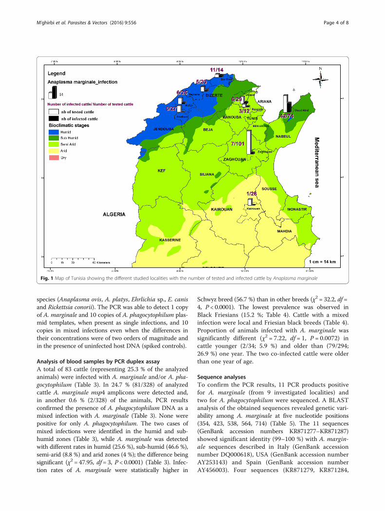
Study design and sampling approachA cross-sectional study was carried out in 9 localities,located in 4 different bioclimatic zones, in northern andcentral Tunisia (humid, sub-humid, semi-arid and arid)where cattle’s breeding is an important economic activity(Fig. 1). All localities have a Mediterranean climate - cool,moist winters and dry, hot summers. Topographic-ally, the areas have rolling hills interspersed withfarmland, grassland, oak woodlands and Mediterraneanscrub (Olea europaea, Pistacia lentiscus, Cistus monspe-liensis, etc.). A total of 80 farms with fewer than 30 ani-mals per farm were chosen randomly as representativeof the local management system on the basis of therecommendations of the State Veterinary Office. Animalhusbandry practices are generally traditional small herdsgrazing on permanent pastures or bush. A total of 328cattle were sampled of which 37.2 % were local breed,32.3 % cross-breeds, 18 % Friesian, 9.2 % Schwytz and3.4 % Holstein. Animals ranged in age between 3 monthsand 13 years, and most were dairy cattle (97.6 %).
Blood sample collection and DNA extractionAnimals were bled once between June and November, aperiod during which they are typically grazing in pasturesand exposed to tick bites. Blood was sampled in tubescontaining ethylenediamine tetraacetic acid and DNA was
extracted using the QIAamp DNA Mini Kit (QIAGEN,Hilden, Germany). DNA yields were determined with aNano-Drop® ND-1000 Spectrophotometer (Nano-DropTechnologies, DE, USA).DNA samples were subjected to duplex PCR assay in
order to detect A. marginale and A. phagocytophilum asdescribed above, and amplicons were resolved in eth-idium bromide-stained agarose (Gellyphor, EuroClone,Milan, Italy) gels (1.5 %) and measured by comparingthem with the with Gene RulerTM 100-bp DNA Ladder(MBI Fermentas, Vilnius, Lithuania) as molecular marker.Gels were photographed using Gel Doc 2000 (Bio-Rad,Hercules, CA, USA).
Sequencing and data analysisThe specificity of the duplex PCR was confirmed by se-quencing PCR amplicons of A. marginale and A. pha-gocytophilum using primers M4-OvMar-F/M4-Mar-Rfor msp4 gene and Msp2-3 F/Msp2-3R for msp2, respect-ively. Thirteen randomly chosen positive PCR productswere purified using the ExoSAP cleanup procedure(Amersham Biosciences, Piscataway, NJ, USA). All nu-cleotide sequences were obtained using the Big DyeTerminator v.3.1 Cycle Sequencing Kit (Applied Biosys-tems, Foster City, CA, USA) and the 3130 automatedsequencer (Applied Biosystems). The sequences were edi-ted and aligned using DNA Baser Sequence Aligner v3.5.4software (Heracle BioSoft SRL, www.DnaBaser.com) toobtain optimal sequence alignment files. A BLAST ana-lysis was made in the NCBI database to retrieve sets ofhomologues exhibiting high scores with the partial msp2and msp4 gene of A. phagocytophilum and A. marginale,respectively.
Statistical analysisThe Chi-square or Fisher's exact tests were used tocompare proportions of positivity in relation with biocli-matic zone, breed and sex. Observed differences wereconsidered significant when the resulting P-value wasless than 0.05.
Nucleotide sequence accession numbersSequence data were deposited in GenBank; accessionnumbers for the partial msp2 and msp4 sequences areKR871275–KR871287.
ResultsPerformance of the duplex PCR assayFragments of the expected size were generated from thetemplate plasmids representing A. marginale (420 bp)and A. phagocytophilum (334 bp); while DNA fromuninfected bovines used as negative control, displayedno evidence of fragment amplification. Similarly, noamplicons were obtained when testing DNA from other
Table 2 Analytical sensitivity of the duplex PCR assay
Plasmid copiesa DNAuninfected cattleb
A. phagocytophilum A. marginale
10 AP N Positive Negative
10 AP Y Positive Negative
1 AP N Negative Negative
1 AP Y Negative Negative
10 AM N Negative Positive
1 AM N Negative Positive
1 AM Y Negative Positive
103 AP + 10 AM N Positive Positive
103 AP + 10 AM Y Positive Positive
10 AP + 103 AM N Positive Positive
10 AP + 103 AM Y Positive PositiveaAP, plasmid with an insert of the msp2 gene fragment of Anaplasmaphagocytophilum; AM, plasmid with an insert of the msp4 gene fragment ofAnaplasma marginalebPresence (Y) or absence (N) in the PCR reaction of DNA extracted from bloodfrom a non-infected cow spiked with the indicated plasmid orplasmid combinations
M’ghirbi et al. Parasites & Vectors (2016) 9:556 Page 3 of 8
species (Anaplasma ovis, A. platys, Ehrlichia sp., E. canisand Rickettsia conorii). The PCR was able to detect 1 copyof A. marginale and 10 copies of A. phagocytophilum plas-mid templates, when present as single infections, and 10copies in mixed infections even when the differences intheir concentrations were of two orders of magnitude andin the presence of uninfected host DNA (spiked controls).
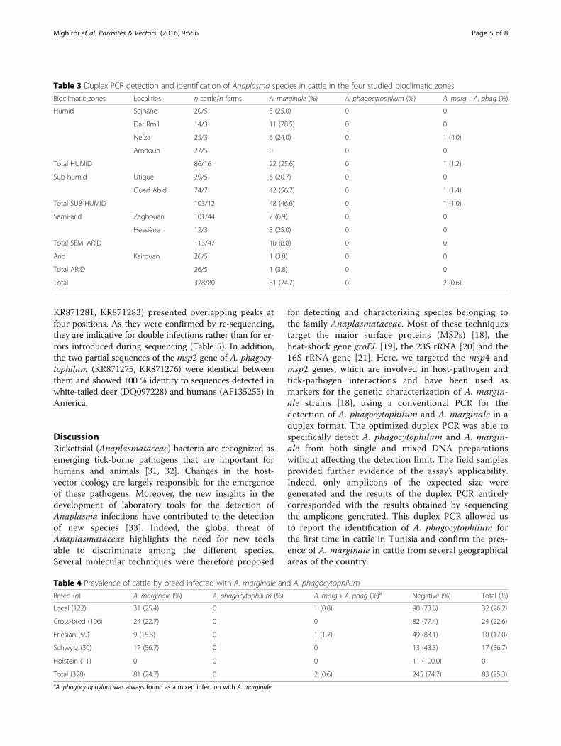
Analysis of blood samples by PCR duplex assayA total of 83 cattle (representing 25.3 % of the analyzedanimals) were infected with A. marginale and/or A. pha-gocytophilum (Table 3). In 24.7 % (81/328) of analyzedcattle A. marginale msp4 amplicons were detected and,in another 0.6 % (2/328) of the animals, PCR resultsconfirmed the presence of A. phagocytophilum DNA as amixed infection with A. marginale (Table 3). None werepositive for only A. phagocytophilum. The two cases ofmixed infections were identified in the humid and sub-humid zones (Table 3), while A. marginale was detectedwith different rates in humid (25.6 %), sub-humid (46.6 %),semi-arid (8.8 %) and arid zones (4 %); the difference beingsignificant (χ2 = 47.95, df = 3, P < 0.0001) (Table 3). Infec-tion rates of A. marginale were statistically higher in
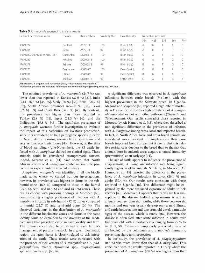
Schwyz breed (56.7 %) than in other breeds (χ2 = 32.2, df =4, P < 0.0001). The lowest prevalence was observed inBlack Friesians (15.2 %; Table 4). Cattle with a mixedinfection were local and Friesian black breeds (Table 4).Proportion of animals infected with A. marginale wassignificantly different (χ2 = 7.22, df = 1, P = 0.0072) incattle younger (2/34; 5.9 %) and older than (79/294;26.9 %) one year. The two co-infected cattle were olderthan one year of age.
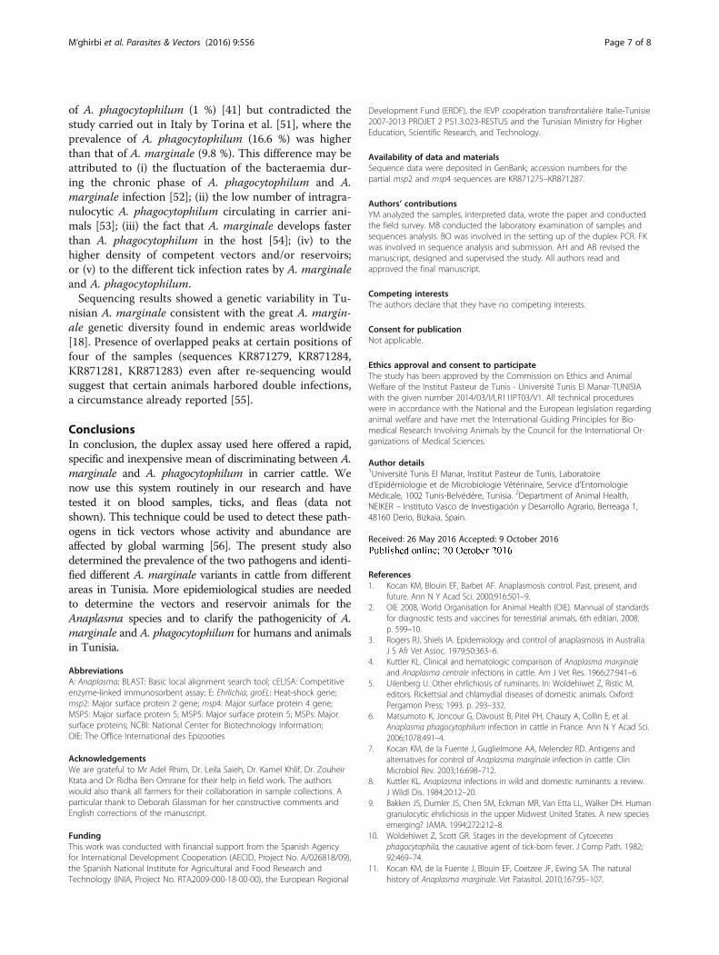
Sequence analysesTo confirm the PCR results, 11 PCR products positivefor A. marginale (from 9 investigated localities) andtwo for A. phagocytophilum were sequenced. A BLASTanalysis of the obtained sequences revealed genetic vari-ability among A. marginale at five nucleotide positions(354, 423, 538, 564, 714) (Table 5). The 11 sequences(GenBank accession numbers KR871277–KR871287)showed significant identity (99–100 %) with A. margin-ale sequences described in Italy (GenBank accessionnumber DQ000618), USA (GenBank accession numberAY253143) and Spain (GenBank accession numberAY456003). Four sequences (KR871279, KR871284,
Fig. 1 Map of Tunisia showing the different studied localities with the number of tested and infected cattle by Anaplasma marginale
M’ghirbi et al. Parasites & Vectors (2016) 9:556 Page 4 of 8
KR871281, KR871283) presented overlapping peaks atfour positions. As they were confirmed by re-sequencing,they are indicative for double infections rather than for er-rors introduced during sequencing (Table 5). In addition,the two partial sequences of the msp2 gene of A. phagocy-tophilum (KR871275, KR871276) were identical betweenthem and showed 100 % identity to sequences detected inwhite-tailed deer (DQ097228) and humans (AF135255) inAmerica.
DiscussionRickettsial (Anaplasmataceae) bacteria are recognized asemerging tick-borne pathogens that are important forhumans and animals [31, 32]. Changes in the host-vector ecology are largely responsible for the emergenceof these pathogens. Moreover, the new insights in thedevelopment of laboratory tools for the detection ofAnaplasma infections have contributed to the detectionof new species [33]. Indeed, the global threat ofAnaplasmataceae highlights the need for new toolsable to discriminate among the different species.Several molecular techniques were therefore proposed
for detecting and characterizing species belonging tothe family Anaplasmataceae. Most of these techniquestarget the major surface proteins (MSPs) [18], theheat-shock gene groEL [19], the 23S rRNA [20] and the16S rRNA gene [21]. Here, we targeted the msp4 andmsp2 genes, which are involved in host-pathogen andtick-pathogen interactions and have been used asmarkers for the genetic characterization of A. margin-ale strains [18], using a conventional PCR for thedetection of A. phagocytophilum and A. marginale in aduplex format. The optimized duplex PCR was able tospecifically detect A. phagocytophilum and A. margin-ale from both single and mixed DNA preparationswithout affecting the detection limit. The field samplesprovided further evidence of the assay’s applicability.Indeed, only amplicons of the expected size weregenerated and the results of the duplex PCR entirelycorresponded with the results obtained by sequencingthe amplicons generated. This duplex PCR allowed usto report the identification of A. phagocytophilum forthe first time in cattle in Tunisia and confirm the pres-ence of A. marginale in cattle from several geographicalareas of the country.
Table 3 Duplex PCR detection and identification of Anaplasma species in cattle in the four studied bioclimatic zones
Bioclimatic zones Localities n cattle/n farms A. marginale (%) A. phagocytophilum (%) A. marg + A. phag (%)
Humid Sejnane 20/5 5 (25.0) 0 0
Dar Rmil 14/3 11 (78.5) 0 0
Nefza 25/3 6 (24.0) 0 1 (4.0)
Amdoun 27/5 0 0 0
Total HUMID 86/16 22 (25.6) 0 1 (1.2)
Sub-humid Utique 29/5 6 (20.7) 0 0
Oued Abid 74/7 42 (56.7) 0 1 (1.4)
Total SUB-HUMID 103/12 48 (46.6) 0 1 (1.0)
Semi-arid Zaghouan 101/44 7 (6.9) 0 0
Hessiène 12/3 3 (25.0) 0 0
Total SEMI-ARID 113/47 10 (8.8) 0 0
Arid Kairouan 26/5 1 (3.8) 0 0
Total ARID 26/5 1 (3.8) 0 0
Total 328/80 81 (24.7) 0 2 (0.6)
Table 4 Prevalence of cattle by breed infected with A. marginale and A. phagocytophilum
Breed (n) A. marginale (%) A. phagocytophilum (%) A. marg + A. phag (%)a Negative (%) Total (%)
Local (122) 31 (25.4) 0 1 (0.8) 90 (73.8) 32 (26.2)
Cross-bred (106) 24 (22.7) 0 0 82 (77.4) 24 (22.6)
Friesian (59) 9 (15.3) 0 1 (1.7) 49 (83.1) 10 (17.0)
Schwytz (30) 17 (56.7) 0 0 13 (43.3) 17 (56.7)
Holstein (11) 0 0 0 11 (100.0) 0
Total (328) 81 (24.7) 0 2 (0.6) 245 (74.7) 83 (25.3)aA. phagocytophylum was always found as a mixed infection with A. marginale
M’ghirbi et al. Parasites & Vectors (2016) 9:556 Page 5 of 8
The obtained prevalence of A. marginale (24.7 %) waslower than that reported in Kansas (37.6 %) [21], India(73.1–36.8 %) [34, 35], Sicily (50 %) [36], Brazil (70.2 %)[37], South African provinces (65–90 %) [38], Texas(82 %) [39] and Costa Rica (56.9 %) [40]. By contrast,this prevalence was higher than those recorded inTurkey (2.8 %) [41], Egypt (21.3 %) [42] and thePhilippines (19.8 %) [43]. The significant prevalence ofA. marginale warrants further investigation to evaluatethe impact of this bacterium on livestock production,since it is considered to be a pathogenic species in cattlein North Africa, causing severe clinical symptoms andvery serious economic losses [44]. However, at the timeof blood sampling (June-November), the 83 cattle in-fected with A. marginale showed no clinical signs. Theseanimals could be considered asymptomatic carriers.Indeed, Sergent et al. [44] have shown that NorthAfrican strains of A. marginale confer an immune pro-tection in experimentally infected animals.Anaplasma marginale was identified in all the biocli-
matic zones where we carried out our investigations,however, its prevalence was highest in farms in the sub-humid zone (46.6 %) compared to those in the humid(25.6 %), semi-arid (8.8 %) and arid (3.8 %) zones. Theseresults concur with previous findings in Morocco [45],demonstrating a higher prevalence of infection with A.marginale in cattle in sub-humid (52 %) zones comparedto humid (22.7 %) and semi-arid zone (20 %). Theobserved variations in the distribution of A. marginalein the different bioclimatic zones and farms in the samelocality could be explained by the diversity of the Ixodi-dae fauna that parasitize cattle in each locality and farm.The difference can also be attributed to each farmer’smanagement of pasture livestock. In a given bioclimaticregion, the latter factor is closely related to tick infest-ation of the cattle. These results were correlated withthe presence of tick vectors of A. marginale and A. pha-gocytophilum, mainly Hyalomma spp., Rhipicephalusspp. and Ixodes spp. [46, 47].
A significant difference was observed in A. marginaleinfections between cattle breeds (P < 0.05), with thehighest prevalence in the Schwytz breed. In Uganda,Magona and Mayende [48] reported a high rate of mortal-ity in Friesian cattle due to a high prevalence of A. margin-ale associated or not with other pathogens (Theileria andTrypanosoma). Our results contradict those reported inMorocco by Ait Hamou et al. [45], where they described anon-significant difference in the prevalence of infectionwith A. marginale among cross, local and imported breeds.In fact, in North Africa, local and cross-breed animals areconsidered more resistant to anaplasmosis than purebreeds imported from Europe. But it seems that this rela-tive resistance is due less to the breed than to the fact thatanimals born in endemic areas acquire a natural immunity(premunition) at an early age [44].The age of cattle appears to influence the prevalence of
anaplasmosis, A. marginale infection rate being signifi-cantly higher in older animals. Similarly, in Morocco, AitHamou et al. [45] reported the difference in the preva-lence of A. marginale infections in calves (26.1 %) andadults (52.4 %). Our results were consistent with thosereported in Uganda [48]. This difference might be ex-plained by the more sustained exposure of adults to tickvectors [49]. Moreover, it appears that calves are less sus-ceptible to the disease. Indeed, anaplasmosis is rare inanimals younger than six months, while those between sixmonths and one year usually develop only a mild illness,and cattle between one and two years old develop multiplesigns of the disease, which is rarely fatal. However, thedisease is often fatal after acute infection in adults overtwo years old, with a mortality risk ranging from 29 % to49 % [7, 50]. Calves are temporarily protected (maternalantibodies) by the colostrum and a mother’s immunity,preventing short-term protection.In our study, the prevalence of A. phagocytophilum
(0.6 %) was much lower than that of A. marginale. Thisconcurred with the results reported in Turkey where theprevalence of A. marginale (2.8 %) was higher than that
Table 5 A. marginale sequencing analysis results
GenBank accession number Locality Blast analysis Similarity (%) Host (Country) Nucleotide positionsa
354 423 538 564 714
KR871277 Dar Rmil AY253143 100 Bison (USA) A G T A C
KR871284 Nefza AY253143 99 Bison (USA) A R T R C
KR871280, KR871285 to KR871287 Oued Abid DQ000618 100 Bison (Italy) G A T G C
KR871282 Hessiène DQ000618 100 Bison (Italy) G A T G C
KR871279 Sejnane DQ000618 99 Bison (Italy) R A T G C
KR871278 Zaghouan AY456003 100 Deer (Spain) G G T A C
KR871281 Utique AY456003 99 Deer (Spain) R G T A C
KR871283 Kairouan DQ000618 99 Cattle (Italy) G A T G Y
Abbreviations: R degenerated nucleotide (A/G), Y degenerated nucleotide (C/T)aNucleotide positions are indicated referring to the complete msp4 gene sequence (e.g. AF428081)
M’ghirbi et al. Parasites & Vectors (2016) 9:556 Page 6 of 8
of A. phagocytophilum (1 %) [41] but contradicted thestudy carried out in Italy by Torina et al. [51], where theprevalence of A. phagocytophilum (16.6 %) was higherthan that of A. marginale (9.8 %). This difference may beattributed to (i) the fluctuation of the bacteraemia dur-ing the chronic phase of A. phagocytophilum and A.marginale infection [52]; (ii) the low number of intragra-nulocytic A. phagocytophilum circulating in carrier ani-mals [53]; (iii) the fact that A. marginale develops fasterthan A. phagocytophilum in the host [54]; (iv) to thehigher density of competent vectors and/or reservoirs;or (v) to the different tick infection rates by A. marginaleand A. phagocytophilum.Sequencing results showed a genetic variability in Tu-
nisian A. marginale consistent with the great A. margin-ale genetic diversity found in endemic areas worldwide[18]. Presence of overlapped peaks at certain positions offour of the samples (sequences KR871279, KR871284,KR871281, KR871283) even after re-sequencing wouldsuggest that certain animals harbored double infections,a circumstance already reported [55].
ConclusionsIn conclusion, the duplex assay used here offered a rapid,specific and inexpensive mean of discriminating between A.marginale and A. phagocytophilum in carrier cattle. Wenow use this system routinely in our research and havetested it on blood samples, ticks, and fleas (data notshown). This technique could be used to detect these path-ogens in tick vectors whose activity and abundance areaffected by global warming [56]. The present study alsodetermined the prevalence of the two pathogens and identi-fied different A. marginale variants in cattle from differentareas in Tunisia. More epidemiological studies are neededto determine the vectors and reservoir animals for theAnaplasma species and to clarify the pathogenicity of A.marginale and A. phagocytophilum for humans and animalsin Tunisia.
AbbreviationsA: Anaplasma; BLAST: Basic local alignment search tool; cELISA: Competitiveenzyme-linked immunosorbent assay; E: Ehrlichia; groEL: Heat-shock gene;msp2: Major surface protein 2 gene; msp4: Major surface protein 4 gene;MSP5: Major surface protein 5; MSP5: Major surface protein 5; MSPs: Majorsurface proteins; NCBI: National Center for Biotechnology Information;OIE: The Office International des Epizooties
AcknowledgementsWe are grateful to Mr Adel Rhim, Dr. Leila Saieh, Dr. Kamel Khlif, Dr. ZouheirKtata and Dr Ridha Ben Omrane for their help in field work. The authorswould also thank all farmers for their collaboration in sample collections. Aparticular thank to Deborah Glassman for her constructive comments andEnglish corrections of the manuscript.
FundingThis work was conducted with financial support from the Spanish Agencyfor International Development Cooperation (AECID, Project No. A/026818/09),the Spanish National Institute for Agricultural and Food Research andTechnology (INIA, Project No. RTA2009-000-18-00-00), the European Regional
Development Fund (ERDF), the IEVP coopération transfrontalière Italie-Tunisie2007-2013 PROJET 2 PS1.3.023-RESTUS and the Tunisian Ministry for HigherEducation, Scientific Research, and Technology.
Availability of data and materialsSequence data were deposited in GenBank; accession numbers for thepartial msp2 and msp4 sequences are KR871275–KR871287.
Authors’ contributionsYM analyzed the samples, interpreted data, wrote the paper and conductedthe field survey. MB conducted the laboratory examination of samples andsequences analysis. BO was involved in the setting up of the duplex PCR. FKwas involved in sequence analysis and submission. AH and AB revised themanuscript, designed and supervised the study. All authors read andapproved the final manuscript.
Competing interestsThe authors declare that they have no competing interests.
Consent for publicationNot applicable.
Ethics approval and consent to participateThe study has been approved by the Commission on Ethics and AnimalWelfare of the Institut Pasteur de Tunis - Université Tunis El Manar-TUNISIAwith the given number 2014/03/I/LR11IPT03/V1. All technical procedureswere in accordance with the National and the European legislation regardinganimal welfare and have met the International Guiding Principles for Bio-medical Research Involving Animals by the Council for the International Or-ganizations of Medical Sciences.
Author details1Université Tunis El Manar, Institut Pasteur de Tunis, Laboratoired’Epidémiologie et de Microbiologie Vétérinaire, Service d’EntomologieMédicale, 1002 Tunis-Belvédère, Tunisia. 2Department of Animal Health,NEIKER – Instituto Vasco de Investigación y Desarrollo Agrario, Berreaga 1,48160 Derio, Bizkaia, Spain.
Received: 26 May 2016 Accepted: 9 October 2016
References1. Kocan KM, Blouin EF, Barbet AF. Anaplasmosis control. Past, present, and
future. Ann N Y Acad Sci. 2000;916:501–9.2. OIE 2008, World Organisation for Animal Health (OIE). Mannual of standards
for diagnostic tests and vaccines for terrestirial animals, 6th editian, 2008,p. 599–10.
3. Rogers RJ, Shiels IA. Epidemiology and control of anaplasmosis in Australia.J S Afr Vet Assoc. 1979;50:363–6.
4. Kuttler KL. Clinical and hematologic comparison of Anaplasma marginaleand Anaplasma centrale infections in cattle. Am J Vet Res. 1966;27:941–6.
5. Uilenberg U. Other ehrlichiosis of ruminants. In: Woldehiwet Z, Ristic M,editors. Rickettsial and chlamydial diseases of domestic animals. Oxford:Pergamon Press; 1993. p. 293–332.
6. Matsumoto K, Joncour G, Davoust B, Pitel PH, Chauzy A, Collin E, et al.Anaplasma phagocytophilum infection in cattle in France. Ann N Y Acad Sci.2006;1078:491–4.
7. Kocan KM, de la Fuente J, Guglielmone AA, Melendez RD. Antigens andalternatives for control of Anaplasma marginale infection in cattle. ClinMicrobiol Rev. 2003;16:698–712.
8. Kuttler KL. Anaplasma infections in wild and domestic ruminants: a review.J Wildl Dis. 1984;20:12–20.
9. Bakken JS, Dumler JS, Chen SM, Eckman MR, Van Etta LL, Walker DH. Humangranulocytic ehrlichiosis in the upper Midwest United States. A new speciesemerging? JAMA. 1994;272:212–8.
10. Woldehiwet Z, Scott GR. Stages in the development of Cytoecetesphagocytophila, the causative agent of tick-born fever. J Comp Path. 1982;92:469–74.
11. Kocan KM, de la Fuente J, Blouin EF, Coetzee JF, Ewing SA. The naturalhistory of Anaplasma marginale. Vet Parasitol. 2010;167:95–107.
M’ghirbi et al. Parasites & Vectors (2016) 9:556 Page 7 of 8

12. Kocan KM, de la Fuente J, Blouin EF, Garcia-Garcia JC. Anaplasma marginale(Rickettsiales: Anaplasmataceae): recent advances in defining host-pathogenadaptations of a tick-borne rickettsia. Parasitol. 2004;129:S285–300.
13. Luther DG, Cox HU, Nelson WO. Comparison of serotests with calfinoculations for detection of carriers in anaplasmosis-vaccinated cattle. Am JVet Res. 1980;41:2085–6.
14. Knowles D, Torioni de Echaide S, Palmer G, McGuire T, Stiller D, McElwain T.Antibody against an Anaplasma marginale MSP5 epitope common to tickand erythrocyte stages identifies persistently infected cattle. J Clin Microbiol.1996;34:2225–30.
15. Dreher UM, de la Fuente J, Hofmann-Lehmann R, Meli ML, Pusterla N, KocanKM, et al. Serologic cross-reactivity between Anaplasma marginale andAnaplasma phagocytophilum. Clin Diagn Lab Immunol. 2005;12:1177–83.
16. Strik NI, Alleman AR, Barbet AF, Sorenson HL, Wamsley HL, Gaschen FP,et al. Characterization of Anaplasma phagocytophilum major surface protein5 and the extent of its cross-reactivity with A. marginale. Clin VaccineImmunol. 2007;14:262–8.
17. Palmer GH, Barbet AF, Cantor GH, McGuire TC. Immunization of cattle withthe MSP-1 surface protein complex induces protection against a structurallyvariant Anaplasma marginale isolate. Infect Immun. 1989;57:3666–9.
18. de la Fuente J, Ruybal P, Mtshali MS, Naranjo V, Shuqing L, Mangold AJ,et al. Analysis of world strains of Anaplasma marginale using major surfaceprotein 1a repeat sequences. Vet Microbiol. 2007;119:382–90.
19. Park HS, Lee JH, Jeong EJ, Park TK, Kim TY, Chae JS, Park JH, Klein TA, JangWJ, Park KH, Lee SH, et al. Differentiation of Anaplasmataceae throughpartial groEL gene analysis. Microbiol Immunol. 2005;49:655–62.
20. Dahmani M, Davoust B, Benterki MS, Fenollar F, Raoult D, Mediannikov O.Development of a new PCR-based assay to detect Anaplasmataceae andthe first report of Anaplasma phagocytophilum and Anaplasma platys incattle from Algeria. Comp Immunol Microbiol Infect Dis. 2015;39:39–45.
21. Reinbold JB, Coetzee JF, Sirigireddy KR, Ganta RR. Detection of Anaplasmamarginale and A. phagocytophilum in bovine peripheral blood samples byduplex real-time reverse transcriptase PCR assay. J Clin Microbiol. 2010;48:2424–32.
22. Hurtado A, Barandika JF, Oporto B, Minguijón E, Povedano I, García-PérezAL. Risks of suffering tick-borne diseases in sheep translocated to a tickinfested area: a laboratory approach for the investigation of an outbreak.Ticks Tick Borne Dis. 2015;6:31–7.
23. Sarih M, M’ghirbi Y, Bouattour A, Gern L, Baranton G, Postic D. Detectionand identification of Ehrlichia spp. in ticks collected in Tunisia and Morocco.J Clin Microbiol. 2005;43:1127–32.
24. M’ghirbi Y, Ghorbel A, Amouri M, Nebaoui A, Haddad S, Bouattour A.Clinical, serological, and molecular evidence of ehrlichiosis andanaplasmosis in dogs in Tunisia. Parasitol Res. 2009;104:767–74.
25. M’Ghirbi Y, Yaïch H, Ghorbel A, Bouattour A. Anaplasma phagocytophilum inhorses and ticks in Tunisia. Parasit Vectors. 2012;5:180–4.
26. Belkahia H, Ben Said M, Sayahi L, Alberti A, Messadi L. Detection of novelstrains genetically related to Anaplasma platys in Tunisian one-humpedcamels (Camelus dromedarius). J Infect Dev Ctries. 2015;9:1117–25.
27. Belkahia H, Ben Said M, Alberti A, Abdi K, Issaoui Z, Hattab D, et al. Firstmolecular survey and novel genetic variants' identification of Anaplasmamarginale, A. centrale and A. bovis in cattle from Tunisia. Infect Genet Evol.2015;34:361–71.
28. Ben Said M, Belkahia H, Alberti A, Zobba R, Bousrih M, Yahiaoui M, et al.Molecular survey of Anaplasma species in small ruminants reveals thepresence of novel strains closely related to A. phagocytophilum in Tunisia.Vector Borne Zoonotic Dis. 2015;15:580–90.
29. Ben Said M, Belkahia H, Karaoud M, Bousrih M, Yahiaoui M, Daaloul-Jedidi M,Messadi L. First molecular survey of Anaplasma bovis in small ruminantsfrom Tunisia. Vet Microbiol. 2015;179:322–6.
30. Zeidner NS, Burkot TR, Massing R, Nicholson WL, Dolan MC, Rutherford JS,et al. Transmission of the agent of human granulocytic ehrlichiosis by Ixodesspinipalpis ticks: evidence of an enzootic cycle of dual infection with Borreliaburgdorferi in Northern Colorado. J Infect Dis. 2000;182:616–9.
31. Rikihisa Y. New findings on members of the family Anaplasmataceae ofveterinary importance. Ann NY Acad Sci. 2006;1078:438–45.
32. Ismail N, Bloch KC, McBride JW. Human ehrlichiosis and anaplasmosis. ClinLab Med. 2010;30:261–92.
33. Doudier B, Olano J, Parola P, Brouqui P. Factors contributing toemergence of Ehrlichia and Anaplasma spp. as human pathogens. VetParasitol. 2010;167:149–54.
34. Singh H, Jyoti Haque M, Singh NK, Rath SS. Molecular detection of Anaplasmamarginale infection in carrier cattle. Ticks Tick Borne Dis. 2012;3:55–8.
35. Sharma A, Singla LD, Tuli A, Kaur P, Bal MS. Detection and assessment ofrisk factors associated with natural concurrent infection of Trypanosomaevansi and Anaplasma marginale in dairy animals by duplex PCR in easternPunjab. Trop Anim Health Prod. 2015;47:251–7.
36. De la Fuente J, Torina A, Caracappa S, Tumeno G, Furla R, Almazan C, KocanKM. Serologic and molecular characterization of Anaplasma species infection infarm animals and ticks from Sicily. Vet Parasitol. 2005;133:357–62.
37. Pohl AE, Cabezas-Cruz A, Flávio M, Ribeiro B, Angélica J, da Silveira G, et al.Detection of genetic diversity of Anaplasma marginale isolates in MinasGerais, Brazil. Rev Bras Parasitol Vet. 2013;22:129–35.
38. Mutshembele AM, Cabezas-Cruzc A, Mtshali MS, Thekisoe OMM, Galindo RC,de la Fuente J. Epidemiology and evolution of the genetic variability ofAnaplasma marginale in South Africa. Ticks Tick borne Dis. 2014;5:624–31.
39. Hairgrove T, Schroeder ME, Budke CM, Rodgersb S, Chungd C, Uetie MW,Bounpheng MA. Molecular and serological in-herd prevalence of Anaplasmamarginale infection in Texas cattle Preventive. Vet Med. 2015;119:1–9.
40. Shebish E, Vemulapalli R, Oseto C. Prevalence and molecular detection ofAnaplasma marginale, Babesia bovis and Babesia bigemina in cattle fromPuntarenas Province, Costa Rica. Vet Parasitol. 2012;188:164–7.
41. Aktas M, Altay K, Dumanli N. Molecular detection and identification ofAnaplasma and Ehrlichia species in cattle from Turkey. Ticks Tick Borne Dis.2011;2:62–5.
42. El-Ashker M, Hotzelb H, Gwidab M, El-Beskawyd M, Silaghi C, Tomaso H.Molecular biological identification of Babesia, Theileria, and Anaplasmaspecies in cattle in Egypt using PCR assays, gene sequence analysis and anovel DNA microarray. Vet Parasitol. 2015;207:329–34.
43. Ybanez AP, Ybanez RHD, Cruz-Flores MJ, Xuenan X, Yokoyama N, InokumaH. High genetic diversity of Anaplasma marginale detected from Philippinecattle. J Vet Med Sci. 2014;76:1009–14.
44. Sergent E, Donatien A, Parrot L, Lestoquard F. Etude sur les PiroplasmosesBovines. Arch Inst Pasteur Algérie. 1945. p. 816.
45. Ait Hamou S, Rahali T, Sahibi H, Belghyti D, Losson B, Goff W, Rhalem A.Molecular and serological prevalence of Anaplasma marginale in cattle ofNorth Central Morocco. Res Vet Sc. 2012;93:1318–23.
46. Bouattour A, Darghouth MA, Daoud A. Distribution and ecology of ticks(Acari: Ixodidae) infesting livestock in Tunisia: an overview of eight yearsfield collection. Parasitologia. 1999;41:5–10.
47. M’ghirbi Y, Hurtado A, Bouattour A. Theileria and Babesia parasites in ticks inTunisia. Transboundary Emerging Dis. 2010;57:49–51.
48. Magona JW, Mayende JSP. Occurrence of concurrent trypanosomosis,theileriosis, anaplasmosis and helminthosis in Friesian, Zebu and Sahiwalcattle in Uganda. J Vet Res. 2002;69:133–40.
49. Bouattour A, Darghouth MA, Ben ML. Cattle infestation by Hyalomma ticksand prevalence of Theileria in Hyalomma. detritum species in Tunisia. VetPar. 1996;65:256–63.
50. Richey EJ. Bovine anaplasmosis. In: The 24th Annual Convention ofProceedings of American Association of Bovine Practices, Orlando,Florida, 1991. p. 3–11.
51. Torina A, Vicente J, Alongi A, Scimeca A, Turla R, Nicosia S, et al. Observedprevalence of tick-borne pathogens in domestic animals in Sicily, Italyduring 2003–2005. Zoon Pub Health. 2007;54:8–15.
52. Kieser ST, Eriks IE, Palmer GH. Cyclic rickettsemia during persistentAnaplasma marginale infection in cattle. Infect Immun. 1990;58:1117–9.
53. Pusterla N, Braun U. Clinical findings in cows after experimental infectionwith Ehrlichia phagocytophila. Zentralbl Veterinarmed. 1997;44:385–90.
54. Hoar BR, Nieto NC, Rhodes DM, Foley JE. Evaluation of sequentialcoinfection with Anaplasma phagocytophilum and Anaplasma marginale incattle. Am J Vet Res. 2008;69:1171–8.
55. Palmer GH, Rurangirwa FR, McElwain TF. Strain composition of the ehrlichiaAnaplasma marginale within persistently infected cattle, a mammalianreservoir for tick transmission. J Clin Microbiol. 2001;39:631–5.
56. Estrada-Pena A. Tick-borne pathogens, transmission rates and climatechange. Front Biosci (Landmark Ed). 2009;14:2674–87.
M’ghirbi et al. Parasites & Vectors (2016) 9:556 Page 8 of 8