An in vivo comparison of the modified Mason-Allen suture technique versus an inclined horizontal
7
An in vivo comparison of the modified Mason-Allen suture technique versus an inclined horizontal mattress suture technique with regard to tendon-to-bone healing: A biomechanical and histologic study in sheep Theodore F. Schlegel, MD, a Richard J. Hawkins, MD, b Chad W. Lewis, PhD, c and A. Simon Turner, BVSc, MS, Dipl ACVS, c Denver, Vail, and Fort Collins, CO The purpose of this study is to examine long-term tendon-to-bone healing, by use of a sheep animal model, after rotator cuff repairs performed with 2 dif- ferent suture techniques: an inclined horizontal mat- tress suture pattern placed with special arthroscopic instrumentation (HMS) and the modified Mason-Allen pattern (MMA). After a pre hoc power analysis, 18 skeletally mature sheep were randomly assigned to either the HMS or MMA repair technique, with con- tralateral limbs used for the control group. At 26 weeks, the animals were euthanized. Six sheep from each group underwent biomechanical testing. Load-to- failure and stiffness results indicated no statistically significant difference between the 2 groups. Avulsion of the tuberosity was the primary mode of failure for both groups. In the remaining 6 sheep, histologic eval- uation demonstrated that, regardless of treatment, the tendon appeared completely healed in the bony trough. Because the long-term biomechanical and his- tologic properties of healed tendons repaired with an HMA technique are equal to those obtained with an MMA technique, the inclined horizontal mattress suture may be appropriate for arthroscopic rotator cuff re- pair. Short-term studies are necessary to determine whether these findings are true early after tendon re- pair, when failure may be most common. (J Shoulder Elbow Surg 2007;16:115-121.) A major concern in rotator cuff repair surgery is failure, particularly in large and massive tears. 3,6 Traditionally, rotator cuff repairs have been per- formed by reapproximating the torn tendon to a bony trough created in the greater tuberosity. 7 It is critical to have sufficient strength at the time of the initial repair to permit early rehabilitation and to improve the likelihood of a successful repair. The modified Mason-Allen suture has been popular for open rotator cuff repairs because of its superior holding power in soft tissue. 4,5 At this time, the majority of all rotator cuff repairs are performed via a classic open or miniopen technique. These approaches make it rela- tively easy to place the modified Mason-Allen suture pattern. Recently, there has been a growing trend toward all-arthroscopic rotator cuff repair. When this tech- nique is being used, it is extremely difficult to place a modified Mason-Allen suture because of the con- straints of the current arthroscopic instrumentation. The simple and horizontal suture patterns are techni- cally more feasible in all-arthroscopic repairs. The strongest possible construct should be created be- cause the initial strength of the tendon attachment to bone is an important factor in the final success of the repair and will ultimately permit more aggressive initial rehabilitation programs. The purpose of this study was to determine whether the long-term results after repair of a horizontal suture technique with special instrumentation is as secure as the modified Mason-Allen technique when reattach- ing tendon to a bony trough in an animal model. MATERIALS AND METHODS After performance of a pre hoc power analysis (power 80%; .05), the right infraspinatus tendon in 18 skeletally mature sheep was reattached into a bony trough. Half of the sheep were randomly assigned to a group in which tendon fixation was performed via the modified Mason-Allen suture (MMA), and the other half underwent fixation by use of an automated suturing device to place a horizontal mattress suture (HMS) (Figure 1). This animal From the a Steadman Hawkins Clinic Denver, Denver, b Steadman Hawkins Sports Medicine Foundation, Vail, and c Colorado State University, Fort Collins. Reprint requests: Theodore F. Schlegel, MD, Steadman Hawkins Clinic Denver, 8200 E Belleview Ave, Suite 615, Greenwood Village, CO 80111. Copyright © 2007 by Journal of Shoulder and Elbow Surgery Board of Trustees. 1058-2746/2007/$32.00 doi:10.1016/j.jse.2006.05.002 115
Transcript of An in vivo comparison of the modified Mason-Allen suture technique versus an inclined horizontal

Attb
TA
TtmftipsetwefsobutttHMmpwpE
F
R
C
1d
n in vivo comparison of the modified Mason-Allen sutureechnique versus an inclined horizontal mattress sutureechnique with regard to tendon-to-bone healing: Aiomechanical and histologic study in sheep
heodore F. Schlegel, MD,a Richard J. Hawkins, MD,b Chad W. Lewis, PhD,c and
. Simon Turner, BVSc, MS, Dipl ACVS,c Denver, Vail, and Fort Collins, COAfTfttrtMcscmtp
anmsTcscbri
ttti
M
�sHwMfi
he purpose of this study is to examine long-termendon-to-bone healing, by use of a sheep animalodel, after rotator cuff repairs performed with 2 dif-
erent suture techniques: an inclined horizontal mat-ress suture pattern placed with special arthroscopicnstrumentation (HMS) and the modified Mason-Allenattern (MMA). After a pre hoc power analysis, 18keletally mature sheep were randomly assigned toither the HMS or MMA repair technique, with con-ralateral limbs used for the control group. At 26eeks, the animals were euthanized. Six sheep fromach group underwent biomechanical testing. Load-to-ailure and stiffness results indicated no statisticallyignificant difference between the 2 groups. Avulsionf the tuberosity was the primary mode of failure foroth groups. In the remaining 6 sheep, histologic eval-ation demonstrated that, regardless of treatment, theendon appeared completely healed in the bonyrough. Because the long-term biomechanical and his-ologic properties of healed tendons repaired with anMA technique are equal to those obtained with anMA technique, the inclined horizontal mattress sutureay be appropriate for arthroscopic rotator cuff re-air. Short-term studies are necessary to determinehether these findings are true early after tendon re-air, when failure may be most common. (J Shoulderlbow Surg 2007;16:115-121.)
rom the aSteadman Hawkins Clinic Denver, Denver, bSteadmanHawkins Sports Medicine Foundation, Vail, and cColorado StateUniversity, Fort Collins.
eprint requests: Theodore F. Schlegel, MD, Steadman HawkinsClinic Denver, 8200 E Belleview Ave, Suite 615, GreenwoodVillage, CO 80111.opyright © 2007 by Journal of Shoulder and Elbow SurgeryBoard of Trustees.
058-2746/2007/$32.00
hoi:10.1016/j.jse.2006.05.002major concern in rotator cuff repair surgery isailure, particularly in large and massive tears.3,6
raditionally, rotator cuff repairs have been per-ormed by reapproximating the torn tendon to a bonyrough created in the greater tuberosity.7 It is criticalo have sufficient strength at the time of the initialepair to permit early rehabilitation and to improvehe likelihood of a successful repair. The modified
ason-Allen suture has been popular for open rotatoruff repairs because of its superior holding power inoft tissue.4,5 At this time, the majority of all rotatoruff repairs are performed via a classic open oriniopen technique. These approaches make it rela-
ively easy to place the modified Mason-Allen sutureattern.
Recently, there has been a growing trend towardll-arthroscopic rotator cuff repair. When this tech-ique is being used, it is extremely difficult to place aodified Mason-Allen suture because of the con-
traints of the current arthroscopic instrumentation.he simple and horizontal suture patterns are techni-ally more feasible in all-arthroscopic repairs. Thetrongest possible construct should be created be-ause the initial strength of the tendon attachment toone is an important factor in the final success of theepair and will ultimately permit more aggressivenitial rehabilitation programs.
The purpose of this study was to determine whetherhe long-term results after repair of a horizontal sutureechnique with special instrumentation is as secure ashe modified Mason-Allen technique when reattach-ng tendon to a bony trough in an animal model.
ATERIALS AND METHODS
After performance of a pre hoc power analysis (power80%; � � .05), the right infraspinatus tendon in 18
keletally mature sheep was reattached into a bony trough.alf of the sheep were randomly assigned to a group inhich tendon fixation was performed via the modifiedason-Allen suture (MMA), and the other half underwent
xation by use of an automated suturing device to place a
orizontal mattress suture (HMS) (Figure 1). This animal115

msto(
O
tkmLmtt
watsMItp
P
edmirw
ri
S
atpbht
B
hcshmKaPmTaC5n
tsm
mattr
116 Schlegel et al J Shoulder Elbow SurgJanuary/February 2007
odel was chosen because the infraspinatus tendon in theheep is similar to the human supraspinatus in width andhickness.2,4,8 The investigation was approved by the Col-rado State University Animal Care and Use Committeeprotocol No. 98-064A-01) (Fort Collins, CO).
perative techniquePreoperative analgesics and antibiotics were adminis-
ered to all animals. General anesthesia was induced withetamine (4 mg/kg) and diazepam (7.5 mg) and wasaintained with halothane (1.5%-3%) in 100% oxygen (2
/min). After aseptic preparation, a 12-cm incision wasade over the right shoulder. The infraspinatus muscle and
endon were isolated. The tendon was then sharply de-ached from the greater tuberosity of the humerus.
A bony trough, 2.0 cm in length and 0.5 cm in depth,as prepared in the proximal humerus with an osteotomend bur. The infraspinatus tendon was sutured into the
rough by use of three No. 2 Ethibond nonabsorbableutures (Ethicon, Somerville, NJ) via either a modifiedason-Allen technique or a horizontal mattress technique.
n both groups, 3 suture anchors were used to secure theendon to the bony trough. Once reattachment was accom-lished, standard closure of soft tissues was performed.
ostoperative protocolAfter surgery, assessment of pain was recorded daily in
ach sheep. All animals received pain management for 3ays. The sheep were temporarily restricted by the attach-ent of a 6-inch softball under the foot of the operative limb
n an attempt to reduce weight-bearing forces. The softballemained in place for 6 weeks, during which the sheep
Figure 1 Comparison of suture patterns commonly usinclined horizontal mattress suture (B), and traditional
ere confined to a small pen. After 6 weeks, the ball was b
emoved, and the animals were allowed unrestricted activ-ties in a larger pen.
ample preparationAt 26 weeks after surgery, the sheep were euthanized in
humane manner with an intravenous overdose of barbi-urate. At necropsy, each infraspinatus muscle-tendon com-lex and its corresponding humerus were dissected fromoth the operative and contralateral control limbs. Afterarvest, each sample was frozen at �30°C and stored athis temperature until biomechanical testing was performed.
iomechanical testingSix of the infraspinatus muscle-tendon complexes and
umeri from each treatment group were used for biome-hanical testing. Each sample was thawed in normal salineolution at 37°C, and nonessential tissue was resected. Theumeral end of each sample was potted in anatomic align-ent with Dyna-Cast (low-temperature thermostat resin;indt Collins, Cleveland, OH). The sample was attached toservohydraulic testing machine (model 809; MTS, Eden
rairie, MN) by use of specialized grips. The lower grip,ade from aluminum, held the potted end of the sample.he upper grip was clamped to the infraspinatus tendon innatomic alignment with the Cryo-Jaw (custom made atSU) design by use of carbon dioxide, frozen to �25°C �°C. Similar to other tendon-healing studies, sutures wereot cut before testing.6
After each sample was prepared and attached to theesting apparatus, a 5-N cyclic preload was applied. Eachample was then loaded to failure by use of the MTSachine, with a rate of distraction of 500 mm/min, as has
r rotator cuff repair: modified Mason-Allen suture (A),ess suture (C).
ed fo
een done in previous work.8–10 The site of failure was
ncswi
S
wpegl
H
siiofiwsdTicli
R
nms
1tNtgtd
ri
t.lcfaHe
epas
tttAatfiBbfi3re
D
uagiueTmipwtttb
twTwao
T
MHC
*�†
�
J Shoulder Elbow Surg Schlegel et al 117Volume 16, Number 1
oted for all specimens. Force and displacement data wereollected at 100 Hz to calculate load to failure, and thelope of the linear portion of the force-displacement curveas measured to determine the stiffness of the bone-tendon
nterface.
tatistical analysisThe mean, SE, and SD of load to failure and stiffness
ere determined. By use of analysis of variance and Tukeyost hoc analysis for equal variances (� � .05), the differ-nces between the HMS group, MMA group, and controlroup were determined for the responses with regard to
oad to failure and stiffness.
istologic evaluationThree specimens from each group were evaluated by a
ingle pathologist blinded to treatment for assessment of thentegrity of the tendon insertion site, the degree of collagenngrowth, and the contribution of periosteal fibers to therganization of the tendon attachment. The specimens werexed in 10% neutral buffered formalin and then decalcifiedith formic acid and resin ion exchange. Sequential 7-�m
ections were obtained from the insertion (repair) site of theecalcified specimen and stained with hematoxylin-eosin.he area of interest was the gap between the stump of thenfraspinatus and the bony trough. There are no specificriteria for scoring soft-tissue repairs to bone, but the histo-ogic evaluation consisted of qualitative assessment of thentegrity of the tendon insertion site.
ESULTS
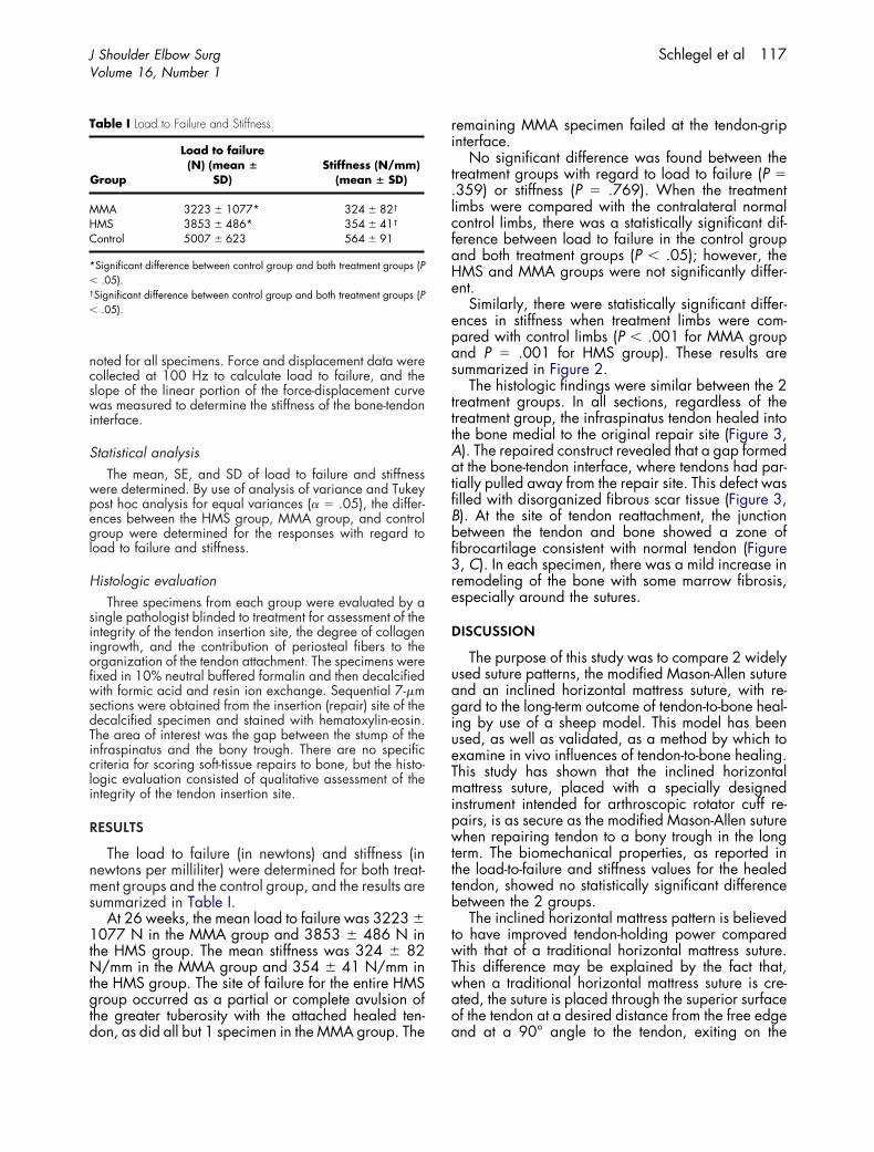
The load to failure (in newtons) and stiffness (inewtons per milliliter) were determined for both treat-ent groups and the control group, and the results are
ummarized in Table I.At 26 weeks, the mean load to failure was 3223 �
077 N in the MMA group and 3853 � 486 N inhe HMS group. The mean stiffness was 324 � 82/mm in the MMA group and 354 � 41 N/mm in
he HMS group. The site of failure for the entire HMSroup occurred as a partial or complete avulsion of
he greater tuberosity with the attached healed ten-
able I Load to Failure and Stiffness
Group
Load to failure(N) (mean �
SD)Stiffness (N/mm)
(mean � SD)
MA 3223 � 1077* 324 � 82†
MS 3853 � 486* 354 � 41†
ontrol 5007 � 623 564 � 91
Significant difference between control group and both treatment groups (P.05).
Significant difference between control group and both treatment groups (P.05).
on, as did all but 1 specimen in the MMA group. The a
emaining MMA specimen failed at the tendon-gripnterface.
No significant difference was found between thereatment groups with regard to load to failure (P �359) or stiffness (P � .769). When the treatmentimbs were compared with the contralateral normalontrol limbs, there was a statistically significant dif-erence between load to failure in the control groupnd both treatment groups (P � .05); however, theMS and MMA groups were not significantly differ-nt.
Similarly, there were statistically significant differ-nces in stiffness when treatment limbs were com-ared with control limbs (P � .001 for MMA groupnd P � .001 for HMS group). These results areummarized in Figure 2.
The histologic findings were similar between the 2reatment groups. In all sections, regardless of thereatment group, the infraspinatus tendon healed intohe bone medial to the original repair site (Figure 3,). The repaired construct revealed that a gap formedt the bone-tendon interface, where tendons had par-
ially pulled away from the repair site. This defect waslled with disorganized fibrous scar tissue (Figure 3,). At the site of tendon reattachment, the junctionetween the tendon and bone showed a zone ofbrocartilage consistent with normal tendon (Figure, C). In each specimen, there was a mild increase inemodeling of the bone with some marrow fibrosis,specially around the sutures.
ISCUSSION
The purpose of this study was to compare 2 widelysed suture patterns, the modified Mason-Allen suturend an inclined horizontal mattress suture, with re-ard to the long-term outcome of tendon-to-bone heal-
ng by use of a sheep model. This model has beensed, as well as validated, as a method by which toxamine in vivo influences of tendon-to-bone healing.his study has shown that the inclined horizontalattress suture, placed with a specially designed
nstrument intended for arthroscopic rotator cuff re-airs, is as secure as the modified Mason-Allen suturehen repairing tendon to a bony trough in the long
erm. The biomechanical properties, as reported inhe load-to-failure and stiffness values for the healedendon, showed no statistically significant differenceetween the 2 groups.
The inclined horizontal mattress pattern is believedo have improved tendon-holding power comparedith that of a traditional horizontal mattress suture.his difference may be explained by the fact that,hen a traditional horizontal mattress suture is cre-ted, the suture is placed through the superior surfacef the tendon at a desired distance from the free edge
nd at a 90° angle to the tendon, exiting on the
iBa
tb
ol gro
118 Schlegel et al J Shoulder Elbow SurgJanuary/February 2007
nferior aspect, once again at a 90° angle (Figure 1,). The suture is then passed parallel to the free edge
Figure 2 Load to failure (A) and stiffness (B) in contr
long the inferior aspect of the tendon at a set dis- t
ance and then through the tendon at a 90° angle tooth the inferior and superior surfaces. This tradi-
up versus MMA and HMS groups.
ional horizontal mattress suture differs from the in-

catseswi1hfwrsi
ca
gSMtmttwi
tspbmbt
n rea
J Shoulder Elbow Surg Schlegel et al 119Volume 16, Number 1
lined horizontal mattress suture created with theutomated suturing device used in this study. With
his instrumentation, the suture enters the superiorurface and then travels obliquely away from thedge of the tendon, ultimately exiting on the under-urface of the tendon at a greater distance away fromhere it started on the superior surface. Thus, an
nclined horizontal mattress suture is created (Figure, C). By the nature of this inclined suture pattern, theolding power is increased because the suture travelsor a longer distance in the tendon when comparedith the traditional horizontal mattress suture. The
esultant vector of the inclined horizontal mattressuture creates a compressive force at the bone-tendonnterface.
Another unique finding in this study was theonsistency (SD) that was seen in the load-to-failure
Figure 3 A, Gap formation and medial attachment ooriginal repair site. C, Zone of fibrocartilage at tendo
nd stiffness results for the horizontal mattress e
roup when compared with the MMA group. TheD in the HMS group was nearly half that in theMA group. This may be explained by the fact that
he suturing device allows for a very reproducibleethod of applying the suture. The automated su-
uring device creates a sewing-machine consis-ency. This reproducibility is not generally createdhen the suture is placed by hand, as is the mod-
fied Mason-Allen suture.Another possible explanation for the lower SD with
he HMS technique has to do with the ability of theuture to slide once placed within the tendon. Thisrovides the advantage of having equal tension onoth limbs of the suture once it is secured to bone. Thisay not always occur when the MMA suture is used,ecause the upper limb of the suture often locks onto
he horizontal limb, creating an opportunity for un-
on to bony trough. B, Disorganized fibrous tissues atttachment site.
f tend
qual tension in the sutures as they are secured to
bp
ggicthcblbtbitgotatortaa
iGwtlspWtbt5otmstoubarstuiUtt
paopsfsdlecmepiatbctaficdmwaSsruibcso
stttsatd
G
R
120 Schlegel et al J Shoulder Elbow SurgJanuary/February 2007
one. The consistency in the limbs of the suture mayrovide a more reproducible repair.
When the biomechanical results in the controlroup were compared with those in both studyroups, there were statistically significant differences
n both load to failure and stiffness. These results areonsistent with other studies, which have shown thathe intermediate biomechanical results after tendonealing are inferior to normal tendons.8,10 The de-reased strength of the healed construct is believed toe a result of early cyclic loading.1 These low-level
oads create an early gap formation at the tendon-one interface in animal models. Histologic evalua-
ion from this study, along with others, confirms thisiologic process, which occurs during early heal-
ng.10 Eventually the tendon heals medially into therough, and a fibrous scar fills in where the originalapping occurred. This most likely creates a weakerverall construct when compared with the normal
endon. This is represented by lower load-to-failurend stiffness values in the repaired tendons. Factors
hat influence this process include the initial strengthf the repaired construct, healing time, and earlyehabilitation or mobilization. It is interesting to notehat this scar filling the gap is consistently formed innimal models but not in human beings. This must beppreciated in interpreting our study results.
With the sheep model, there are limitations regard-ng restriction of motion after rotator cuff repairs.erber et al4 reported that, without prevention of fulleight-bearing, most repairs failed regardless of the
echnique that was used. For this reason, they be-ieved that the only way to protect the repair was touspend the animal in a hanging device, as well as tolace a softball under the hoof of the involved limb.ith this device, the sheep did not bear full weight on
he limb but touched the ground, avoiding full weight-earing and reducing the rotational forces between
he limb and the ground. The ball was removed afterweeks. Although this was a preliminary study with-
ut sufficient statistical power, the implications werehat postoperative protection from tension overloaday be necessary for successful healing. In our re-
earch institution, the Animal Care and Use Commit-ee would not allow us to suspend the animal as partf an immobilization technique. Instead, we havesed a postoperative protocol in which a 6-inch soft-all is fixed under the foot of the operative limb in anttempt to reduce weight-bearing forces. The softballemained in place for 6 weeks, during which theheep were confined to a small pen. After 6 weeks,he ball was removed, and the animals were allowednrestricted activities in a larger pen. Other researchnstitutions conducting the same type of study in thenited States have published studies using this iden-
ical restriction. It is important to note that the loads at
he repair site with this approach are unknown.An all-arthroscopic technique for rotator cuff re-airs can be technically challenging. The theoreticdvantages of this technique compared with the openr miniopen procedure are decreased postoperativeain and decreased postoperative morbidity.9 Theuccess of tendon-to-bone healing relies on manyactors, one of which is believed to be the initialecurity of the repair. It should be noted that our studyid not evaluate initial security but only addressed
ong-term repair. In the long term, any initial differ-nces in suture method could be masked, as signifi-ant scar tissue fills the gap in all cases in this animalodel. Further study evaluating these approaches atarlier time points, between 1 and 8 weeks, mayrovide further important information on the compar-
son of these approaches. Nevertheless, for an all-rthroscopic repair, the ability to reattach the torn
endon to bone and create a secure construct haseen limited by the current techniques. At this time, itan be quite difficult to perform an all-arthroscopicechnique because of challenges related to creatingppropriate suture pattern placement in tendon. Inact, the ability to use a modified Mason-Allen suturen an arthroscopic repair is considered significantlyhallenging by even the best arthroscopists. With theevelopment of new instrumentation, the surgeonay be able to place a suture easily arthroscopically,hich provides fixation that is as secure as thatchieved with the present open treatment methods.ince the completion of this study, we have nowtarted to use this suture device in our arthroscopicotator cuff repairs. This instrumentation has alloweds to place a desired suture pattern easily and quicklyn our early human clinical protocols. Armed with theasic science results, we have now become moreonfident in our all-arthroscopic repairs and havetarted a prospective study to evaluate our clinicalutcomes.
Future research is necessary to develop anchoringystems that will allow secure fixation via a knotlessechnique. We are presently using an anchoring sys-em that has been developed to allow for a knotlessechnique. This anchor allows us to create ideal ten-ion with our repair and secure fixation. This knotlessnchor is compatible with our current automated su-
ure device, creating an increased sense of confi-ence in our arthroscopic rotator cuff repairs.
We gratefully acknowledge Tyler Richardson and Jennaodfrey for their technical assistance.
EFERENCES
1. Burkhart SS, Fischer SP, Nottage WM, Esch JC, Barber FA,Doctor D, et al. Tissue fixation security in transosseous rotator cuffrepairs: a mechanical comparison of simple versus mattress su-
tures. Arthroscopy 1996;12:704-8.
1
J Shoulder Elbow Surg Schlegel et al 121Volume 16, Number 1
2. France EP, Paulos LE, Harner CD, Straight CB. Biomechanicalevaluation of rotator cuff fixation methods. Am J Sports Med1989;17:176-81.
3. Galatz LM, Ball CM, Teefey SA, Middleton WD, Yamaguchi K.The outcome and repair integrity of completely arthroscopicallyrepaired large and massive rotator cuff tears. J Bone Joint Surg Am2004:86:219-24.
4. Gerber C, Schneeberger AG, Perren SM, Nyffeler RW. Experi-mental rotator cuff repair. A preliminary study. J Bone Joint SurgAm 1999;81:1281-9.
5. Gerber C, Schneeberger AG, Beck M, Schlegel U. Mechanicalstrength of repairs of the rotator cuff. J Bone Joint Surg Br 1994;76:371-80.
6. Harryman DT II, Mack LA, Wang KY, Jackins SE, Richardson ML,Matsen FA III. Repairs of the rotator cuff. Correlation of the
functional results with integrity of the cuff. J Bone Joint Surg Am1991;73:982-9.
7. McLaughlin HL. Rupture of the rotator cuff. J Bone Joint Surg Am1962;44:979-83.
8. Rodeo SA, Arnoczky SP, Torzilli PA, Hidaka C, Warren RF.Tendon-healing in a bone tunnel. A biomechanical and histolog-ical study in the dog. J Bone Joint Surg Am 1993;75:1795-803.
9. Severud EL, Ruotolo C, Abbott DD, Nottage WM. All-arthroscopic versus mini-open rotator cuff repair: a long-termretrospective outcome comparison. Arthroscopy 2003;19:234-8.
0. St Pierre P, Olson EJ, Elliott JJ, O’Hair KC, McKinney LA, Ryan J.Tendon-healing to cortical bone compared with healing to a
cancellous trough. A biomechanical and histological evaluationin goats. J Bone Joint Surg Am 1995;77:1858-66.